Embed Size (px)
Citation preview
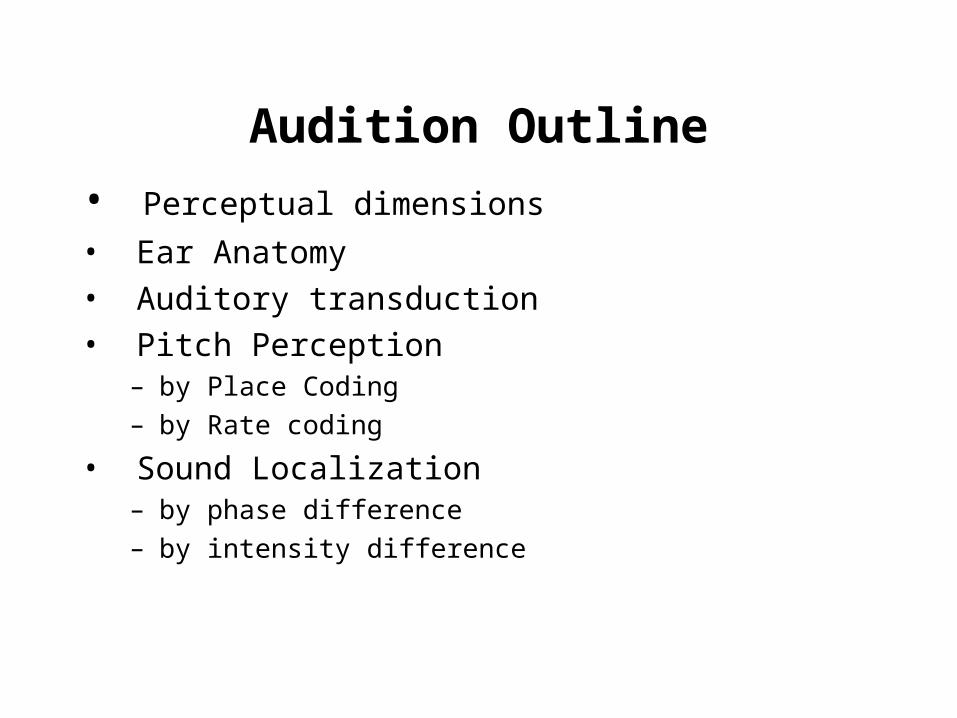
Audition Outline
• Perceptual dimensions
• Ear Anatomy • Auditory transduction• Pitch Perception
– by Place Coding– by Rate coding
• Sound Localization– by phase difference– by intensity difference
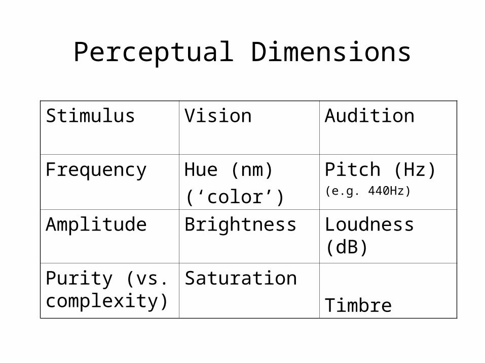
Perceptual Dimensions
Stimulus Vision Audition
Frequency Hue (nm)
(‘color’)
Pitch (Hz)(e.g. 440Hz)
Amplitude Brightness Loudness (dB)
Purity (vs. complexity)
Saturation
Timbre
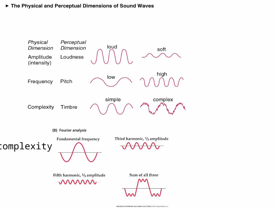
complexity
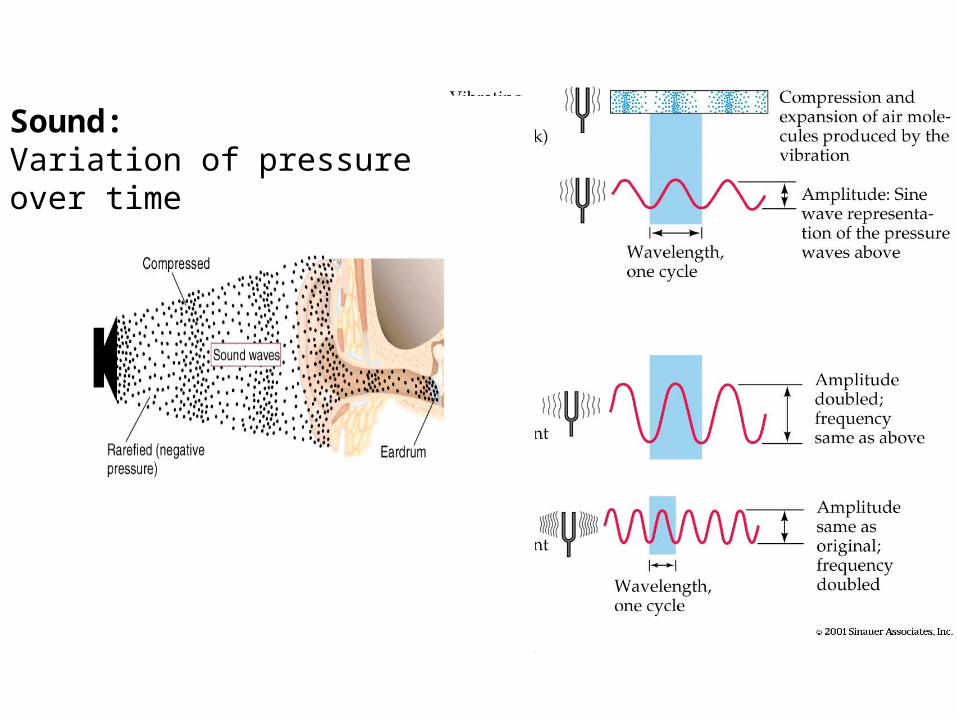
Sound: Variation of pressure over time
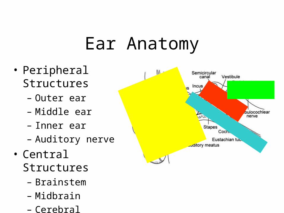
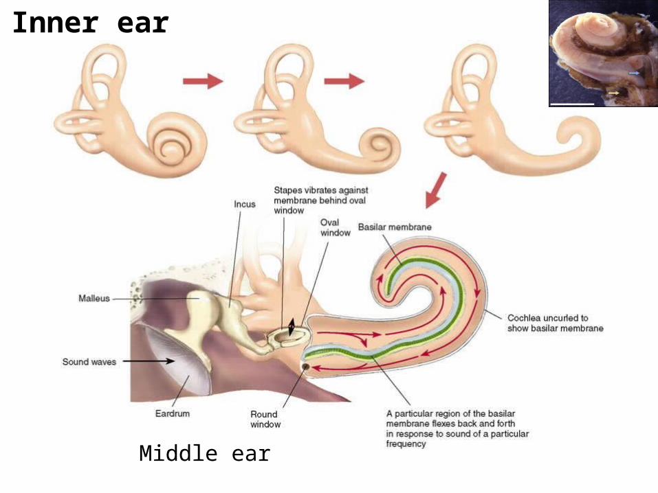
Ear Anatomy• Peripheral Structures
– Outer ear– Middle ear– Inner ear– Auditory nerve
• Central Structures– Brainstem– Midbrain– Cerebral
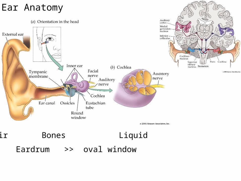
Air Bones Liquid
Eardrum >> oval window
Ear Anatomy
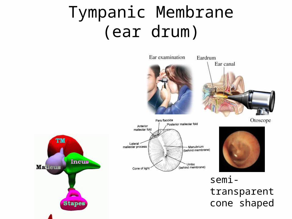
Tympanic Membrane (ear drum)
semi-transparent cone shaped
http://www.qub.ac.uk/cskills/Ears.htmHow to use an otoscope
http://medweb.uwcm.ac.uk/otoscopy/Default.htm
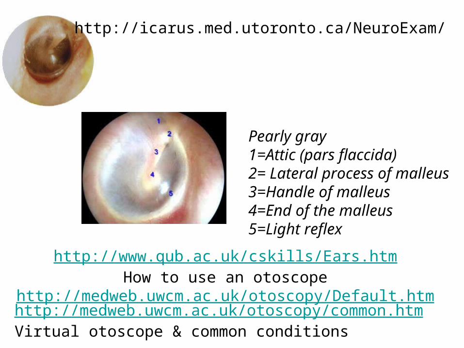
Pearly gray1=Attic (pars flaccida) 2= Lateral process of malleus 3=Handle of malleus 4=End of the malleus 5=Light reflex
http://medweb.uwcm.ac.uk/otoscopy/common.htmVirtual otoscope & common conditions
http://icarus.med.utoronto.ca/NeuroExam/
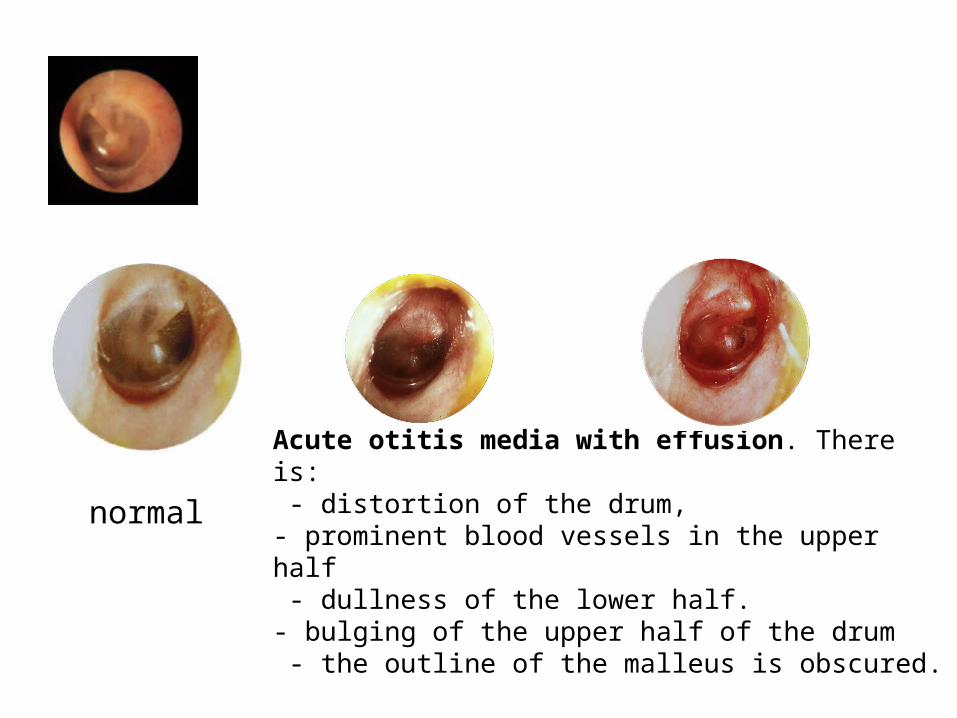
normalAcute otitis media with effusion. There is: - distortion of the drum, - prominent blood vessels in the upper half - dullness of the lower half. - bulging of the upper half of the drum - the outline of the malleus is obscured.
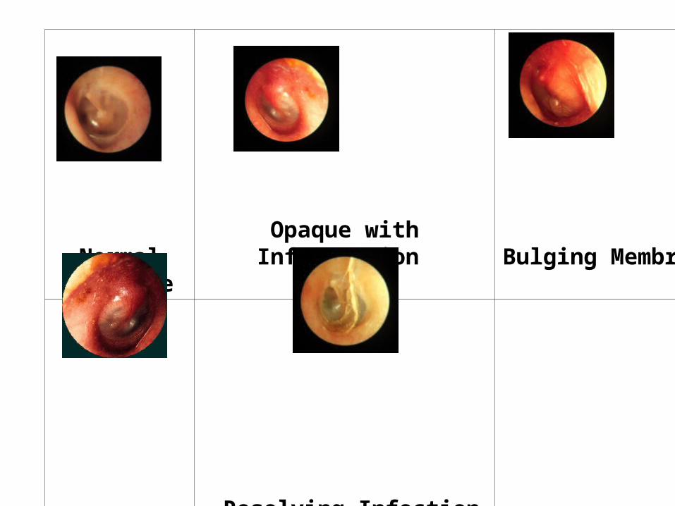
Normal
Membrane
Opaque with Inflammation
Bulging Membrane
Chronic
Inflammation
Resolving Infection
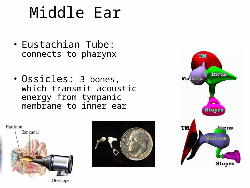
Middle Ear
• Eustachian Tube: connects to pharynx
• Ossicles: 3 bones, which transmit acoustic energy from tympanic membrane to inner ear
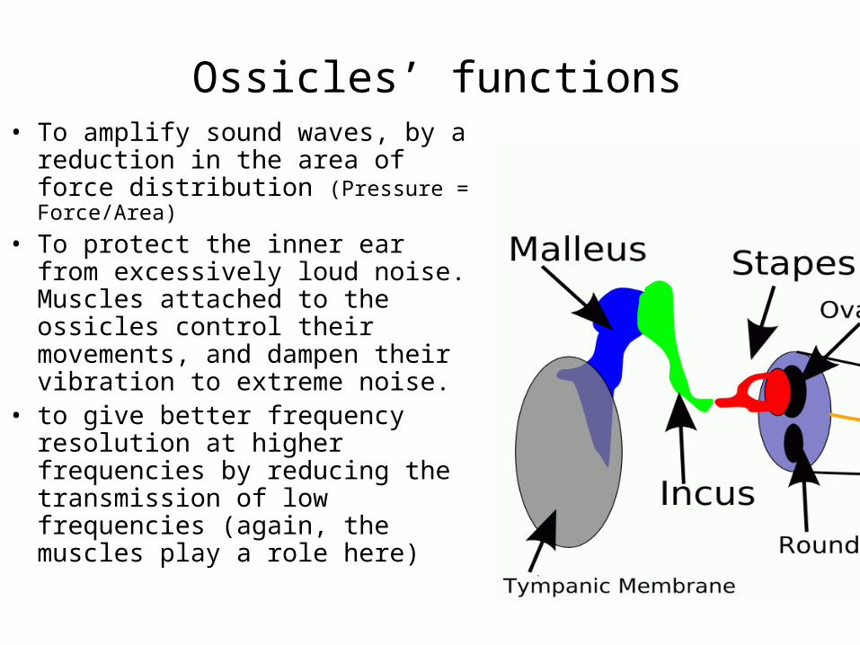
Ossicles’ functions• To amplify sound waves, by a
reduction in the area of force distribution (Pressure = Force/Area)
• To protect the inner ear from excessively loud noise. Muscles attached to the ossicles control their movements, and dampen their vibration to extreme noise.
• to give better frequency resolution at higher frequencies by reducing the transmission of low frequencies (again, the muscles play a role here)
Middle ear
Inner ear
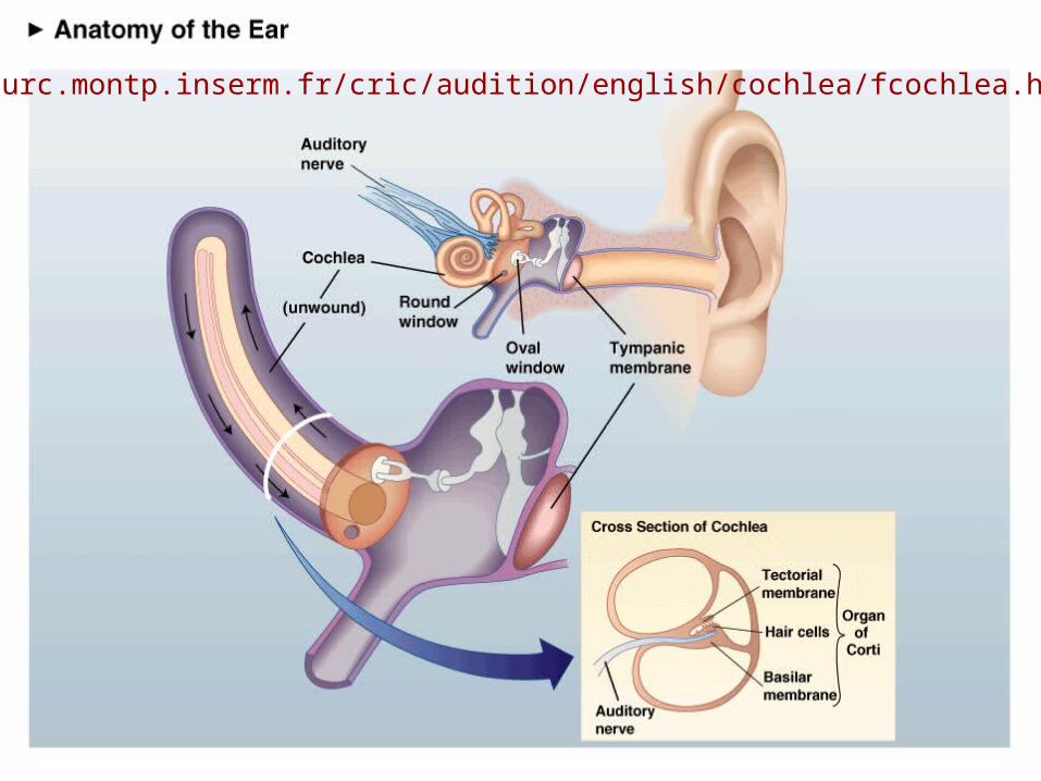
www.iurc.montp.inserm.fr/cric/audition/english/cochlea/fcochlea.htm
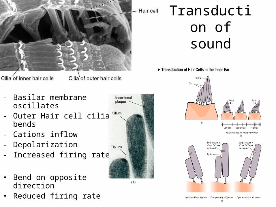
Transduction of sound
- Basilar membrane oscillates- Outer Hair cell cilia bends - Cations inflow- Depolarization- Increased firing rate
• Bend on opposite direction• Reduced firing rate
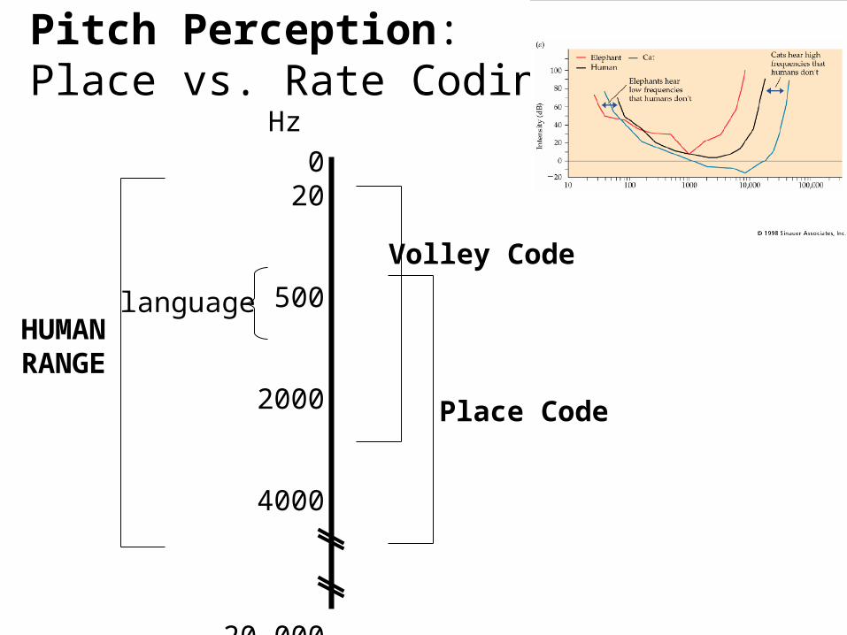
020
500
2000
4000
20,000
30,000
HUMANRANGE
Volley Code
Place Code
Hz
language
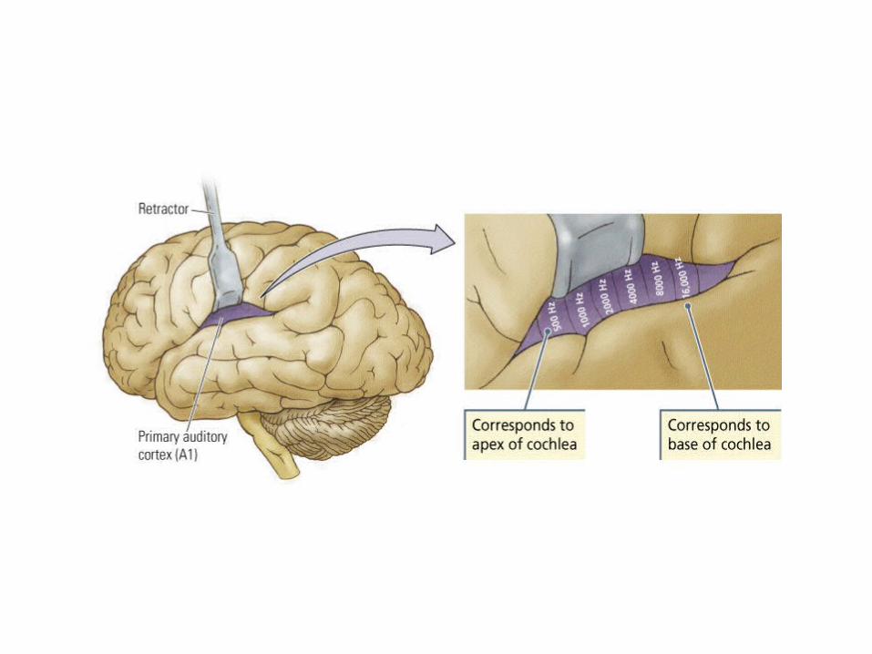
Pitch Perception: Place vs. Rate Coding
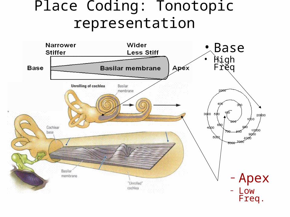
Place Coding: Tonotopic representation
• Base• High Freq
– Apex– Low Freq.
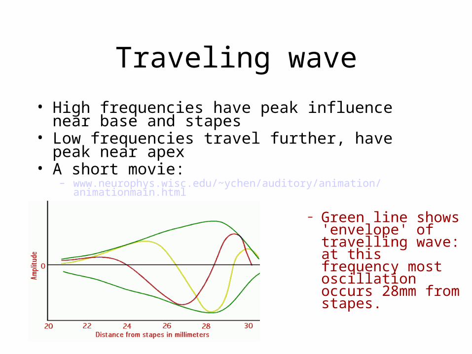
Traveling wave
• High frequencies have peak influence near base and stapes
• Low frequencies travel further, have peak near apex• A short movie:
– www.neurophys.wisc.edu/~ychen/auditory/animation/animationmain.html
– Green line shows 'envelope' of travelling wave: at this frequency most oscillation occurs 28mm from stapes.
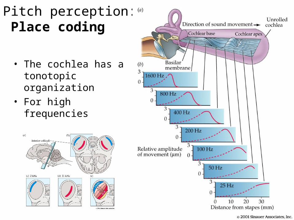
Pitch perception: Place coding
• The cochlea has a tonotopic organization
• For high frequencies
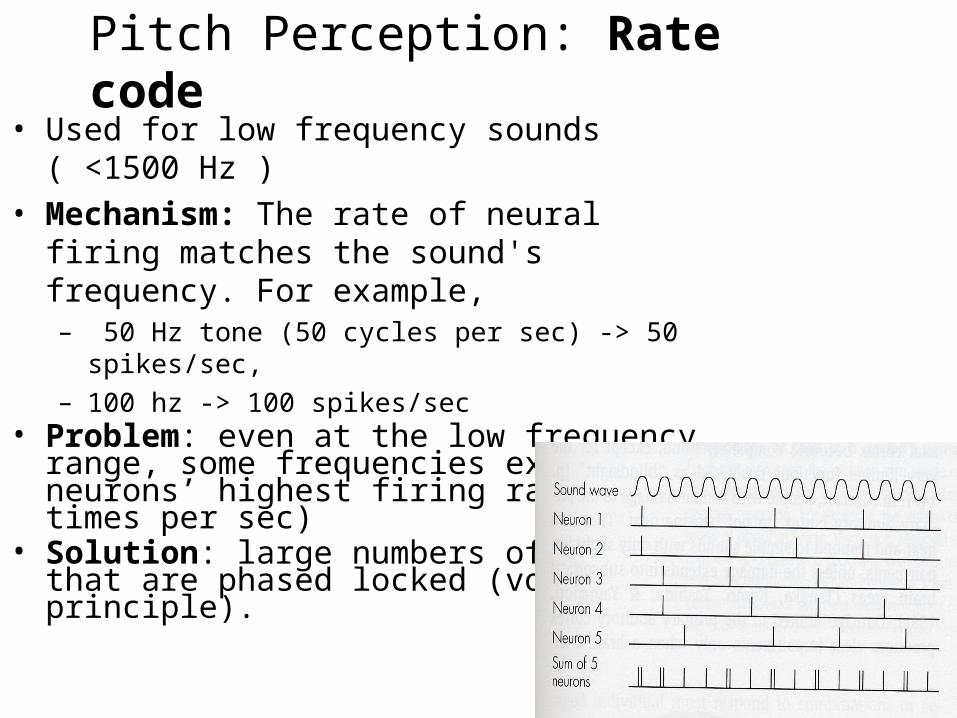
Pitch Perception: Rate code • Used for low frequency sounds ( <1500 Hz ) • Mechanism: The rate of neural firing matches
the sound's frequency. For example,– 50 Hz tone (50 cycles per sec) -> 50 spikes/sec, – 100 hz -> 100 spikes/sec
• Problem: even at the low frequency range, some frequencies exceed neurons’ highest firing rate (200 times per sec)
• Solution: large numbers of neurons that are phased locked (volley principle).
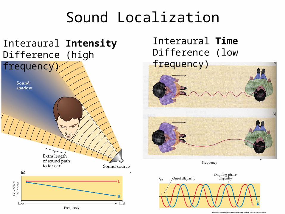
Sound Localization
Interaural Intensity Difference (high frequency)
Interaural Time Difference (low frequency)

Delay Lines – Interaural Time Difference (ITD)
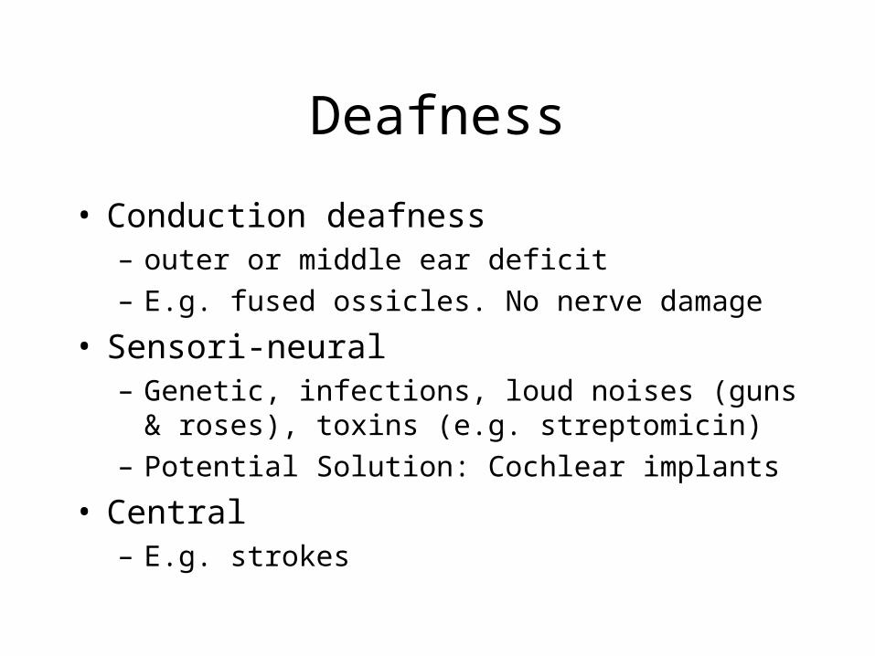
Deafness
• Conduction deafness– outer or middle ear deficit– E.g. fused ossicles. No nerve damage
• Sensori-neural– Genetic, infections, loud noises (guns & roses),
toxins (e.g. streptomicin)– Potential Solution: Cochlear implants
• Central– E.g. strokes
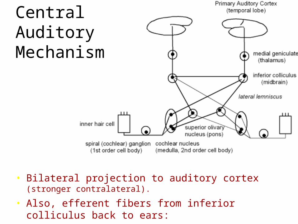
• Bilateral projection to auditory cortex (stronger contralateral).
• Also, efferent fibers from inferior colliculus back to ears: •they attenuate motion of the middle ear bones (dampen loud sounds)
Central Auditory Mechanism
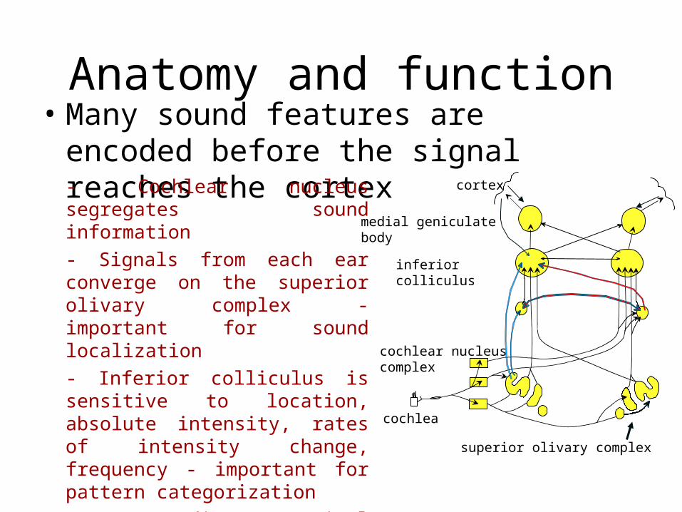
Anatomy and function• Many sound features are encoded before
the signal reaches the cortex
- Cochlear nucleus segregates sound information
- Signals from each ear converge on the superior olivary complex - important for sound localization
- Inferior colliculus is sensitive to location, absolute intensity, rates of intensity change, frequency - important for pattern categorization
- Descending cortical influences modify the input from the medial geniculate nucleus - important as an adaptive ‘filter’
inferior colliculus
medial geniculate body
cortex
superior olivary complex
cochlea
cochlear nucleuscomplex
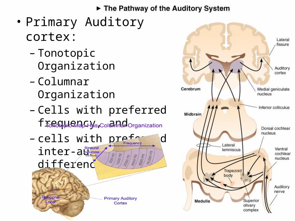
• Primary Auditory cortex:– Tonotopic Organization– Columnar Organization– Cells with preferred
frequency, and – cells with preferred inter-
aural time difference
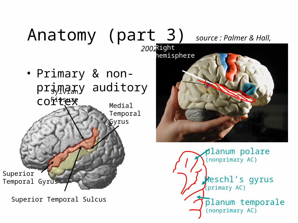
Anatomy (part 3) source : Palmer & Hall, 2002
• Primary & non-primary auditory cortex
Sylvian Fissure
Superior Temporal Gyrus
Superior Temporal Sulcus
Medial Temporal Gyrus
Right hemisphere
Heschl’s gyrus (primary AC)
planum temporale (nonprimary AC)
planum polare (nonprimary AC)
Spare slides
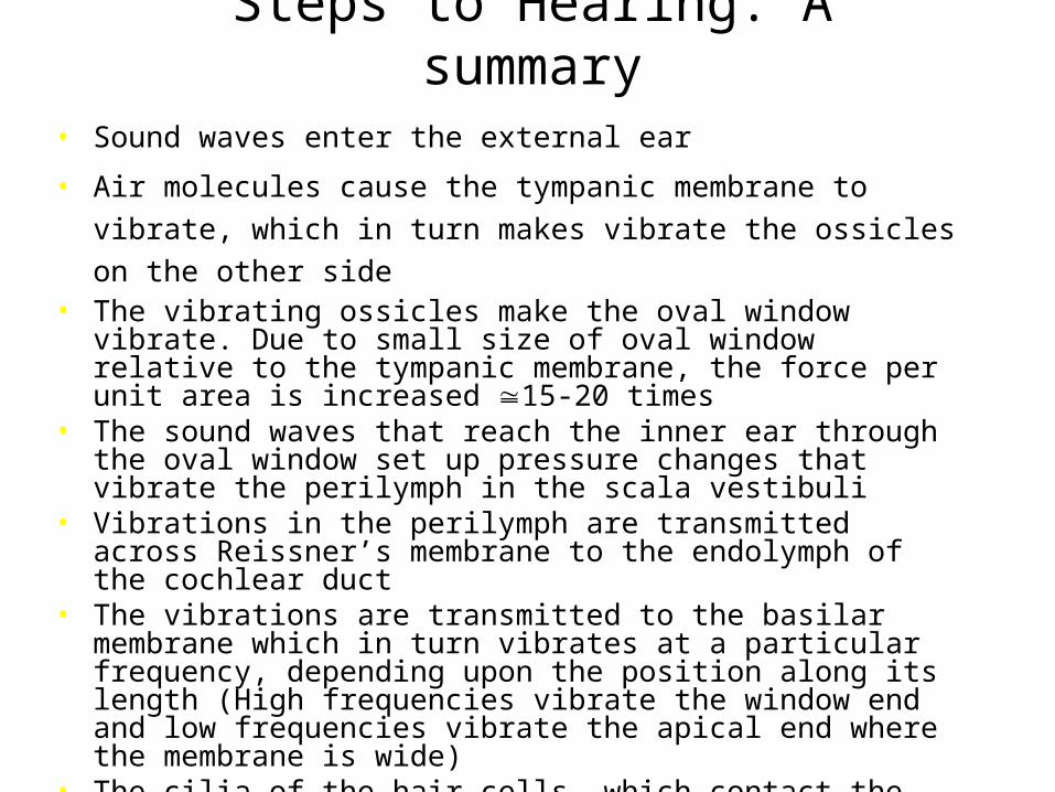
Steps to Hearing: A summary• Sound waves enter the external ear
• Air molecules cause the tympanic membrane to vibrate, which in
turn makes vibrate the ossicles on the other side • The vibrating ossicles make the oval window vibrate. Due to small
size of oval window relative to the tympanic membrane, the force per unit area is increased 15-20 times
• The sound waves that reach the inner ear through the oval window set up pressure changes that vibrate the perilymph in the scala vestibuli
• Vibrations in the perilymph are transmitted across Reissner’s membrane to the endolymph of the cochlear duct
• The vibrations are transmitted to the basilar membrane which in turn vibrates at a particular frequency, depending upon the position along its length (High frequencies vibrate the window end and low frequencies vibrate the apical end where the membrane is wide)
• The cilia of the hair cells, which contact the overlying tectorial membrane, bend as the basilar membrane vibrates Displacement of the stereocilia in the direction of the tallest stereocilia is excitatory and in the opposite direction is inhibitory
• The actions are transmitted along the cochlear branch of the vestibulocochlear nerve, activating auditory pathways in the central nervous system, eventually terminating in the auditory area of the temporal lobe of the cerebral cortex
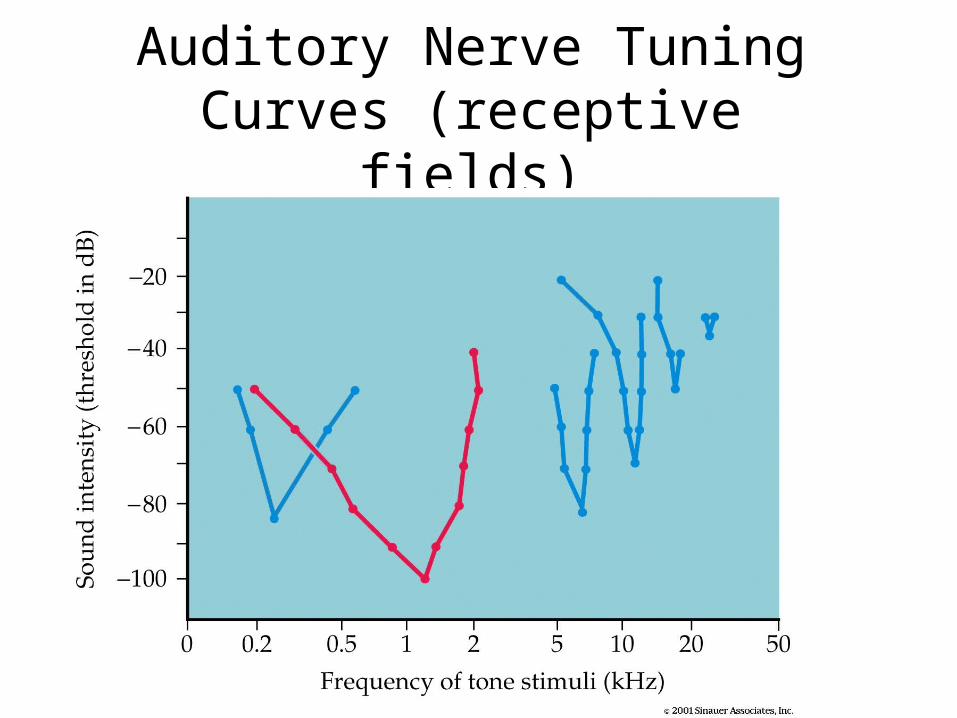
Auditory Nerve Tuning Curves (receptive fields)
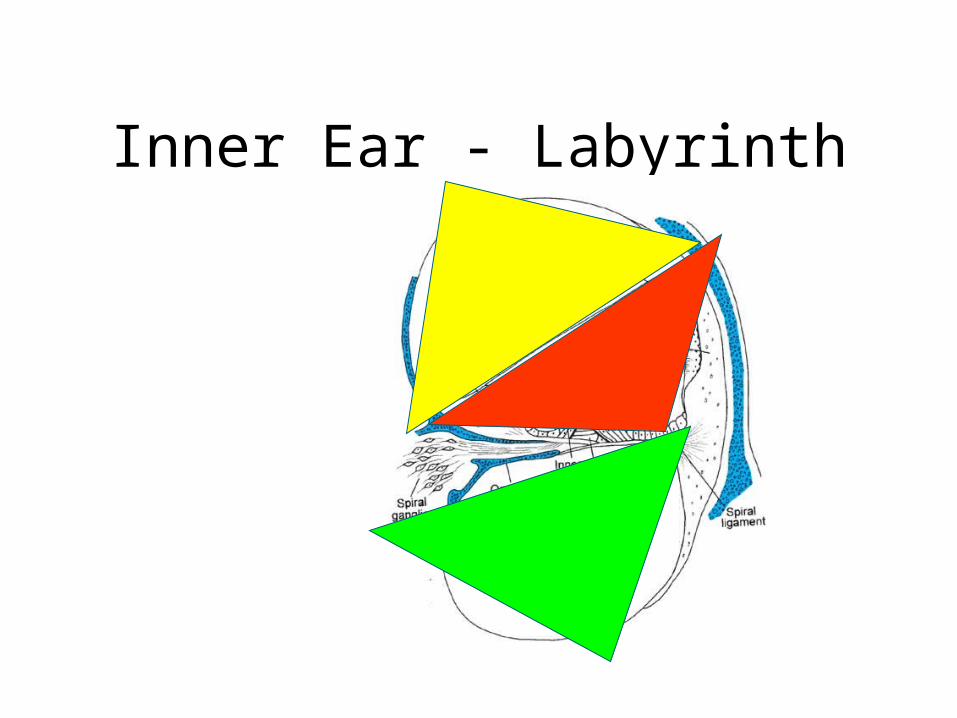
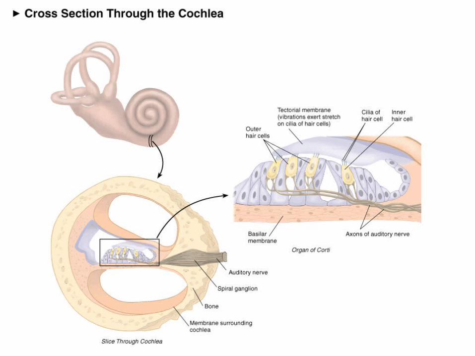
Inner Ear - Labyrinth
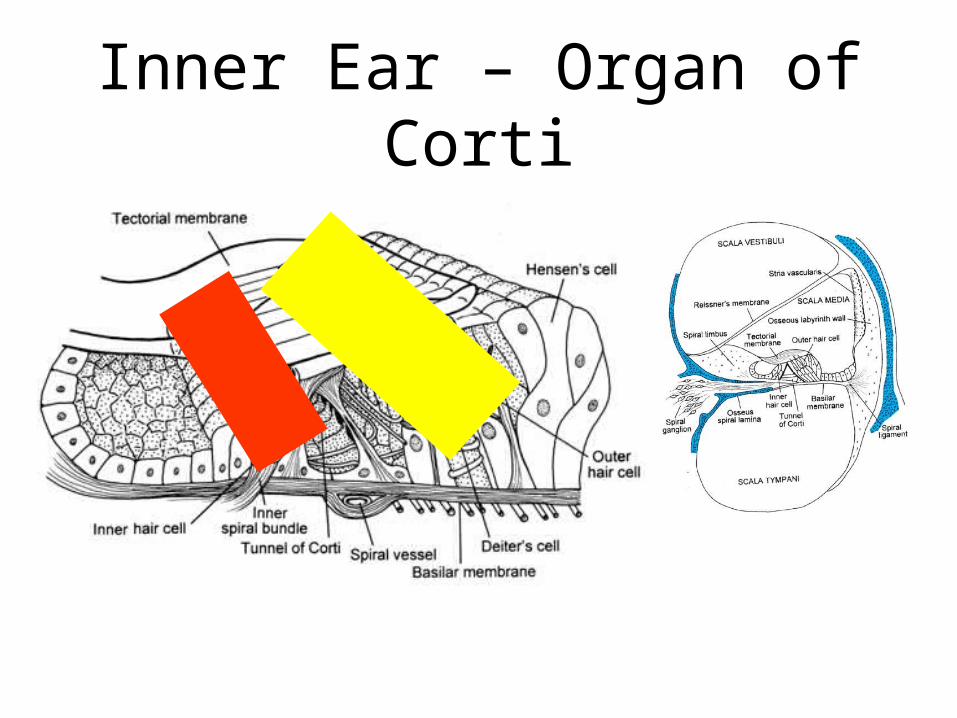
Inner Ear – Organ of Corti