Embed Size (px)
Citation preview

AtypicalAtypicalPneumoniaPneumonia
BY BY
Annerie HattinghAnnerie Hattingh
26/08/0926/08/09

Introduction:Introduction:
• Pneumonia caused by atypical pathogens• Typical pathogens usually includes: - Strep. pneumonia - Haemophilus pneumonia - Klebsiella pneumonia• Does not respond to the usual antibiotics• Causes a milder form of pneumonia (hence the term
“walking pneumonia”)• Characterized by a more drawn out coarse of
symptoms

• Legionella + SARS are exceptions to the above – both can be very severe infections
• Typical pneumonia can come on more quickly + with more severe early sx
• The arbitrary classification of typical vs. atypical pneumonia is of limited clinical value
• Literature now shows that a primary pathogen may co-exist with a secondary one, further blurring this distinction
IntroductionIntroduction::

Introduction:Introduction:
Causes:
“Classical” atypical pneumonias:
1.) Mycoplasma pneumonia
2.) Chlamydia pneumonia
3.) Legionella pneumonia

Introduction:Introduction:
Causes:Other micro-organisms that cause similar patterns
of presentation:
1.) Chlamydia psittaci (exposure to birds)
2.) Coxiella burnetti (presenting as Q fever)
3.) Viral pneumonias - Influenza A
- SARS
- RSV
- Adenoviridae
- Varicella pneumonitis

Epidemiology:Epidemiology:
• It is thought that the 3 main atypical pathogens might be implicated in up to 40% of CAP
• The precise incidence is not known• Often not identified in clinical practice due to lack of
readily available, reliable standardized tests to confirm dx
• By age 20, 50% of people in the USA have detectable levels of Antibodies to Chlamydia
pneumonia

Risk Factors:Risk Factors:
• Mycoplasma + Chlamydia spread by person-to-person contact
- spread most common in closed populations e.g.
schools, offices + military barracks
• Legionellae found most commonly in fresh water + man-made H2O systems

Risk Factors:Risk Factors:
- sources of contaminated H2O includes:
* showers
* condensers
* whirlpools
* cooling towers
* respiratory equipment
* air conditioning systems

Risk Factors:Risk Factors:
• Other risk factors include:
- young, healthy people
- cigarette smoking
- lung disease (like COPD)
- weakened immune system (e.g. chronic steroid
use or HIV)

Presentation:Presentation:
Mycoplasma pneumonia:Mycoplasma pneumonia:
• Gram neg bacteria with no true cell wall
• Frequent cause of CAP in adults + children
• Prevalence in adults with pneumonia 2 – 30%
• Tends to be endemic, occurring @ 4-7yr intervals

Presentation:Presentation:
Mycoplasma pneumonia:Mycoplasma pneumonia:Clinical Features:Clinical Features:
• Symptomatic / asymp
• Gradual onset (over few days – weeks)
• Prodrome of “flu-like” symptoms

Presentation:Presentation:
Mycoplasma pneumonia:Mycoplasma pneumonia:Clinical Features:Clinical Features:
• Including: - headache - malaise - fever - non prod. Cough - sore throat

Presentation:Presentation:
Mycoplasma pneumonia:Mycoplasma pneumonia:Clinical Features:Clinical Features:
• Objective AbN on physical exam are minimal in contrast to the pt’s reported symptoms
• Present like many of common viral illnesses BUT persistence + progression of sx help to mark it out

Presentation:Presentation:
Mycoplasma pneumonia:Mycoplasma pneumonia:
Extrapulm. Manifestations/Complications:Extrapulm. Manifestations/Complications:
• Can involve: CNS, Blood, Skin, CVS, Joints, GIT

Presentation:Presentation:
Mycoplasma pneumonia:Mycoplasma pneumonia:Extrapulm. Manifestations/Complications:Extrapulm. Manifestations/Complications:
Neurological compl.- Aseptic meningitis- Cerebellar ataxia- Transverse myelitis- Peripheral neuropathy

Presentation:Presentation:
Mycoplasma pneumonia:Mycoplasma pneumonia:Extrapulm. Manifestations/Complications:Extrapulm. Manifestations/Complications:
• Neurological manifestations are infrequent• Usually found in kids, if seen• Associated with increased morbidity + mortality• Antecedent resp. infection not always present

Presentation:Presentation:
Mycoplasma pneumonia:Mycoplasma pneumonia:Extrapulm. Manifestations/Complications:Extrapulm. Manifestations/Complications:
Hematological compl.• Hemolytic anemia• IgM antibodies to erythrocyte membrane I antigen
are present• Produces a cold agglutinin response that leads to
hemolysis

Presentation:Presentation:
Mycoplasma pneumonia:Mycoplasma pneumonia:Extrapulm. Manifestations/Complications:Extrapulm. Manifestations/Complications:
Dermatological compl.
Include rashes such as:
1. Erythema multiforme
2. Erythema nodosum
3. Urticaria

Presentation:Presentation:
Mycoplasma pneumonia:Mycoplasma pneumonia:Extrapulm. Manifestations/Complications:Extrapulm. Manifestations/Complications:
Cardiac involvement:
1. Pericarditis
2. Myocarditis

Presentation:Presentation:
Mycoplasma pneumonia:Mycoplasma pneumonia:Extrapulm. Manifestations/Complications:Extrapulm. Manifestations/Complications:
Joint involvent: (occationately described)
1. Arthralgia
2. Arthritis

Presentation:Presentation:
Mycoplasma pneumonia:Mycoplasma pneumonia:Extrapulm. Manifestations/Complications:Extrapulm. Manifestations/Complications:
GIT symptoms:
1. N + V
2. Diarrhea
3. Pancreatitis (rarely)

Presentation:Presentation:
Chlamydia:Chlamydia:
• Genus Chlamydia includes 3 species that infect humans: - C. psittaci
- C. trachomatis
- C. pneumonia
• Small, coccoid, Gram neg bacteria that resemble rickettsiae
Presentation:Presentation:
Chlamydia:Chlamydia:
Chlamydia trachomatis - seen in newborn infants
during delivery
- has been ass. with
pneumonia in adults
Presentation:Presentation:
Chlamydia:Chlamydia:
Chlamydia psittaci: • Ornithosis is a systemic infection often acc. by
pneumonia• Common in birds + some domestic animals• Pet shop employees + poultry workers @ risk• Other systems involved: CNS
(meningoencephalitis) + CVS (cult. neg. endocarditis)

Presentation:Presentation:
Chlamydia pneumonia:Chlamydia pneumonia:
• Prevalence varies by yr + geographic setting• Causes 5-15% of all CAP• Repeat infection is common• Gradual onset which may show improvement
before worsening again• Incubation 3-4 weeks• Initial non-specific URTI Sx lead to bronchitic/
pneumonic features
Presentation:Presentation:
Chlamydia pneumonia:Chlamydia pneumonia:
• Most infected remains quite well + asymptomatic• Can cause prolonged, acute bronchitis with
prod. cough• Hoarseness + headache are common features• Fever relatively uncommon• Sx may drag on for weeks/months despite course
of appropriate antibiotics

Presentation:Presentation:
Chlamydia pneumonia:Chlamydia pneumonia:
• Clinical severity usually caused by a secondary pathogen or co-existing illness e.g. diabetes
• Complications:
1. Sinusitis, otitis media
2. New onset asthma after acute infection
3. Endocarditis, myocarditis

Presentation:Presentation:
Legionella pneumonia:Legionella pneumonia:• Aerobic, motile, non-encapsulated, Gram neg
bacilli• Tends to be the most severe of the atypical
pneumonias• Focal outbreaks centered around poorly
maintained air conditioning / humidification systems
• Incubation 2-10 days• Initial mild headache, myalgia leading to fever,
chills + rigors

Presentation:Presentation:
Legionella pneumonia:Legionella pneumonia:
• Minimally prod. cough • Dyspnoea, pleuritic pain + hemoptysis are not
uncommon• Extra pulmonary legionellosis is rare but can be
severe• CVS most common extrapulm. site causing
myocarditis, pericarditis + endocarditis• Also pancreatitis, peritonitis, glomerulonephritis +
focal neurological deficit

Diagnosis:Diagnosis:
• CXR findings are usually non-specific and difficult to distinguish from other pneumonias
• Chest signs on examination minimal
• Rx of suspected atypical pneumonias should be empirical
• Cultures + serologic tests are not routinely available in laboratories
Diagnosis:Diagnosis:
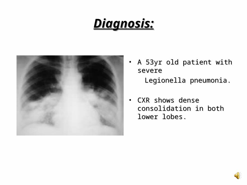
• A 53yr old patient with severe A 53yr old patient with severe
Legionella pneumonia. Legionella pneumonia.
• CXR shows dense CXR shows dense consolidation in both lower consolidation in both lower lobes.lobes.

Diagnosis:Diagnosis:
• A 40yr old patient with A 40yr old patient with Chlamydia pneumonia.Chlamydia pneumonia.
• CXR shows multifocal, patchy CXR shows multifocal, patchy consolidation in the right consolidation in the right upper, middle and lower lobes.upper, middle and lower lobes.

Diagnosis:Diagnosis:
• A 38yr old patient with A 38yr old patient with Mycoplasma pneumonia.Mycoplasma pneumonia.
• CXR shows a vague, ill CXR shows a vague, ill defined opacity in the left lower defined opacity in the left lower lobe.lobe.

Cause of pneumonia:
Mycoplasma pneumoniae
Legionella pneumophilaChlamydophila (Chlamydia) pneumoniae
Blood tests
May be raised WCC or rarely evidence of haemolytic anaemia. ESR may be elevated. Serology titres and complement fixation tests/ELISA can help to confirm the diagnosis.
FBC may show left shift. Severe cases may have DIC evident on FBC/INR. Hyponatraemia may occur due to syndrome of inappropriate ADH secretion. Urea/creatinine can be raised if complicated by renal failure or dehydration. LFTs often non-specifically deranged. CK may be elevated in rhabdomyolysis. Serological tests on blood or urine may be used to confirm diagnosis.
Usually non-specific and unhelpful. Serology titres or polymerase chain reaction tests may be used to confirm the diagnosis.
CXR
Usually single lower-lobe bronchopneumonia pattern with lobar consolidation rare. Other possible patterns include atelectasis, nodular infiltration akin to TB/sarcoidosis, hilar adenopathy and rarely pleural effusion.
50% have pleural effusion. Patchy alveolar infiltrates may be seen. CXR can take up to 4 months to return to normal and may initially progress despite therapy.
Usually lower-lobe single subsegmental infiltrate. Pleural effusion found in up to a quarter of cases. Can progress to ARDS. CXR changes may take up to 3 months to resolve.
ABGs may be checked to assess respiratory function in acute, severe cases of community-acquired pneumonia. Similarly, blood cultures should be taken to aid subsequent
microbiological diagnosis. In cases of atypical pneumonia where there is evidence of focal or global cerebral impairment, an LP should be considered.

Management:Management:
• Severe cases should be admitted
• Atypical pneumonias usually Rx as for other
CAP, at least initially
• No evidence that routinely giving antibiotics active against atypical organisms leads to better outcomes in non-severe CAP

Management:Management:
• Macrolides, such as Erythromycin, Clarithromycin + Azithromycin have been shown to be effective in the Rx of all 3 organisms
• Erythromycin tends to be less well tolerated + only few trails demonstrates its efficacy in the Rx of Legionella
• Severe Legionella infections may require rifampicin + a macrolide
• Tetracycline, Doxycycline + Fluoroquinolones are also effective
• Recommened duration of therapy usually 2-3 weeks

THE END
QUESTIONS??

References:References:
1.1. Shakeel Amanullah:Shakeel Amanullah: Atypical Bacterial Pneumonia; Atypical Bacterial Pneumonia; eMed. March 2008.eMed. March 2008.
2.2. www.patient.co.uk: www.patient.co.uk: Atypical Pneumonias; Jan. 2007.Atypical Pneumonias; Jan. 2007.
3.3. www.thirdage.comwww.thirdage.com: Encyclopedia – Atypical : Encyclopedia – Atypical Pneumonia (Mycoplasma and Viral) (Walking Pneumonia (Mycoplasma and Viral) (Walking Pneumonia); May 2008.Pneumonia); May 2008.
4.4. Rosen’s Emergency Medicine Online: Rosen’s Emergency Medicine Online: Community Community Acquired PneumoniaAcquired Pneumonia


![l & E x perimenta l i n ic lp Journal of Clinical ... · the so-called atypical pneumonia, which is a type of pneumonia with an overall prevalence of 22.7% [1]. It is also a causative](https://img.pdfslide.us/doc/110x75/6082e1604c6d78134f366804/l-e-x-perimenta-l-i-n-ic-lp-journal-of-clinical-the-so-called-atypical.jpg)