Embed Size (px)
Citation preview
Indexed through the National Library of Medicine(PubMed/MEDLINE), PubMed Central (PMC), and EMBASE
ON THE WEB:hematologyandoncology.net
November 2016 Volume 14, Issue 11, Supplement 11
Atypical Hemolytic Uremic Syndrome (aHUS): Essential Aspects of an Accurate Diagnosis
Authors
Jeffrey Laurence, MDProfessor of MedicineDivision of Hematology and Medical OncologyNew York Presbyterian Hospital andWeill Cornell Medical CollegeNew York, New York
Hermann Haller, MDProfessor of MedicineDepartment of Nephrology and HypertensionHannover Medical School (MHH)Hannover, Germany
Pier Mannuccio Mannucci, MDProfessor of MedicineIRCCS Cà Granda Foundation Maggiore Policlinico HospitalMilan, Italy
Masaomi Nangaku, MD, PhDProfessor of MedicineDivision of Nephrology and EndocrinologyThe University of Tokyo Graduate School of MedicineTokyo, Japan
Manuel Praga, MDProfessor of MedicineDivision of NephrologyComplutense UniversityMadrid, Spain
Santiago Rodriguez de Cordoba, PhDProfessor, Department of Cellular and Molecular MedicineEl Centro de Investigaciones Biológicas and El Centro de Investigación Biomédica en Red de Enfermedades RarasMadrid, Spain
2 Clinical Advances in Hematology & Oncology Volume 14, Issue 11, Supplement 11 November 2016
R E V I E W A R T I C L E
fragmented red blood cells or schistocytes on peripheral blood smear, low haptoglobin levels, elevated lactate dehydrogenase (LDH) and indirect bilirubin, and a decline in baseline hemoglobin—accompanied by thrombocytopenia. These laboratory changes must occur in concert with clinical involvement of at least 1 organ system, the most common sites being the central nervous system, the kidneys, and the gastrointestinal tract. DIC, usually
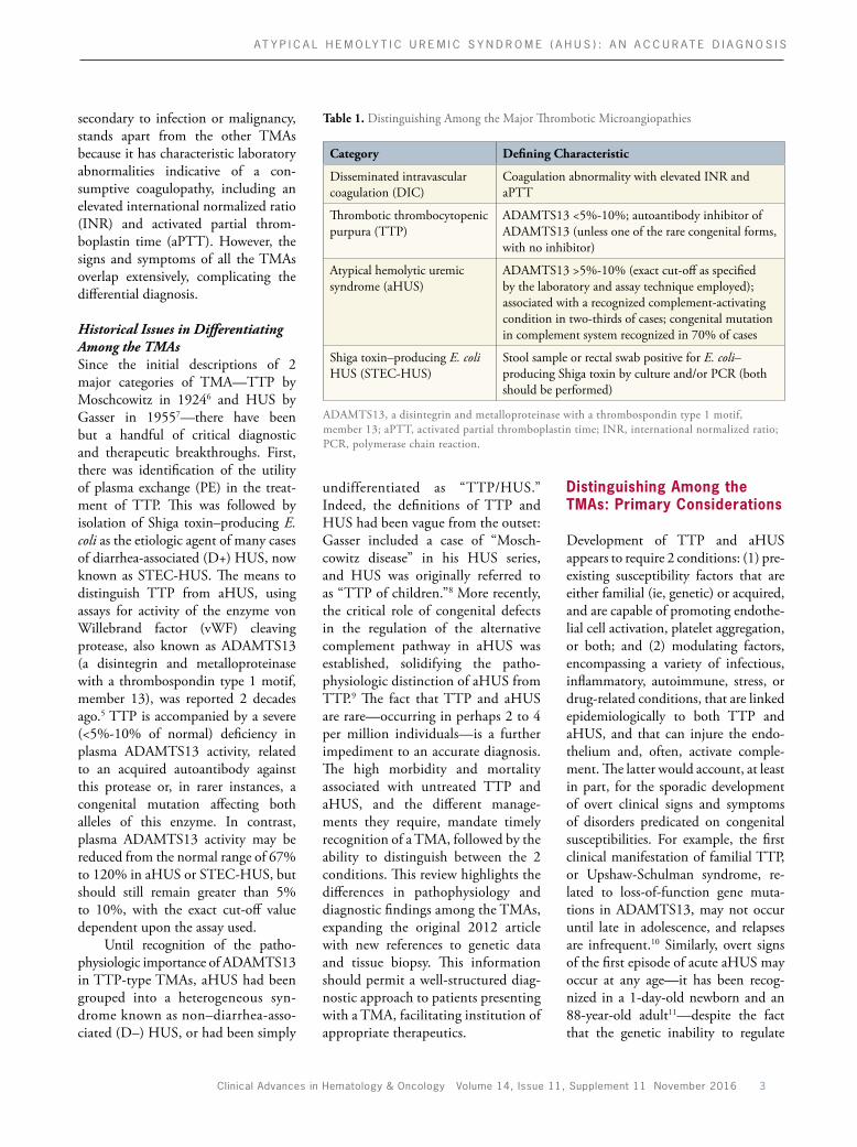
angiopathy (TMA) that is clinically very similar to aHUS.25 However, a major challenge per sists: how to make an efficient and accurate differential diagnosis among the TMAs.
Table 1 lists the 4 major TMAs: aHUS, TTP, Shiga toxin–producing Escherichia coli (STEC)HUS, and disseminated intravascular coagulation (DIC). Clinical recognition of any TMA requires the documentation of microangiopathic hemolysis—with
Abstract: Atypical hemolytic uremic syndrome (aHUS), a thrombotic microangiopathy (TMA), is a rare, life-threatening,
systemic disease. When unrecognized or inappropriately treated, aHUS has a high degree of morbidity and mortality.
aHUS results from chronic, uncontrolled activity of the alternative complement pathway, which activates platelets and
damages the endothelium. Two-thirds of aHUS cases are associated with an identifiable complement-activating condition.
aHUS is clinically very similar to the other major TMAs: Shiga toxin–producing Escherichia coli (STEC)-HUS, thrombotic
thrombocytopenic purpura (TTP), and disseminated intravascular coagulation (DIC). The signs and symptoms of all the
TMAs overlap, complicating the differential diagnosis. Clinical identification of a TMA requires documentation of micro-
angiopathic hemolysis accompanied by thrombocytopenia. DIC must be recognized and treated before it is possible to
discriminate among the other 3 major TMAs. STEC-HUS can be excluded through testing for Shiga toxin–producing E. coli.
aHUS can be distinguished from TTP on the basis of ADAMTS13 (a disintegrin and metalloproteinase with a thrombos-
pondin type 1 motif, member 13) activity, with a severe decrease characteristic of TTP. This test, as both an activity assay
and an inhibitor assay, should be ordered before the initiation of plasma therapy in any patient presenting with a TMA.
Finally, it is important to recognize that aHUS remains a clinical diagnosis, but in complex scenarios, tissue biopsy may be
a useful adjunct in diagnosis.
DisclaimerFunding for this supplement has been provided by Alexion Pharmaceuticals, Inc. Support of this supplement does not imply the supporter’s agreement with the views expressed herein. Every effort has been made to ensure that drug usage and other information are presented accurately; however, the ultimate responsibility rests with the prescribing physician. Millennium Medical Publishing, Inc, and the participants shall not be held responsible for errors or for any consequences arising from the use of information contained herein. Readers are strongly urged to consult any relevant primary literature. No claims or endorsements are made for any drug or compound at present under clinical investigation.
©2016 Millennium Medical Publishing, Inc., 611 Broadway, Suite 310, New York, NY 10012. Printed in the USA. All rights reserved, including the right of reproduction, in whole or in part, in any form.
Atypical Hemolytic Uremic Syndrome (aHUS): Essential Aspects of an Accurate DiagnosisJeffrey Laurence, MD, Hermann Haller, MD, Pier Mannuccio Mannucci, MD, Masaomi Nangaku, MD, PhD, Manuel Praga, MD, and Santiago Rodriguez de Cordoba, PhD
Introduction
In October 2012, this journal published a review on “Making the diagnosis” of atypical hemolytic uremic syn drome (aHUS).1 Recent advances have enabled the implementation of directed therapy, with dramatic declines in morbidity and mortality over the historical interventions used in thrombotic thrombocytopenic purpura (TTP), a thrombotic micro
Clinical Advances in Hematology & Oncology Volume 14, Issue 11, Supplement 11 November 2016 3
A T Y P I C A L H E M O LY T I C U R E M I C S Y N D R O M E ( A H U S ) : A N A C C U R A T E D I A G N O S I S
Distinguishing Among the TMAs: Primary Considerations
Development of TTP and aHUS appears to require 2 conditions: (1) preexisting susceptibility factors that are either familial (ie, genetic) or acquired, and are capable of promoting endothelial cell activation, platelet aggregation, or both; and (2) modulating factors, encompassing a variety of infectious, inflammatory, autoimmune, stress, or drugrelated conditions, that are linked epi demiologically to both TTP and aHUS, and that can injure the endothelium and, often, activate complement. The latter would account, at least in part, for the sporadic development of overt clinical signs and symptoms of disorders predicated on congenital susceptibilities. For example, the first clinical manifestation of familial TTP, or UpshawSchulman syndrome, re lated to lossoffunction gene mutations in ADAMTS13, may not occur until late in adolescence, and relapses are infrequent.10 Similarly, overt signs of the first episode of acute aHUS may occur at any age—it has been recognized in a 1dayold newborn and an 88yearold adult11—despite the fact that the genetic inability to regulate
undifferentiated as “TTP/HUS.” Indeed, the definitions of TTP and HUS had been vague from the outset: Gasser included a case of “Moschcowitz disease” in his HUS series, and HUS was originally referred to as “TTP of children.”8 More recently, the critical role of congenital defects in the regulation of the alternative complement pathway in aHUS was established, solidifying the pathophysiologic distinction of aHUS from TTP.9 The fact that TTP and aHUS are rare—occurring in perhaps 2 to 4 per million individuals—is a further impediment to an accurate diagnosis. The high morbidity and mortality associated with untreated TTP and aHUS, and the different managements they require, mandate timely recognition of a TMA, followed by the ability to distinguish between the 2 conditions. This review highlights the differences in pathophysiology and diagnostic findings among the TMAs, expanding the original 2012 article with new references to genetic data and tissue biopsy. This information should permit a wellstructured diagnostic approach to patients presenting with a TMA, facilitating institution of appropriate therapeutics.
secondary to infection or malignancy, stands apart from the other TMAs because it has characteristic laboratory abnormalities indicative of a consumptive coagulopathy, including an elevated international normalized ratio (INR) and activated partial thromboplastin time (aPTT). However, the signs and symptoms of all the TMAs overlap extensively, complicating the differential diagnosis.
Historical Issues in Differentiating Among the TMAsSince the initial descriptions of 2 major categories of TMA—TTP by Moschcowitz in 19246 and HUS by Gasser in 19557—there have been but a handful of critical diagnostic and therapeutic breakthroughs. First, there was identification of the utility of plasma exchange (PE) in the treatment of TTP. This was followed by isolation of Shiga toxin–producing E. coli as the etiologic agent of many cases of diarrheaassociated (D+) HUS, now known as STECHUS. The means to distinguish TTP from aHUS, using assays for activity of the enzyme von Willebrand factor (vWF) cleaving protease, also known as ADAMTS13 (a disintegrin and metalloproteinase with a thrombospondin type 1 motif, member 13), was reported 2 decades ago.5 TTP is accompanied by a severe (<5%10% of normal) deficiency in plasma ADAMTS13 activity, related to an acquired autoantibody against this protease or, in rarer instances, a congenital mutation affecting both alleles of this enzyme. In contrast, plasma ADAMTS13 activity may be reduced from the normal range of 67% to 120% in aHUS or STECHUS, but should still remain greater than 5% to 10%, with the exact cutoff value dependent upon the assay used.
Until recognition of the path o physiologic importance of ADAMTS13 in TTPtype TMAs, aHUS had been grouped into a hetero geneous syndrome known as non–diarrheaassociated (D–) HUS, or had been simply
Table 1. Distinguishing Among the Major Thrombotic Microangiopathies
Category Defining Characteristic
Disseminated intravascular coagulation (DIC)
Coagulation abnormality with elevated INR and aPTT
Thrombotic thrombocytopenic purpura (TTP)
ADAMTS13 <5%10%; autoantibody inhibitor of ADAMTS13 (unless one of the rare congenital forms, with no inhibitor)
Atypical hemolytic uremic syndrome (aHUS)
ADAMTS13 >5%10% (exact cutoff as specified by the laboratory and assay technique employed); associated with a recognized complementactivating condition in twothirds of cases; congenital mutation in complement system recognized in 70% of cases
Shiga toxin–producing E. coli HUS (STECHUS)
Stool sample or rectal swab positive for E. coli– producing Shiga toxin by culture and/or PCR (both should be performed)
ADAMTS13, a disintegrin and metalloproteinase with a thrombospondin type 1 motif, member 13; aPTT, activated partial thromboplastin time; INR, international normalized ratio; PCR, polymerase chain reaction.
4 Clinical Advances in Hematology & Oncology Volume 14, Issue 11, Supplement 11 November 2016
R E V I E W A R T I C L E
aHUS.”19,20 This distinction is important, as the conditions listed above can all lead to the signs and symptoms of a TMA that is simply another manifestation of that disease process itself, such as systemic lupus erythematosus (SLE) or malignant hypertension, or associated with pregnancy, drug therapy, or other conditions. One must first treat that initial disease process adequately. If the TMA does not resolve, then consider that it unmasked aHUS, which should then be viewed as the primary cause of patient morbidity and therefore treated. Making this distinction may require special diagnostic tools, including tissue biopsy, as discussed in section VI.
Given the pathophysiologic differences between aHUS and TTP, one might think that diagnostic criteria to distinguish among the TMAs would be relatively simple to apply. Often they are, and this is critical clinically, as it will guide management decisions. Just as mortality from TTP declined from more than 90% to less than 10% with institution of appropriate treatment—therapeutic PE21—outcome is highly unfavorable in inadequately treated aHUS. Up to 50% of patients progress to endstage renal disease (ESRD) within a year, and 25% die during the acute phase, despite extensive PE.8,22 The importance of rapid diagnosis of patients with aHUS cannot be stressed more highly.2,23
I. Seven Steps to Consider in Reaching a Specific TMA Diagnosis
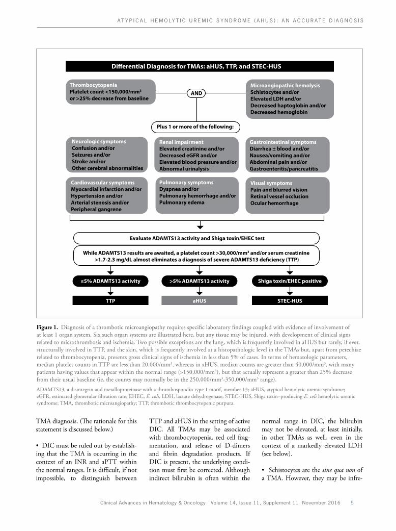
1. Recognize a TMA. As outlined in Table 1 and Figure 1, clinical recognition of a TMA involves documentation of the principle laboratory criteria for microangiopathic hemolytic anemia—schistocytes on peripheral blood smear, elevated LDH, low haptoglobin, elevated indirect bilirubin, and a decline in baseline hemoglobin—accompanied by thrombocytopenia. Not all of these changes are necessary to make a
the alternative complement pathway is present at birth.
The vast majority of TTP cases are idiopathic; disease susceptibility results from an acquired, autoantibodymediated deficiency of ADAMTS13. This leads to propagation of platelet aggregates, related to the inability to cleave long tethers of platelets bound to ultra–highmolecularweight multimers of vWF, which requires an intact ADAMTS13. The resultant systemic, uncontrolled microthrombus formation is clinically devastating.12 By contrast, in the vast majority of aHUS cases, susceptibility factors are familial, not acquired. As discussed below, they
are genetic defects in complement and complement regulatory proteins that permit unregulated amplification of the alternative complement pathway. The consequences are massive terminal complement pathway activation with generation of C5a (a potent anaphylatoxin) and C5b9 (known as membrane attack complex [MAC]), triggering inflammation, platelet activation, platelet aggregation, erythrocyte lysis, endothelial cell injury, and fibrin microthrombus formation throughout the microvasculature.13 Any TMA can be associated with activation of the alternative complement system. It is rare, however, for the TMA to be sustained, except when this activation cannot be regulated—as seen in aHUS.14
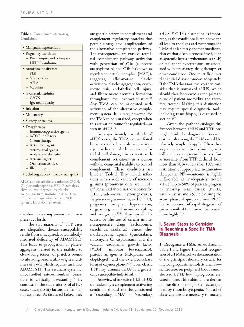
In approximately twothirds of aHUS cases, the TMA is manifested by a recognized complementactivating condition, which causes endothelial cell damage in concert with complement activation, in a person with the congenital inability to control complement. These conditions are listed in Table 2. They include infections with a wide variety of microorganisms (prominent ones are H1N1 influenza and those in the vaccines for H1N1, adenovirus, cytomegalovirus, Streptococcus pneumoniae, and STEC), pregnancy, malignant hypertension, surgery, organ and tissue transplant, and malignancy.15,16 They can also be caused by the use of certain immunosuppressive drugs (cyclosporine, tacrolimus, sirolimus), cancer chemotherapeutic agents (gemcitabine, mitomycin C, cisplatinum, and the vascular endothelial growth factor [VEGF] inhibitor bevacizumab), platelet antagonists (ticlopidine and clopidogrel), and the extendedrelease form of oxymorphone.15,16 Even classic TTP may unmask aHUS in a genetically susceptible individual.17,18
As reviewed in Section III.2, aHUS unmasked by a complementactivating condition should not be considered a “secondary TMA” or “secondary
Table 2. ComplementActivating Conditions
• Malignant hypertension
• Pregnancyassociated – Preeclampsia and eclampsia – HELLP syndrome
• Autoimmune diseases – SLE – Scleroderma – APLS – Vasculitis
• Glomerulonephritis – C3GN – IgA nephropathy
• Infection
• Malignancy
• Surgery or trauma
• Drug therapy – Immunosuppressive agents – mTOR inhibitors – Chemotherapy – Antitumor agents – Antimalarial agents – Antiplatelet therapies – Antiviral agents – Oral contraceptives – Illicit drugs
• Solid organ/bone marrow transplant
APLS, antiphospholipid syndrome; C3GN, C3 glomerulon ephritis; HELLP, hemolysis, elevated liver enzymes, low platelet count; IgA, immunoglobulin A; mTOR, mammalian target of rapamycin; SLE, systemic lupus erythematosus.
Clinical Advances in Hematology & Oncology Volume 14, Issue 11, Supplement 11 November 2016 5
A T Y P I C A L H E M O LY T I C U R E M I C S Y N D R O M E ( A H U S ) : A N A C C U R A T E D I A G N O S I S
normal range in DIC, the bilirubin may not be elevated, at least initially, in other TMAs as well, even in the context of a markedly elevated LDH (see below).
• Schistocytes are the sine qua non of a TMA. However, they may be infre
TTP and aHUS in the setting of active DIC. All TMAs may be associated with thrombocytopenia, red cell fragmentation, and release of Ddimers and fibrin degradation products. If DIC is present, the underlying condition must first be corrected. Although indirect bilirubin is often within the
TMA diagnosis. (The rationale for this statement is discussed below.)
• DIC must be ruled out by establishing that the TMA is occurring in the context of an INR and aPTT within the normal ranges. It is difficult, if not impossible, to distinguish between
Figure 1. Diagnosis of a thrombotic microangiopathy requires specific laboratory findings coupled with evidence of involvement of at least 1 organ system. Six such organ systems are illustrated here, but any tissue may be injured, with development of clinical signs related to microthrombosis and ischemia. Two possible exceptions are the lung, which is frequently involved in aHUS but rarely, if ever, structurally involved in TTP, and the skin, which is frequently involved at a histopathologic level in the TMAs but, apart from petechiae related to thrombocytopenia, presents gross clinical signs of ischemia in less than 5% of cases. In terms of hematologic parameters, median platelet counts in TTP are less than 20,000/mm3, whereas in aHUS, median counts are greater than 40,000/mm3, with many patients having values that appear within the normal range (>150,000/mm3), but that actually represent a greater than 25% decrease from their usual baseline (ie, the counts may normally be in the 250,000/mm3350,000/mm3 range).
ADAMTS13, a disintegrin and metalloproteinase with a thrombospondin type 1 motif, member 13; aHUS, atypical hemolytic uremic syndrome; eGFR, estimated glomerular filtration rate; EHEC, E. coli; LDH, lactate dehydrogenase; STECHUS, Shiga toxin–producing E. coli hemolytic uremic syndrome; TMA, thrombotic microangiopathy; TTP, thrombotic thrombocytopenic purpura.
Di�erential Diagnosis for TMAs: aHUS, TTP, and STEC-HUS
ThrombocytopeniaPlatelet count <150,000/mm3 or >25% decrease from baseline
Microangiopathic hemolysisSchistocytes and/or Elevated LDH and/orDecreased haptoglobin and/or Decreased hemoglobin
AND
Plus 1 or more of the following:
Neurologic symptomsConfusion and/orSeizures and/orStroke and/orOther cerebral abnormalities
Renal impairmentElevated creatinine and/orDecreased eGFR and/orElevated blood pressure and/orAbnormal urinalysis
Gastrointestinal symptomsDiarrhea ± blood and/orNausea/vomiting and/orAbdominal pain and/orGastroenteritis/pancreatitis
Cardiovascular symptomsMyocardial infarction and/orHypertension and/orArterial stenosis and/orPeripheral gangrene
Pulmonary symptomsDyspnea and/orPulmonary hemorrhage and/orPulmonary edema
Visual symptomsPain and blurred visionRetinal vessel occlusionOcular hemorrhage
Evaluate ADAMTS13 activity and Shiga toxin/EHEC test
While ADAMTS13 results are awaited, a platelet count >30,000/mm3 and/or serum creatinine >1.7-2.3 mg/dL almost eliminates a diagnosis of severe ADAMTS13 de�ciency (TTP)
≤5% ADAMTS13 activity >5% ADAMTS13 activity Shiga toxin/EHEC positive
TTP aHUS STEC-HUS
6 Clinical Advances in Hematology & Oncology Volume 14, Issue 11, Supplement 11 November 2016
R E V I E W A R T I C L E
tually never directly involved in TTP, but frequently involved in untreated aHUS.26,27
4. Consider the absolute platelet count and serum creatinine values. The patient’s presenting platelet count and serum creatinine can serve as a guide to whether TTP or aHUS is the more likely diagnosis.28 In an “uncomplicated” case, a platelet count greater than 30,000/mm3 at presentation is highly unusual in TTP, but classic for aHUS.20,29 (An “uncomplicated” case is one that is not linked to an ongoing complementactivating condition, such as autoimmune disease, infection, can cer, and use of certain medications, all of which can unmask aHUS, but themselves may alter platelet counts and renal function.) TTP is a plateletconsumptive disorder, and median platelet counts are less than 20,000/mm3. In aHUS, however, typical fibrin microthrombi, rather than plateletrich clots, predominate, with less platelet consumption. The median platelet count in aHUS is 30,000/mm3 to 40,000/mm3. Indeed, platelet counts within the normal range occur in up to 20% of aHUS cases at presentation, reflecting the fact that those “normal” numbers may still indicate a greater than 25% change from the patient’s usual baseline.30
In terms of serum creatinine, values greater than 2.3 mg/dL (200 μmol/L) are unusual in TTP. The adjusted odds ratio for aHUS vs TTP is 9.1 for serum creatinine values exceeding 1.7 mg/dL to 2.3 mg/dL (150200 μmol/L).20,29
5. Evaluate ADAMTS13 activity. TTP can be distinguished from aHUS on the basis of ADAMTS13 testing. Reductions to less than 5% to 10% of normal activity levels are characteristic of TTP, but not seen in aHUS or STECHUS. An increasing number of university hospitals are conducting these assays inhouse, permitting results to be available within hours. It is
quent on initial presentation, overlapping with levels in healthy controls of 1 or less per highpower microscopic field. Intact reticuloendothelial and splenic function is capable of clearing red cells with damaged membranes—eg, schistocytes—from the periphery for several days, despite other laboratory and clinical manifestations of a TMA. This argues for daily review of the peripheral blood smear to evaluate changes in schistocyte frequency. Biopsy may be useful in the absence of peripheral schistocytes, as characteristic features of a TMA in tissue can still be present, as discussed in section VI.
An elevated LDH, usually at least 2 times the upper limit of normal range,24 is also characteristic of a TMA. At disease onset, its rise is a consequence of both hemolysis and tissue ischemia. Once PE has been initiated, LDH levels may decline dramatically in both TTP and aHUS, yet usually do not normalize in aHUS with PE alone. LDH isoenzyme analysis has shown that a substantial portion of LDH elevation in a TMA may be attributable to its release from tissues damaged as a result of microthrombosisassociated ischemia, which is not corrected by PE in aHUS.25 This can also account for the fact that in an acute TMA, LDH elevations may be far out of proportion to the degree of red cell destruction suggested by minimal initial changes in indirect bilirubin or hemoglobin.
• Decreased or undetectable serum haptoglobin levels are classic for any hemolytic anemia. However, haptoglobin may fall within the normal range despite an active TMA. This relates to the role of haptoglobin as an acutephase reactant, and underlies the recommendation to not utilize haptoglobin as a diagnostic criterion for a TMA.24
2. Rule out STEC-HUS. In the presence of a TMA and diarrhea, STEC needs to be evaluated, whether or not
gross blood is present in the stool. Given the variability in competence among laboratories, both polymerase chain reaction (PCR) and culturebased assays for Shiga toxin–producing E. coli must be employed, using stool or a rectal swab. It is important to determine if the laboratory tests for more than the common O157:H7 STEC variant, as recent outbreaks in Europe have been linked to a much less common isolate, O104:H4.14
Gastrointestinal signs and sym ptoms cannot be relied upon to distinguish among the TMAs. Bloody diarrhea is a classic sign of STECHUS, but approximately 30% of aHUS and TTP cases involve diarrhea, which can be bloody, perhaps as a consequence of colonic microinfarcts. The infectious pathogens responsible for diarrhea are among the most potent activators of the alternative complement pathway. They may unmask aHUS, and prolong its course, as breaching of intestinal epithelial barriers leads to microbial translocation and a positive feedback loop for complement activation. Indeed, STECHUS that does not respond to usual supportive care may unmask aHUS in a susceptible patient.14
3. Document clinical involvement of at least 1 organ system. Laboratory abnormalities consistent with a TMA must be accompanied by clinical signs linked to at least 1 organ system. Figure 1 lists the 6 most common sites, but both aHUS and TTP can affect any tissue. In approximately 20% of initial aHUS presentations, there is little to no involvement in terms of serum creatinine, despite the word uremic in the disease’s name, although microscopic hematuria and microalbuminuria are usually present. In contrast, classic TTP involves the kidneys in more than 50% of cases, although with much less severity than aHUS or HUS. The one possible exception to universal tissue involvement in the TMAs relates to the lung, which is vir
Clinical Advances in Hematology & Oncology Volume 14, Issue 11, Supplement 11 November 2016 7
A T Y P I C A L H E M O LY T I C U R E M I C S Y N D R O M E ( A H U S ) : A N A C C U R A T E D I A G N O S I S
a typical case of TTP with high titer antiADAMTS13 IgG autoantibodies, it is highly unlikely that 1 or 2 cycles of PE can raise levels of enzyme activity from a TTP diagnostic level of less than 5% to greater than 20% (HanMou Tsai, MD, personal communication). However, this possibility is still an important consideration. The major effect of plasma infusions or PE in TTP is restoration of a functional ADAMTS13 enzyme. After several cycles of plasma, particularly in the setting of a lowtiter ADAMTS13 inhibitor, the patient will have some exogenous enzymes derived from donor plasma.
Alternatively, what if the treating physician believes that the diagnosis of TTP is firm despite ADAMTS13 levels greater than 5% to 10%, based on his or her interpretation of certain published studies? In one series, ADAMTS13 activity of less than 5% was seen in only 33% of patients with “idiopathic TTP.”32 In a parallel study, 29% of patients diagnosed with idiopathic or secondary TTP “responsive” to PE did not have a severe ADAMTS13 deficiency.33 Furthermore, some hematologists reject the term aHUS as it “lacks both specificity and a suggestion of cause,” with “nonspecific diagnostic criteria,” including microangiopathic hemolytic anemia, thrombocytopenia, and ADAMTS13 activity at or greater than 5%, which “may also occur in all other primary TMA syndromes.”34 If there is any doubt about the diagnosis of aHUS, despite ADAMTS13 levels greater than 5% to 10%, additional methods to distinguish among the TMAs should be rapidly pursued. Four strategies to further solidify the diagnosis of aHUS are outlined below.
III. Additional Methods to Distinguish Between TTP and aHUS
1. Response to PE. A clinically significant response to PE involves improvement or complete correction in the
TMA.21 If an apheresis station is not immediately available, and renal function permits, fresh frozen plasma (FFP) infusions may be initiated instead, awaiting eventual PE. Plasma therapy is continued pending ADAMTS13 activity results. Based on those results, there are 3 possibilities. The first 2 are straightforward:
1. TMA in the setting of an ADAMTS13 activity less than 5%. If the ADAMTS13 activity is less than 5% of normal control levels (or <10% in some assays), the diagnosis is TTP, and therapeutic PE should be continued. The presence of acquired inhibitors of ADAMTS13, most commonly immunoglobulin G (IgG) autoantibodies, should also be evaluated. They are detectable in 80% to 90% of TTP patients with acquired ADAMTS13 deficiency,31 but are not present in patients with congenital ADAMTS13 deficiency. Additional treatments for recalcitrant TTP, including various immunosuppressive regimens, should be considered if only minor responses in laboratory and clinical abnormalities defining the TMA are seen after 3 to 9 PEs, each representing replacement of 1.0 to 1.5 plasma volumes or approximately 20 to 40 L of FFP. (The meaning of “response” is discussed below.)
2. TMA in the setting of an ADAMTS13 activity greater than 5% to 10%. If the ADAMTS13 activity is greater than 5% to 10%; there are no complicating, untreated complementactivating conditions present (in that scenario, see section III.2); and cobalamin C deficiency has been excluded then the diagnosis is aHUS. Plasmabased treatment should be stopped.
3. TMA in the setting of an unknown/unreliable ADAMTS13 res ult. But what if an ADAMTS13 assay was not obtained, and the patient has undergone several cycles of PE? In
critical to draw blood for ADAMTS13 analysis prior to instituting PE, as interpretation of values obtained after initiation of plasma therapy is difficult. (This concern is expanded upon in Section II.) One should also recognize that any disease process that injures endothelium thereby releases vWF multimers into the circulation, which can bind to plasma ADAMTS13 and reduce levels to below the normal range for a healthy individual. However, absent TTP, these conditions will not reduce ADAMTS13 activity to the TTP diagnostic range of less than 5% to 10%, nor would they be linked to an ADAMTS13 inhibitor.
6. Cobalamin C deficiency–associ-ated TMA, diagnosed by high plasma levels of homocysteine and methyl-malonic acid, should also be consid-ered once STEC-HUS and TTP have been ruled out by the above tests. This is particularly pertinent if the reticulocyte count is low, although the proinflammatory state characteristic of aHUS may also be associated with a low reticulocyte count.
7. Review the kidney biopsy, if obtained for reasons other than defining a TMA, or consider obtain-ing a biopsy of the kidney or other organs/tissues in a complicated case. aHUS and TTP are clinical diagnoses. However, histopathology and immunohistochemistry (IHC) may be useful in helping to identify a TMA in difficult cases, as outlined in sections III.2 and VI.
II. Distinguishing Between aHUS and TTP: Additional Considerations
A new patient presenting with laboratory and clinical signs of a TMA, recognized by the criteria in the first 2 rows of Figure 1, usually begins treatment with plasma therapy. PE, rather than plasma infusion, is the initial standard of care for an undifferentiated
8 Clinical Advances in Hematology & Oncology Volume 14, Issue 11, Supplement 11 November 2016
R E V I E W A R T I C L E
H (CFH) and complement factor I (CFI), the 2 most commonly mutated genes in aHUS, to effect those transient changes.8,39 But, unlike the use of PE in TTP, serum creatinine rarely normalizes, and the risk of ESRD and death is not altered.
One can document this differential response to PE in TTP vs aHUS in the laboratory. Normalization of the platelet count following PE in a TTP patient results in normally functioning platelets, with little expression of activation markers, such as Pselectin, as assessed by flow cytometry. In contrast, PE also often leads to normalization of platelet counts in an aHUS patient, but platelet activation persists, with high expression of Pselectin40 reflected clinically in continued organ system involvement.41
In summary, if a patient diagnosed as having TTP has a limited
parameters of Figure 1. A complete response is defined by the following: normalization of the hemoglobin, LDH, and platelet count, and a decrease of at least 25% from baseline in serum creatinine after plasma therapy has been completed. Guidelines as to the median times for response to PE for the various signs and symptoms of TTP have been published.35
Response may also be defined in terms of the amount of plasma required. The first randomized study of PE vs plasma infusion in TTP (defined clinically and by serum creatinine <1.6 mg/dL, but without the benefit of ADAMTS13 testing), demonstrated the superiority of PE over plasma infusion.21 Fortyseven percent of those receiving PE had a complete response after the first treatment cycle, which involved an average of 21.5 ± 7.8 L of FFP exchanged throughout 9 days.
In an additional 31% of patients, 1 or 2 further cycles of PE were required to effect a complete response.21 This is similar to many later trials of PE, where remissions were obtained with a mean number of PEs of 19 ± 17 in one study,36 and a median of 9 PEs (mean cumulative infused FFP of 43 ± 77 L) in another.37
However, approximately 80% of patients with aHUS may also have dramatic responses to PE, based upon normalization in platelet count, hemoglobin, and haptoglobin, and a decline, but often not a normalization, in LDH.2,13 Yet tissue damage persists, and maintenance of even those responses usually requires continued PE.13 The variability in initial response rates is dependent upon the nature of the complementrelated mutation involved,13,38 as FFP contains sufficient quantities of complement factor
Platelet Count Recovery (platelet count >150,000/µL
by day 21)
LDH Normalization (normal LDH
by day 21)
Renal Function Recovery (normal creatinine level
by day 21)
Pati
ents
(%)
100
75
50
25
0
ADAMTS13 activity >5% (n=22) ADAMTS13 activity ≤5% (n=22)
71%
95%
62%
95%
25%
71%
Deaths
23%
0%n=15/21 n=21/22 n=13/21 n=21/22 n=3/12 n=5/7 n=5/22
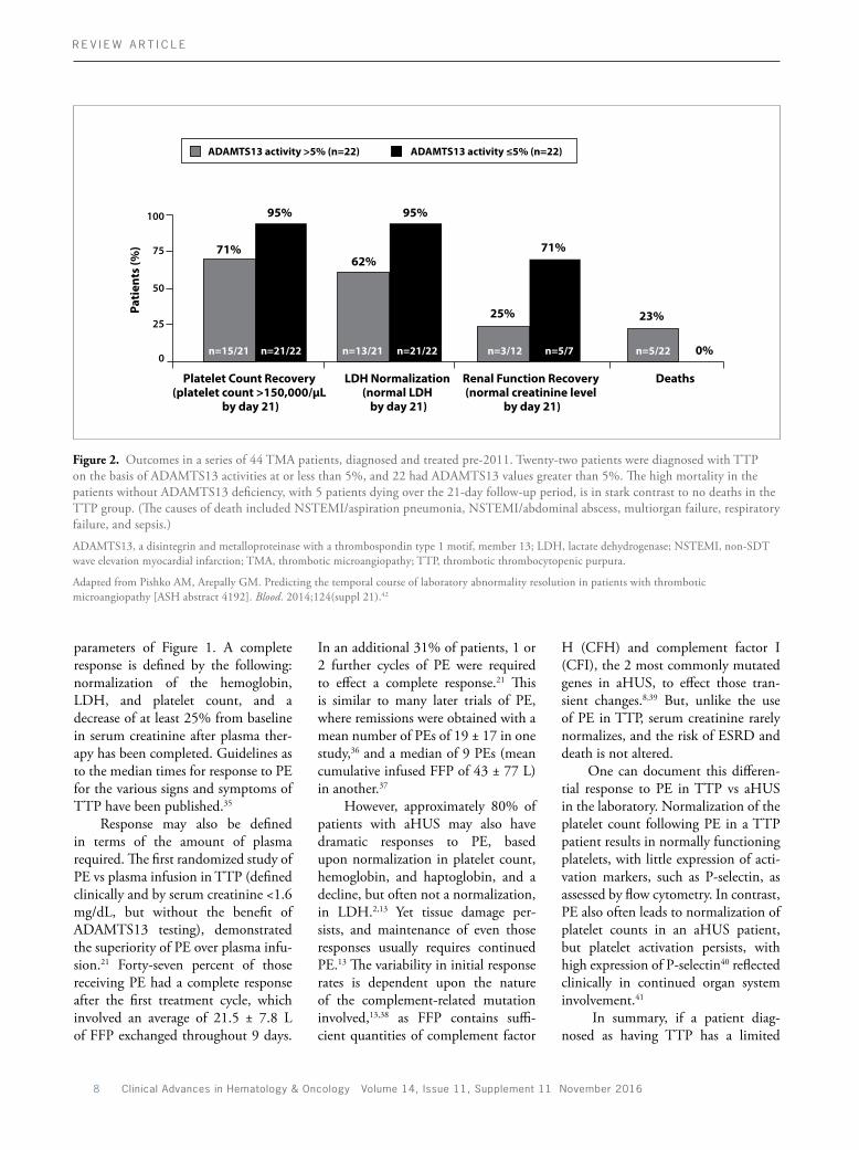
Figure 2. Outcomes in a series of 44 TMA patients, diagnosed and treated pre2011. Twentytwo patients were diagnosed with TTP on the basis of ADAMTS13 activities at or less than 5%, and 22 had ADAMTS13 values greater than 5%. The high mortality in the patients without ADAMTS13 deficiency, with 5 patients dying over the 21day followup period, is in stark contrast to no deaths in the TTP group. (The causes of death included NSTEMI/aspiration pneumonia, NSTEMI/abdominal abscess, multiorgan failure, respiratory failure, and sepsis.)
ADAMTS13, a disintegrin and metalloproteinase with a thrombospondin type 1 motif, member 13; LDH, lactate dehydrogenase; NSTEMI, nonSDT wave elevation myocardial infarction; TMA, thrombotic microangiopathy; TTP, thrombotic thrombocytopenic purpura.
Adapted from Pishko AM, Arepally GM. Predicting the temporal course of laboratory abnormality resolution in patients with thrombotic microangiopathy [ASH abstract 4192]. Blood. 2014;124(suppl 21).42
Clinical Advances in Hematology & Oncology Volume 14, Issue 11, Supplement 11 November 2016 9
A T Y P I C A L H E M O LY T I C U R E M I C S Y N D R O M E ( A H U S ) : A N A C C U R A T E D I A G N O S I S
response to PE, or is requiring quantities of plasma exceeding those outlined above for TTP, it is prudent to reevaluate the diagnosis and consider aHUS. It is critical to recognize that the majority of aHUS patients treated with PE alone may have a complete or nearcomplete hematologic remission yet go on to develop ESRD or die. That is, they may lose an organ, or die, but do so with normal lab numbers. This was illustrated by a retrospective review of TMA patients treated with PE at Duke University from 2007 to mid2013.42 The “ADAMTS13deficiency” group included patients with a TMA occurring in the context of ADAMTS13 less than 5%—those with TTP; whereas the patients in the “without–ADAMTS13deficiency” group had a TMA and ADAMTS13
greater than 5%—what we would now recognize as aHUS. Cases linked to chemotherapeutic agents or bone marrow transplant were excluded. All patients were treated with PE. There were no deaths reported among the 22 patients in the “ADAMTS13deficiency” group, but 5 deaths among the 22 patients in the “without–ADAMTS13deficiency” cohort throughout an observation period of 21 days (Figure 2). In addition, 71% of patients with ADAMTS13 deficiency normalized their serum creatinine by day 21, compared with only 25% of the patients without severe ADAMTS13 deficiency (Figure 2). Yet while 95% of patients with ADAMTS13 deficiency normalized their platelet counts and LDH, so did approximately 70% of patients without
ADAMTS13 deficiency.42 These data were recently corroborated by a review of 186 adult patients entered into the Harvard TMA Research Collaborative registry with a TMA thought to be clinically “suggestive of TTP,” but with ADAMTS13 activities exceeding 10%.43 The authors concluded that outcomes were not improved by the use of PE in this setting.
2. Diagnosing aHUS in the setting of a complement-activating condition. An additional source of misdiagnosis or delay in diagnosis may be failure to consider that a variety of conditions that activate complement, both physiologic and pathologic, can present with signs and symptoms characteristic of a TMA. As outlined in Table 2, these conditions include pregnancy;
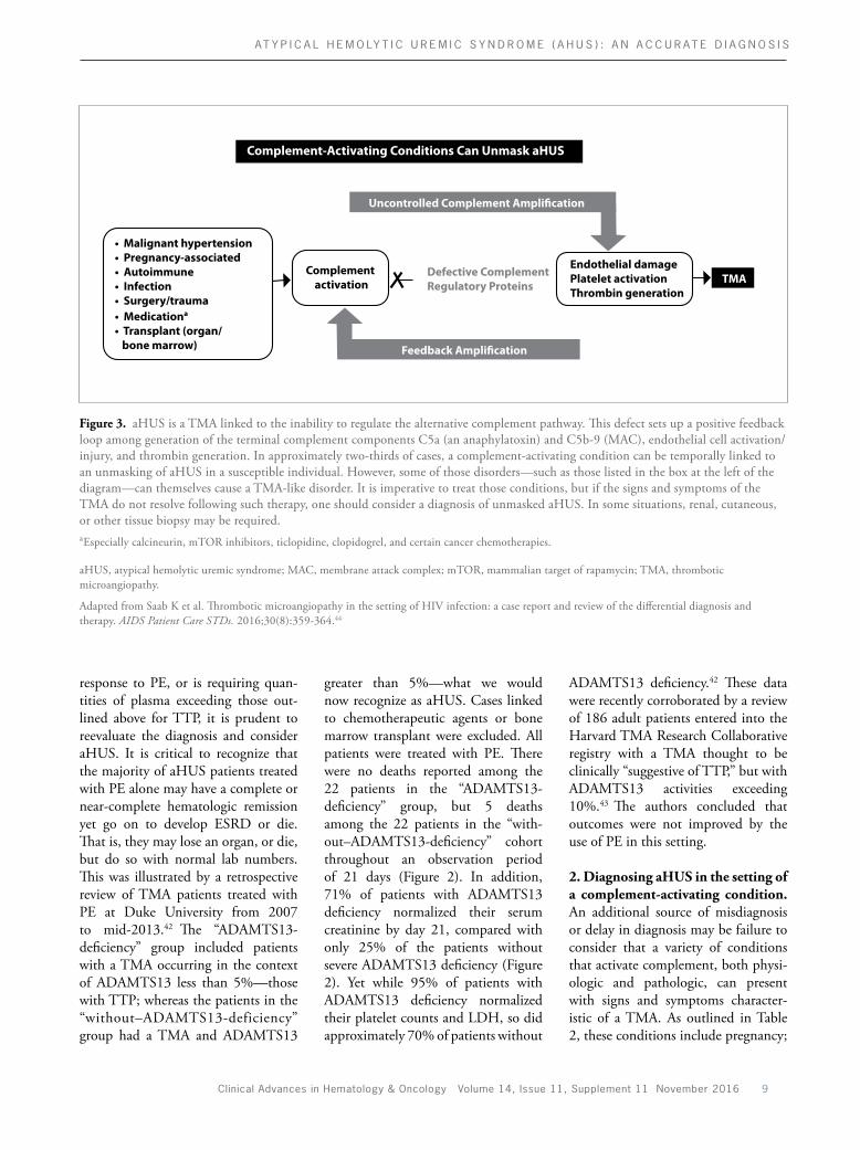
Complement-Activating Conditions Can Unmask aHUS
Uncontrolled Complement Ampli�cation
Endothelial damagePlatelet activationThrombin generation
Complement activation
Gastrointestinal symptoms
• Malignant hypertension• Pregnancy-associated• Autoimmune• Infection• Surgery/trauma• Medicationa
• Transplant (organ/ bone marrow)
Defective Complement Regulatory Proteins
TMA
Feedback Ampli�cation
Figure 3. aHUS is a TMA linked to the inability to regulate the alternative complement pathway. This defect sets up a positive feedback loop among generation of the terminal complement components C5a (an anaphylatoxin) and C5b9 (MAC), endothelial cell activation/injury, and thrombin generation. In approximately twothirds of cases, a complementactivating condition can be temporally linked to an unmasking of aHUS in a susceptible individual. However, some of those disorders—such as those listed in the box at the left of the diagram—can themselves cause a TMAlike disorder. It is imperative to treat those conditions, but if the signs and symptoms of the TMA do not resolve following such therapy, one should consider a diagnosis of unmasked aHUS. In some situations, renal, cutaneous, or other tissue biopsy may be required. aEspecially calcineurin, mTOR inhibitors, ticlopidine, clopidogrel, and certain cancer chemotherapies.
aHUS, atypical hemolytic uremic syndrome; MAC, membrane attack complex; mTOR, mammalian target of rapamycin; TMA, thrombotic microangiopathy.
Adapted from Saab K et al. Thrombotic microangiopathy in the setting of HIV infection: a case report and review of the differential diagnosis and therapy. AIDS Patient Care STDs. 2016;30(8):359364.44
10 Clinical Advances in Hematology & Oncology Volume 14, Issue 11, Supplement 11 November 2016
R E V I E W A R T I C L E
acteristic of malignant hypertension, is a not uncommon feature of aHUS (Manuel Praga, MD, personal observation).
• Bone marrow transplant: TMAs persisting despite discontinuation of graftvshost disease (GVHD) prophylaxis involving an mTOR or calcineurin inhibitor, and resolution of an underlying infection or GVHD, if present, are often aHUS.48
• Drugs: TMAs temporally related to certain chemotherapeutic agents, VEGF inhibitors, antiGVHD medications, and extendedrelease opi ates, which do not resolve following withdrawal of those agents, are usually aHUS, not TTP. TMAs temporally related to the antiplatelet agent ticlopidine, and possibly also to clopidogrel, may unmask aHUS in a genetically susceptible individual even if ADAMTS13 activity and inhibitor levels are classic for a TTPtype TMA.18 Indeed, whenever thrombin is generated in an acute coagulation disorder, there is a positive feedback loop between thrombin generation; C5 cleavage, as thrombin functions as a C5 convertase49; and formation of C5a and C5b9. In an individual genetically susceptible to aHUS, TTP could thereby unmask aHUS, and it is the latter disease process that may be responsible for the clinical se quelae.17,18
IV. Complement Genetics
The complement system is an essential part of innate immunity, with critical roles in response to pathogens and the removal of cell debris and immune complexes. Complement is activated through the classic, lectin, and alternative pathways, resulting in the formation of unstable bimolecular complexes called C3 convertases. C3 can hydrolyze spontaneously into C3(H2O), a molecule that mimics the C3 cleavage product C3b and confers upon the alternative pathway the
• Finally, discontinue or change medications linked to aHUS. In terms of the calcineurin or mammalian target of rapamycin (mTOR) inhibitors, this is typically for at least one halflife (78 days and 34 days, respectively). In addition, treat the underlying complementactivating condition for as long as your experience suggests you should continue that intervention. If your treatments are working and the TMA has resolved, then no other therapy would be required. However, bear in mind the initial differential diagnosis that led to consideration of a TMA unmasked by the complementactivating condition, in case the TMA should recur. Alternatively, if those treatments do not mitigate the TMA, and TTP has been ruled out by ADAMTS13 testing, then conclude that aHUS is involved.
Major examples of a complementactivating condition are:
• Pregnancy: TMAs occurring during pregnancy, which resolve following pregnancy termination, are virtually always TTP (with approximately onequarter previously undiagnosed con genital TTP cases).46 TMAs occurring late in the third trimester, or postpartum, are usually aHUS.46 TMAs of HELLP syndrome that occur late in preg nancy and do not resolve once the preg nancy was terminated are usually aHUS.
• SLE: What appeared to be exacerbation of lupus nephritis in a patient with known SLE was instead aHUSlinked renal failure, with evidence of microthrombi on renal biopsy.
• Malignant hypertension: Renal function should improve in the vast majority of patients with associated acute kidney injury once blood pressure is brought under control.47 Persistence of anemia, thrombocytopenia, and renal injury should raise the suspicion of aHUS unmasked by the malignant hypertension. In addition, grade III to IV hypertensive retinopathy, char
the hemolysis, elevated liver enzymes, and low platelets (HELLP) syndrome; infection; autoimmune diseases, such as SLE and scleroderma; malignant hypertension; organ and tissue transplant; and the use of certain medications. If those signs and symptoms of TMA do not resolve once the complementactivating condition has been treated and medications associated with aHUS have been discontinued or changed to other drugs, recognize that it may have unmasked aHUS or TTP and undertake appropriate diagnostic procedures to investigate that possibility.
Figure 3 illustrates a general scheme by which one can consider the possibility of aHUS arising in the setting of what appears to be an acute presentation of a new complementactivating condition, or exacerbation of an existing one.44
• First, establish the existence of a microangiopathic hemolytic anemia. This is usually based on interpretation of the peripheral blood smear, with documentation of schistocytes and thrombocytopenia, as defined in Figure 1. Renal biopsy is usually not performed, even if there appears to be minimal risk of thrombosis linked to the use of platelet transfusions to perform the biopsy.45 However, a TMA can be diagnosed on renal biopsy in the absence of peripheral blood schistocytes, based on the clinical setting and findings of subendothelial edema in arterioles and glomerular capillary loops, which may or may not be associated with luminal fibrin microthrombi. This subject, and the utility of staining for C5b9, is discussed in detail in section VI.
• Second, eliminate TTP if the ADAMTS13 is greater than 5% to 10%.
• Third, as outlined below, recognize that certain complementactivating conditions are much more likely to be linked to one type of TMA.
Clinical Advances in Hematology & Oncology Volume 14, Issue 11, Supplement 11 November 2016 11
A T Y P I C A L H E M O LY T I C U R E M I C S Y N D R O M E ( A H U S ) : A N A C C U R A T E D I A G N O S I S
Mukherjee, in his monumental work The Gene, stated that a mutation “. . . can provide no real information about a disease or disorder. The definition of disease rests, rather, on the specific disabilities caused by an incongru-ity between an individual’s genetic endowment and his or her current environment. . . .”56
In making a decision to order genetic testing, one must recognize that it may take months. As noted above, 30% of the time a negative result will be obtained, often with the puzzling comment that a genetic variant in a complement gene “of unknown significance” is present.57 But it can be of value in family genetic counseling, as carriers might be closely monitored during conditions triggering marked complement activation, such as surgery, trauma, infection, malignancy, and pregnancy. Concerning longterm prognosis, certain mutations, particularly in MCP, may be associated with milder disease and fewer relapses.22
V. Circulating Complement Levels
Measurements of complement and soluble complement regulatory protein levels in plasma or serum are unreliable markers for aHUS. A low C3 level accompanied by a normal or elevated C4 level would be classic for activation of the alternative complement pathway, which underlies aHUS, but this pattern is only occasionally observed. Serum C3 is normal in up to 80% of aHUS patients, and C3 and C4 levels are too variable for diagnostic purposes.30,58 Complement can also be activated in TTP, leading to elevated levels of C5a and C5b9, as in aHUS.59 Measurement of C5b9 in urine or properly processed plasma—recognizing the need for rapid freezing of a plasma sample, given the short halflife of C5b9—may help identify a TMA, but it cannot distinguish a primary TMA from a related complementactivating condition, nor TTP from aHUS.59
ability to bypass a particular activator. It is always “on,” at a low level. Under physiologic conditions, this lowlevel activation is controlled by complement regulatory proteins, both soluble and present on the surface of most eukaryotic cells. Pathogens do not usually have complement regulators on their surfaces to inhibit this spontaneous activation. Incorporation of C3b molecules to surfacebound C3 convertases then generates C5 convertases, cleaving C5 and leading to formation of C5a (an anaphylatoxin) and C5b9 (MAC), setting up an inflammatory response and destroying the pathogen. Regulation of such alternative complement pathway activation is a complex process involving 2 soluble proteins, CFH and CFI, previously mentioned in the context of FFP infusions, and several membranebound proteins: mem brane cofactor protein (MCP; CD46), thrombomodulin (THBD), complement receptor 1 (CR1; CD35), and decayaccelerating factor (DAF; CD55). The activity of these molecules preserves complement homeostasis and prevents endothelial cell activation and injury, platelet activation and aggregation, and inflammation. Many of these proteins are encoded by genes within a cluster known as RCA (Regulator of Complement Activation) on human chromosome 1q32.50
Over the past 15 years, the analy sis of hundreds of aHUS patients through international collaborative studies has established that approximately 70% carry identifiable genetic abnormalities that alter the regulation of the alternative pathway. These mutations are heterozygous in approximately 90% of cases, and include pathogenic variants in CFH, CD46, CFI, C3, complement factor B (CFB), THBD, CFHR1, CFHR3, diacylglycerol kinaseε (DGKE), and plasminogen.51 Most lead to loss of protein function, with the exception of those in C3 and CFB, identified in 15% of aHUS cases, which are gainoffunction mutations.22,38 There may be an acquired component in 7% to 10%
of aHUS patients, with antiCFH autoantibodies leading to decreased CFH function.22,52 More than 90% of these patients are homozygous for a common polymorphism that deletes CFHR3 and CFHR1 genes. An aHUS patient with such mutations should be evaluated for an antiCFH autoantibody.52
Mutations cannot be identified in cases of aHUS 30% of the time.51 Expansion of genetic analyses among patients with aHUS, particularly those enrolled in international aHUS registries, and pursuit of laboratory studies to functionally characterize genetic variants not yet recognized as pathogenic, will be critical to improving diagnostic criteria for aHUS. For example, genotyping for risk hap lotypes CFHH3 and MCPggaac should be pursued, along with evaluation of copy number variation, hybrid genes, and other complex genomic rearrangements.5355
It is understandable that failure to identify a recognized mutation in a patient with aHUS might generate uncertainties regarding diagnosis and/or treatment duration, but it should not. Not finding a mutation functionally characterized in the literature as pathogenic does not exclude dysregulation of the alternative pathway, nor prove that a genetic component is not involved.
Incomplete penetrance must also be considered. A key paradigm in using genotyping to diagnose a clinical disease (the phenotype) is the realization that gene mutations or genetic variants are not the sole determinants of phenotype. The environment can be critical to the incomplete penetrance of aHUS. For example, this may involve the type and degree of daily pathogen exposure in different cities or countries of residence; physiologic triggers of complement activation, such as pregnancy; and chance, linked to divergent epigenetic modifications or random fluctuations in unrecognized complement or thrombosis regulatory molecules. Dr Siddhartha
12 Clinical Advances in Hematology & Oncology Volume 14, Issue 11, Supplement 11 November 2016
R E V I E W A R T I C L E
antemortem. The gingiva, rectum, or skin are the suggested sites to sample, whether or not there is a visible lesion.
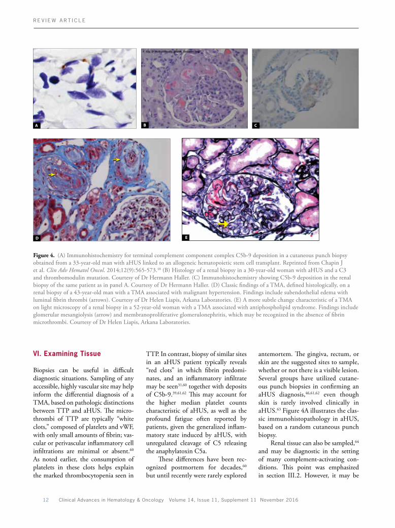
Several groups have utilized cutaneous punch biopsies in confirming an aHUS diagnosis,46,61,62 even though skin is rarely involved clinically in aHUS.63 Figure 4A illustrates the classic immunohistopathology in aHUS, based on a random cutaneous punch biopsy.
Renal tissue can also be sampled,64 and may be diagnostic in the setting of many complementactivating conditions. This point was emphasized in section III.2. However, it may be
TTP. In contrast, biopsy of similar sites in an aHUS patient typically reveals “red clots” in which fibrin predominates, and an inflammatory infiltrate may be seen31,60 together with deposits of C5b9.39,61,62 This may account for the higher median platelet counts characteristic of aHUS, as well as the profound fatigue often reported by patients, given the generalized inflammatory state induced by aHUS, with unregulated cleavage of C5 releasing the anaphylatoxin C5a.
These differences have been recognized postmortem for decades,60 but until recently were rarely explored
VI. Examining Tissue
Biopsies can be useful in difficult diagnostic situations. Sampling of any accessible, highly vascular site may help inform the differential diagnosis of a TMA, based on pathologic distinctions between TTP and aHUS. The microthrombi of TTP are typically “white clots,” composed of platelets and vWF, with only small amounts of fibrin; vascular or perivascular inflammatory cell infiltrations are minimal or absent.60 As noted earlier, the consumption of platelets in these clots helps explain the marked thrombocytopenia seen in
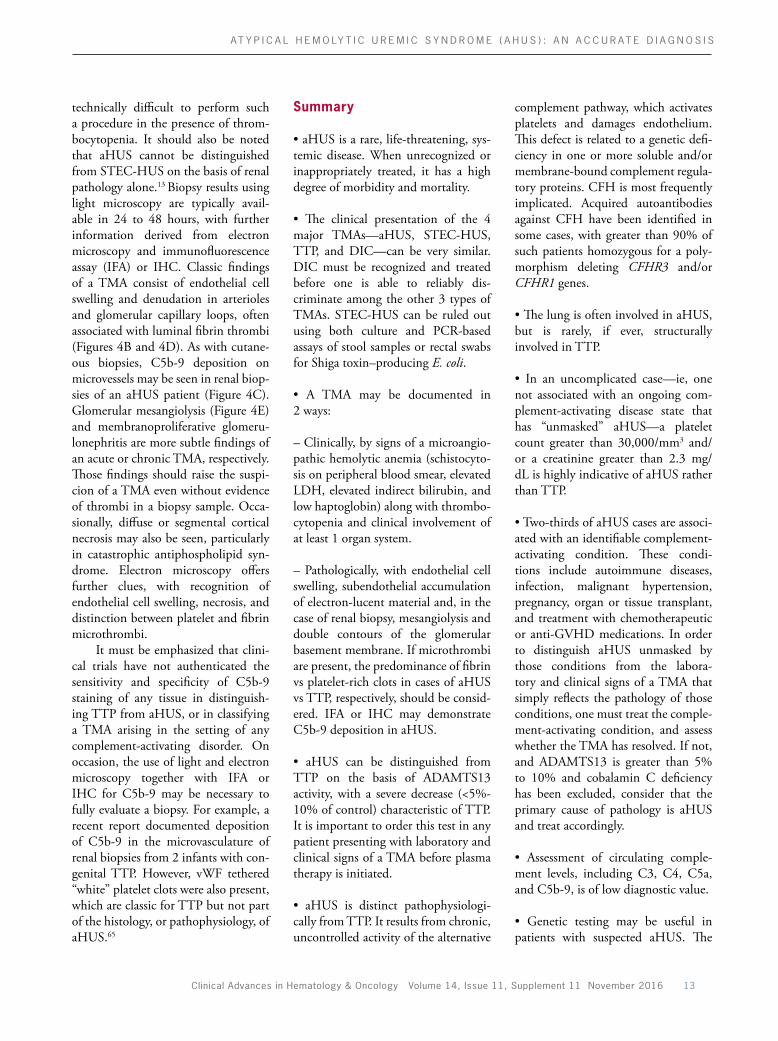
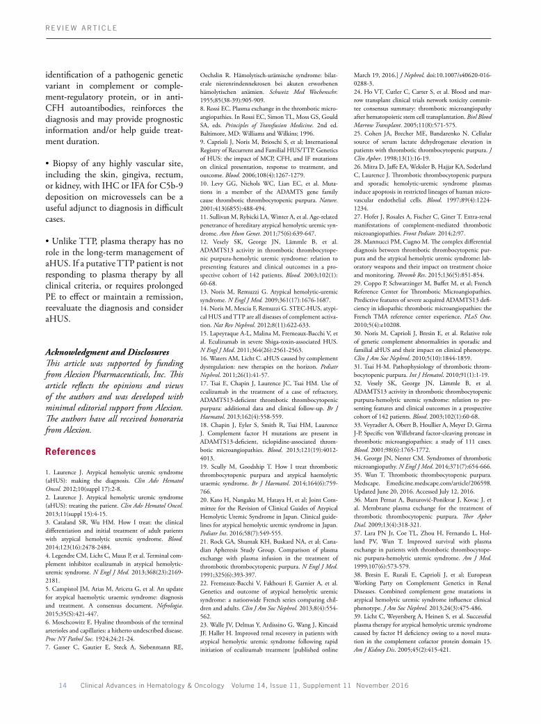
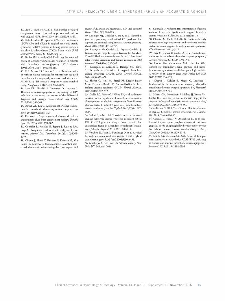
Figure 4. (A) Immunohistochemistry for terminal complement component complex C5b9 deposition in a cutaneous punch biopsy obtained from a 33yearold man with aHUS linked to an allogeneic hematopoietic stem cell transplant. Reprinted from Chapin J et al. Clin Adv Hematol Oncol. 2014;12(9):565573.48 (B) Histology of a renal biopsy in a 30yearold woman with aHUS and a C3 and thrombomodulin mutation. Courtesy of Dr Hermann Haller. (C) Immunohistochemistry showing C5b9 deposition in the renal biopsy of the same patient as in panel A. Courtesy of Dr Hermann Haller. (D) Classic findings of a TMA, defined histologically, on a renal biopsy of a 43yearold man with a TMA associated with malignant hypertension. Findings include subendothelial edema with luminal fibrin thrombi (arrows). Courtesy of Dr Helen Liapis, Arkana Laboratories. (E) A more subtle change characteristic of a TMA on light microscopy of a renal biopsy in a 52yearold woman with a TMA associated with antiphospholipid syndrome. Findings include glomerular mesangiolysis (arrow) and membranoproliferative glomerulonephritis, which may be recognized in the absence of fibrin microthrombi. Courtesy of Dr Helen Liapis, Arkana Laboratories.
A B C
D E
Clinical Advances in Hematology & Oncology Volume 14, Issue 11, Supplement 11 November 2016 13
A T Y P I C A L H E M O LY T I C U R E M I C S Y N D R O M E ( A H U S ) : A N A C C U R A T E D I A G N O S I S
technically difficult to perform such a procedure in the presence of thrombocytopenia. It should also be noted that aHUS cannot be distinguished from STECHUS on the basis of renal pathology alone.13 Biopsy results using light microscopy are typically available in 24 to 48 hours, with further information derived from electron microscopy and immunofluorescence assay (IFA) or IHC. Classic findings of a TMA consist of endothelial cell swelling and denudation in arterioles and glomerular capillary loops, often associated with luminal fibrin thrombi (Figures 4B and 4D). As with cutaneous biopsies, C5b9 deposition on microvessels may be seen in renal biopsies of an aHUS patient (Figure 4C). Glomerular mesangiolysis (Figure 4E) and membranoproliferative glomerulonephritis are more subtle findings of an acute or chronic TMA, respectively. Those findings should raise the suspicion of a TMA even without evidence of thrombi in a biopsy sample. Occasionally, diffuse or segmental cortical necrosis may also be seen, particularly in catastrophic antiphospholipid syndrome. Electron microscopy offers further clues, with recognition of endothelial cell swelling, necrosis, and distinction between platelet and fibrin microthrombi.
It must be emphasized that clinical trials have not authenticated the sensitivity and specificity of C5b9 staining of any tissue in distinguishing TTP from aHUS, or in classifying a TMA arising in the setting of any complementactivating disorder. On occasion, the use of light and electron microscopy together with IFA or IHC for C5b9 may be necessary to fully evaluate a biopsy. For example, a recent report documented deposition of C5b9 in the microvasculature of renal biopsies from 2 infants with congenital TTP. However, vWF tethered “white” platelet clots were also present, which are classic for TTP but not part of the histology, or pathophysiology, of aHUS.65
Summary
• aHUS is a rare, lifethreatening, systemic disease. When unrecognized or inappropriately treated, it has a high degree of morbidity and mortality.
• The clinical presentation of the 4 major TMAs—aHUS, STECHUS, TTP, and DIC—can be very similar. DIC must be recognized and treated before one is able to reliably discriminate among the other 3 types of TMAs. STECHUS can be ruled out using both culture and PCRbased assays of stool samples or rectal swabs for Shiga toxin–producing E. coli.
• A TMA may be documented in 2 ways:
– Clinically, by signs of a microangiopathic hemolytic anemia (schistocytosis on peripheral blood smear, elevated LDH, elevated indirect bilirubin, and low haptoglobin) along with thrombocytopenia and clinical involvement of at least 1 organ system.
– Pathologically, with endothelial cell swelling, subendothelial accumulation of electronlucent material and, in the case of renal biopsy, mesangiolysis and double contours of the glomerular basement membrane. If microthrombi are present, the predominance of fibrin vs plateletrich clots in cases of aHUS vs TTP, respectively, should be considered. IFA or IHC may demonstrate C5b9 deposition in aHUS.
• aHUS can be distinguished from TTP on the basis of ADAMTS13 activity, with a severe decrease (<5%10% of control) characteristic of TTP. It is important to order this test in any patient presenting with laboratory and clinical signs of a TMA before plasma therapy is initiated.
• aHUS is distinct pathophysiologically from TTP. It results from chronic, uncontrolled activity of the alternative
complement pathway, which activates platelets and damages endothelium. This defect is related to a genetic deficiency in one or more soluble and/or membranebound complement regulatory proteins. CFH is most frequently implicated. Acquired autoantibodies against CFH have been identified in some cases, with greater than 90% of such patients homozygous for a polymorphism deleting CFHR3 and/or CFHR1 genes.
• The lung is often involved in aHUS, but is rarely, if ever, structurally involved in TTP.
• In an uncomplicated case—ie, one not associated with an ongoing complementactivating disease state that has “unmasked” aHUS—a platelet count greater than 30,000/mm3 and/or a creatinine greater than 2.3 mg/dL is highly indicative of aHUS rather than TTP.
• Twothirds of aHUS cases are associated with an identifiable complementactivating condition. These conditions include autoimmune diseases, infection, malignant hypertension, pregnancy, organ or tissue transplant, and treatment with chemotherapeutic or antiGVHD medications. In order to distinguish aHUS unmasked by those conditions from the laboratory and clinical signs of a TMA that simply reflects the pathology of those conditions, one must treat the complementactivating condition, and assess whether the TMA has resolved. If not, and ADAMTS13 is greater than 5% to 10% and cobalamin C deficiency has been excluded, consider that the primary cause of pathology is aHUS and treat accordingly.
• Assessment of circulating complement levels, including C3, C4, C5a, and C5b9, is of low diagnostic value.
• Genetic testing may be useful in patients with suspected aHUS. The
14 Clinical Advances in Hematology & Oncology Volume 14, Issue 11, Supplement 11 November 2016
R E V I E W A R T I C L E
March 19, 2016.] J Nephrol. doi:10.1007/s4062001602883.24. Ho VT, Cutler C, Carter S, et al. Blood and marrow transplant clinical trials network toxicity committee consensus summary: thrombotic microangiopathy after hematopoietic stem cell transplantation. Biol Blood Marrow Transplant. 2005;11(8):571575.25. Cohen JA, Brecher ME, Bandarenko N. Cellular source of serum lactate dehydrogenase elevation in patients with thrombotic thrombocytopenic purpura. J Clin Apher. 1998;13(1):1619.26. Mitra D, Jaffe EA, Weksler B, Hajjar KA, Soderland C, Laurence J. Thrombotic thrombocytopenic purpura and sporadic hemolyticuremic syndrome plasmas induce apoptosis in restricted lineages of human microvascular endothelial cells. Blood. 1997;89(4):12241234.27. Hofer J, Rosales A, Fischer C, Giner T. Extrarenal manifestations of complementmediated thrombotic microangiopathies. Front Pediatr. 2014;2:97.28. Mannucci PM, Cugno M. The complex differential diagnosis between thrombotic thrombocytopenic purpura and the atypical hemolytic uremic syndrome: laboratory weapons and their impact on treatment choice and monitoring. Thromb Res. 2015;136(5):851854.29. Coppo P, Schwarzinger M, Buffet M, et al; French Reference Center for Thrombotic Microangiopathies. Predictive features of severe acquired ADAMTS13 deficiency in idiopathic thrombotic microangiopathies: the French TMA reference center experience. PLoS One. 2010;5(4):e10208.30. Noris M, Caprioli J, Bresin E, et al. Relative role of genetic complement abnormalities in sporadic and familial aHUS and their impact on clinical phenotype. Clin J Am Soc Nephrol. 2010;5(10):18441859.31. Tsai HM. Pathophysiology of thrombotic thrombocytopenic purpura. Int J Hematol. 2010;91(1):119.32. Vesely SK, George JN, Lämmle B, et al. ADAMTS13 activity in thrombotic thrombocytopenic purpurahemolytic uremic syndrome: relation to presenting features and clinical outcomes in a prospective cohort of 142 patients. Blood. 2003;102(1):6068.33. Veyradier A, Obert B, Houllier A, Meyer D, Girma JP. Specific von Willebrand factorcleaving protease in thrombotic microangiopathies: a study of 111 cases. Blood. 2001;98(6):17651772.34. George JN, Nester CM. Syndromes of thrombotic microangiopathy. N Engl J Med. 2014;371(7):654666.35. Wun T. Thrombotic thrombocytopenic purpura. Medscape. Emedicine.medscape.com/article/206598. Updated June 20, 2016. Accessed July 12, 2016.36. Marn Pernat A, ButurovićPonikvar J, Kovac J, et al. Membrane plasma exchange for the treatment of thrombotic thrombocytopenic purpura. Ther Apher Dial. 2009;13(4):318321.37. Lara PN Jr, Coe TL, Zhou H, Fernando L, Holland PV, Wun T. Improved survival with plasma exchange in patients with thrombotic thrombocytopenic purpurahemolytic uremic syndrome. Am J Med. 1999;107(6):573579.38. Bresin E, Rurali E, Caprioli J, et al; European Working Party on Complement Genetics in Renal Diseases. Combined complement gene mutations in atypical hemolytic uremic syndrome influence clinical phenotype. J Am Soc Nephrol. 2013;24(3):475486.39. Licht C, Weyersberg A, Heinen S, et al. Successful plasma therapy for atypical hemolytic uremic syndrome caused by factor H deficiency owing to a novel mutation in the complement cofactor protein domain 15. Am J Kidney Dis. 2005;45(2):415421.
Oechslin R. Hämolytischurämische syndrome: bilaterale nierenrindennekrosen bei akuten erworbenen hämolytischen anämien. Schweiz Med Wochenschr. 1955;85(3839):905909.8. Rossi EC. Plasma exchange in the thrombotic microangiopathies. In Rossi EC, Simon TL, Moss GS, Gould SA, eds. Principles of Transfusion Medicine. 2nd ed. Baltimore, MD: Williams and Wilkins; 1996.9. Caprioli J, Noris M, Brioschi S, et al; International Registry of Recurrent and Familial HUS/TTP. Genetics of HUS: the impact of MCP, CFH, and IF mutations on clinical presentation, response to treatment, and outcome. Blood. 2006;108(4):12671279.10. Levy GG, Nichols WC, Lian EC, et al. Mutations in a member of the ADAMTS gene family cause thrombotic thrombocytopenic purpura. Nature. 2001;413(6855):488494.11. Sullivan M, Rybicki LA, Winter A, et al. Agerelated penetrance of hereditary atypical hemolytic uremic syndrome. Ann Hum Genet. 2011;75(6):639647.12. Vesely SK, George JN, Lämmle B, et al. ADAMTS13 activity in thrombotic thrombocytopenic purpurahemolytic uremic syndrome: relation to presenting features and clinical outcomes in a prospective cohort of 142 patients. Blood. 2003;102(1): 6068.13. Noris M, Remuzzi G. Atypical hemolyticuremic syndrome. N Engl J Med. 2009;361(17):16761687.14. Noris M, Mescia F, Remuzzi G. STECHUS, atypical HUS and TTP are all diseases of complement activation. Nat Rev Nephrol. 2012;8(11):622633.15. Lapeyraque AL, Malina M, FremeauxBacchi V, et al. Eculizumab in severe Shigatoxinassociated HUS. N Engl J Med. 2011;364(26):25612563.16. Waters AM, Licht C. aHUS caused by complement dysregulation: new therapies on the horizon. Pediatr Nephrol. 2011;26(1):4157.17. Tsai E, Chapin J, Laurence JC, Tsai HM. Use of eculizumab in the treatment of a case of refractory, ADAMTS13deficient thrombotic thrombocytopenic purpura: additional data and clinical followup. Br J Haematol. 2013;162(4):558559.18. Chapin J, Eyler S, Smith R, Tsai HM, Laurence J. Complement factor H mutations are present in ADAMTS13deficient, ticlopidineassociated thrombotic microangiopathies. Blood. 2013;121(19):40124013.19. Scully M, Goodship T. How I treat thrombotic thrombocytopenic purpura and atypical haemolytic uraemic syndrome. Br J Haematol. 2014;164(6):759766.20. Kato H, Nangaku M, Hataya H, et al; Joint Committee for the Revision of Clinical Guides of Atypical Hemolytic Uremic Syndrome in Japan. Clinical guidelines for atypical hemolytic uremic syndrome in Japan. Pediatr Int. 2016;58(7):549555.21. Rock GA, Shumak KH, Buskard NA, et al; Canadian Apheresis Study Group. Comparison of plasma exchange with plasma infusion in the treatment of thrombotic thrombocytopenic purpura. N Engl J Med. 1991;325(6):393397.22. FremeauxBacchi V, Fakhouri F, Garnier A, et al. Genetics and outcome of atypical hemolytic uremic syndrome: a nationwide French series comparing children and adults. Clin J Am Soc Nephrol. 2013;8(4):554562.23. Walle JV, Delmas Y, Ardissino G, Wang J, Kincaid JF, Haller H. Improved renal recovery in patients with atypical hemolytic uremic syndrome following rapid initiation of eculizumab treatment [published online
identification of a pathogenic genetic variant in complement or complementregulatory protein, or in antiCFH autoantibodies, reinforces the diagnosis and may provide prognostic information and/or help guide treatment duration.
• Biopsy of any highly vascular site, including the skin, gingiva, rectum, or kidney, with IHC or IFA for C5b9 deposition on microvessels can be a useful adjunct to diagnosis in difficult cases.
• Unlike TTP, plasma therapy has no role in the longterm management of aHUS. If a putative TTP patient is not responding to plasma therapy by all clinical criteria, or requires pro longed PE to effect or maintain a remission, reevaluate the diagnosis and consider aHUS.
Acknowledgment and DisclosuresThis article was supported by funding from Alexion Pharmaceuticals, Inc. This article reflects the opinions and views of the authors and was developed with minimal editorial support from Alexion. The authors have all received honoraria from Alexion.
References
1. Laurence J. Atypical hemolytic uremic syndrome (aHUS): making the diagnosis. Clin Adv Hematol Oncol. 2012;10(suppl 17):28.2. Laurence J. Atypical hemolytic uremic syndrome (aHUS): treating the patient. Clin Adv Hematol Oncol. 2013;11(suppl 15):415.3. Cataland SR, Wu HM. How I treat: the clinical differentiation and initial treatment of adult patients with atypical hemolytic uremic syndrome. Blood. 2014;123(16):24782484.4. Legendre CM, Licht C, Muus P, et al. Terminal complement inhibitor eculizumab in atypical hemolyticuremic syndrome. N Engl J Med. 2013;368(23):21692181. 5. Campistol JM, Arias M, Ariceta G, et al. An update for atypical haemolytic uraemic syndrome: diagnosis and treatment. A consensus document. Nefrologia. 2015;35(S):421447.6. Moschcowitz E. Hyaline thrombosis of the terminal arterioles and capillaries: a hitherto undescribed disease. Proc NY Pathol Soc. 1924;24:2124.7. Gasser C, Gautier E, Steck A, Siebenmann RE,
Clinical Advances in Hematology & Oncology Volume 14, Issue 11, Supplement 11 November 2016 15
A T Y P I C A L H E M O LY T I C U R E M I C S Y N D R O M E ( A H U S ) : A N A C C U R A T E D I A G N O S I S
57. Kavanagh D, Anderson HE. Interpretation of genetic variants of uncertain significance in atypical hemolytic uremic syndrome. Kidney Int. 2012;81(1):1113.58. Ohanian M, Cable C, Halka K. Eculizumab safely reverses neurologic impairment and eliminates need for dialysis in severe atypical hemolytic uremic syndrome. Clin Pharmacol. 2011;3:512.59. Réti M, Farkas P, Csuka D, et al. Complement activation in thrombotic thrombocytopenic purpura. J Thromb Haemost. 2012;10(5):791798.60. Hosler GA, Cusumano AM, Hutchins GM. Thrombotic thrombocytopenic purpura and hemolytic uremic syndrome are distinct pathologic entities. A review of 56 autopsy cases. Arch Pathol Lab Med. 2003;127(7):834839.61. Chapin J, Weksler B, Magro C, Laurence J. Eculizumab in the treatment of refractory idiopathic thrombotic thrombocytopenic purpura. Br J Haematol. 2012;157(6):772774.62. Magro CM, Momtahen S, Mulvey JJ, Yassin AH, Kaplan RB, Laurence JC. Role of the skin biopsy in the diagnosis of atypical hemolytic uremic syndrome. Am J Dermatopathol. 2015;37(5):349356.63. Ardissino G, Tel F, Testa S, et al. Skin involvement in atypical hemolytic uremic syndrome. Am J Kidney Dis. 2014;63(4):652655.64. Canaud G, Kamar N, Anglicheau D, et al. Eculizumab improves posttransplant thrombotic microangiopathy due to antiphospholipid syndrome recurrence but fails to prevent chronic vascular changes. Am J Transplant. 2013;13(8):21792185.65. Tati R, Kristoffersson AC, Ståhl AL, et al. Complement activation associated with ADAMTS13 deficiency in human and murine thrombotic microangiopathy. J Immunol. 2013;191(5):21842193.
review of diagnosis and treatments. Clin Adv Hematol Oncol. 2014;12(9):565573.49. Krisinger MJ, Goebeler V, Lu Z, et al. Thrombin generates previously unidentified C5 products that support the terminal complement activation pathway. Blood. 2012;120(8):17171725.50. Rodríguez de Córdoba S, EsparzaGordillo J, Goicoechea de Jorge E, LopezTrascasa M, SánchezCorral P. The human complement factor H: functional roles, genetic variations and disease associations. Mol Immunol. 2004;41(4):355367.51. Rodríguez de Córdoba S, Hidalgo MS, Pinto S, Tortajada A. Genetics of atypical hemolytic uremic syndrome (aHUS). Semin Thromb Hemost. 2014;40(4):422430.52. Skerka C, Józsi M, Zipfel PF, DragonDurey MA, FremeauxBacchi V. Autoantibodies in haemolytic uraemic syndrome (HUS). Thromb Haemost. 2009;101(2):227232.53. Challis RC, Araujo GS, Wong EK, et al. A de novo deletion in the regulators of complement activation cluster producing a hybrid complement factor H/complement factor Hrelated 3 gene in atypical hemolytic uremic syndrome. J Am Soc Nephrol. 2016;27(6):16171624.54. Valoti E, Alberti M, Tortajada A, et al. A novel atypical hemolytic uremic syndromeassociated hybrid CFHR1/CFH gene encoding a fusion protein that antagonizes factor Hdependent complement regulation. J Am Soc Nephrol. 2015;26(1):209219.55. Venables JP, Strain L, Routledge D, et al. Atypical haemolytic uraemic syndrome associated with a hybrid complement gene. PLoS Med. 2006;3(10):e431.56. Mukherjee S. The Gene: An Intimate History. New York, NY: Scribner; 2016.
40. Licht C, Pluthero FG, Li L, et al. Plateletassociated complement factor H in healthy persons and patients with atypical HUS. Blood. 2009;114(20):45384545.41. Licht C, Muus P, Legendre CM, et al. Eculizumab (ECU) safety and efficacy in atypical hemolytic uremic syndrome (aHUS) patients with long disease duration and chronic kidney disease (CKD): 2year results [ASH abstract 985]. Blood. 2012;120(suppl 21).42. Pishko AM, Arepally GM. Predicting the temporal course of laboratory abnormality resolution in patients with thrombotic microangiopathy [ASH abstract 4192]. Blood. 2014;124(suppl 21).43. Li A, Makar RS, Hurwitz S, et al. Treatment with or without plasma exchange for patients with acquired thrombotic microangiopathy not associated with severe ADAMTS13 deficiency: a propensity scorematched study. Transfusion. 2016;56(8):20692077.44. Saab KR, Elhadad S, Copertino D, Laurence J. Thrombotic microangiopathy in the setting of HIV infection: a case report and review of the differential diagnosis and therapy. AIDS Patient Care STDS. 2016;30(8):359364. 45. Otrock ZK, Liu C, Grossman BJ. Platelet transfusion in thrombotic thrombocytopenic purpura. Vox Sang. 2015;109(2):168172.46. Fakhouri F. Pregnancyrelated thrombotic microangiopathies: clues from complement biology. Transfus Apher Sci. 2016;54(2):199202.47. González R, Morales E, Segura J, Ruilope LM, Praga M. Longterm renal survival in malignant hypertension. Nephrol Dial Transplant. 2010;25(10):32663272.48. Chapin J, Shore T, Forsberg P, Desman G, Van Besien K, Laurence J. Hematopoietic transplantassociated thrombotic microangiopathy: case report and


![Official Title: A MULTICENTER, OPEN-LABEL, PHASE III STUDY ... · atypical hemolytic uremic syndrome [aHUS]) was observed in . 3. 2 patients receiving ... bleed information, including](https://img.pdfslide.us/doc/110x75/5f343f8217d7f5103034834b/official-title-a-multicenter-open-label-phase-iii-study-atypical-hemolytic.jpg)