Embed Size (px)
Citation preview
Atypical Femoral Fractures: A multimodality review of radiographic features and
complications
Catherine Lang, Robert Bleakney, Linda Probyn, Leon Lenchik, Angela M.
Cheung
Disclosure
• I have had no affiliation with a pharmaceutical, medical device or communications organization within the previous 24 months (financial or otherwise).
Learning Objectives 1. Review imaging features of the case definition
of atypical femoral fractures (AFFs) – Outlined by the 2013 ASBMR Task Force
2. Illustrate radiographic features of AFFs through various modalities – Plain films, CT, MRI, bone scan, ultrasound, single
energy scan of the femur, and DXA
3. Illustrate complications of AFFs – Fracture progression, delayed healing, bilateral
fractures, and hardware failure

Background
• Treatment of osteoporosis with long-term bisphosphonate therapy is increasingly associated with AFFs
• AFFs often have nonspecific clinical symptoms – Radiologists must recognize their features across all
imaging modalities – Plain films may not be in the initial workup
Background
• AFFs are often linked to complications 1. Progression from incomplete to complete fracture 2. Fracture of the contralateral femur 3. Delayed fracture healing
• AFFs may be treated with hardware either prophylactically or for complete fracture fixation
Case Definition
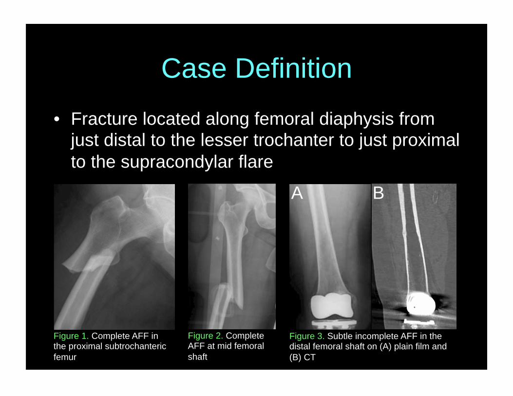
• Fracture located along femoral diaphysis from just distal to the lesser trochanter to just proximal to the supracondylar flare
Figure 1. Complete AFF in the proximal subtrochanteric femur
Figure 2. Complete AFF at mid femoral shaft
Figure 3. Subtle incomplete AFF in the distal femoral shaft on (A) plain film and (B) CT
A B
Major Features
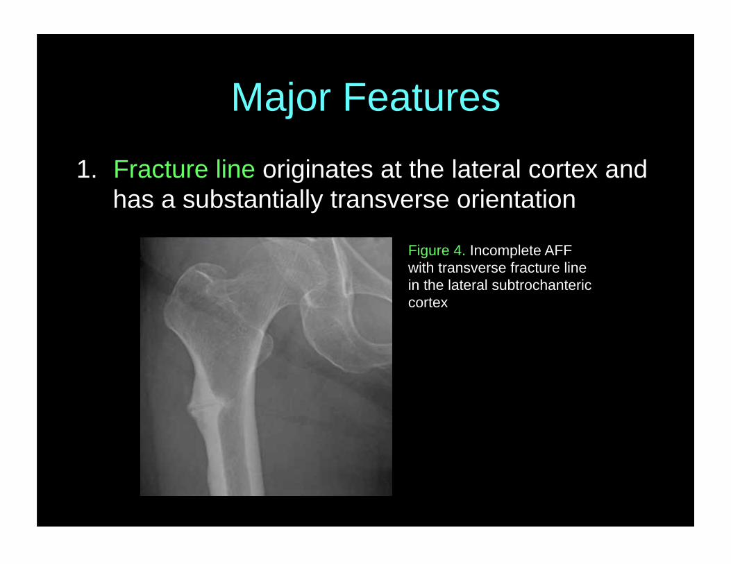
1. Fracture line originates at the lateral cortex and has a substantially transverse orientation
Figure 4. Incomplete AFF with transverse fracture line in the lateral subtrochanteric cortex
Major Features
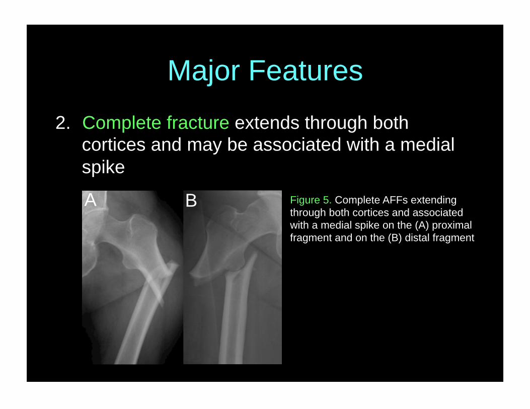
2. Complete fracture extends through both cortices and may be associated with a medial spike
Figure 5. Complete AFFs extending through both cortices and associated with a medial spike on the (A) proximal fragment and on the (B) distal fragment
A B
Major Features
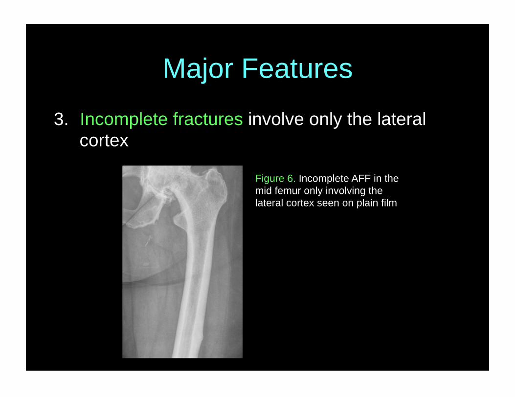
3. Incomplete fractures involve only the lateral cortex
Figure 6. Incomplete AFF in the mid femur only involving the lateral cortex seen on plain film
Major Features
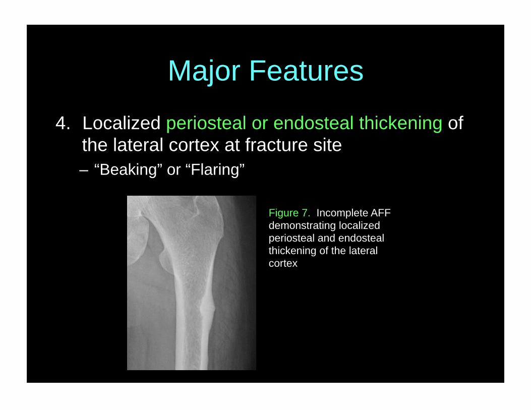
4. Localized periosteal or endosteal thickening of the lateral cortex at fracture site – “Beaking” or “Flaring”
Figure 7. Incomplete AFF demonstrating localized periosteal and endosteal thickening of the lateral cortex
Major Features
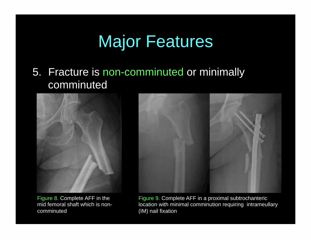
5. Fracture is non-comminuted or minimally comminuted
Figure 9. Complete AFF in a proximal subtrochanteric location with minimal comminution requiring intrameullary (IM) nail fixation
Figure 8. Complete AFF in the mid femoral shaft which is non-comminuted
Minor Features
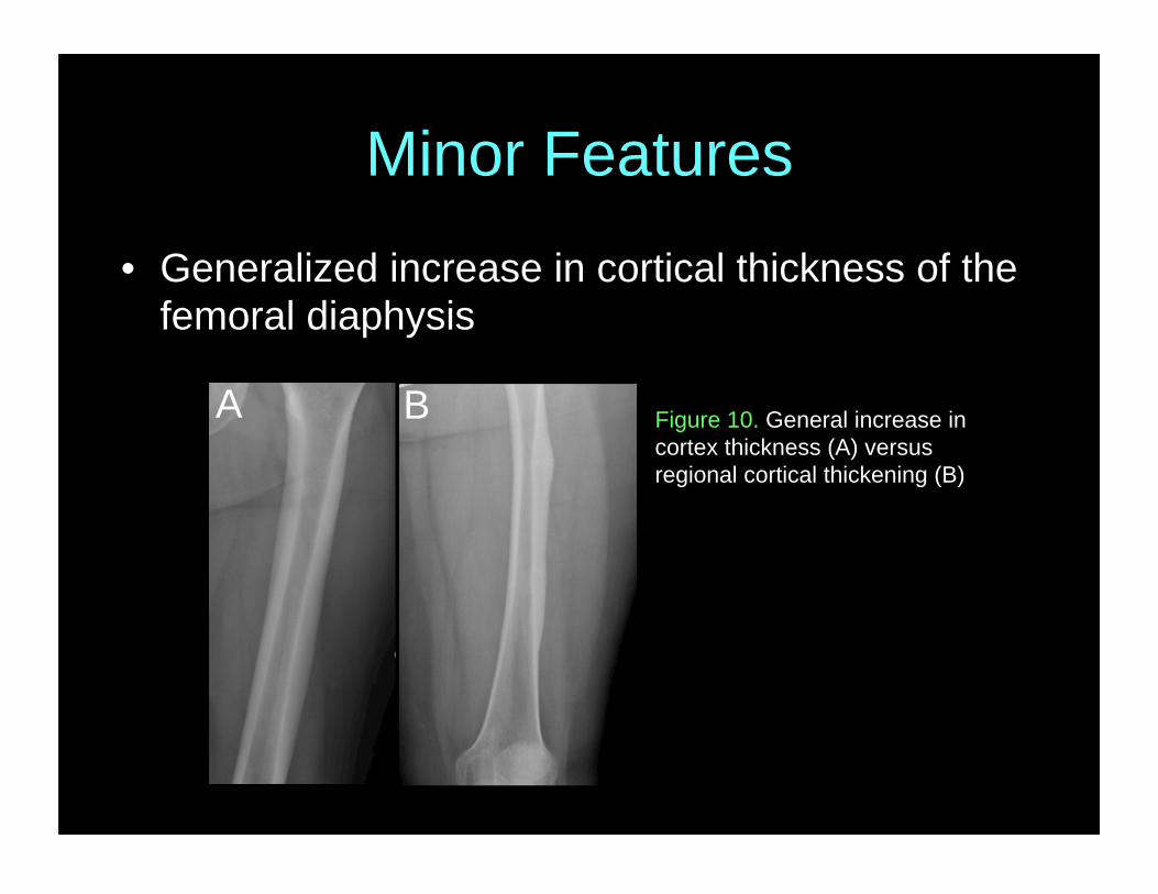
• Generalized increase in cortical thickness of the femoral diaphysis
Figure 10. General increase in cortex thickness (A) versus regional cortical thickening (B)
A B
Minor Features
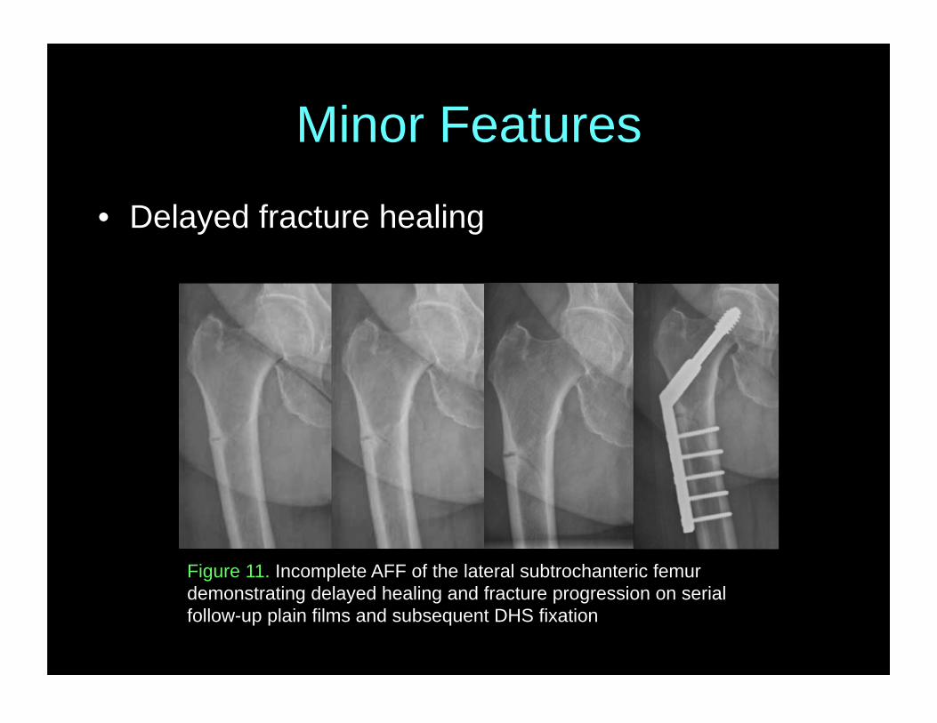
• Delayed fracture healing
Figure 11. Incomplete AFF of the lateral subtrochanteric femur demonstrating delayed healing and fracture progression on serial follow-up plain films and subsequent DHS fixation
Minor Features
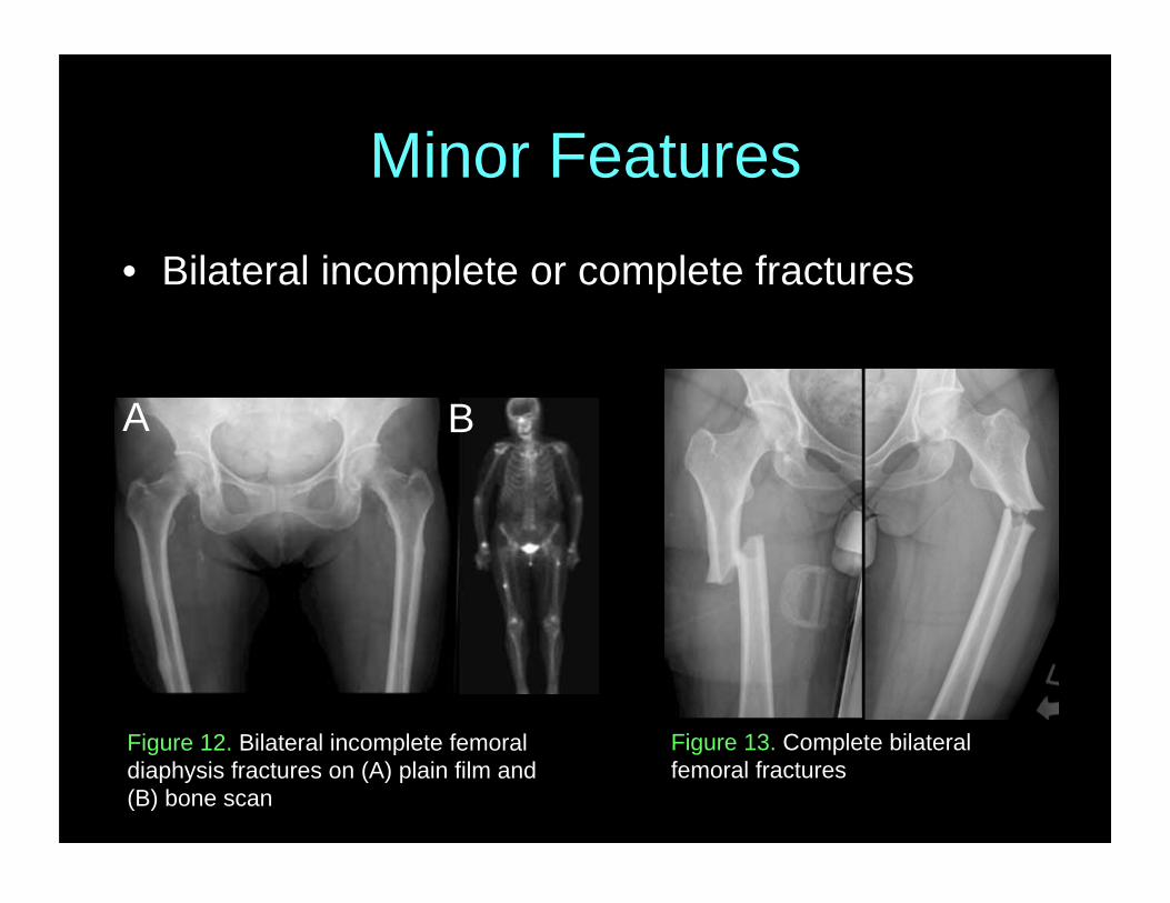
• Bilateral incomplete or complete fractures
Figure 13. Complete bilateral femoral fractures
Figure 12. Bilateral incomplete femoral diaphysis fractures on (A) plain film and (B) bone scan
A B
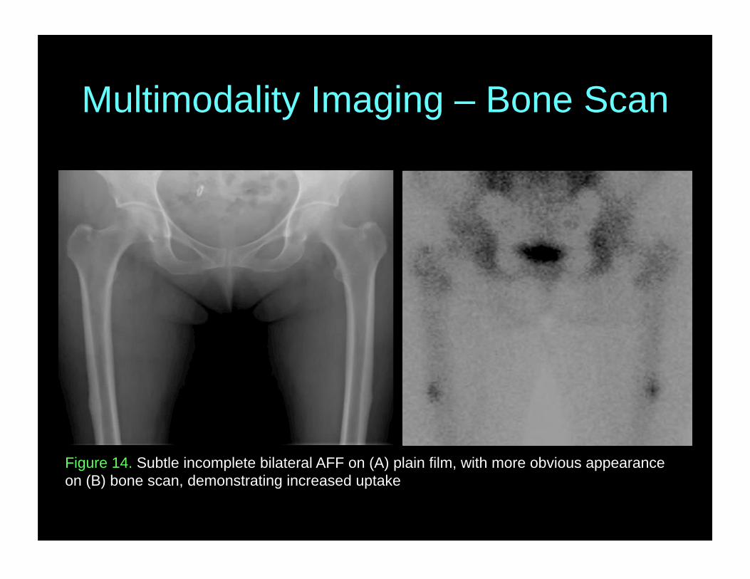
Multimodality Imaging – Bone Scan
Figure 14. Subtle incomplete bilateral AFF on (A) plain film, with more obvious appearance on (B) bone scan, demonstrating increased uptake
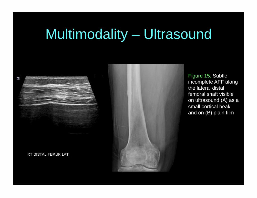
Multimodality – Ultrasound
Figure 15. Subtle incomplete AFF along the lateral distal femoral shaft visible on ultrasound (A) as a small cortical beak and on (B) plain film
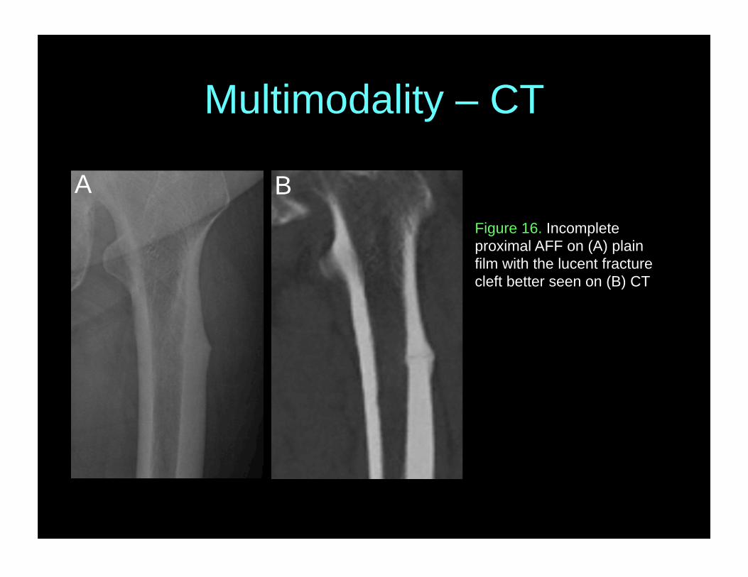
Multimodality – CT
A BFigure 16. Incomplete proximal AFF on (A) plain film with the lucent fracture cleft better seen on (B) CT
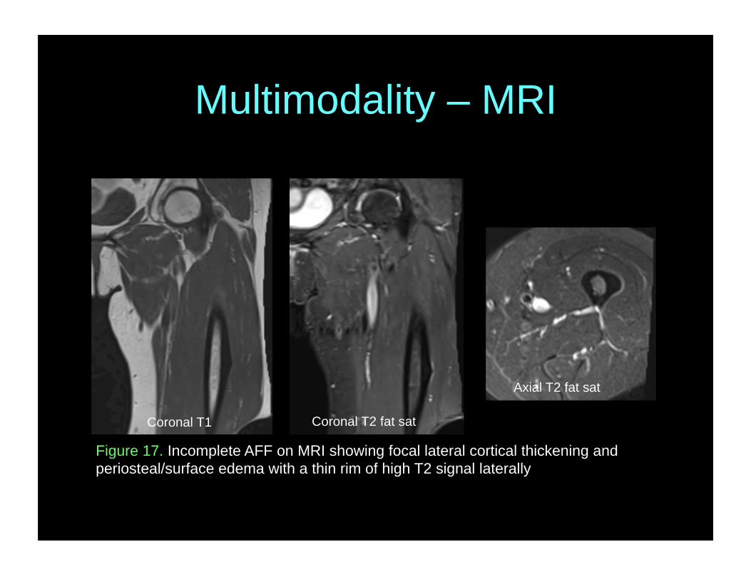
Multimodality – MRI
Figure 17. Incomplete AFF on MRI showing focal lateral cortical thickening and periosteal/surface edema with a thin rim of high T2 signal laterally
Coronal T1 Coronal T2 fat sat
Axial T2 fat sat
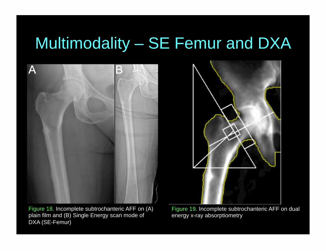
Multimodality – SE Femur and DXA
Figure 18. Incomplete subtrochanteric AFF on (A) plain film and (B) Single Energy scan mode of DXA (SE-Femur)
A B
Figure 19. Incomplete subtrochanteric AFF on dual energy x-ray absorptiometry
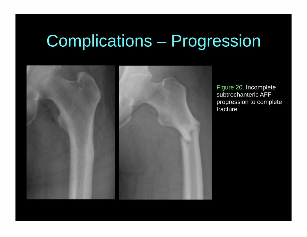
Complications – Progression
Figure 20. Incomplete subtrochanteric AFF progression to complete fracture
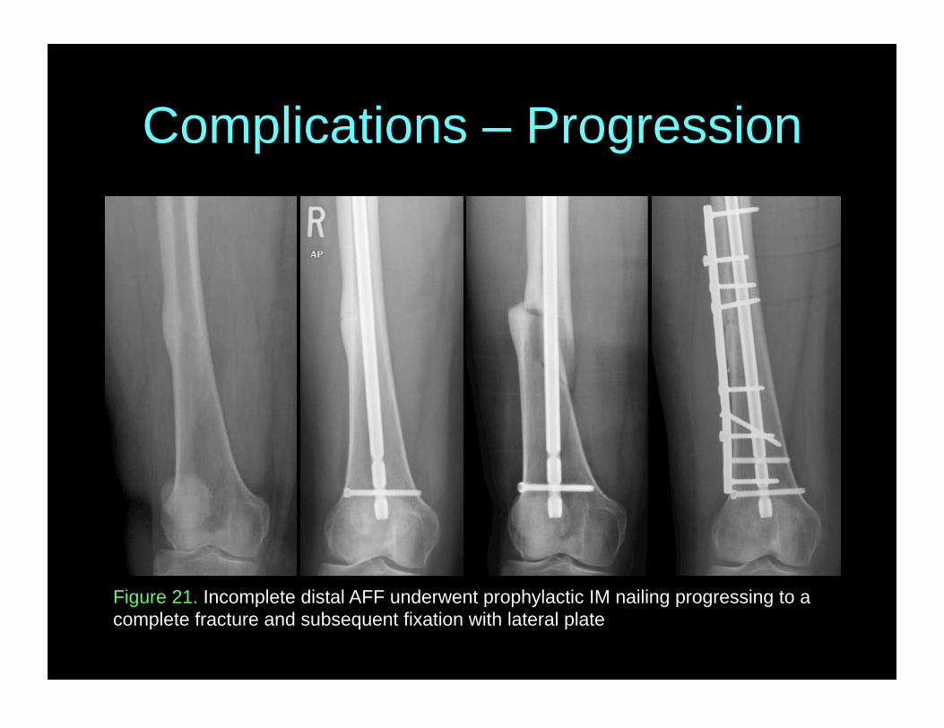
Complications – Progression
Figure 21. Incomplete distal AFF underwent prophylactic IM nailing progressing to a complete fracture and subsequent fixation with lateral plate
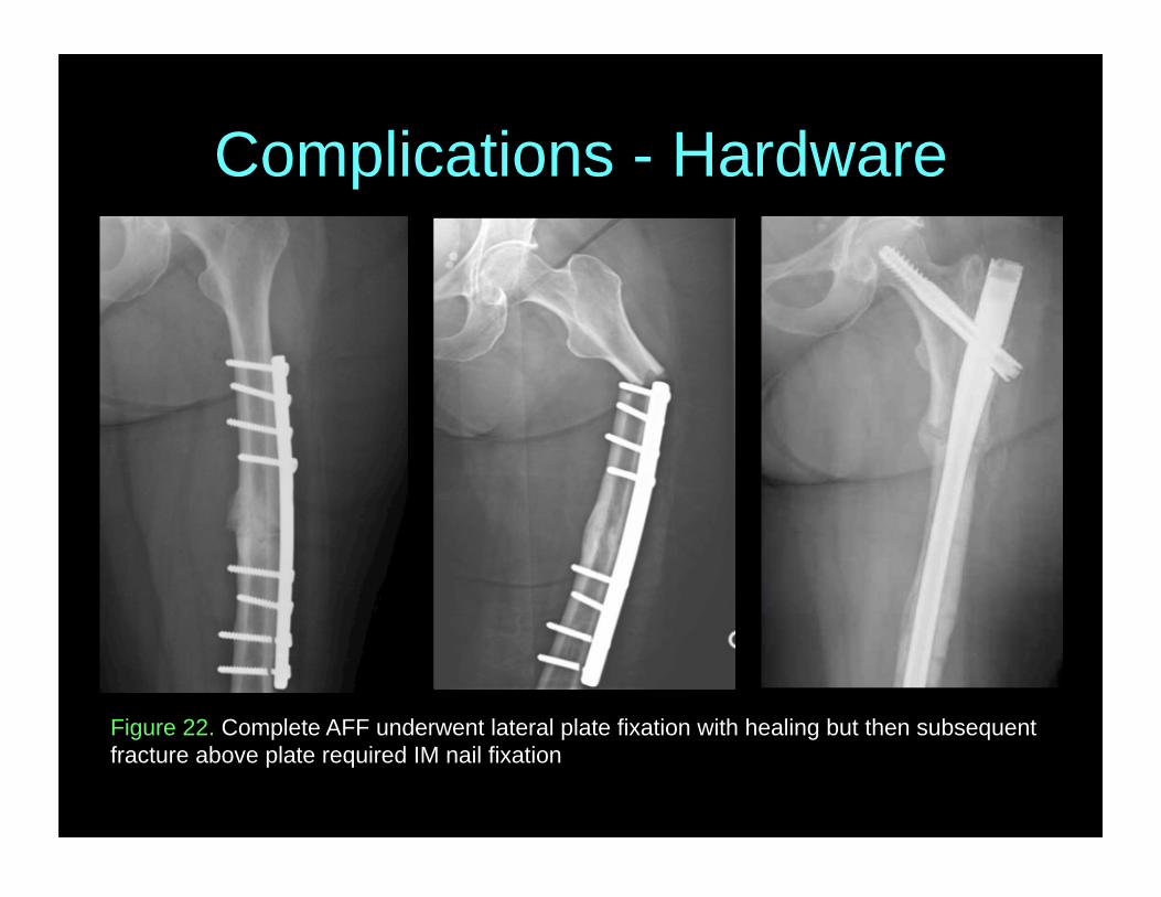
Complications - Hardware
Figure 22. Complete AFF underwent lateral plate fixation with healing but then subsequent fracture above plate required IM nail fixation
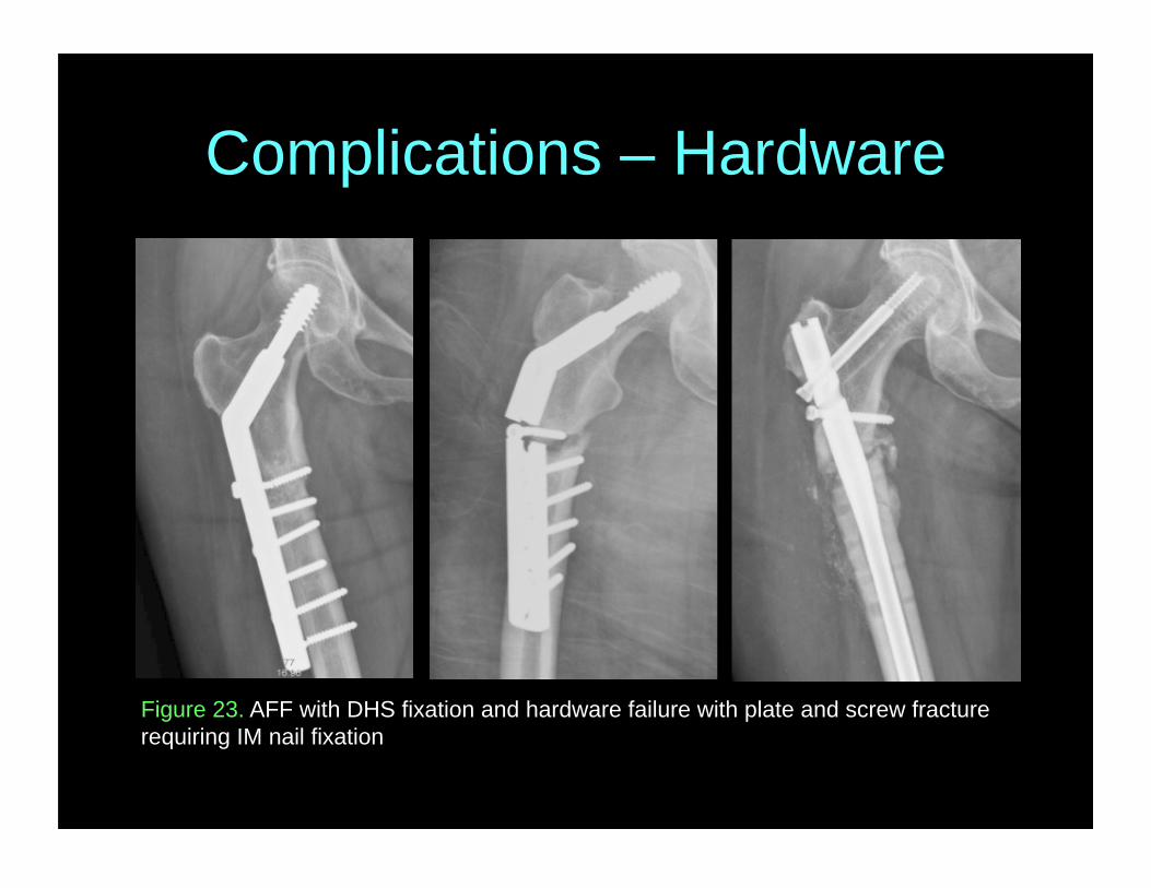
Complications – Hardware
Figure 23. AFF with DHS fixation and hardware failure with plate and screw fracture requiring IM nail fixation
Conclusion
• AFFs have characteristic radiographic features – May present in varying combinations – Can be seen on all imaging modalities
• Radiologists should be familiar with these varying and potentially subtle findings to better diagnose AFFs and their complications
References 1. Shane E, Burr D, Abrahamsen B, et al. Atypical
subtrochanteric and diaphyseal femoral fractures: Second report of a task force of the American Society for Bone and Mineral Research. J Bone Miner Res 2014; 29 (1):1-23.
2. Shane E, Burr D, Ebeling PR, Abrahamsen B, Adler RA, Brown TD, et al. Atypical subtrochanteric and diaphyseal femoral fractures: report of the task force of the American Society for Bone and Mineral Research. J Bone Miner Res 2010;25(11):2267-94.