Embed Size (px)
Citation preview
JOURNAL OF VIROLOGY,0022-538X/98/$04.0010
Feb. 1998, p. 965–974 Vol. 72, No. 2
Copyright © 1998, American Society for Microbiology
Attenuation of the Vaccine Oka Strain of Varicella-ZosterVirus and Role of Glycoprotein C in Alphaherpesvirus
Virulence Demonstrated in the SCID-hu MouseJENNIFER F. MOFFAT,1 LEIGH ZERBONI,1 PAUL R. KINCHINGTON,2 CHARLES GROSE,3
HIDETO KANESHIMA,4 AND ANN M. ARVIN1*
Department of Pediatrics, Stanford University School of Medicine, Stanford, California 943051; Department ofOphthalmology, University of Pittsburgh, Pittsburgh, Pennsylvania 152132; Department of Pediatrics,
University of Iowa, Iowa City, Iowa 522423; and SyStemix, Inc., Palo Alto, California 943044
Received 22 August 1997/Accepted 4 November 1997
The SCID-hu mouse implanted with human fetal tissue is a novel model for investigating human viralpathogenesis. Infection of human skin implants was used to investigate the basis for the clinical attenuationof the varicella-zoster virus (VZV) strain, V-Oka, from which the newly licensed vaccine is made. Thepathogenicity of V-Oka was compared with that of its parent, P-Oka, another low-passage clinical isolate,strain Schenke (VZV-S), and VZV-Ellen, a standard laboratory strain. The role of glycoprotein C (gC) ininfectivity for human skin was assessed by using gC-negative mutants of V-Oka and VZV-Ellen. Whereas allof these VZV strains replicated well in tissue culture, only low-passage clinical isolates were fully virulent inskin, as shown by infectious virus yields and analysis of implant tissues for VZV DNA and viral proteinsynthesis. The infectivity of V-Oka in skin was impaired compared to that of P-Oka, providing the first evidenceof a virologic basis for the clinical attenuation of V-Oka. The infectivity of V-Oka was further diminished in theabsence of gC expression. All strains except gC-Ellen retained some capacity to replicate in human skin, butcell-free virus was recovered only from implants infected with P-Oka or VZV-S. Although VZV is closely relatedto herpes simplex virus type 1 (HSV-1) genetically, experiments in the SCID-hu model revealed differences intropism for human cells that correlated with differences in VZV and HSV-1 disease. VZV caused extensiveinfection of epidermal and dermal skin cells, while HSV-1 produced small, superficial lesions restricted to theepidermis. As in VZV, gC expression was a determinant for viral replication in skin. VZV infects human CD41
and CD81 T cells in thymus/liver implants, but HSV-1 was detected only in epithelial cells, with no evidenceof lymphotropism. These SCID-hu mouse experiments show that the clinical attenuation of the varicellavaccine can be attributed to decreased replication of V-Oka in skin and that tissue culture passage alonereduces the ability of VZV to infect human skin in vivo. Furthermore, gC, which is dispensable for replicationin tissue culture, plays a critical role in the virulence of the human alphaherpesviruses VZV and HSV-1 forhuman skin.
Varicella-zoster virus (VZV) is a human alphaherpesvirusthat causes varicella, or chickenpox, as the primary infection insusceptible individuals (2). The critical events in the pathogen-esis of primary VZV infection include inoculation of respira-tory mucosa, the occurrence of cell-associated viremia, and thetransfer of infectious virus to skin, resulting in the character-istic vesicular exanthem (27, 28, 36, 39). Like other alphaher-pesviruses, VZV establishes latency in sensory ganglia (12, 32).VZV reactivation from the latent state causes herpes zoster,manifesting as a localized rash in a unilateral, dermatomaldistribution that is often associated with severe neuropathicpain (2, 44).
A live attenuated varicella vaccine was developed to reducethe morbidity due to VZV infection and is the first herpesvirusvaccine licensed for use in humans (29). The varicella vaccineis derived from the Oka strain, a Japanese clinical isolate,which was attenuated by passage in semipermissive guinea pigembryo fibroblasts (42). While most healthy children andadults who are given the varicella vaccine develop immunity
without experiencing any signs of disease, the virologic basisfor this clinical attenuation of the vaccine Oka strain (V-Oka)is not known. Further evidence for the attenuation of V-Okawas seen in children vaccinated by intranasal inoculation, thepresumed route of natural infection, who developed immuneresponses without signs of disease (6). The pattern of replica-tion of V-Oka in tissue culture cells resembles that of otherVZV strains, and restriction endonuclease analysis of genomicDNA does not reveal obvious differences between V-Oka andother geographically related VZV isolates (17, 30). In fact, it isnot necessary to assume that the absence of clinical symptomsafter immunization is due to an intrinsic altered virulence ofV-Oka. Alternatively, early immunologic responses that areinduced by subcutaneous administration of the vaccine virus,which is not the natural route of VZV inoculation, or by thenoninfectious, viral protein content of the vaccine could mod-ulate the course of V-Oka replication in vaccine recipients (5).
The SCID-hu mouse model provides a unique opportunityto examine the attenuation of V-Oka and other aspects ofVZV pathogenesis (35). Virus-cell interactions can be assessedin intact human tissues independently of the effects of the hostimmune response on viral replication (1, 4, 23, 34). The inoc-ulation of human skin implants in SCID-hu mice with a low-passage clinical isolate of VZV induced histopathologicchanges typical of VZV skin lesions observed in patients with
* Corresponding author. Mailing address: Department of Pediat-rics, Stanford University School of Medicine, Stanford, CA 94305-5208. Phone: (650) 723-5682. Fax: (650) 725-8040. E-mail: [email protected].
965
on August 28, 2017 by guest
http://jvi.asm.org/
Dow
nloaded from
varicella or herpes zoster (9, 35). VZV replicated in CD41 andCD81 human T cells in thymus/liver (thy/liv) implants inSCID-hu mice, demonstrating that it must be classified as alymphotropic as well as a neurotropic herpesvirus (12, 13, 32,35). Although reduced infectivity of V-Oka for T cells couldhave accounted for its attenuation, V-Oka was as infectious forhuman T cells as the low-passage clinical isolate of VZV.Therefore, the first objective of these experiments was to de-termine whether a change in virulence for human skin couldprovide a virologic explanation for the clinical attenuation ofV-Oka.
Diminished expression of glycoprotein C (gC) has been pro-posed as a genetic factor that may be related to the clinicalattenuation of V-Oka (24). gC is one of six known VZV en-velope glycoproteins and has 34% amino acid homology toherpes simplex virus type 1 (HSV-1) gC (25). HSV-1 gC bindsto heparan sulfate on the cell surface and to the C3b compo-nent of complement (16, 21). While HSV-1 gC is dispensablefor infection in vitro, it mediates binding to the apical surfaceof polarized MDCK cells (8, 40). Similarly, VZV gC is notrequired for replication, and its synthesis is variable when VZVisolates are grown in tissue culture (11, 24). VZV variants thatdo not produce any detectable gC arise with a low frequency invitro and can be isolated by repeated plaque purification.Whether this phenomenon has implications for VZV virulencein vivo is not known. Therefore, our second objective was tocompare the infectivity for human skin of gC-negative strainsof VZV, including a derivative of V-Oka and of a standardlaboratory virus, the Ellen strain, with that of their respectiveparent strains, in vivo. Third, to define similarities or differ-ences between VZV and HSV-1, which is the prototype of thealphaherpesviruses, we examined the tropism of these virusesfor human cells in skin and thy/liv implants in the SCID-humodel.
The comparative analysis of V-Oka and the parent Okastrain (P-Oka) along with other VZV strains in the SCID-humouse model established that V-Oka has a diminished capacityto replicate in human skin, in the absence of any modulation byhost responses. The clinical attenuation of V-Oka can be ex-plained by this decreased virulence for human skin, a charac-teristic which is associated with prolonged passage in tissueculture cells and is independent of a requirement for passagein nonhuman cells. Although the attenuation of V-Oka was notattributable to decreased gC expression, gC was a specificvirulence determinant in VZV infection of human skin cells, aswell as for the epidermal cell tropism of HSV-1. The findingthat VZV gC is required for effective viral replication in skin isthe first evidence of an essential role for any VZV gene prod-uct in the pathogenesis of human infection. Since all of theVZV strains that we evaluated were indistinguishable in theirpatterns of replication in tissue culture cells, these experimentsalso demonstrated that whether virus strains differ in pathoge-nicity and whether particular genes are critical for virus-cellinteractions must be determined in intact tissue in vivo.
MATERIALS AND METHODS
SCID-hu mice. Male homozygous C.B-17 scid/scid mice were bred and main-tained at SyStemix, Inc, Palo Alto, Calif. When the mice were 8 weeks old,human skin from 18- to 23-week fetuses was introduced subcutaneously asfull-thickness dermal grafts. The tissue was allowed to engraft for 3 to 5 weeksbefore use. Human fetal tissues were obtained with informed consent accordingto federal and state regulations and were screened for human immunodeficiencyvirus.
The general care of the experimental animals used for this study was done inaccordance with National Institutes of Health guidelines for laboratory animalsand in compliance with the Animal Welfare Act (Public Law 94-279). Thisspecific project was reviewed and approved by the Stanford University Admin-istrative Panel on Laboratory Animal Care.
Viral strains and culture conditions. The VZV strains included a low-passageclinical isolate, strain Schenke (VZV-S), P-Oka, V-Oka, a gC-minus Oka variant(gC2-Oka), VZV Ellen strain (VZV-Ellen), and a gC-minus Ellen variant (gC2-Ellen). VZV-S was recovered from a cutaneous lesion and passaged twice inhuman foreskin fibroblasts and four times in MRC-5 cells. P-Oka was isolatedfrom a child with varicella, passaged six times in human foreskin fibroblasts, andstored at 270°C (42). V-Oka is the varicella vaccine strain manufactured byMerck & Co., Inc.; it was derived from P-Oka by growth at low temperature(32°C) and passaged 11 times in human embryonic lung cells, 12 times in guineapig embryo fibroblasts, once in WI-38 cells, and 9 times in MRC-5 cells. gC2-Okais a naturally occurring variant which was plaque purified from V-Oka and waskindly provided by Lawrence Gelb, Washington University, St. Louis, Mo. VZV-Ellen is a standard laboratory strain passaged more than 100 times since itsisolation in 1964 (7, 41); gC2-Ellen is a plaque-purified variant of VZV-Ellen,previously designated L-N strain (24). Before inoculation into skin implants, allVZV strains were passed three times in MRC-5 cells in minimal essential me-dium (Mediatech, Washington, D.C.) supplemented with 50 IU of penicillin, 50mg of streptomycin (Pen/Strep; ICN Biomedicals, Inc., Costa Mesa, Calif.), and0.5 mg of amphotericin B (Fungizone; Flow Laboratories, McLean, Va.) with10% fetal calf serum (FCS; Tissue Culture Biologicals, Tulare, Calif.) (tissueculture medium [TCM]). The monolayer was trypsinized; the cells were counted,centrifuged, resuspended, and briefly stored on ice before injection into theSCID-hu mouse implants; mock-infected implants were injected with an equalnumber of uninfected MRC-5 cells. The titer of each inoculum was determinedby infectious focus assay and was approximately 2 3 105 to 4 3 105 PFU/ml foreach VZV isolate.
HSV-1 strains included KOS, a gC-minus deletion mutant designated DgC2-3,and the rescued virus DgC2-3rev, constructed by Herold and colleagues (21),which were kindly provided by Curtis R. Brandt, University of Wisconsin, Mad-ison. HSV-1 strains were passaged once in MRC-5 cells before injection intoSCID-hu mouse skin or thy/liv implants; plaque titrations were done in Vero cellsat the time of inoculation and resulted in approximately 6.0 3 105 to 6.2 3 105
PFU/ml for each HSV strain.Inoculation of skin implants. Mice were anesthetized with a solution of 5%
(wt/vol) ketamine (Aveco Co., Inc., Fort Dodge, Iowa) and 2.5% (wt/vol) xyla-zine (LyphoMed, Inc., Rosemont, Ill.) in phosphate-buffered saline (PBS; 140mM NaCl, 2.7 mM KCl, 15 mM Na2HPO4, 1.5 mM KH2PO4 [pH 7.6]) byintraperitoneal injection. Bilateral skin implants were exposed through a 1-cmdorsal, midsagittal incision. The inoculum (approximately 10 ml) was injectedinto the graft by using a 27-gauge needle. At 14, 21, and 28 days after inoculation,the implants were dissected from the murine skin and divided; one 2.0-mmcentral slice was fixed in 4% paraformaldehyde for histology and in situ hybrid-ization, approximately one half was frozen in PBS at 220°C for Western blotanalysis, and the other half was placed in SPGA buffer (218 mM sucrose, 3.8 mMKH2PO4, 7.2 mM K2HPO4, 4.9 mM sodium glutamate, 1% bovine albumin, 10%FCS) for virus isolation. Each implant was minced and vortexed thoroughly in 1.0ml of SPGA buffer; an aliquot of the suspension was titered directly, and asecond aliquot was filtered through a 0.45-mm-pore-size membrane before titra-tion to detect cell-free virus. In some experiments, tissue was placed in 2.5%glutaraldehyde for electron microscopy analysis.
HSV-1-infected skin implants were harvested 6 days after inoculation. Onehalf of each implant was minced, resuspended in 1.0 ml of TCM, frozen in a dryice-ethanol bath, thawed, and sonicated for 30 s. The cell debris was removed bycentrifugation, and the supernatants were titered in a plaque assay on Vero cells.The other half of the tissue block was fixed for histology and in situ hybridizationas described above. Thy/liv implants were inoculated with HSV-1 as previouslydescribed for VZV inoculation (35). Infected implants were harvested at 2, 4, 6,and 7 days after inoculation and then prepared for titration and histology.Apoptotic T cells in HSV-infected thy/liv implants were detected by using an InSitu Cell Death Detection Kit, AP (Boehringer Mannheim, Indianapolis, Ind.)according the instructions provided.
Infectious focus assay. Virus titrations of infected MRC-5 cell inocula, skinimplant suspensions, or cell-free filtrates of skin implant suspensions were donewith specimens serially diluted 10-fold in TCM with 5% FCS. A 0.1-ml cellsuspension, mixed with 1.5 3 105 Vero cells in 0.9 ml of TCM, was added to24-well plates in triplicate. The plates were incubated for 6 days at 37°C in 5%CO2; 1.0 ml of fresh TCM with 5% FCS was added on day 3. Following aspirationof the supernatant, the wells were flooded with crystal violet stain (5% ethanol,5% formaldehyde, and 0.13% crystal violet in PBS) for 2 to 5 min. The stain wasaspirated, the wells were air dried, and plaques were counted in an inverted lightmicroscope (magnification, 340). The level of detection of the infectious focusassay was 10 PFU per specimen.
Western blot analysis. One half of each infected skin implant was minced toa paste and sonicated for 1 min in detergent extract buffer containing proteaseinhibitors (10 mM Tris [pH 7.4], 150 mM NaCl, 0.5% Triton X-100, 4 mMPefabloc SC [Boehringer Mannheim], and 0.2 U of aprotinin per ml). Followingstandard techniques, the skin extract supernatants were separated by sodiumdodecyl sulfate (SDS)-polyacrylamide gel electrophoresis (PAGE) in 7.5% gels,transferred to Immobilon-P polyvinylidene difluoride membranes (Millipore,Bedford, Mass.), and stained with amido black (1% amido black [naphthol blueblack], 45% methanol, 10% acetic acid) to reveal total protein before Westernblot analysis was performed. The amount of total protein in 15 ml of each sample
966 MOFFAT ET AL. J. VIROL.
on August 28, 2017 by guest
http://jvi.asm.org/
Dow
nloaded from
was equivalent and was verified by staining the membranes with amido black. Fordetection of gC, infected cell lysates were analyzed by SDS-PAGE and trans-ferred to membranes in a similar manner.
VZV proteins were detected with a high-titer polyclonal human immuneserum, gC was detected in infected cell lysates with a high-titer polyclonal humanmonospecific serum (gift of P. Kinchington), and a secondary goat anti-humanimmunoglobulin G-horseradish peroxidase conjugate was used for enhancedchemiluminscence (ECL) detection of bound antibodies. ECL reagents (Amer-sham, Buckinghamshire, England) were added, and the blots were immediatelyexposed to a phosphorimager screen for exactly 1 h. The screen was scanned witha Bio-Rad (Hercules, Calif.) GS-505 Molecular Imager System and analyzedwith Molecular Analyst software (Bio-Rad). The intensity of bands was quanti-tated in density units for each test sample, and the statistical significance ofdifferences from controls was determined with Statview II software (AbacusConcepts, Inc., Berkeley, Calif.).
Histology and in situ hybridization. Skin implants were fixed in 4% parafor-maldehyde overnight at 4°C, embedded in paraffin, cut into 3-mm sections, andstained with hematoxylin and eosin. Unstained sections were deparaffinated inxylene and rehydrated in graded ethanols before use. In situ hybridization wasdone as described previously (35). Briefly, sections were probed with a 12.9-kbbiotinylated plasmid, pVZV-C, that consists of a pBR322 vector carrying theHindIII fragment C of VZV genomic DNA. A negative control probe consistingof pBR322 vector alone was used at the same concentration as the VZV-specificprobe. The probe used for in situ hybridization in HSV experiments was abiotinylated plasmid containing the EcoRI A fragment of HSV-1 genome clonedin pACYC184. Hybridization was detected with a streptavidin-alkaline phospha-tase conjugate and visualized with nitroblue tetrazolium salt and 5-bromo-4-chloro-3-indolylphosphate p-toluidine salt. The tissue sections were counter-stained with hematoxylin and examined by light microscopy.
Electron microscopy. Skin implants infected with VZV-S were recovered 21days after inoculation, and a 2- by 5-mm piece was placed in 2.5% glutaraldehydein 0.1 M sodium phosphate (pH 7.2) for 24 h. Postfixation transmission electronmicroscopy procedures were performed as previously described (20).
Northern blots. VZV-S, P-Oka, V-Oka, and gC2-Oka, VZV-Ellen, and gC2-Ellen were evaluated for transcription of glycoprotein genes by using a standardNorthern blot procedure. RNA was prepared and analyzed as described previ-ously (26), with some modifications. Briefly, 80% confluent monolayers ofMeWo cells on 75-cm2 flats were infected with different VZV strains by overlayof infected cells at 1 infected cell per 20 uninfected cells and incubated at 35°Cfor 56 h. Total cell RNA was prepared by the guanidinium isothiocyanate-phenolmethod (10). From the extracted RNA, mRNA was prepared by using a Micro-FastTrack kit (InVitrogen Corp., Inc., Carlsbad, Calif.). For Northern blot anal-yses, 1.5 mg of each RNA was electrophoresed on formaldehyde-denaturingagarose gels and transferred to a GeneScreen membrane (NEN DupontNemours, Inc., Boston, Mass.) by capillary action. RNA was fixed to the mem-branes by UV cross-linking, using the automatic setting on a UV Stratalinker(Stratagene Inc., La Jolla, Calif.).
RNA blots were prehybridized and hybridized to probes as described previ-ously and exposed to a phosphorimager screen for exactly 24 h (26). The screenwas scanned with a Bio-Rad GS-505 Molecular Imager System and analyzed withMolecular Analyst software (Bio-Rad). The intensity of bands was quantitated indensity units for each test sample, and ratios of glycoprotein RNA transcriptswere determined. All probes were double-stranded DNA fragments labeled tohigh specific activity by using oligonucleotide-primed repair synthesis and [a-32P]dCTP (3,000 Ci/mmol). Each specific probe was generated from previouslyderived DNA clones from VZV strain Scott, a low-passage clinical isolate (25).VZV gC-specific probes were generated by using a BstN1-AhdI fragment, rep-resenting bp 19480 to 21116. VZV gE-specific probes were generated from aSmaI-BglI fragment, representing bp 117870 to 115712 (approximately 100 bp ofthis probe overlaps with the sequences encoding the putative open reading frame[ORF] 69). The VZV gB-specific probe was generated from a mixture of aBamHI and an NsiI-BamHI fragment representing bp 56994 to 59326. The VZVgH-specific probe was generated from a PstI-SalI fragment representing bp66583 to 69349 (approximately 520 bp of this fragment overlaps with the codingregion of ORF 38). The VZV ORF 13-specific probe was generated from aBst107I fragment representing positions 17547 to 19040, and the ORF 15 probewas generated from a BamHI-NdeI fragment representing bp 21264 to 23062.
Restriction enzyme analysis. Nucleocapsids were purified from VZV-infectedcells, resuspended in STE buffer (1.0 M Tris-Cl, 0.5 M EDTA, 2% SDS),proteinase K (EM Science) at 1.0 mg/ml was added, and the solution wasincubated for 30 min at 50°C (41). VZV DNA was extracted once with Tris-saturated phenol, once with 1:1 phenol-chloroform containing 2% isoamyl alco-hol, and once with chloroform alone. The DNA was ethanol precipitated andresuspended in Tris-EDTA buffer. Genomic VZV DNA samples were cleavedwith restriction endonucleases HpaI, BglII, EcoRI, and BamHI (New EnglandBiolabs, Beverly, Mass.) and subjected to electrophoresis in 0.8% agarose gels.DNA bands were visualized with ethidium bromide.
Sequencing of gC promoter region. VZV genomic DNA was used for PCRamplification of the gC promoter region. A 504-bp fragment spanning the inter-genic region between VZV ORFs 14 and 15 was amplified by using primers59-GGGTGTGGGTTGAGATTC-39 and 59-CAGGGTTTTGCCGTTTTA-39,which map to codons 21039 and 21543 of the VZV genomic sequence (14). Both
strands of amplified product from VZV-S, P-Oka, V-Oka, gC2-Oka, VZV-Ellen,and gC2-Ellen were sequenced by using an Applied Biosystems automatedsequencing apparatus (model 373A, version 2.0.1S).
RESULTS
Unique characteristics of VZV infection of human skin. Ourprevious experiments in the SCID-hu mouse model demon-strated that infection of human skin with VZV-S, a low-pas-sage clinical isolate, results in the formation of lesions typicalof varicella (35). P-Oka, the parent of V-Oka, shares this vir-ulence phenotype (Fig. 1). After 21 days, infection with P-Okahad spread deep into the dermis, balloon cells were prevalent,and acellular material from degenerated cells was enclosed bya keratinized surface layer. In situ hybridization of these le-sions revealed VZV DNA within glandular cells and fibroblastsof the dermis (Fig. 1a). Further analysis of skin implants in-fected with VZV-S, representing wild-type virus, by electronmicroscopy showed that virions were carried to the cell surfacein a cytoplasmic vacuole, egressed through the cell membrane,and dispersed from the surface of the cell (Fig. 2), indicatingthat the highly cell-associated pattern of VZV replication intissue culture does not reflect virus-cell interactions in vivo(17). Remnants of collapsed transport vacuoles were clearlyvisible beneath virions on the cell surface. Many mature virionsproduced in skin were enclosed in a membrane bilayer andwere intact morphologically, in contrast to the predominanceof defective VZV particles observed in vitro. The infectivity ofcell-free virions produced in skin was confirmed by infectiousfocus assay.
Tissue culture passage of VZV reduces the synthesis andrelease of infectious virus in human skin in vivo. P-Oka andVZV-S, representing low-passage clinical isolates, were com-pared with V-Oka and VZV-Ellen to determine whethergrowth in tissue culture altered the virulence of VZV in vivo.V-Oka was less infective for human skin than P-Oka orVZV-S. When the infectious virus yields from skin implantswere compared at 14, 21, and 28 days, the titer of V-Oka inunfiltered cell suspensions was approximately two-thirds lessthan the titer of P-Oka at each time point (Fig. 3). By day 21,the mean titer in six skin specimens infected with V-Oka was1,600 PFU (range, 0 to 2,600 PFU), whereas the mean titer was6,200 PFU (range, 4,300 to 9,700 PFU) in six implants infectedwith P-Oka. The difference in mean titer was statistically sig-nificant (P 5 0.002, Student’s t test). In addition to decreasedproduction of intracellular virus, V-Oka did not replicate intwo of six skin implants.
The patterns of V-Oka and P-Oka replication in human skinalso differed with respect to the release of virus from infectedcells. Cell-free virus was detected in filtered suspensions of skininfected with P-Oka at 14, 21, and 28 days, whereas no cell-freevirus could be detected in implants infected with V-Oka at anytime point. The maximum release of virus was observed on day21, when five of six implants inoculated with P-Oka had con-centrations of cell-free virus ranging from 67 to 2,200 PFU;titers declined by day 28 as the infection progressed to causeextensive necrosis of the implant.
VZV-Ellen, a standard laboratory strain which has beenpassaged extensively in human tissue culture cell lines, was alsosignificantly less virulent in skin implants than the low-passageclinical isolates, P-Oka and VZV-S. Cell-associated infectiousvirus was recovered from only one of eight skin implants in-fected with VZV-Ellen. VZV-Ellen was also less infective thanV-Oka even though V-Oka was specifically passaged in non-human cells to derive a vaccine strain.
Cell-associated infectious virus recovered from implants in-
VOL. 72, 1998 VZV PATHOGENESIS IN HUMAN SKIN IMPLANTS IN SCID-hu MICE 967
on August 28, 2017 by guest
http://jvi.asm.org/
Dow
nloaded from
fected with V-Oka or VZV-Ellen was reinoculated directlyinto fresh skin implants to determine whether a single passagein vivo restored wild-type levels of infectivity. After 21 days,protein synthesis was detected in one of four implants inocu-lated with V-Oka although none of the implants yielded infec-tious virus (data not shown). No viral protein synthesis wasdetected by Western blotting, and no infectious virus was cul-tured from any of four implants inoculated with VZV-Ellenthat had undergone one passage in skin implants.
Tissue culture passage is associated with decreased viralprotein synthesis by VZV in human skin in vivo. Synthesis ofviral proteins in the range of 70 to 120 kDa was quantitated byphosphorimager analysis of Western blots prepared from ex-tracts of infected skin implants and incubated with polyclonalantiserum to VZV. Inoculation of skin implants with P-Oka orVZV-S resulted in equivalent synthesis of viral proteins at day21 (Fig. 4). Based on the mean of values measured in 10
implants inoculated with each strain, both P-Oka and VZV-Sproduced levels of VZV protein of approximately 2,000 densityunits (P 5 0.19, Student’s t test). In contrast, inoculation withV-Oka resulted in protein levels of approximately 1,000 densityunits in 10 implants, which was significantly less than for P-Okaor VZV-S (P 5 0.01). In five experiments, protein synthesis byV-Oka was undetectable by Western blotting in 4 of 14 of skinimplants derived from the same donor, infected with an equiv-alent inoculum, and harvested 21 days after injection. Two ofeight implants injected with VZV-Ellen showed trace amountsof viral protein by Western blotting. The mean density unitsnumbered 11, which is just above the level of detection of theassay and was significantly less than for V-Oka (P 5 0.03) aswell as P-Oka and VZV-S (P , 0.01). No VZV protein syn-thesis was detectable in experiments using standard autora-diography of Western blots prepared from skin extracts inoc-ulated with VZV-Ellen.
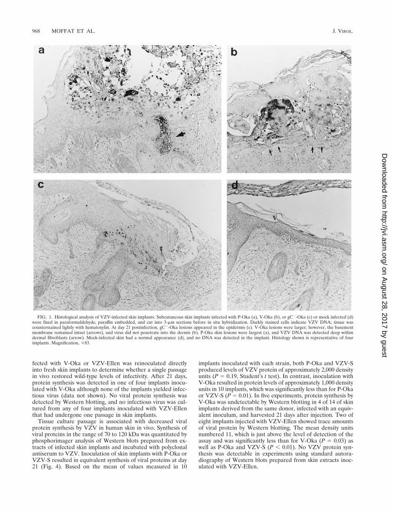
FIG. 1. Histological analysis of VZV-infected skin implants. Subcutaneous skin implants infected with P-Oka (a), V-Oka (b), or gC2-Oka (c) or mock infected (d)were fixed in paraformaldehyde, paraffin embedded, and cut into 3-mm sections before in situ hybridization. Darkly stained cells indicate VZV DNA; tissue wascounterstained lightly with hematoxylin. At day 21 postinfection, gC2-Oka lesions appeared in the epidermis (c). V-Oka lesions were larger; however, the basementmembrane remained intact (arrows), and virus did not penetrate into the dermis (b). P-Oka skin lesions were largest (a), and VZV DNA was detected deep withindermal fibroblasts (arrow). Mock-infected skin had a normal appearance (d), and no DNA was detected in the implant. Histology shown is representative of fourimplants. Magnification, 383.
968 MOFFAT ET AL. J. VIROL.
on August 28, 2017 by guest
http://jvi.asm.org/
Dow
nloaded from
Effect of gC expression on VZV replication in skin. Theinfectivities for skin of gC2-Oka and gC2-Ellen strains, whichare two naturally occurring variants of VZV that fail to syn-thesize gC, were compared with each parent strain. Beforeinoculation of skin implants, transcription and translation ofgC by each strain were evaluated by Northern and Westernblot analyses. A marked decrease in the transcription of gCmRNA by gC2-Oka and gC2-Ellen was documented in com-parison with VZV-S, P-Oka, V-Oka, and VZV-Ellen; tran-scription of gC in P-Oka, VZV-S, V-Oka, and VZV-Ellen fromthe 1.9-kb transcript, the predominant coding transcript, was
equivalent, with small differences due to variability of cyto-pathic effect (CPE) at time of harvest (Fig. 5A). The low levelof transcription of the 2.5-kb mRNA by P-Oka did not affectsynthesis of gC (Fig. 6). CPE and yields of cell-associatedinfectious virus in vitro were indistinguishable for all strains.Transcription of gE, gB, and gH was evaluated and comparedto that of gC to ensure that differences in infectivity of the gC2
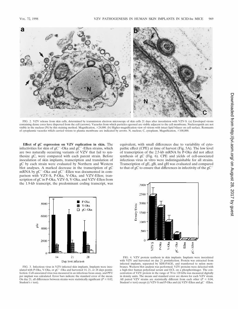
FIG. 2. VZV release from skin cells, determined by transmission electron microscopy of skin cells 21 days after inoculation with VZV-S. (a) Enveloped vironscontaining dense cores have dispersed from the cell (arrows). Vacuoles from which particles egressed are visible adjacent to the cell membrane. Nucleocapsids are notvisible in the nucleus (N) by this staining method. Magnification, 324,000. (b) Higher-magnification view of virions with intact lipid bilayer on cell surface. Remnantsof cytoplasmic vacuoles which carried virions to plasma membrane are indicated by arrows. N, nucleus; C, cytoplasm. Magnification, 3108,000.
FIG. 3. Infectious virus in VZV-infected skin implants. Implants were inoc-ulated with P-Oka, V-Oka, or gC2-Oka and harvested 14, 21, or 28 days postin-fection. Cell-associated virus was measured in an infectious focus assay, and PFUper implant was calculated. Error bars indicate the standard error of the mean.On day 21, all differences between strains were statistically significant (P # 0.02,Student’s t test).
FIG. 4. VZV protein synthesis in skin implants. Implants were inoculatedwith VZV and harvested on day 21 postinfection. Protein was extracted frominfected implants, separated by SDS-PAGE, and transferred to nylon mem-branes. Western blot analysis was performed; VZV proteins were detected witha high-titer human polyclonal serum and ECL on a phosphorimager. The con-centration of VZV protein in the range of 70 to 120 kDa was measured digitallyin density units. The means and standard error are shown for each VZV strain.All paired VZV strains are statistically different from each other (P # 0.04,Student’s t test) except (i) VZV-S and P-Oka and (ii) VZV-Ellen and gC2-Ellen.
VOL. 72, 1998 VZV PATHOGENESIS IN HUMAN SKIN IMPLANTS IN SCID-hu MICE 969
on August 28, 2017 by guest
http://jvi.asm.org/
Dow
nloaded from
derivatives for skin were not associated with disrupted mRNAtranscription of other major viral glycoprotein genes; no alter-ations were observed (Fig. 5B). To confirm that the absence ofgC transcription by gC2-Oka and gC2-Ellen was not a globaldefect in transcription of that region of the genome, Northernblot analyses detecting ORFs 13 and 15 were performed. Inagreement with a detailed transcriptional analysis of this re-gion described by others (31), all strains tested synthesizedequivalent amounts of ORF 13 and ORF 15 mRNAs (data notshown). By Western blotting, P-Oka, VZV-S, V-Oka, andVZV-Ellen produced the characteristic 105-kDa gC protein inMRC-5 cells, with some variability in quantity and size which istypical of this glycoprotein (11, 24). In contrast, gC expressioncould not be detected in cells infected with gC2-Oka or gC2-Ellen in vitro (Fig. 6). Restriction enzyme digest patterns wereidentical when DNA preparations of P-Oka, VZV-S, V-Oka,VZV-Ellen, gC2-Ellen, and gC2-Oka DNA were analyzed byusing HpaI, BglII, EcoRI, and BamHI (data not shown).
Sequencing of the gC promoter regions of P-Oka, V-Oka,gC2-Oka, VZV-Ellen, and gC2-Ellen strains showed no dif-ferences that could account for the reduced transcription of gCmRNA in gC2-Oka and gC2-Ellen. The putative 145-bp gCpromoter regions of all strains tested were identical to theconsensus VZV genomic DNA sequence (Dumas strain) (14);this homology extended into the coding regions of ORFs 14and 15 (data not shown).
The absence of gC expression was associated with a further
decrease in the already diminished capacity of V-Oka to rep-licate in human skin. Some cell-associated infectious virus wasrecovered after inoculation of skin implants with gC2-Oka, butthe concentration of virus was significantly lower than yieldsfrom skin infected with P-Oka or V-Oka, from which it wasderived (Fig. 3). At day 21, a mean of 143 PFU of cell-associ-ated virus was cultured from six implants infected with gC2-Oka, compared to 6,200 PFU of P-Oka (P 5 0.0005) and 1,600PFU of V-Oka (P 5 0.02) (Fig. 3). Infectious virus was recov-ered in unfiltered cell suspensions from five of the six implantsinoculated with gC2-Oka, but no cell-free virus was detected.The level of VZV protein expression in skin implants infectedwith gC2-Oka was 308 density units, which was significantlyless than protein synthesis detected after inoculation with P-Oka or V-Oka (P # 0.02) (Fig. 4).
The decreased replication and VZV protein expression inimplants infected with gC2-Oka was confirmed by histologyand in situ hybridization (Fig. 1). Sections of infected skinstained with hematoxylin and eosin showed that gC2-Oka pro-duced small lesions in the epidermis, compared to the largevesicular areas of necrosis extending deep into the dermis inskin infected with V-Oka and P-Oka. Mock-infected skin hada normal appearance. Viral DNA was detected by in situ hy-bridization only in the superficial keratinocyte layer of theepidermis in gC2-Oka-infected skin (Fig. 1c), while V-Oka andP-Oka DNA was present in lower layers of the dermis thatconsist primarily of fibroblasts (Fig. 1a and b). VZV DNA wasnot detected in the mock-infected control (Fig. 1d).
Given the marked inhibition of the virulence of VZV-Ellen,the failure to express gC by gC2-Ellen could not be implicatedas the cause of any further decrease in infectivity for humanskin. In two experiments, infectious virus was not recoveredfrom any of nine implants inoculated with gC2-Ellen, andVZV protein synthesis was not detected by Western blot inthese implants by standard autoradiography. The enhancedsensitivity of phosphorimager analysis revealed VZV proteinsin eight implants inoculated with gC2-Ellen and harvested atday 21, at an average of 14 density units, which was similar tothe minimal synthesis of VZV proteins in implants infectedwith the parent strain, VZV-Ellen, in vivo (Fig. 4).
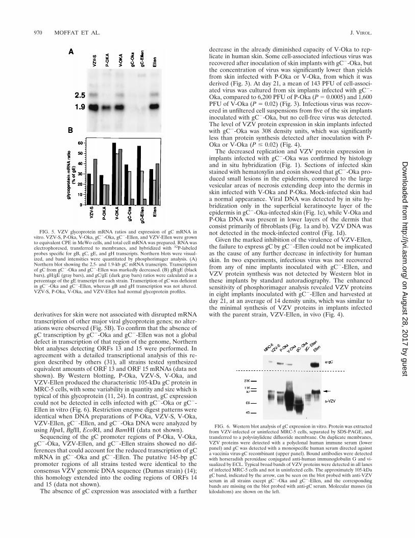
FIG. 6. Western blot analysis of gC expression in vitro. Protein was extractedfrom VZV-infected or uninfected MRC-5 cells, separated by SDS-PAGE, andtransferred to a polyvinylidene difluoride membrane. On duplicate membranes,VZV proteins were detected with a polyclonal human immune serum (lowerpanel) and gC was detected with a monospecific human serum directed againsta vaccinia virus-gC recombinant (upper panel). Bound antibodies were detectedwith horseradish peroxidase conjugated anti-human immunoglobulin G and vi-sualized by ECL. Typical broad bands of VZV proteins were detected in all lanesof infected MRC-5 cells and not in uninfected cells. The approximately 105-kDagC band, indicated by the arrow, can be seen on the blot probed with anti-VZVserum in all strains except gC2-Oka and gC2-Ellen, and the correspondingbands are missing on the blot probed with anti-gC serum. Molecular masses (inkilodaltons) are shown on the left.
FIG. 5. VZV glycoprotein mRNA ratios and expression of gC mRNA invitro. VZV-S, P-Oka, V-Oka, gC2-Oka, gC2-Ellen, and VZV-Ellen were grownto equivalent CPE in MeWo cells, and total cell mRNA was prepared. RNA waselectrophoresed, transferred to membranes, and hybridized with 32P-labeledprobes specific for gB, gC, gE, and gH transcripts. Northern blots were visual-ized, and band intensities were quantitated by phosphorimager analysis. (A)Northern blot showing the 2.5- and 1.9-kb gC mRNA transcripts. Transcriptionof gC from gC2-Oka and gC2-Ellen was markedly decreased. (B) gB/gE (blackbars), gH/gE (gray bars), and gC/gE (dark gray bars) ratios were calculated as apercentage of the gE transcript for each strain. Transcription of gC was deficientin gC2-Oka and gC2-Ellen, whereas gB and gH transcription was not altered.VZV-S, P-Oka, V-Oka, and VZV-Ellen had normal glycoprotein profiles.
970 MOFFAT ET AL. J. VIROL.
on August 28, 2017 by guest
http://jvi.asm.org/
Dow
nloaded from
Comparison of VZV and HSV-1 infectivity in thy/liv im-plants and effect of gC expression on HSV-1 replication inhuman skin. Thy/liv implants were inoculated with HSV-1KOS, the gC deletion strain DgC2-3, and its gC-positive rever-tant DgC2-3rev. Replication peaked on day 4, and there was nodifference in virus yields between the three strains (Table 1). Insitu hybridization showed HSV-1 DNA in large cells scatteredthroughout the thy/liv implant (Fig. 7). Combined in situ hy-bridization and immunohistochemistry using an antikeratinmonoclonal antibody showed that HSV DNA was present incortical epithelial cells (data not shown). The presence orabsence of gC did not affect HSV infectivity for cortical epi-thelial cells. Colocalization experiments using a monoclonalantibody to T-cell or macrophage markers showed no HSVDNA in these cell types. T cells were depleted in regions ofmost pronounced cytopathology, and many had undergoneapoptosis, demonstrated by enzymatic in situ labeling of apo-ptosis-induced DNA strand breaks (data not shown).
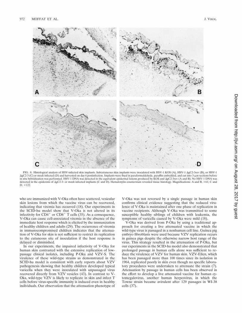
DgC2-3, the gC deletion strain of HSV-1 KOS, was tested inskin implants to determine whether this HSV protein, which ishomologous to VZV gC, was a virulence factor in human skincells (Fig. 8). Skin implants were infected with the gC deletionstrain, the gC-expressing revertant strain DgC2-3rev, or theparent strain KOS in duplicate experiments. The implantswere harvested on day 6 after inoculation and examined forinfectious virus yields, histopathologic changes, and localiza-tion of viral DNA by in situ hybridization. Virus was recovered
from six of eight implants infected with HSV-1 KOS, with amean titer of 5.1 3 106 PFU, and from eight of eight implantsinfected with the revertant, with a mean titer of 3.6 3 107 PFU.HSV-1 DgC2-3, the gC deletion strain, was recovered from 3 of10 infected implants, with a mean titer of 1.3 3 106 PFU, whileno infectious virus was detected in the remaining seven im-plants (Table 1).
When skin implants were inoculated with HSV-1 KOS or therevertant DgC2-3rev, the virus caused superficial lesions re-stricted to the epidermis whereas VZV DNA was detected indermal layers (Fig. 1a and 8A and B). No lesion formation orother pathologic changes were observed, and no HSV DNAwas detected in skin implants inoculated with the HSV-1 gCdeletion strain. With respect to histologic appearance and insitu hybridization analysis, the implants were indistinguishablefrom mock-infected controls (Fig. 8C and D). The skin implantshown in Fig. 8C was one of the three implants from whichvirus was recovered, yet no evidence for viral replication wasseen in the tissue. The cellular site of replication of the gCdeletion strain in the three skin implants from which HSV-1DgC2-3 was recovered was likely to have been the membraneof murine origin which forms around the subcutaneous skinimplants.
DISCUSSION
These experiments using human skin implants in theSCID-hu mouse model have provided the first opportunity todocument differences in the pathogenicity of VZV strains thatare indistinguishable by the characteristics of their replicationin tissue culture cells in vitro. The most important observationis that V-Oka, the varicella vaccine strain, is attenuated in itsinfectivity for human skin by virologic measures, includingreduced yields of infectious virus and decreased viral proteinsynthesis. The diminished virulence of V-Oka for human skinas demonstrated in the SCID-hu mouse model explains severalaspects of the clinical experience with this newly licensed her-pesvirus vaccine. Vaccine-related rashes are rare in healthychildren, but immunocompromised children with leukemia
TABLE 1. Replication of HSV-1 KOS and the HSV-1 gC deletionmutant in thy/liv and skin implants
Implant tissue
Mean log PFU/implant 6 SE (no. of implants thatyielded virus/no. analyzed)a
KOS DgC2-3rev DgC2-3
Thy/liv 6.6 6 1.2 (2/2) 5.4 6 1.2 (2/2) 6.5 6 0.4 (3/3)Skin 6.7 6 0.3 (6/8) 7.6 6 0.8 (8/8) 6.1 6 0.5 (3/10)
a Implants were inoculated with HSV-1 strains and harvested on day 4 (thy/liv)or day 6 (skin) after infection, and titers in cell lysates were determined.
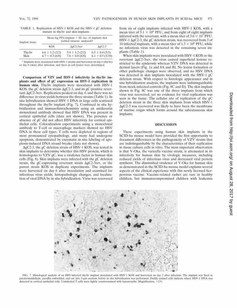
FIG. 7. Histological analysis of an HSV-infected thy/liv implant inoculated with HSV-1 KOS and harvested on day 2 after infection. The implant was fixed inparaformaldehyde, paraffin embedded, and cut into 3-mm sections before in situ hybridization was performed. Darkly stained cells indicate where HSV-1 DNA wasdetected in cortical epithelial cells. Uninfected T cells were lightly counterstained with hematoxylin. Magnification, 3131.
VOL. 72, 1998 VZV PATHOGENESIS IN HUMAN SKIN IMPLANTS IN SCID-hu MICE 971
on August 28, 2017 by guest
http://jvi.asm.org/
Dow
nloaded from
who are immunized with V-Oka often have scattered, vesicularskin lesions from which the vaccine virus can be recovered,indicating that viremia has occurred (18). Our experiments inthe SCID-hu model show that V-Oka is not altered in itsinfectivity for CD41 or CD81 T cells (35). As a consequence,V-Oka can cause cell-associated viremia in the absence of theimmediate host response which is elicited by the immunizationof healthy children and adults (29). The occurrence of viremiain immunocompromised children indicates that the attenua-tion of V-Oka for skin is not sufficient to restrict its replicationto the cutaneous site of inoculation if the host response isdelayed or diminished.
In our experiments, the impaired infectivity of V-Oka forhuman skin contrasted with the extensive replication of low-passage clinical isolates, including P-Oka and VZV-S. Thevirulence of these wild-type strains as demonstrated in theSCID-hu model is consistent with early reports about VZVpathogenesis showing that healthy children developed typicalvaricella when they were inoculated with unpassaged virusrecovered directly from VZV vesicles (43). In contrast to V-Oka, wild-type VZV is likely to replicate in skin and infect Tcells before virus-specific immunity is induced even in healthyindividuals. Our observation that the attenuation phenotype of
V-Oka was not reversed by a single passage in human skinconfirms clinical evidence suggesting that the reduced viru-lence of V-Oka is maintained after one phase of replication invaccine recipients. Although V-Oka was transmitted to somesusceptible healthy siblings of children with leukemia, thesymptoms of varicella caused by V-Oka were mild (18).
V-Oka was derived from P-Oka by using a traditional ap-proach for creating a live attenuated vaccine in which thewild-type virus is passaged in a nonhuman cell line. Guinea pigembryo fibroblasts were used because VZV replication occursin guinea pigs despite the otherwise narrow host range of thevirus. This strategy resulted in the attenuation of P-Oka, butour experiments in the SCID-hu model also demonstrated thatprolonged passage in human cells alone was sufficient to re-duce the virulence of VZV for human skin. VZV-Ellen, whichhas been passaged more than 100 times since its isolation in1964, replicated poorly in skin even though no specific labora-tory procedures were undertaken to attenuate the strain (7).Attenuation by passage in human cells has been observed inthe effort to develop a live attenuated vaccine for human cy-tomegalovirus, another human herpesvirus, in which theTowne strain became avirulent after 129 passages in WI-38cells (37).
FIG. 8. Histological analysis of HSV-infected skin implants. Subcutaneous skin implants were inoculated with HSV-1 KOS (A), HSV-1 DgC2-3rev (B), or HSV-1DgC2-3 (C) or mock infected (D) and harvested on day 6 postinfection. Implants were fixed in paraformaldehyde, paraffin embedded, and cut into 3-mm sections beforein situ hybridization was performed. HSV-1 DNA was detected in the equivalent epidermal lesions produced by KOS and DgC2-3rev (A and B). No HSV-1 DNA wasdetected in the epidermis of DgC2-3- or mock-infected implants (C and D). Hematoxylin counterstain revealed tissue histology. Magnifications: A and B, 361; C andD, 3122.
972 MOFFAT ET AL. J. VIROL.
on August 28, 2017 by guest
http://jvi.asm.org/
Dow
nloaded from
One of the distinguishing characteristics of VZV is thehighly cell-associated nature of its replication in vitro (2, 20).In contrast, infectious virus was released from skin infectedwith P-Oka and VZV-S in vivo. Similarly, vesicular fluid fromskin lesions of patients with varicella and herpes zoster con-tains intact, cell-free virus (17). By electron microscopy, themorphology of VZV virions produced in skin implants in theSCID-hu model differed markedly from the pattern of defec-tive particles and virion degradation observed in tissue culturecells. V-Oka, like other VZV strains, produces a mixture ofviral particles in embryonic lung fibroblasts, many of whichhave aberrant capsids, are not enveloped, and are thereforenot infectious (19, 20). Nevertheless, despite adjusting the in-oculum to equivalent titers of infectious virus, V-Oka was lessinfective for human skin than its parent strain. Prolonged pas-sage in tissue culture cells appears to induce virologic changesthat have a significant impact on VZV virulence for humanepidermal and dermal cells in vivo since only the low-passageclinical isolates, P-Oka and VZV-S, were fully virulent in skinimplants. These changes occurred without any major genomicalterations detectable by restriction enzyme analysis usingHpaI, BglII, EcoRI, and BamHI.
The tropism of VZV for human T cells and its infectivity forskin are both essential elements of its pathogenicity in humandisease. These experiments in the SCID-hu mouse model dem-onstrated that these tropisms are mediated by different viru-lence determinants. V-Oka was indistinguishable from low-passage VZV in its peak titers for CD41 and CD81 T cells,whereas its pathogenic potential in human skin was reducedsubstantially, as assessed by the extent of the cutaneous lesions,viral protein synthesis, infectious virus yields, and release ofinfectious virus. This altered infectivity of V-Oka for humanskin in vivo was not predictive of a corresponding attenuationof the same VZV strain with respect to its T-cell tropism.
The comparison of V-Oka and its derivative strain, gC2-Oka, demonstrated that gC is a specific determinant for VZVvirulence in skin, although the reduced infectivity of V-Okarelative to P-Oka indicates that gC expression is not the onlyfactor. The analysis of viral localization by in situ hybridizationshowed that lack of gC expression was associated with a failureof VZV to penetrate through the epidermal cell layer intodermal fibroblasts. Although T-cell tropism was preserved, de-letion of gC may have other attenuating effects on VZV, inaddition to reduced skin infectivity. As is true of other alpha-herpesviruses, VZV gC is not required for replication in tissueculture. In HSV-1, gC binds to a proteoglycan receptor on theapical surface of polarized MDCK cells but is not required forHSV-1 entry via the basolateral surface (40). VZV spreads bycell fusion in nonpolarized tissue culture cells, which probablyaccounts for the fact that VZV gC is dispensable and its ex-pression is variable in vitro. In contrast, the requirement forVZV gC expression for infectivity in skin implants in SCID-humice suggests that gC is involved in viral interactions withpolarized epidermal cells in intact skin in vivo. That gC expres-sion is a virulence determinant for alphaherpesvirus replica-tion in human skin was further substantiated by the failure ofthe HSV-1 gC deletion mutant DgC2-3 to replicate in epider-mal cells in skin implants. In vivo, both VZV gC and HSV-1 gCcould affect viral transport across the basal lamina and into thetightly joined cells of the epidermis. Enhanced infectivity forpolarized cells is likely to be the important common patho-genic function of gC in VZV and HSV-1 since other activitiesof the alphaherpesvirus gC homologs, such as increasing infec-tivity by binding to heparan sulfate on the cell surface orbinding to the C3b component of complement, do not appearto be shared by VZV (11, 15, 16, 21, 22, 23a, 33, 38, 46). Of
note, the transcriptional inactivation of VZV gC in the gC2-Oka and gC2-Ellen variants and their altered virulence in vivooccurred despite the presence of an intact gC promoter se-quence. Despite this extensive analysis of gC transcription andthe ORF 14 regulatory region, additional mutations that aroseelsewhere in the genomes of the gC2-Oka and gC2-Ellenstrains cannot be excluded as contributing factors in thesestrains’ loss of infectivity for skin. A possible explanation forthe absence of ORF 14 transcription is that one or moretransactivating proteins are involved in regulation of its pro-moter (31). A detailed analysis of ORF 14 transcription byLing et al. showed that the 1.9-kb transcript is the predominantcoding transcript and is sufficient to code for the entire gCprotein (31). Levels of the 2.5-kb transcript, a minor speciespolyadenylated at an alternative site within ORF 13, may varyat the late stages of infection when transcriptional controlstarts to erode. This could account for the absence of genera-tion of the 2.5-kb transcript by P-Oka.
The comparative analysis of VZV and HSV-1 replication inthe SCID-hu model revealed significant differences in cell tro-pisms that correlate with clinical observations about theirpathologic effects. Although T cells within thy/liv implants arehighly permissive for VZV replication, HSV-1 was detectedonly in nonlymphoid, cortical epithelial cells. Varicella is char-acterized by the appearance of scattered cutaneous vesicles,while primary HSV-1 infection is associated with localized,mucocutaneous lesions (2). This widespread rash is a conse-quence of the lymphocyte-associated viremia caused by VZVduring primary infection, whereas HSV viremia is an unusualcomplication (3, 35). Immunocompromised patients are alsosusceptible to cell-associated viremia during VZV reactivation,which is a rare occurrence during HSV reactivation (45).HSV-1 can infect activated T-cells in vitro, and abortive rep-lication in T cells was not not excluded in the thy/liv implantexperiments. In SCID-hu mice, VZV caused extensive necrosisin deeper dermal layers, but HSV-1 necrosis was confined tothe epidermis of skin implants. Similarly, the initial cutaneouslesions produced by primary VZV infection extend into thedermis and often cause scarring before an immune responsedevelops, while HSV-1 lesions remain superficial.
In general, the documentation of virulence phenotypesamong VZV strains that are indistinguishable in vitro, includ-ing the differentiation of V-Oka from its parent, and the dif-ferences in cell tropisms of VZV and HSV-1 underscore therelevance of the SCID-hu model for studies of viral pathogen-esis and the development of live attenuated viral vaccines.
ACKNOWLEDGMENTS
J.F.M. was supported by a training grant from the Division of De-velopmental and Neonatal Biology (HD07249) and NRSA AI09195.The work was supported by Public Health Service grants AI20459,AI36884, and P01-CA49605 to A.M.A. and RO1-EY09397 and P30-EY08098 to P.R.K.
We thank Marvin Sommer, Judie Boisvert, and Jean-Paul Vergnesfor technical assistance.
REFERENCES
1. Aldrovandi, G. M., G. Feuer, L. Gao, B. Jamieson, M. Kristeva, I. S. Y. Chen,and J. A. Zack. 1993. The SCID-hu mouse as a model for HIV-1 infection.Nature 363:732–736.
2. Arvin, A. M. 1996. Varicella-zoster virus, p. 2547–2585. In B. N. Fields, D. N.Knipe, and P. M. Howley (ed.), Virology, Lippincott-Raven Publishers, Phil-adelphia, Pa.
3. Arvin, A. M., C. M. Koropchak, B. R. G. Williams, F. C. Grumet, and S. K.Foung. 1986. Early immune response in healthy and immunocompromisedsubjects with primary varicella-zoster virus infection. J. Infect. Dis. 154:422–429.
4. Auwaerter, P. G., H. Kaneshima, J. M. McCune, G. Wiegand, and D. E.
VOL. 72, 1998 VZV PATHOGENESIS IN HUMAN SKIN IMPLANTS IN SCID-hu MICE 973
on August 28, 2017 by guest
http://jvi.asm.org/
Dow
nloaded from
Griffin. 1996. Measles virus infection of thymic epithelium in the SCID-humouse leads to thymocyte apoptosis. J. Virol. 70:3734–3740.
5. Bergen, R. E., P. S. Diaz, and A. M. Arvin. 1990. The immunogenicity of theOka/Merck varicella vaccine in relation to infectious varicella-zoster virusand relative viral antigen content. J. Infect. Dis. 162:1049–1054.
6. Bogger-Goren, S., K. Baba, P. Hurley, H. Yabuuchi, M. Takahashi, and P. L.Ogra. 1982. Antibody response to varicella-zoster virus after natural orvaccine-induced infection. J. Infect. Dis. 146:260–265.
7. Brunell, P. A., and H. L. Casey. 1964. Crude tissue culture antigen fordetermination of varicella-zoster complement fixing antibody. Public HealthRep. 79:839–842.
8. Centifanto-Fitzgerald, Y. M., T. Yamaguchi, H. E. Kaufman, M. Tognon,and B. Roizman. 1982. Ocular disease pattern induced by herpes simplexvirus is genetically determined by a specific region of viral DNA. J. Exp.Med. 155:475–489.
9. Cheatham, W. J., T. H. Weller, T. F. J. Dolan, and J. C. Dower. 1956.Varicella: report of two fatal cases with necropsy, virus isolation, and sero-logic studies. Am. J. Pathol. 32:1015–1035.
10. Chomczynski, P., and N. Sacchi. 1987. Single step method of RNA isolationby acid guanidinium thiocyanate-phenol-chloroform extraction. Anal. Bio-chem. 162:156–159.
11. Cohen, J. I., and K. E. Seidel. 1994. Absence of varicella-zoster virus (VZV)glycoprotein V does not alter growth of VZV in vitro or sensitivity to heparin.J. Gen. Virol. 75:3087–3093.
12. Croen, K., J. Ostrove, L. Dragovic, and S. Straus. 1988. Patterns of geneexpression and sites of latency in human nerve ganglia are different forvaricella-zoster and herpes simplex viruses. Proc. Natl. Acad. Sci. USA85:9773–9777.
13. Croen, K., and S. Straus. 1991. Varicella-zoster virus latency. Annu. Rev.Microbiol. 4:2391–99.
14. Davison, A. J., and J. E. Scott. 1986. The complete DNA sequence ofvaricella-zoster virus. J. Gen. Virol. 67:1759–1816.
15. Flynn, S. J., and P. Ryan. 1996. The receptor-binding domain of pseudora-bies virus glycoprotein gC is composed of multiple discrete units that arefunctionally redundant. J. Virol. 70:1355–1364.
16. Friedman, H. M., G. H. Cohen, R. J. Eisenberg, C. A. Seidel, and D. B. Cines.1984. Glycoprotein C of HSV-1 functions as a C3b receptor on infectedendothelial cells. Nature 309:633–635.
17. Gelb, L. D. 1993. Varicella-zoster virus: molecular biology, p. 257–279. In B.Roizman, R. J. Whitley, and C. Lopez (ed.), The human herpes viruses.Raven Press, New York, N.Y.
18. Gershon, A. A., P. LaRussa, I. Hardy, S. Steinberg, and S. Silverstein. 1992.Varicella vaccine: the American experience. J. Infect. Dis. 166(Suppl. 1):S63–S68.
19. Grose, C. 1996. Pathogenesis of infection with varicella vaccine, p. 489–505.In R. W. Ellis and C. J. White (ed.), Infectious disease clinics of NorthAmerica. W. B. Saunders Co., Philadelphia, Pa.
20. Harson, R., and C. Grose. 1995. Egress of varicella-zoster virus from themelanoma cell: a tropism for the melanocyte. J. Virol. 69:4994–5010.
21. Herold, B. C., D. WuDunn, N. Soltys, and P. G. Spear. 1991. Glycoprotein Cof herpes simplex virus type 1 plays a principal role in the adsorption of virusto cells and in infectivity. J. Virol. 65:1090–1098.
22. Huemer, H. P., C. Larcher, and N. E. Coe. 1992. Pseudorabies virus glyco-protein-III derived from virions and infected cells binds to the 3rd compo-nent of complement. Virus Res. 23:271–280.
23. Kaneshima, H., C.-C. Shih, R. Namikawa, L. Rabin, G. Outzen, S. G.Machado, and J. M. McCune. 1991. Human immunodeficiency virus infec-tion of human lumph nodes in the SCID-hu mouse. Proc. Natl. Acad. Sci.USA 88:4523–4527.
23a.Kinchington, P., and G. Cohen. Personal communication.24. Kinchington, P. R., P. Ling, M. Pensiero, A. Gershon, J. Hay, and W. T.
Ruyechan. 1990. A Possible role for glycoprotein gpV in the pathogenesis ofvaricella-zoster virus, p. 83–91. In C. Lopez (ed.), Immunobiology and pro-phylaxis of human herpesvirus infections. Plenum Press, New York, N.Y.
25. Kinchington, P. R., J. Remenick, J. M. Ostrove, S. E. Straus, W. T.Ruyechan, and J. Hay. 1986. Putative glycoprotein gene of varicella-zostervirus with variable copy numbers of a 42-base-pair repeat sequence hashomology to herpes simplex virus glycoprotein C. J. Virol. 59:660–668.
26. Kinchington, P. R., J.-P. Vergnes, P. Defechereux, J. Piette, and S. E. Turse.1994. Transcriptional mapping of the varicella-zoster virus regulatory genesencoding open reading frames 4 and 63. J. Virol. 68:3570–3581.
27. Koropchak, C. M., P. S. Diaz, and A. M. Arvin. 1989. Investigation ofvaricella-zoster virus infection of lymphocytes by in situ hybridization. J. Vi-rol. 63:2392–2395.
28. Koropchak, C. M., G. Graham, J. Palmer, M. Winsberg, S. F. Ting, M.Wallace, C. G. Prober, and A. M. Arvin. 1991. Investigation of varicella-zoster virus infection by polymerase chain reaction in the immunocompetenthost with acute varicella. J. Infect. Dis. 163:1016–1022.
29. Krause, P. R., and D. M. Klinman. 1995. Efficacy, immunogenicity, safety,and use of live attenuated chickenpox vaccine. J. Pediatr. 127:518–525.
30. LaRussa, P., O. Lungu, I. Hardy, A. Gershon, S. P. Steinberg, and S.Silverstein. 1992. Restriction fragment length polymorphism of polymerasechain reaction products from vaccine and wild-type varicella-zoster virusisolates. J. Virol. 66:1016–1020.
31. Ling, P., P. R. Kinchington, W. T. Ruychan, and J. Hay. 1991. A detailedanalysis of transcripts mapping to varicella zoster virus gene 14 (glycoproteinV). Virology 184:625–635.
32. Mahalingam, R., M. Wellish, W. Wolf, A. N. Dueland, R. Cohrs, A. Vafai,and D. Gilden. 1990. Latent VZV DNA in human trigeminal and thoracicganglia. N. Engl. J. Med. 323:627–631.
33. Mettenleiter, T. C., L. Zsak, F. Zuckermann, N. Sugg, H. Kern, and T.Ben-Porat. 1990. Interaction of glycoprotein gIII with a cellular heparinlikesubstance mediates adsorption of pseudorabies virus. J. Virol. 64:278–286.
34. Mocarski, E. S., M. Bonyhadi, S. Salimi, J. M. McCune, and H. Kaneshima.1993. Human cytomegalovirus in a SCID-hu mouse: thymic epithelial cellsare prominent targets of viral replication. Proc. Natl. Acad. Sci. USA 90:104–108.
35. Moffat, J. F., M. D. Stein, H. Kaneshima, and A. M. Arvin. 1995. Tropism ofvaricella-zoster virus for human CD41 and CD81 T lymphocytes and epi-dermal cells in SCID-hu mice. J. Virol. 69:5236–5242.
36. Ozaki, T., T. Ichikawa, Y. Matsui, H. Kondo, T. Nagai, Y. Asano, K. Ya-manishi, and M. Takahashi. 1986. Lymphocyte-associated viremia in vari-cella. J. Med. Virol. 19:249–253.
37. Plotkin, S. A., S. E. Starr, H. M. Friedman, E. Gonczol, and R. E. Weibel.1989. Protective effects of Towne cytomegalovirus vaccine against low-pas-sage cytomegalovirus administered as a challenge. J. Infect. Dis. 159:860–865.
38. Robbins, A. K., M. E. Whealy, R. J. Watson, and L. W. Enquist. 1986.Pseudorabies virus gene encoding glycoprotein gIII is not essential forgrowth in tissue culture. J. Virol. 59:635–645.
39. Sawyer, M. H., Y. N. Wu, C. J. Chamberlin, C. Burgos, S. K. Brodine, W. A.Bowler, A. LaRocco, E. C. Oldfield III, and M. R. Wallace. 1992. Detectionof varicella-zoster virus DNA in the oropharynx and blood of patients withvaricella. J. Infect. Dis. 166:886–888.
40. Sears, A. E., B. S. McGwire, and B. Roizman. 1991. Infection of polarizedMDCK cells with herpes simplex virus 1: two asymmetrically distributed cellreceptors interact with different viral proteins. Proc. Natl. Acad. Sci. USA88:5087–5091.
41. Straus, S., H. S. Aulakh, W. T. Ruychan, J. Hay, T. A. Casey, G. F. VandeWoude, J. Owens, and H. A. Smith. 1981. Structure of varicella-zoster virusDNA. J. Virol. 40:516–525.
42. Takahashi, M., T. Otsuka, Y. Okuno, Y. Asano, T. Yazaki, and S. Isomura.1974. Live vaccine used to prevent the spread of varicalla in children inhospital. Lancet ii:1288–1290.
43. von Bokay, J. 1892. Das Auftreten der Schafblattern uter besonderen Um-standen. Ungerer’s Arch. Med. 1:159.
44. Whitley, R. J. 1990. In G. Galasso, R. Whitley, and T. C. Merigan (ed.),Antiviral agents and viral diseases of man, p. 235–264. Raven Press, NewYork, N.Y.
45. Wilson, A., M. Sharp, C. M. Koropchak, S. F. Ting, and A. M. Arvin. 1992.Subclinical varicella-zoster virus viremia, herpes zoster and recovery of T-lymphocyte responses to varicella-zoster viral antigens after allogeneic andautologous bone marrow transplantation. J. Infect. Dis. 165:119–126.
46. Zhu, Z., M. D. Gershon, R. Ambron, C. Gabel, and A. A. Gershon. 1995.Infection of cells by varicella zoster virus: inhibition of viral entry by man-nose 6-phosphate and heparin. Proc. Natl. Acad. Sci. USA 92:3546–3550.
974 MOFFAT ET AL. J. VIROL.
on August 28, 2017 by guest
http://jvi.asm.org/
Dow
nloaded from