Embed Size (px)
Citation preview
Attention-Gated Networksfor Improving Ultrasound Scan Plane Detection
Jo Schlemper1, Ozan Oktay1, Liang Chen1, Jacqueline Matthew2,Caroline Knight2, Bernhard Kainz1, Ben Glocker1, and Daniel Rueckert1
1Biomedical Image Analysis Group, Imperial College London, London, UK2King’s College London, London, [email protected]
Abstract
In this work, we apply an attention-gated network to real-time automated scanplane detection for fetal ultrasound screening. Scan plane detection in fetal ul-trasound is a challenging problem due the poor image quality resulting in lowinterpretability for both clinicians and automated algorithms. To solve this, wepropose incorporating self-gated soft-attention mechanisms. A soft-attention mech-anism generates a gating signal that is end-to-end trainable, which allows thenetwork to contextualise local information useful for prediction. The proposedattention mechanism is generic and it can be easily incorporated into any existingclassification architectures, while only requiring a few additional parameters. Weshow that, when the base network has a high capacity, the incorporated attentionmechanism can provide efficient object localisation while improving the overallperformance. When the base network has a low capacity, the method greatly out-performs the baseline approach and significantly reduces false positives. Lastly,the generated attention maps allow us to understand the model’s reasoning process,which can also be used for weakly supervised object localisation.
1 Introduction
Fetal ultrasound screening is an important diagnostic protocol to detect abnormal fetal development.Abnormal development is one of the leading causes for perinatal mortality world-wide. Duringscreening examination, multiple anatomically standardised [15] scan planes are used to obtainbiometric measurements as well as identifying abnormalities such as lesions. While 2D ultrasound isthe preferred approach for examination due to its low cost and real-time capabilities, ultrasound suffersfrom low signal-to-noise ratio and image artefacts. As such, diagnostic accuracy and reproducibilityis limited and requires a high level of expert knowledge and training. Therefore, automated scan planedetection algorithms can help training experts, facilitate non-expert examination, support consistentdata acquisition and make diagnostics more robust.
Automated scan plane detection poses many challenges: Firstly, during the examination, the majorityof time is spent exploring the present anatomy. As such, there are a large number of backgroundlabels and a significant class imbalance must be considered. Secondly, even if the object of interestis localised, it may not have reached the ideal scanning plane for diagnosis and hence the framemay be labelled as background; Therefore, in addition to understanding the global context, it isessential to understand the small differences in local structures to detect a correct plane. In the past,several approaches were proposed [28, 3], however, they are computationally expensive and cannotbe deployed for the real-time application.
1st Conference on Medical Imaging with Deep Learning (MIDL 2018), Amsterdam, The Netherlands.
In recent years, deep learning and convolutional neural networks (CNNs) have become popu-lar approaches for a variety of medical image classification problems, including classification ofAlzheimer’s disease [20], lung nodule in CT/X-ray [34], skin lesion [4, 7], anatomy [19] and theviews for echo-cardiograms [13].An extensive list of applications can be found in [9, 29]. In [2] theauthors propose a CNN architecture called Sononet to solve the standard plane classification problemduring fetal ultrasound examination. The proposed approach achieves very good performance inreal-time plane detection, retrospective frame retrieval (retrieving the most relevant frame) and weaklysupervised object localisation. However, despite its success, the method suffers from relatively lowprecision and especially struggles differentiating anatomically related cardiac views. We argue thatthe reason for this is that Sononet is good at aggregating global information but it cannot preservelocal information well. Moreover, the heuristics employed for the object localisation requires guidedbackpropagation, which limits the object localisation speed that can be achieved.
In fact, we claim that the inability to exploit local information is a common problem in medical imageanalysis: in many of these scenarios, typically, the object of interest is very small (e.g. lesions, localdeformity, etc.) compared to the size of the input image, which can be high resolution 2D, 3D or 4Ddata. Such situation requires to tackle the object detection and classification problem as a two-stageprocess. In this work, we introduce soft-attention in the context of medical image classification.Attention is a modular mechanism that allows to efficiently exploit localised information, which alsoprovides soft object localisation during forward pass. In this work, we demonstrate the usefulness ofsuch attention mechanism by applying the proposed approach to improve the scan plane detection forfetal ultrasound screening.
1.1 Related Work
Attention mechanisms were first popularised in the context of natural language processing [21], suchas machine translation [1, 12]. In these settings often recurrent neural networks are employed tomodel a sequence of text. In particular, given a sequence of text and a current word, a task is toextract a next word in a sentence generation or translation. The idea of attention mechanisms is togenerate a context vector which assigns weights on the input sequence. Thus, the signal highlightsthe salient feature of the sequence conditioned on the current word while suppressing the irrelevantcounter-parts, making the prediction more contextualised. Attention mechanisms can further beseparated into two types: soft-attention and hard-attention. In soft-attention, continuous functions(e.g. soft-max) are used to assign the attention weight on the input, making it fully differentiable.In comparison, hard-attention models propose specific words by sampling from the weights. As thesampling operation is not differentiable, hard-attention is trained using the gradient of the likelihoodterm generated by Monte-Carlo sampling [26].
In computer vision, attention mechanisms are applied to a variety of problems, including imageclassification [6, 23, 32], segmentation [18], action recognition [10, 16, 24], image captioning [26, 11],and visual question answering [27, 14]. In the context of medical image analysis, attention modelshave been exploited for medical report generation [31, 30] as well as joint image and text classification[25]. However, for standard medical image classification, despite the importance of local information,only a handful of works use attention mechanisms [17, 5]. In these methods, either bounding boxlabels are available to guide the attention, or the local context is extracted by a hard-attention model(i.e. region proposal followed by hard-cropping). In our work, we propose incorporating self-gating,a soft-attention approach that is end-to-end trainable. This also does not require any bounding boxlabels and backpropagation-based saliency map generation as in [2].
1.2 Contributions
• We introduce a self-gated, soft-attention mechanism in the context of pure medical imageclassification. We apply the proposed model to real-time fetal ultrasound scan plane detectionand show its superior classification performance over the baseline approach.
• We demonstrate that the proposed attention mechanism provides fine-scale attention mapsthat can be visualised, with minimal computational overhead, which is a crucial step towardsexplainable deep learning.
• Finally, we show that attention maps can used for fast (weakly-supervised) object localisation,demonstrating that the attended features indeed correlates to the anatomy of interest.
2
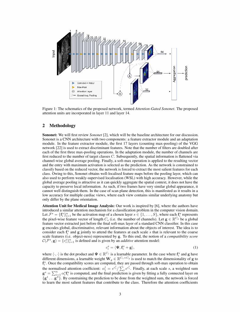
Figure 1: The schematics of the proposed network, termed Attention-Gated Sononet. The proposedattention units are incorporated in layer 11 and layer 14.
2 Methodology
Sononet: We will first review Sononet [2], which will be the baseline architecture for our discussion.Sononet is a CNN architecture with two components: a feature extractor module and an adaptationmodule. In the feature extractor module, the first 17 layers (counting max-pooling) of the VGGnetwork [22] is used to extract discriminant features. Note that the number of filters are doubled aftereach of the first three max-pooling operations. In the adaptation module, the number of channels arefirst reduced to the number of target classes C. Subsequently, the spatial information is flattened viachannel-wise global average pooling. Finally, a soft-max operation is applied to the resulting vectorand the entry with maximum activation is selected as the prediction. As the network is constrained toclassify based on the reduced vector, the network is forced to extract the most salient features for eachclass. Owing to this, Sononet obtains well-localised feature maps before the pooling layer, which canalso used to perform weakly-supervised localisation (WSL) with high accuracy. However, while theglobal average pooling is attractive as it can quickly aggregate the spatial context, it does not have thecapacity to preserve local information. As such, if two frames have very similar global appearance, itcannot well distinguish them. In the case of scan plane detection, this is manifested as it results in alow accuracy for multiple cardiac views, where each view contains similar underlying anatomy butonly differ by the plane orientation.
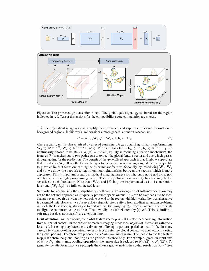
Attention Unit for Medical Image Analysis: Our work is inspired by [6], where the authors haveintroduced a similar attention mechanism for a classification problem in the computer vision domain.Let Fs = {fsi }ni=1 be the activation map of a chosen layer s ∈ {1, . . . , S}, where each fsi representsthe pixel-wise feature vector of length Cs (i.e. the number of channels). Let g ∈ RCg be a globalfeature vector extracted just before the final soft-max layer of a standard CNN classifier. In this caseg encodes global, discriminative, relevant information about the objects of interest. The idea is toconsider each fsi and g jointly to attend the features at each scale s that is relevant to the coarsescale features (i.e. object-ness) represented by g. To this end, the notion of a compatibility scoreC(Fs,g) = {csi}ni=1 is defined and is given by an additive attention model:
csi = 〈Ψ, fsi + g〉, (1)
where 〈·, ·〉 is the dot product and Ψ ∈ RCs is a learnable parameter. In the case where fsi and g havedifferent dimensions, a learnable weight Wg ∈ RCs×Cg is used to match the dimensionality of g tofsi . Once the compatibility scores are computed, they are passed through soft-max operation to obtainthe normalised attention coefficient: αli = ec
li/
∑i ecli . Finally, at each scale s, a weighted sum
gs =∑ni=1 α
si fsi is computed, and the final prediction is given by fitting a fully connected layer on
{g1 . . .gS}. By constraining the prediction to be done from the weighted sum, the network is forcedto learn the most salient features that contribute to the class. Therefore the attention coefficients
3
..
Global Feature Map g
Feature Map ℱs
f si
gI
Compatibility Score C Normalisation σ2
Attention Map α Cs
W
H
αsi
..
Attended Feature Map gs
Attention Unit
csi Ψ Wfσ1 f s
i Wg gI bg bΨ+ + +=
×Cint
Cs
× 1Cs× 1Cg
×Cint
Cg × 1Cint
1 × 11 × 1 1 × Cint
Compatibility Score C( , g)f si
Figure 2: The proposed grid attention block. The global gate signal gI is shared for the regionindicated in red. Tensor dimensions for the compatibility score computation are shown.
{αli} identify salient image regions, amplify their influence, and suppress irrelevant information inbackground regions. In this work, we consider a more general attention mechanism:
csi = Ψσ1 (Wf fsi + Wgg + bg) + bψ, (2)
where a gating unit is characterised by a set of parameters Θatt containing: linear transformationsWf ∈ RCint×C , Wg ∈ RCint×Cg , Ψ ∈ RCint and bias terms bψ ∈ R , bg ∈ RCint . σ1 is anonlinearity chosen to be ReLU: σ1(x) = max(0,x). By introducing attention mechanism, thefeatures Fs branches out to two paths: one to extract the global feature vector and one which passesthrough gating for the prediction. The benefit of the generalised approach is that firstly, we speculatethat introducing Wf allows the fine-scale layer to focus less on generating a signal that is compatibleto g, which helps it focus on learning the discriminant features. Secondly, by introducing Wf , Wg
and σ1, we allow the network to learn nonlinear relationships between the vectors, which is moreexpressive. This is important because in medical imaging, images are inherently noisy and the regionof interest is often highly non-homogeneous. Therefore, a linear compatibility function may be toosensitive to such fluctuation. Note that {Wf} and {Ψ,bψ} are implemented as 1× 1 convolutionlayer and {Wg,bg} is a fully connected layer.
Similarly, for normalising the compatibility coefficients, we also argue that soft-max operation maynot be the optimal approach as it typically produces sparse output. This can be over-sensitive to localchanges even though we want the network to attend to the region with high variability. An alternativeis a sigmoid unit. However, we observe that a sigmoid often suffers from gradient saturation problems.As such, the best working strategy is to first subtract the mini{αsi}ni=1 from all attention coefficientsto align the minimum value to be 0. Then, we divide each element by
∑i α
si . This is similar to
soft-max but does not sparsify the attention map.
Grid Attention: As seen above, the global feature vector g is a 1D vector incorporating informationfrom all spatial context. In the context of medical imaging, since most objects of interest are extremelylocalised, flattening may have the disadvantage of losing important spatial context. In fact in manycases, a few max-pooling operations are sufficient to infer the global context without explicitly usingthe global pooling. Therefore, we propose a grid attention mechanism. The idea is to use the featuremap just before the global pooling as the gridded instance of g. For example, given an input sizeof Nx ×Ny, after r max pooling operations, the tensor size is reduced to Nx/(2r)×Ny/(2r). Togenerate the attention map, we upsample the coarse grid to match the spatial resolution of Fs (see
4
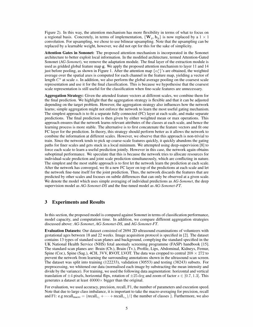
Figure 2). In this way, the attention mechanism has more flexibility in terms of what to focus ona regional basis. Concretely, in terms of implementation, {Wg,bg} is now replaced by a 1 × 1convolution. For upsampling, we chose to use bilinear upsampling. Note that the upsampling can bereplaced by a learnable weight, however, we did not opt for this for the sake of simplicity.
Attention Gates in Sononet: The proposed attention mechanism is incorporated in the Sononetarchitecture to better exploit local information. In the modified architecture, termed Attention-GatedSononet (AG-Sononet), we remove the adaptation module. The final layer of the extraction module isused as gridded global feature map g. We apply the proposed attention mechanism to layer 11 and 14just before pooling, as shown in Figure 1. After the attention map {αsi}’s are obtained, the weightedaverage over the spatial axes is computed for each channel in the feature map, yielding a vector oflength Cs at scale s. In addition, we also perform the global average pooling on the coarsest scalerepresentation and use it for the final classification. This is because we hypothesise that the coarsestscale representation is still useful for the classification when fine-scale features are unnecessary.
Aggregation Strategy: Given the attended feature vectors at different scales, we combine them forthe final prediction. We highlight that the aggregation strategy is flexible and that it can be adjusteddepending on the target problem. However, the aggregation strategy also influences how the networklearns; simple aggregation might not enforce the network to learn the most useful gating mechanism.The simplest approach is to fit a separate fully connected (FC) layer at each scale, and make separatepredictions. The final prediction is then given by either weighted mean or max operations. Thisapproach ensures that the network learns relevant attributes of the classes at each scale, and hence thelearning process is more stable. The alternative is to first concatenate the feature vectors and fit oneFC layer for the prediction. In theory, this strategy should perform better as it allows the network tocombine the information at different scales. However, we observe that this approach is non-trivial totrain. Since the network tends to pick up coarse-scale features quickly, it quickly abandons the gatingpaths for finer scales and gets stuck in a local minimum. We attempted using deep-supervision [8] toforce each scale to learn a useful prediction jointly. However in this case, the network again obtainssuboptimal performance. We speculate that this is because the network tries to allocate resources forindividual-scale prediction and joint scale prediction simultaneously, which are conflicting in nature.The simplest and the most stable approach is to first let the network learn the prediction at each scale.After the network has converged, we fit a new FC layer on top of the predictions at each scale and letthe network fine-tune itself for the joint prediction. Thus, the network discards the features that arepredicted by other scales and focuses on subtle differences that can only be observed at a given scale.We denote the model which uses simple averaging of individual predictions as AG-Sononet, the deepsupervision model as AG-Sononet-DS and the fine-tuned model as AG-Sononet-FT.
3 Experiments and Results
In this section, the proposed model is compared against Sononet in terms of classification performance,model capacity, and computation time. In addition, we compare different aggregation strategiesdiscussed above: AG-Sononet, AG-Sononet-DS, and AG-Sononet-FT.
Evaluation Datasets: Our dataset consisted of 2694 2D ultrasound examinations of volunteers withgestational ages between 18 and 22 weeks. Image acquisition protocol is specified in [2]. The datasetcontains 13 types of standard scan planes and background, complying the standard specified in theUK National Health Service (NHS) fetal anomaly screening programme (FASP) handbook [15].The standard scan planes are: Brain (Cb.), Brain (Tv.), Profile, Lips, Abdominal, Kidneys, Femur,Spine (Cor.), Spine (Sag.), 4CH, 3VV, RVOT, LVOT. The data was cropped to central 208× 272 toprevent the network from learning the surrounding annotations shown in the ultrasound scan screen.The dataset was split into training (122233), validation (30553) and testing (38243) subsets. Forpreprocessing, we whitened our data (normalised each image by substracting the mean intensity anddivide by the variance). For training, we used the following data augmentation: horizontal and verticaltranslation of ±4 pixels, horizontal flips, rotation of ±25 deg and zoom of factor s ∈ [0.7, 1.3]. Thisgenerates a dataset at least 40000× bigger than the original.
For evaluation, we used accuracy, precision, recall, F1, the number of parameters and execution speed.Note that due to large class imbalance, it is important to take the macro-averaging for precision, recalland F1: e.g recallmacro = (recallc1 + · · ·+ recallcn)/{ the number of classes }. Furthermore, we also
5
qualitatively study the attention map generated to highlight that the network indeed attends salientlocal regions.
Training: Note that due to the nature of fetal ultrasound screening, the background label dominatesthe dataset. Due to large class imbalance, the training is not straightforward. In addition, backgroundframes could contain the anatomy of interest, yet it might be classified as background as the plane isnot a standard plane. Therefore, an appropriate ratio between all classes and background is important.We used a weighted sampling strategy: the sampling “probability” of an image from class c is givenby 1/nc, where nc is the number of images in class c. For the background label, we used 13/nc,where 13 is the number of the standard scan planes. In this way, we expect to see one backgroundimage for every standard scan plane. We used cross entropy loss and the network was optimisedusing Stochastic Gradient Descent with Nesterov momentum (ρ = 0.9). The initial learning rate wasset to 0.1, which was subsequently reduced by a factor of 0.1 for every 100 epoch. We also used awarm-start learning rate of 0.01 for the first 5 epochs. Each network was trained for 300 epochs. Thebatch size was set to 64. `2 weight regularisation was used with λ = 10−4.
Implementation Details: We modified the baseline Sononet architecture slightly: instead of using2 convolution layers for the first 2 feature scales and 3 convolution layers for the last 3 featurescales, we used 3 layers for the first 3 and 2 layers for the last 2 feature scales. The architecture forAG-sononet is shown in 1. As discussed, training AG-sononet is slightly more tricky as the optimalgating mechanism may not be necessarily learnt. However, we observed that the simplest approach toachieve the desired gating mechanism was to initialise AG-Sononet with a partially trained Sononet.We compare our models with different capacities, with initial number of features 8, 16 and 32. Ourimplementation in PyTorch library is publicly available1.
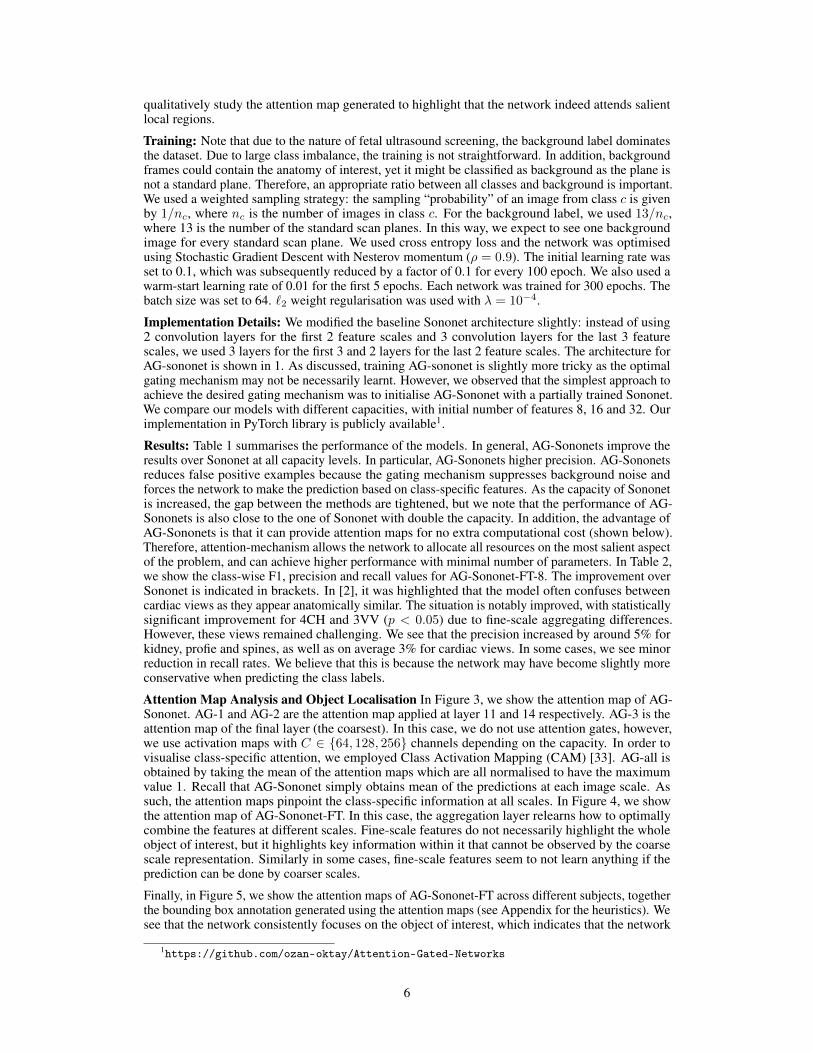
Results: Table 1 summarises the performance of the models. In general, AG-Sononets improve theresults over Sononet at all capacity levels. In particular, AG-Sononets higher precision. AG-Sononetsreduces false positive examples because the gating mechanism suppresses background noise andforces the network to make the prediction based on class-specific features. As the capacity of Sononetis increased, the gap between the methods are tightened, but we note that the performance of AG-Sononets is also close to the one of Sononet with double the capacity. In addition, the advantage ofAG-Sononets is that it can provide attention maps for no extra computational cost (shown below).Therefore, attention-mechanism allows the network to allocate all resources on the most salient aspectof the problem, and can achieve higher performance with minimal number of parameters. In Table 2,we show the class-wise F1, precision and recall values for AG-Sononet-FT-8. The improvement overSononet is indicated in brackets. In [2], it was highlighted that the model often confuses betweencardiac views as they appear anatomically similar. The situation is notably improved, with statisticallysignificant improvement for 4CH and 3VV (p < 0.05) due to fine-scale aggregating differences.However, these views remained challenging. We see that the precision increased by around 5% forkidney, profie and spines, as well as on average 3% for cardiac views. In some cases, we see minorreduction in recall rates. We believe that this is because the network may have become slightly moreconservative when predicting the class labels.
Attention Map Analysis and Object Localisation In Figure 3, we show the attention map of AG-Sononet. AG-1 and AG-2 are the attention map applied at layer 11 and 14 respectively. AG-3 is theattention map of the final layer (the coarsest). In this case, we do not use attention gates, however,we use activation maps with C ∈ {64, 128, 256} channels depending on the capacity. In order tovisualise class-specific attention, we employed Class Activation Mapping (CAM) [33]. AG-all isobtained by taking the mean of the attention maps which are all normalised to have the maximumvalue 1. Recall that AG-Sononet simply obtains mean of the predictions at each image scale. Assuch, the attention maps pinpoint the class-specific information at all scales. In Figure 4, we showthe attention map of AG-Sononet-FT. In this case, the aggregation layer relearns how to optimallycombine the features at different scales. Fine-scale features do not necessarily highlight the wholeobject of interest, but it highlights key information within it that cannot be observed by the coarsescale representation. Similarly in some cases, fine-scale features seem to not learn anything if theprediction can be done by coarser scales.
Finally, in Figure 5, we show the attention maps of AG-Sononet-FT across different subjects, togetherthe bounding box annotation generated using the attention maps (see Appendix for the heuristics). Wesee that the network consistently focuses on the object of interest, which indicates that the network
1https://github.com/ozan-oktay/Attention-Gated-Networks
6
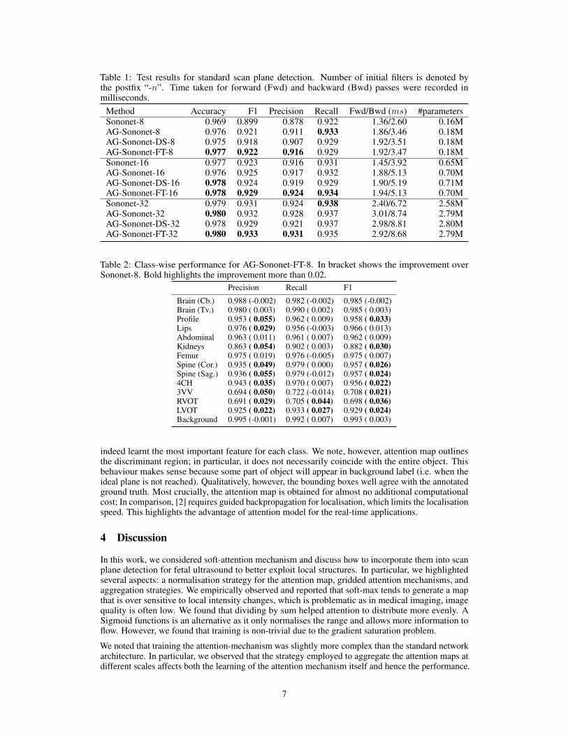
Table 1: Test results for standard scan plane detection. Number of initial filters is denoted bythe postfix “-n”. Time taken for forward (Fwd) and backward (Bwd) passes were recorded inmilliseconds.
Method Accuracy F1 Precision Recall Fwd/Bwd (ms) #parametersSononet-8 0.969 0.899 0.878 0.922 1.36/2.60 0.16MAG-Sononet-8 0.976 0.921 0.911 0.933 1.86/3.46 0.18MAG-Sononet-DS-8 0.975 0.918 0.907 0.929 1.92/3.51 0.18MAG-Sononet-FT-8 0.977 0.922 0.916 0.929 1.92/3.47 0.18MSononet-16 0.977 0.923 0.916 0.931 1.45/3.92 0.65MAG-Sononet-16 0.976 0.925 0.917 0.932 1.88/5.13 0.70MAG-Sononet-DS-16 0.978 0.924 0.919 0.929 1.90/5.19 0.71MAG-Sononet-FT-16 0.978 0.929 0.924 0.934 1.94/5.13 0.70MSononet-32 0.979 0.931 0.924 0.938 2.40/6.72 2.58MAG-Sononet-32 0.980 0.932 0.928 0.937 3.01/8.74 2.79MAG-Sononet-DS-32 0.978 0.929 0.921 0.937 2.98/8.81 2.80MAG-Sononet-FT-32 0.980 0.933 0.931 0.935 2.92/8.68 2.79M
Table 2: Class-wise performance for AG-Sononet-FT-8. In bracket shows the improvement overSononet-8. Bold highlights the improvement more than 0.02.
Precision Recall F1
Brain (Cb.) 0.988 (-0.002) 0.982 (-0.002) 0.985 (-0.002)Brain (Tv.) 0.980 ( 0.003) 0.990 ( 0.002) 0.985 ( 0.003)Profile 0.953 ( 0.055) 0.962 ( 0.009) 0.958 ( 0.033)Lips 0.976 ( 0.029) 0.956 (-0.003) 0.966 ( 0.013)Abdominal 0.963 ( 0.011) 0.961 ( 0.007) 0.962 ( 0.009)Kidneys 0.863 ( 0.054) 0.902 ( 0.003) 0.882 ( 0.030)Femur 0.975 ( 0.019) 0.976 (-0.005) 0.975 ( 0.007)Spine (Cor.) 0.935 ( 0.049) 0.979 ( 0.000) 0.957 ( 0.026)Spine (Sag.) 0.936 ( 0.055) 0.979 (-0.012) 0.957 ( 0.024)4CH 0.943 ( 0.035) 0.970 ( 0.007) 0.956 ( 0.022)3VV 0.694 ( 0.050) 0.722 (-0.014) 0.708 ( 0.021)RVOT 0.691 ( 0.029) 0.705 ( 0.044) 0.698 ( 0.036)LVOT 0.925 ( 0.022) 0.933 ( 0.027) 0.929 ( 0.024)Background 0.995 (-0.001) 0.992 ( 0.007) 0.993 ( 0.003)
indeed learnt the most important feature for each class. We note, however, attention map outlinesthe discriminant region; in particular, it does not necessarily coincide with the entire object. Thisbehaviour makes sense because some part of object will appear in background label (i.e. when theideal plane is not reached). Qualitatively, however, the bounding boxes well agree with the annotatedground truth. Most crucially, the attention map is obtained for almost no additional computationalcost; In comparison, [2] requires guided backpropagation for localisation, which limits the localisationspeed. This highlights the advantage of attention model for the real-time applications.
4 Discussion
In this work, we considered soft-attention mechanism and discuss how to incorporate them into scanplane detection for fetal ultrasound to better exploit local structures. In particular, we highlightedseveral aspects: a normalisation strategy for the attention map, gridded attention mechanisms, andaggregation strategies. We empirically observed and reported that soft-max tends to generate a mapthat is over sensitive to local intensity changes, which is problematic as in medical imaging, imagequality is often low. We found that dividing by sum helped attention to distribute more evenly. ASigmoid functions is an alternative as it only normalises the range and allows more information toflow. However, we found that training is non-trivial due to the gradient saturation problem.
We noted that training the attention-mechanism was slightly more complex than the standard networkarchitecture. In particular, we observed that the strategy employed to aggregate the attention maps atdifferent scales affects both the learning of the attention mechanism itself and hence the performance.
7
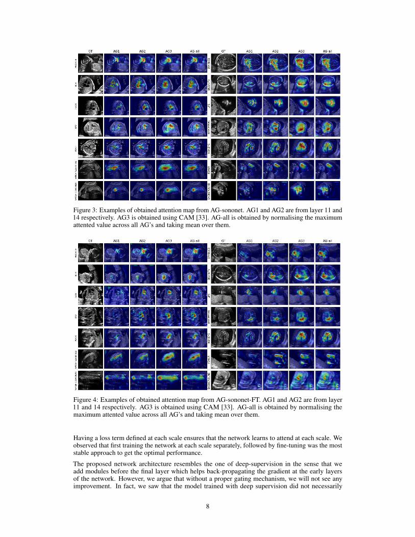
Figure 3: Examples of obtained attention map from AG-sononet. AG1 and AG2 are from layer 11 and14 respectively. AG3 is obtained using CAM [33]. AG-all is obtained by normalising the maximumattented value across all AG’s and taking mean over them.
Figure 4: Examples of obtained attention map from AG-sononet-FT. AG1 and AG2 are from layer11 and 14 respectively. AG3 is obtained using CAM [33]. AG-all is obtained by normalising themaximum attented value across all AG’s and taking mean over them.
Having a loss term defined at each scale ensures that the network learns to attend at each scale. Weobserved that first training the network at each scale separately, followed by fine-tuning was the moststable approach to get the optimal performance.
The proposed network architecture resembles the one of deep-supervision in the sense that weadd modules before the final layer which helps back-propagating the gradient at the early layersof the network. However, we argue that without a proper gating mechanism, we will not see anyimprovement. In fact, we saw that the model trained with deep supervision did not necessarily
8
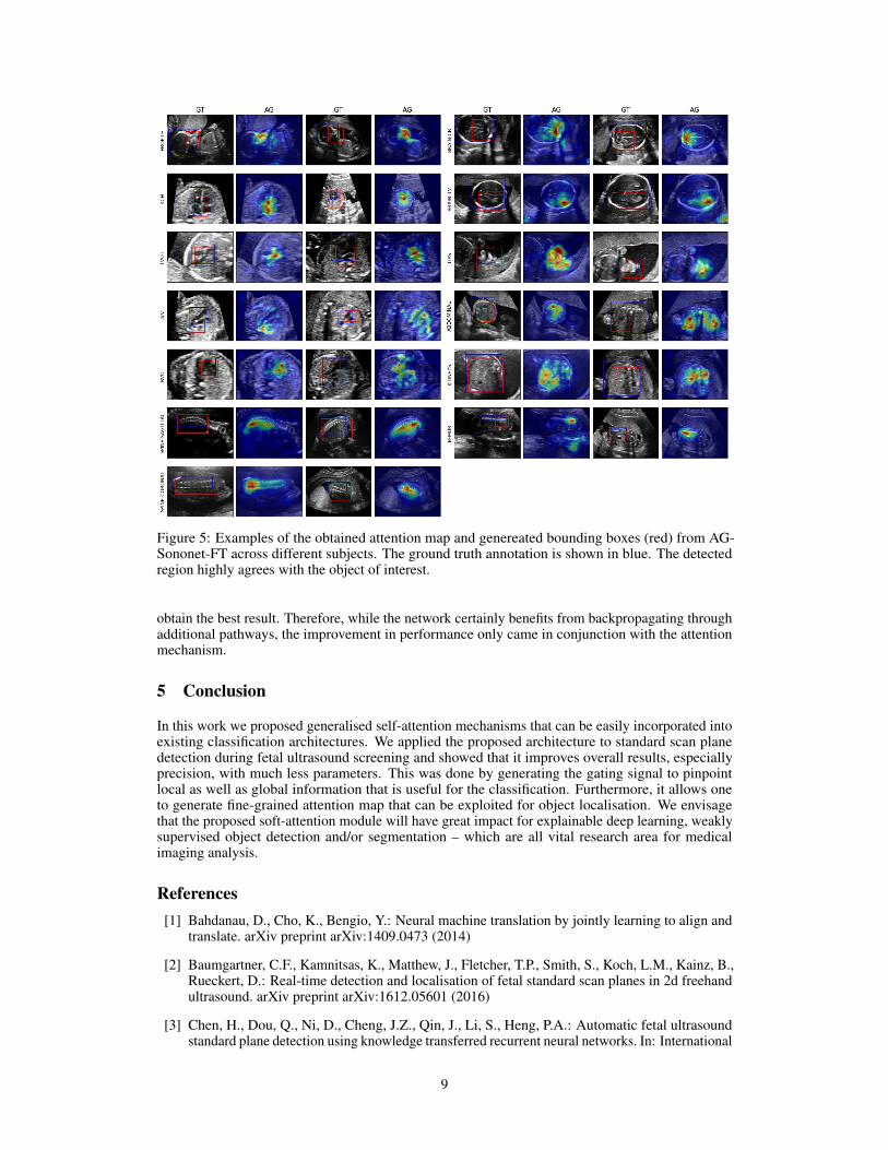
Figure 5: Examples of the obtained attention map and genereated bounding boxes (red) from AG-Sononet-FT across different subjects. The ground truth annotation is shown in blue. The detectedregion highly agrees with the object of interest.
obtain the best result. Therefore, while the network certainly benefits from backpropagating throughadditional pathways, the improvement in performance only came in conjunction with the attentionmechanism.
5 Conclusion
In this work we proposed generalised self-attention mechanisms that can be easily incorporated intoexisting classification architectures. We applied the proposed architecture to standard scan planedetection during fetal ultrasound screening and showed that it improves overall results, especiallyprecision, with much less parameters. This was done by generating the gating signal to pinpointlocal as well as global information that is useful for the classification. Furthermore, it allows oneto generate fine-grained attention map that can be exploited for object localisation. We envisagethat the proposed soft-attention module will have great impact for explainable deep learning, weaklysupervised object detection and/or segmentation – which are all vital research area for medicalimaging analysis.
References[1] Bahdanau, D., Cho, K., Bengio, Y.: Neural machine translation by jointly learning to align and
translate. arXiv preprint arXiv:1409.0473 (2014)
[2] Baumgartner, C.F., Kamnitsas, K., Matthew, J., Fletcher, T.P., Smith, S., Koch, L.M., Kainz, B.,Rueckert, D.: Real-time detection and localisation of fetal standard scan planes in 2d freehandultrasound. arXiv preprint arXiv:1612.05601 (2016)
[3] Chen, H., Dou, Q., Ni, D., Cheng, J.Z., Qin, J., Li, S., Heng, P.A.: Automatic fetal ultrasoundstandard plane detection using knowledge transferred recurrent neural networks. In: International
9
Conference on Medical Image Computing and Computer-Assisted Intervention. pp. 507–514.Springer (2015)
[4] Esteva, A., Kuprel, B., Novoa, R.A., Ko, J., Swetter, S.M., Blau, H.M., Thrun, S.: Dermatologist-level classification of skin cancer with deep neural networks. Nature 542(7639), 115 (2017)
[5] Guan, Q., Huang, Y., Zhong, Z., Zheng, Z., Zheng, L., Yang, Y.: Diagnose like a radiologist:Attention guided convolutional neural network for thorax disease classification. arXiv preprintarXiv:1801.09927 (2018)
[6] Jetley, S., Lord, N.A., Lee, N., Torr, P.: Learn to pay attention. In: International Conference onLearning Representations (2018), https://openreview.net/forum?id=HyzbhfWRW
[7] Kawahara, J., Hamarneh, G.: Multi-resolution-tract cnn with hybrid pretrained and skin-lesiontrained layers. In: International Workshop on Machine Learning in Medical Imaging. pp.164–171. Springer (2016)
[8] Lee, C.Y., Xie, S., Gallagher, P., Zhang, Z., Tu, Z.: Deeply-supervised nets. In: ArtificialIntelligence and Statistics. pp. 562–570 (2015)
[9] Litjens, G.J.S., Kooi, T., Bejnordi, B.E., Setio, A.A.A., Ciompi, F., Ghafoorian, M., van derLaak, J.A.W.M., van Ginneken, B., Sánchez, C.I.: A survey on deep learning in medical imageanalysis. CoRR abs/1702.05747 (2017), http://arxiv.org/abs/1702.05747
[10] Liu, J., Wang, G., Hu, P., Duan, L.Y., Kot, A.C.: Global context-aware attention lstm networksfor 3d action recognition. In: CVPR (2017)
[11] Lu, J., Xiong, C., Parikh, D., Socher, R.: Knowing when to look: Adaptive attention via Avisual sentinel for image captioning. CoRR abs/1612.01887 (2016), http://arxiv.org/abs/1612.01887
[12] Luong, M., Pham, H., Manning, C.D.: Effective approaches to attention-based neural machinetranslation. CoRR abs/1508.04025 (2015), http://arxiv.org/abs/1508.04025
[13] Madani, A., Arnaout, R., Mofrad, M., Arnaout, R.: Fast and accurate view classification ofechocardiograms using deep learning. npj Digital Medicine 1(1), 6 (2018)
[14] Nam, H., Ha, J., Kim, J.: Dual attention networks for multimodal reasoning and matching.CoRR abs/1611.00471 (2016), http://arxiv.org/abs/1611.00471
[15] NHS Screening Programmes: Fetal Anomaly Screen Programme Handbook. NHS (2015)
[16] Pei, W., Baltrusaitis, T., Tax, D.M.J., Morency, L.: Temporal attention-gated model for robust se-quence classification. CoRR abs/1612.00385 (2016), http://arxiv.org/abs/1612.00385
[17] Pesce, E., Ypsilantis, P.P., Withey, S., Bakewell, R., Goh, V., Montana, G.: Learning to detectchest radiographs containing lung nodules using visual attention networks. arXiv preprintarXiv:1712.00996 (2017)
[18] Ren, M., Zemel, R.S.: End-to-end instance segmentation and counting with recurrent attention.CoRR abs/1605.09410 (2016), http://arxiv.org/abs/1605.09410
[19] Roth, H.R., Lee, C.T., Shin, H.C., Seff, A., Kim, L., Yao, J., Lu, L., Summers, R.M.: Anatomy-specific classification of medical images using deep convolutional nets. In: Biomedical Imaging(ISBI), 2015 IEEE 12th International Symposium on. pp. 101–104. IEEE (2015)
[20] Sarraf, S., DeSouza, D.D., Anderson, J., Tofighi, G.: Deepad: Alzheimer’s disease classificationvia deep convolutional neural networks using mri and fmri. bioRxiv (2017), https://www.biorxiv.org/content/early/2017/01/14/070441
[21] Shen, T., Zhou, T., Long, G., Jiang, J., Pan, S., Zhang, C.: Disan: Directional self-attentionnetwork for rnn/cnn-free language understanding. arXiv preprint arXiv:1709.04696 (2017)
[22] Simonyan, K., Zisserman, A.: Very deep convolutional networks for large-scale image recogni-tion. arXiv preprint arXiv:1409.1556 (2014)
10
[23] Wang, F., Jiang, M., Qian, C., Yang, S., Li, C., Zhang, H., Wang, X., Tang, X.: Residualattention network for image classification. arXiv preprint arXiv:1704.06904 (2017)
[24] Wang, X., Girshick, R., Gupta, A., He, K.: Non-local neural networks. arXiv preprintarXiv:1711.07971 (2017)
[25] Wang, X., Peng, Y., Lu, L., Lu, Z., Summers, R.M.: Tienet: Text-image embedding networkfor common thorax disease classification and reporting in chest x-rays. CoRR abs/1801.04334(2018), http://arxiv.org/abs/1801.04334
[26] Xu, K., Ba, J., Kiros, R., Cho, K., Courville, A., Salakhudinov, R., Zemel, R., Bengio, Y.:Show, attend and tell: Neural image caption generation with visual attention. In: InternationalConference on Machine Learning. pp. 2048–2057 (2015)
[27] Yang, Z., He, X., Gao, J., Deng, L., Smola, A.J.: Stacked attention networks for image questionanswering. CoRR abs/1511.02274 (2015), http://arxiv.org/abs/1511.02274
[28] Yaqub, M., Kelly, B., Papageorghiou, A.T., Noble, J.A.: Guided random forests for identificationof key fetal anatomy and image categorization in ultrasound scans. In: International Conferenceon Medical Image Computing and Computer-Assisted Intervention. pp. 687–694. Springer(2015)
[29] Zaharchuk, G., Gong, E., Wintermark, M., Rubin, D., Langlotz, C.: Deep learning in neuroradi-ology. American Journal of Neuroradiology (2018)
[30] Zhang, Z., Chen, P., Sapkota, M., Yang, L.: Tandemnet: Distilling knowledge from medicalimages using diagnostic reports as optional semantic references. In: International Conference onMedical Image Computing and Computer-Assisted Intervention. pp. 320–328. Springer (2017)
[31] Zhang, Z., Xie, Y., Xing, F., McGough, M., Yang, L.: Mdnet: A semantically and visuallyinterpretable medical image diagnosis network. CoRR abs/1707.02485 (2017), http://arxiv.org/abs/1707.02485
[32] Zhao, B., Feng, J., Wu, X., Yan, S.: A survey on deep learning-based fine-grained objectclassification and semantic segmentation. International Journal of Automation and Computing14(2), 119–135 (2017)
[33] Zhou, B., Khosla, A., Lapedriza, A., Oliva, A., Torralba, A.: Learning deep features fordiscriminative localization. In: Computer Vision and Pattern Recognition (CVPR), 2016 IEEEConference on. pp. 2921–2929. IEEE (2016)
[34] Zhu, W., Liu, C., Fan, W., Xie, X.: Deeplung: Deep 3d dual path nets for automated pulmonarynodule detection and classification. CoRR abs/1801.09555 (2018), http://arxiv.org/abs/1801.09555
11
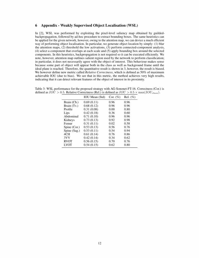
6 Appendix - Weakly Supervised Object Localisation (WSL)
In [2], WSL was performed by exploiting the pixel-level saliency map obtained by guilded-backpropagation, followed by ad-hoc procedure to extract bounding boxes. The same heuristics canbe applied for the given network, however, owing to the attention map, we can device a much efficientway of performing object localisation. In particular, we generate object location by simply: (1) blurthe attention maps, (2) threshold the low activations, (3) perform connected-component analysis,(4) select a component that overlaps at each scale and (5) apply bounding box around the selectedcomponents. In this heuristics, backpropagation is not required so it can be executed efficiently. Wenote, however, attention map outlines salient region used by the network to perform classification;in particular, it does not necessarily agree with the object of interest. This behaviour makes sensebecause some part of object will appear both in the class as well as background frame until theideal plane is reached. Therefore, the quantitative result is shown in 3, however, the result is biased.We however define new metric called Relative Correctness, which is defined as 50% of maximumachievable IOU (due to bias). We see that in this metric, the method achieves very high results,indicating that it can detect relevant features of the object of interest in its proximity.
Table 3: WSL performance for the proposed strategy with AG-Sononet-FT-16. Correctness (Cor.) isdefined as IOU > 0.5. Relative Correctness (Rel.) is defined as IOU > 0.5×max(IOUclass).
IOU Mean (Std) Cor. (%) Rel. (%)
Brain (Cb.) 0.69 (0.11) 0.96 0.96Brain (Tv.) 0.68 (0.12) 0.96 0.96Profile 0.31 (0.08) 0.00 0.80Lips 0.42 (0.18) 0.36 0.60Abdominal 0.71 (0.10) 0.96 0.96Kidneys 0.73 (0.13) 0.92 0.98Femur 0.31 (0.11) 0.02 0.58Spine (Cor.) 0.53 (0.13) 0.56 0.76Spine (Sag.) 0.53 (0.11) 0.54 0.944CH 0.61 (0.14) 0.76 0.863VV 0.42 (0.14) 0.34 0.62RVOT 0.56 (0.15) 0.70 0.76LVOT 0.54 (0.15) 0.62 0.80
12