Embed Size (px)
Citation preview

AtSERPIN1 is an inhibitor of the metacaspase AtMC1-mediatedcell death and autocatalytic processing in planta
Saul Lema Asqui1, Dominique Vercammen2,3, Irene Serrano4, Marc Valls5, Susana Rivas4, Frank Van
Breusegem2,3,6,7, Frank L. Conlon8,9,10,11, Jeffery L. Dangl12,13,14,15,16 and N�uria S. Coll1
1Centre for Research in Agricultural Genomics (CRAG), CSIC-IRTA-UAB-UB, Campus UAB, Bellaterra, Barcelona 08193, Spain; 2Department of Plant Systems Biology, VIB, Ghent 9052,
Belgium; 3Department of Plant Biotechnology and Bioinformatics, Ghent University, Ghent 9052, Belgium; 4LIPM, Universit�e de Toulouse, INRA, CNRS, Castanet-Tolosan, France;
5Department of Genetics, Universitat de Barcelona and Centre for Research in Agricultural Genomics (CSIC-IRTA-UAB-UB) Edifici CRAG, Campus UAB, Bellaterra, Catalonia 08193,
Spain; 6Department of Medical Protein Research, VIB, Ghent 9000, Belgium; 7Department of Biochemistry, Ghent University, Ghent 9000, Belgium; 8Department of Biology, University of
North Carolina, Chapel Hill, NC 27599, USA; 9Department of Genetics, University of North Carolina, Chapel Hill, NC 27599, USA; 10McAllister Heart Institute, University of North
Carolina, Chapel Hill, NC 27599, USA; 11Lineberger Cancer Center, University of North Carolina, Chapel Hill, NC 27599, USA; 12Department of Biology, University of North Carolina,
Chapel Hill, NC 27599-3280, USA; 13Howard Hughes Medical Institute, University of North Carolina, Chapel Hill, NC 27599-3280, USA; 14Curriculum in Genetics and Molecular Biology,
University of North Carolina, Chapel Hill, NC 27599-3280, USA; 15Carolina Center for Genome Sciences, University of North Carolina, Chapel Hill, NC 27599-3280, USA; 16Department
of Microbiology and Immunology, University of North Carolina, Chapel Hill, NC 27599-3280, USA
Author for correspondence:Nuria S. Coll
Tel: +34 93 5606600Email: [email protected]
Received: 20 June 2016
Accepted: 16 December 2016
New Phytologist (2017)doi: 10.1111/nph.14446
Key words: hypersensitive response (HR),metacaspase, plant–pathogen interactions,programmed cell death, protease, serpin.
Summary
� The hypersensitive response (HR) is a localized programmed cell death phenomenon that
occurs in response to pathogen recognition at the site of attempted invasion. Despite more
than a century of research on HR, little is known about how it is so tightly regulated and how
it can be contained spatially to a few cells.� AtMC1 is an Arabidopsis thaliana plant metacaspase that positively regulates the HR. Here,
we used an unbiased approach to identify new AtMC1 regulators. Immunoaffinity purification
of AtMC1-containing complexes led us to the identification of the protease inhibitor AtSer-
pin1.� Our data clearly showed that coimmunoprecipitation between AtMC1 and AtSerpin1 and
formation of a complex between them was lost upon mutation of the AtMC1 catalytic site,
and that the AtMC1 prodomain was not required for the interaction. AtSerpin1 blocked
AtMC1 self-processing and inhibited AtMC1-mediated cell death. Our results constitute an
in vivo example of a Serpin acting as a suicide inhibitor in plants, reminiscent of the activity of
animal or viral serpins on immune/cell death regulators, including caspase-1.� These results indicate a conserved function of a protease inhibitor on cell death regulators
from different kingdoms with unrelated modes of action (i.e. caspases vs metacaspases).
Introduction
Metacaspases are a family of proteases present in plants, fungiand protozoa (Uren et al., 2000). They are members of the clanCD of cysteine proteases, featuring a unique tertiary structure ter-med the caspase-hemoglobinase fold that encloses a conservedcysteine–histidine catalytic dyad (Aravind & Koonin, 2002).Members of this superfamily also include caspases, animal cys-teine proteases with aspartate specificity that have essential rolesin inflammation and cell death. Metacaspases have typically beencompared with caspases, but research has shown that despite theiroverall active site configuration, their mode of action might beradically different. First, they have different substrate sequencecleavage requirements: lysine or arginine for metacaspases andaspartic acid for caspases; second, metacaspase activity is notblocked by caspase inhibitors; and third, according to their
structure, metacaspases cannot form dimers the way caspases do(Salvesen et al., 2016). Regardless of these differences, metacas-pases have been shown to act as cell death regulators (Coll et al.,2010, 2014; Tsiatsiani et al., 2011; Wrzaczek et al., 2015),although it is not clear how they exert this function or how theyare regulated.
Arabidopsis metacaspases are the best characterized amongplants. The Arabidopsis genome encodes nine metacaspases,AtMC1–AtMC3 (type I) and AtMC4–AtMC9 (type II). Themain difference between type I and type II metacaspases is thepresence (type I)/absence (type II) of an N-terminal prodomain.According to the crystal structure of the Trypanosomas type Imetacaspase (McLuskey et al., 2012), the prodomain rests as a lidon top of the catalytic fold, presumably precluding substrateaccess until cleavage or a conformational change occurs. In agree-ment with that proposal, the prodomains of AtMC1 and AtMC2
� 2017 The Authors
New Phytologist� 2017 New Phytologist Trust
New Phytologist (2017) 1www.newphytologist.com
Research

were shown to negatively regulate their function (Coll et al.,2010).
We previously demonstrated that AtMC1 is a positive regu-lator of pathogen-triggered hypersensitive response (HR) celldeath (Coll et al., 2010), a plant reaction that takes placelocally at the site of attempted pathogen attack upon recogni-tion of the invader. In this context, catalytic integrity wascritical for AtMC1 cell death function. Conditional overex-pression of AtMC1 resulted in ectopic cell death, which indi-cated that a tight regulation of this protein must be in placein order to prevent uncontrolled cell death. So far, two nega-tive regulators of AtMC1 have been identified: AtMC2 andthe negative regulator of plant defense and HR, LSD1 (Diet-rich et al., 1994). AtMC1 interacted with LSD1 through itsprodomain, whereas AtMC1 and AtMC2 did not interactwith each other and the details of their interplay remain tobe clarified (Coll et al., 2010).
Here, we used an unbiased approach to identify new regulatorsof AtMC1 activity. Immunoaffinity purification of AtMC1-containing complexes led us to the identification of the proteaseinhibitor AtSerpin1. Our data clearly showed that coimmunopre-cipitation between AtMC1 and AtSerpin1 was dependent on anintact AtMC1 catalytic site and the prodomain was not requiredfor the interaction. Furthermore, AtSerpin1 blocked AtMC1 self-processing and inhibited AtMC1-mediated cell death. Together,our findings uncover AtSerpin1 as a bona fide AtMC1 inhibitorin planta.
Materials and Methods
Plant material and growth conditions
All experiments were performed using Arabidopsis thaliana (L.)Heynh.accession Col-0. Mutant atmc1 and the transgenic atmc1PAtMC1::AtMC1-HA and lsd1 atmc1 PAtMC1::AtMC1-HA werepreviously described in Coll et al. (2010). Mutant atserpin1 andtransgenic Col-0 35S::AtSerpin1-HA lines were described inLampl et al. (2010). Arabidopsis was grown under short-day con-ditions (9 : 15 h, 22 : 20°C, light : dark).
Nicotiana benthamiana was grown under long-day conditions(16 : 8 h, 25 : 22°C, light : dark).
DNA constructs
To obtain the PAtMC1::HA-AtMC1 construct, the product of anoverlapping PCR using AtMC1 promoter and HA-AtMC1 wasdirectionally cloned into pENTR/D/TOPO Gateway vector(Invitrogen) and recombined into the plant binary Gateway-compatible vector pGWB1 (Nakagawa et al., 2007).
The AtSerpin1 full-length cDNA was directionally cloned intopENTR/D/TOPO Gateway vector (Invitrogen) and recombinedinto the plant binary Gateway-compatible vector pGWB641 toobtain 35S::AtSerpin1-YFP or pGWB642 to obtain 35S::YFP-AtSerpin1 (Nakamura et al., 2010).
For subcellular localization experiments, fusion of fluorescentproteins to AtMC1, AtMC1-DN, AtMC1-CA and AtSerpin1
was performed using a multisite GATEWAY cloning strategy(Invitrogen) described previously (Gu & Innes, 2011). Briefly,the full-length open reading frames of AtMC1, AtMC1-DN,AtMC1-CA and AtSerpin1 were cloned into the donor vectorpBSDONR P1-P4 (an ampicillin-resistant vector derived frompDONR221 P1-P4 from Invitrogen) (Gu & Innes, 2011) usingthe BP cloning Kit (Invitrogen). C-terminal eGFP (Cormacket al., 1996) and C-terminal red fluorescent protein (RFP)(Campbell et al., 2002) were cloned into the entry vectorpBSDONR P4r-P2. To fuse AtMC1, AtMC1-DN, AtMC1-CAand AtSerpin1 with the epitope tags, the P1–P4 clones wererecombined with corresponding P4r-P2 and the desired destina-tion vectors using Gateway LR clonase II (Invitrogen). ForAtMC1, AtMC1-DN, and AtMC1-CA, the earlier describedpBSDONR constructs were recombined with the destinationvector pEarleyGate100 (Earley et al., 2006). For AtSerpin1, thecorresponding pBSDONR constructs were recombined with thesteroid-inducible destination vector pBAV154 (Vinatzer et al.,2006).
Plasmids were transformed into Agrobacterium tumefaciensstrain GV3101 (pMP90) by electroporation and plated intoselective Luria-Bertani (LB) agar plates.
Stable transformation of Arabidopsis thaliana
atmc1 PAtMC1::AtMC1-HA plants were transformed with 35S::AtSerpin1-YFP using Agrobacterium tumefaciens (GV3101)-mediated floral dip as previously described (Clough & Bent,1998). Homozygous double transgenic lines were selected onMurashige & Skoog (MS) media supplemented with 20 lg ml�1
Basta (glufosinate-ammonium). atmc1 plants were transformedwith PAtMC1::HA-AtMC1 using A. tumefaciens (GV3101)-mediated floral dip as previously described (Clough & Bent,1998). Homozygous transgenic lines were selected on MS mediasupplemented with 50 lg ml�1 hygromycin.
Immunoisolation of protein complexes from Arabidopsis
Homozygous atmc1 or lsd1 atmc1 PAtMC1::AtMC1-HA were usedfor immunoaffinity purification 24 h after spraying them with300 lMBTH as previously described (Coll et al., 2010). All mate-rials and reagents for immunoisolation were from Thermo FisherScientific (Waltham, MA, USA), unless otherwise stated.AtMC1–HA complexes were immunoisolated using magneticbeads (M-270 epoxy Dynabeads) conjugated to a monoclonal HAantibody (MMS-101P MONO HA.11; Covance, Princeton, NJ,USA). For conjugation, 100 lg of antibody were first washed andconcentrated to the final volume of 100 ll by three rounds ofadding 500 ll of phosphate-buffered saline and centrifugation at9000 g for 15 min at 4°C using a centrifugal filter (Amicon;Merck Millipore, Billerica, MA, USA). One hundred micrograms(100 ll) of clean antibody were added to 18 mg of magnetic beadswashed with 0.1M sodium phosphate buffer (pH 7.4). Then,120 ll of 3 M ammonium sulfate and 120 ll of 0.1 M sodiumphosphate buffer (pH 7.4) were added. Beads were incubatedovernight at 30°C on an orbital shaker. In parallel, 10 g of leaves
New Phytologist (2017) � 2017 The Authors
New Phytologist� 2017 New Phytologist Trustwww.newphytologist.com
Research
NewPhytologist2

were snap-frozen in liquid nitrogen and cryogenically lysed usinga Retsch MM 301 Mixer Mill (20 cycles of 180 s at 30 Hz)(Retsch, Newtown, PA, USA). The frozen powder was resus-pended in cold lysis buffer (20 mM K-HEPES pH 7.4, 110 mMCH3CO2K, 2 mM MgCl2, 0.1% Tween-20, 1 lM ZnCl2, 1 lMCaCl2), supplemented with protease inhibitor cocktail (Roche).Lysates were homogenized using a polytron (two cycles of 15 s).The cell lysate was centrifuged at 1000 g at 4°C for 10 min. Thesupernatant was collected and filtered through a syringe-driven5 lm filter to remove any particles from lysate that did not pellet.The protein concentration of the lysate was measured, to adjust allsamples to the same concentration. A fraction of the lysate wasreserved to run on a sodium dodecyl sulfate–polyacrylamide gelelectrophoresis (SDS-PAGE) (total protein).
Beads were equilibrated by washing three times with lysisbuffer. After that, 1 ml of lysate was added to the beads. Beadswere then incubated at 4°C on an orbital shaker. After 1 h, tubeswere placed on a Dynal (Thermo Fisher Scientific, Waltham,MA, USA) magnetic rack, flowthrough was discarded and beadswere washed five times with lysis buffer. Forty microliters of elu-tion buffer (49 NuPAGE LDS Sample Buffer and 209NuPAGE Sample Reducing Buffer; Thermo Fisher Scientific)supplemented with 2 ll of 1 M iodoacetamide were added persample and incubated 1 h at room temperature. Subsequently,samples were transferred to 70°C to elute proteins off the beads.Eluted proteins were partially separated at 150 V on a NuPAGE3–12% 1-mm-thick Bis-Tris protein gel using NuPAGE MOPSrunning buffer, supplemented with NuPAGE Antioxidant in theinner gel chamber. The gel was stained using Coomassie Sim-plyBlue SafeStain (Thermo Fisher Scientific). Each lane wasexcised and divided in eight fragments. Fragments were individu-ally analyzed by mass spectrometry.
Protein characterization using mass spectrometry
Excised SDS-PAGE gel bands were in-gel-digested with trypsin.The extracted peptides were separated on a nanoAcquity HPLCsystem (Waters Corp., Milford, MA, USA) with a 360 lmOD9 75 lm ID analytical column (14 cm of Magic 5 lmC18AQ resin; Michrom Biosciences, Bruker Corp., Billerica,MA, USA). The liquid chromatography (LC) system was directlyconnected through an electrospray ionization source interfaced toan LTQ Orbitrap Velos ion trap mass spectrometer (ThermoFisher Scientific) controlled by XCALIBUR software (v.2.1.0.1140;Thermo Fisher Scientific) and operated in the data-dependentmode in which the initial MS scan recorded the mass to charge(m/z) ratios of ions over the range 400–2000. Raw files weresearched using MASCOT (v.2.3.02; Matrix Science, WyndhamPlace, UK). Search parameters included peptide mass toleranceof 10 ppm and fragment ion tolerance of 0.8 mass units.
Isolated protein complexes were analyzed by mass spectrometryas previously described (Kaltenbrun et al., 2013). Briefly, tandemmass spectra were extracted by PROTEOME DISCOVERER (ThermoFisher Scientific), and all MS/MS samples were analyzed withSEQUEST (v.1.2.0.208; Thermo Fisher Scientific), set up to searchthe Arabidopsis UniProt-SwissProt protein sequence database,
assuming digestion pattern with trypsin. SCAFFOLD (v. Scaf-fold_3_00_06; Proteome Software Inc., Portland, OR, USA) wasused to validate MS/MS-based peptide and proteinidentifications. Peptide sequences were deemed a match if theycould be established at > 95.0% probability as specified by thePEPTIDEPROPHET algorithm (Keller et al., 2002). In turn, proteinidentifications were deemed a match if they could be established at> 99.0% probability by the PROTEINPROPHET algorithm and haveat least one sequenced peptide.
The mass spectrometry proteomics data have been deposited tothe ProteomeXchange Consortium via the PRIDE partner reposi-tory with the dataset identifier PXD005134 and 10.6019/PXD005134. The description of all files uploaded to Pro-teomeXchange can be found in Supporting Information Table S1.
Transient protein expression in N. benthamiana
Transient A. tumefaciens-mediated transformation ofN. benthamiana leaves was performed as previously described(Coll et al., 2010). Whole N. benthamiana leaves (c. 500 mgeach) transiently expressing the constructs to test together withthe anti-silencing vector p19 (Voinnet et al., 2003) – Dex::AtMC1-HA + 35S::p19 (OD600 0.2 + 0.1), Dex::AtMC1-HA + 35S::AtSerpin1 + 35S::p19 (OD600 0.2 + 0.4 + 0.1), Dex::AtMC1-ΔN-HA + 35S::p19 (OD600 0.4 + 0.1), Dex::AtMC1-ΔN-HA + 35S::AtSerpin1 + 35S::p19 (OD600 0.4 + 0.4 + 0.1),Dex::AtMC1-CA-HA + 35S::p19 (OD600 0.2 + 0.1), Dex::AtMC1-CA-HA + 35S::AtSerpin1 + 35S::p19 (OD600 0.2 +0.4 + 0.1), 35S::AtSerpin1 + 35S::p19 (OD600 0.4 + 0.1) – werefrozen in liquid nitrogen before further processing for proteinextraction.
Colocalization experiment
Agrobacterium tumefaciens cultures carrying the indicated con-structs were grown and resuspended in water at OD600 = 0.8(Wroblewski et al., 2005). For coexpression of multiple con-structs, suspensions were mixed in equal ratios. Bacterial suspen-sion mixtures were infiltrated using a needleless syringe. Sampleswere collected for microscopic imaging 40 h after infiltration.
Confocal laser scanning microscopy
Confocal laser scanning microscopy was performed on a LeicaSP2 AOBS inverted confocal microscope (Leica Microsystems,Wetzlar, Germany) equipped with a 940, numerical aperture-1.2 water objective. eGFP fusion was excited with a 488 nmArgon laser and detected using a 505–530 bandpass emission fil-ter. RFP fusions were excited using a 561 nm He-Ne laser anddetected using a custom 595–620 nm bandpass emission filter.
Coimmunoprecipitation assays and protein analysis
Frozen samples were ground using a mortar and pestle on 3 ml oflysis buffer (200 mM K-HEPES pH 7.4, 1.1 M C2H3KO,20 mM MgCl2, 1% Tween-20, 10 lM ZnCl2, 10 lM CaCl2),
� 2017 The Authors
New Phytologist� 2017 New Phytologist TrustNew Phytologist (2017)
www.newphytologist.com
NewPhytologist Research 3

supplemented with 5 mM dithiothreitol (DTT), and proteaseinhibitor cocktail (Roche). Homogenized samples were filteredthrough miracloth (Millipore) and collected in 15 ml tubes. Sam-ples were then centrifuged for 15 min at 4°C and 7000 g to sepa-rate the cell debris from the total protein extract.
For coimmunoprecipitation, total protein extracts were dilutedto 2 mg ml�1 and incubated with 50 ll of anti green fluorescentprotein (GFP) magnetic beads (MACS; Miltenyi Biotec, Ber-gisch, Gladbach, Germany), for 2 h at 4°C under constant rota-tion. Bound proteins were eluted according to the manufacturer’sinstructions. Twenty-five micrograms of total protein (input), anequal volume of flowthrough (unbound) and 20 ll of eluate wereloaded onto an SDS-PAGE gel. Immunoblots were performedusing 1 : 5000 anti-GFP mouse monoclonal antibody (clone B-2;Santa Cruz Biotechnology, Dallas, TX, USA) or 1 : 5000 mono-clonal anti-HA-HRP (clone 3F10; Roche).
To test binding between AtMC1 and AtSerpin1, we followedthe protocol established by Roberts et al. (2011). In essence, pro-tein extraction was performed using a Laemmli buffer (120 mMTris-HCl pH 6.8, 4% SDS (w/v), 15% glycerol (v/v), 0,02%bromophenol (w/v)) with or without DTT (5 mM) as reducingagent. Proteins were then separated on 10% or 7.5% SDS-PAGEgels and probed with 1 : 5000 monoclonal anti-HA-HRP (clone3F10; Roche) or 1 : 5000 anti-GFP mouse monoclonal antibody(clone B-2; Santa Cruz Biotechnology).
Chemical treatments
Dexamethasone was applied to N. benthamiana leaf surfacesusing cottonballs to induce expression of AtMC1 forms under thecontrol of the dexamethasone promoter (Coll et al., 2010) 48 hafter agroinfiltration. Leaves were treated with 0.2 lM dexam-ethasone and samples were collected 24 h later.
Three-week-old Arabidopsis plants were sprayed with 150 lMbenzol(1,2,3)thiadiazole-7-carbothioic acid S-methyl ester.
The proteasome inhibitor MG-132 (2 lM in 0.2% dimethylsulfoxide; Sigma-Aldrich) was applied on N. benthamiana leafsurfaces 24 h after dexamethasone treatment and samples werecollected 12 h later.
Cell death analyses
Ion leakage assays were carried out using 3-wk-oldN. benthamiana plants transiently expressing different proteincombinations (see the ‘Transient protein expression inN. benthamiana’ subsection earlier). At least four leaves per com-bination were used. Fifteen disks were extracted per leaf with acork borer (7 mm diameter) and placed on a plate with distilledwater during 1 h. After that, 10 disks were placed in a flaskcontaining 10 ml of distilled water (six replicates per sample).Conductivity was measured over time using a hand electricalconductivitymeter (FG3-FIVEGO, Schwerzenbach,Switzerland).
Trypan blue staining of N. benthamiana leaves was performedby collecting whole leaves in 50 ml tubes (each leaf in a separatetube) 72 h after cell death induction and covered with a total of
35 ml of a 1 : 3 dilution of trypan blue stock solution (Keoghet al., 1980). The tubes were incubated in previously boiled waterfor 15 min, and then cleared overnight with chloral hydrate onan orbital shaker. Pictures were taken 72 h after cell death induc-tion. Pictures were also processed adding a binary mask usingIMAGEJ (v.1.50i; National Institues of Health, Bethesda, MD,USA).
Arabidopsis single cell death assay was performed according toColl et al. (2010).
Results
Two forms of AtMC1 coexist in plants: full length and aprodomain-less
We previously showed that removal of AtMC1 prodomainenhances its pro-death activity in Arabidopsis (Coll et al., 2010).To determine whether prodomain removal occurs in nature, wecompared the processing pattern of N-terminally vs C-terminallytagged protein from transgenic plants expressing HA-AtMC1 orAtMC1-HA under the control of AtMC1 promoter (PAtMC1::HA-AtMC1 or PAtMC1::AtMC1-HA, respectively) (Fig. S1).Immunoblot using an anti-HA antibody clearly showed thatAtMC1-HA is present in both its full-length form (41 kDa) anda fragment of c. 36 kDa, presumably corresponding to an auto-processed form (Fig. 1). By contrast, in plants expressing HA-AtMC1 only the full-length form of the protein could bedetected by anti-HA immunoblot. This supports the idea thatthe spontaneously formed smaller AtMC1 fragment correspondsto the prodomain-less version of the protein, indicative ofN-terminal processing.
Immunoaffinity isolation identified the protease inhibitorAtSerpin1 as part of AtMC1-containing protein complexes
In order to identify regulators of AtMC1 activity under nativeconditions, we performed immunoaffinity purification ofAtMC1-containing complexes using rosette leaves from 4-wk-oldatmc1 plants expressing AtMC1 under the control of its own pro-moter (atmc1 PAtMC1::AtMC1-HA, Fig. S1). Immunopurifiedproteins were partially resolved using SDS-PAGE (Fig. S2) andin-gel-digested with trypsin. Analysis was performed using nLC-
Fig. 1 The prodomain of the metacaspase Arabidopsis thaliana AtMC1 iscleaved in plants. Immunoblot using anti-HA antibodies of threeindependent atmc1 PAtMC1::HA-AtMC1 T1 lines (7–9) compared withhomozygous atmc1 PAtMC1::HA-AtMC1. Black and white arrows indicatefull-length and cleaved AtMC1, respectively.
New Phytologist (2017) � 2017 The Authors
New Phytologist� 2017 New Phytologist Trustwww.newphytologist.com
Research
NewPhytologist4

tandem MS (MS/MS) on an LTQ Orbitrap Velos. Two indepen-dent biological replicates were performed. Raw MS/MS spectrawere first analyzed by SEQUEST database searches (PROTEOME DIS-
COVERER) and loaded into SCAFFOLD for further analysis. Proteinidentifications from all replicates were filtered using stringentconfidence parameters (see the Materials and Methods section).Through this analysis we detected 154–1300 proteins per condi-tion, among which we identified the protease inhibitor AtSer-pin1. AtSerpin1 was not identified in the experimental controlsused (Table S2).
AtSerpin1 inhibits AtMC1 autoprocessing in vivo
Conditional overexpression of AtMC1 in N. benthamiana leavesresulted in accumulation of both full-length and processed formssimilar to when expressed in Arabidopsis under the control of itsnative promoter (Fig. S3). AtMC1 cleavage was inhibited bymutation of the predicted catalytic site (Fig. 2b, AtMC1-CA),indicative of autoprocessing. AtSerpin1 coexpression withAtMC1 also blocked autocatalytic processing of AtMC1 (Figs 2a,S3). Absence of processed AtMC1 is consistent with the idea thatAtSerpin1 acts as an inhibitor of AtMC1 cleavage in planta.
Interestingly, AtSerpin1 caused a sharp decrease in the levels ofAtMC1-CA (Fig. 2b). To explain this observation, we hypothe-sized that AtSerpin1 might bind and/or alter the structure of theAtMC1 catalytic mutant, ultimately leading to its proteasomaldegradation. To test whether the reduction in AtMC1-CA pro-tein levels caused by AtSerpin1 coexpression was proteasome-dependent, we treated agroinfiltrated N. benthamiana leaves with
the proteasome inhibitor MG-132 or left them untreated. Asshown in Fig. 2(b) the severe reduction of AtMC1-CA levelscaused by AtSerpin1 coexpression could be totally reverted byproteasome inhibition. However, the AtSerpin1-dependentdegradation of AtMC1-CA might partly occur during proteinextraction as a result of the reducing conditions caused by DTT.The data also indicated that, when expressed alone, AtMC1-CAis also partly degraded by the proteasome, as the levels increaseafter MG-132 treatment when compared with untreated leaves.By contrast, the native full-length and processed forms of AtMC1do not seem to be subjected to proteasome-mediated degradation(Fig. 2b).
AtMC9 was shown to cleave AtSerpin1 in vitro at the pre-dicted cleavage site (R351, corresponding to the predicted reac-tive center loop of AtSerpin1) (Vercammen et al., 2004).Similarly, we observed that AtSerpin1 was cleaved by AtMC1(Fig. 3). This cleavage was partly dependent on an intact catalyticsite, as the levels of the cleaved fragment were lower or notdetectable when YFP-AtSerpin1 (Fig. S4b) or AtSerpin1-YFP(Figs 3, S4a) was coexpressed with AtMC1-CA. In this experi-mental system, endogenous N. benthamiana proteases, includingmetacaspases, may also have the capacity to cleave AtSerpin1 asindicated by the cleavage products that appear on the sampleexpressing AtSerpin1 alone (Fig. 3, lane 5).
AtSerpin1 colocalizes and coimmunoprecipitates withAtMC1
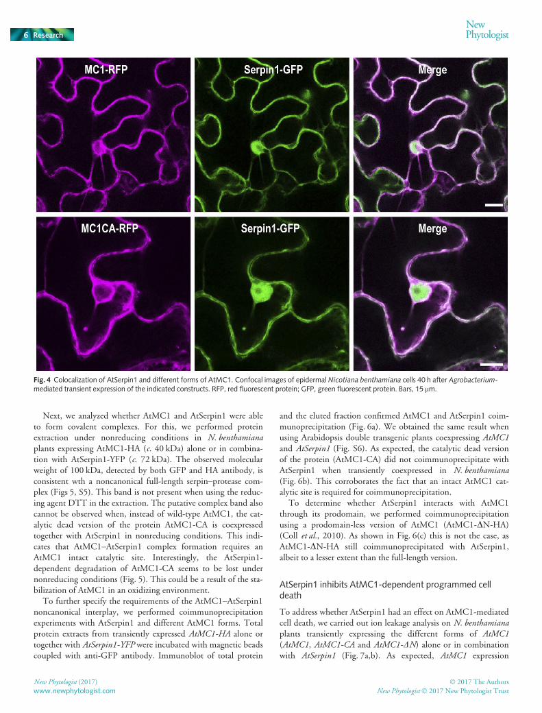
To assess whether AtSerpin1 and AtMC1 colocalize, we obtainedfluorescently tagged versions of both proteins (AtSerpin1-GFPand AtMC1-RFP) and tested their subcellular localization underconfocal laser scanning microscopy. As shown in Fig. 4, bothproteins colocalize in the cytoplasm. In addition, AtSerpin-GFP,but not AtMC1, is visualized in the nucleus of N. benthamianacells when transiently overexpressed, probably as the result ofGFP cleavage.
(a)
(b)
Fig. 2 AtSerpin1 inhibits AtMC1 autoprocessing and enhancesproteasome-mediated degradation of the catalytic dead version of theprotein (AtMC1-C220A). Wild-type and catalytic dead (CA) AtMC1-HAversions were transiently expressed in Nicotiana benthamiana leaves aloneor in combination with AtSerpin1-YFP. Leaves were treated with 2 lMMG-132 (+) or left untreated (�). Proteins were extracted 12 h later andeither Coomassie-stained or immunoblotted using anti-HA antibodies todetect, respectively, AtMC1-HA (a) or AtMC1-CA-HA (b). Black andwhite arrows indicate full-length and cleaved AtMC1, respectively.
Fig. 3 AtSerpin1 is cleaved by AtMC1. AtSerpin1-YFP alone or incombination with AtMC1-HA or AtMC1-CA-HA was transiently expressedin Nicotiana benthamiana leaves. Total proteins were extracted and 50 lgwere either Coomassie-stained or immunoblotted using anti-GFPantibody. The black arrowhead indicates full-length AtSerpin1, whereasthe white arrowhead points at the putative AtSerpin1 cleaved form. YFP,yellow fluorescent protein.
� 2017 The Authors
New Phytologist� 2017 New Phytologist TrustNew Phytologist (2017)
www.newphytologist.com
NewPhytologist Research 5
Next, we analyzed whether AtMC1 and AtSerpin1 were ableto form covalent complexes. For this, we performed proteinextraction under nonreducing conditions in N. benthamianaplants expressing AtMC1-HA (c. 40 kDa) alone or in combina-tion with AtSerpin1-YFP (c. 72 kDa). The observed molecularweight of 100 kDa, detected by both GFP and HA antibody, isconsistent wth a noncanonical full-length serpin–protease com-plex (Figs 5, S5). This band is not present when using the reduc-ing agent DTT in the extraction. The putative complex band alsocannot be observed when, instead of wild-type AtMC1, the cat-alytic dead version of the protein AtMC1-CA is coexpressedtogether with AtSerpin1 in nonreducing conditions. This indi-cates that AtMC1–AtSerpin1 complex formation requires anAtMC1 intact catalytic site. Interestingly, the AtSerpin1-dependent degradation of AtMC1-CA seems to be lost undernonreducing conditions (Fig. 5). This could be a result of the sta-bilization of AtMC1 in an oxidizing environment.
To further specify the requirements of the AtMC1–AtSerpin1noncanonical interplay, we performed coimmunoprecipitationexperiments with AtSerpin1 and different AtMC1 forms. Totalprotein extracts from transiently expressed AtMC1-HA alone ortogether with AtSerpin1-YFP were incubated with magnetic beadscoupled with anti-GFP antibody. Immunoblot of total protein
and the eluted fraction confirmed AtMC1 and AtSerpin1 coim-munoprecipitation (Fig. 6a). We obtained the same result whenusing Arabidopsis double transgenic plants coexpressing AtMC1and AtSerpin1 (Fig. S6). As expected, the catalytic dead versionof the protein (AtMC1-CA) did not coimmunoprecipitate withAtSerpin1 when transiently coexpressed in N. benthamiana(Fig. 6b). This corroborates the fact that an intact AtMC1 cat-alytic site is required for coimmunoprecipitation.
To determine whether AtSerpin1 interacts with AtMC1through its prodomain, we performed coimmunoprecipitationusing a prodomain-less version of AtMC1 (AtMC1-DN-HA)(Coll et al., 2010). As shown in Fig. 6(c) this is not the case, asAtMC1-DN-HA still coimmunoprecipitated with AtSerpin1,albeit to a lesser extent than the full-length version.
AtSerpin1 inhibits AtMC1-dependent programmed celldeath
To address whether AtSerpin1 had an effect on AtMC1-mediatedcell death, we carried out ion leakage analysis on N. benthamianaplants transiently expressing the different forms of AtMC1(AtMC1, AtMC1-CA and AtMC1-DN) alone or in combinationwith AtSerpin1 (Fig. 7a,b). As expected, AtMC1 expression
Fig. 4 Colocalization of AtSerpin1 and different forms of AtMC1. Confocal images of epidermal Nicotiana benthamiana cells 40 h after Agrobacterium-mediated transient expression of the indicated constructs. RFP, red fluorescent protein; GFP, green fluorescent protein. Bars, 15 lm.
New Phytologist (2017) � 2017 The Authors
New Phytologist� 2017 New Phytologist Trustwww.newphytologist.com
Research
NewPhytologist6

caused an increase in ectopic cell death over time. This cell deathwas also observed when expressing the DN form of the proteinand was almost completely abolished in leaves expressing the cat-alytic dead version of AtMC1 (AtMC1-CA). AtSerpin1 clearlyblocked AtMC1- and AtMC1-DN-dependent cell death, furthersupporting the idea that it acts as a bona fide inhibitor of AtMC1activity.
In order to genetically substantiate this claim, we monitoredcell death on atmc1, atserpin1 and atmc1 atserpin1 mutant plantsand plants overexpressing AtSerpin1 (AtSerpin1-HA) comparedwith the wild-type. As a cell death trigger, we used Pseudomonassyringae pv tomato expressing the type III effector avrRpm1 (PtoDC3000(avrRpm1)), which causes AtMC1-dependent HR celldeath mediated by the RPM1 receptor (Coll et al., 2010). Two-week-old plants were infected with Pto DC3000(avrRpm1) andcell death was quantified using a single cell death assay (Collet al., 2010, 2014). As previously observed, the lack of atmc1resulted in a sharp decrease in RPM1-mediated cell death(Fig. 7c). Double atmc1 atserpin1 mutants showed reduced celldeath levels, lower than the wild-type but higher than atmc1plants, whereas atserpin1 mutants behaved similarly to the wild-type. Together, these data suggests that AtSerpin1 acts as a nega-tive regulator of cell death mediated by AtMC1 via (an) addi-tional protease(s) in Arabidopsis.
Discussion
AtMC1 is an autocatalytically active protease in planta
In the past we speculated that AtMC1 and the animalinflammatory caspase-1, may share certain functional similarities(Coll et al., 2010, 2014). This was based on the following facts:analogous catalytic domain structure; both are positive regulatorsof cell death induced upon immune receptor activation; presenceof a prodomain that contains cell death-related motifs; and both
are negatively regulated by an inactive member of their family(caspase-11 in the case of caspase-1 and AtMC2 in the case ofAtMC1).
In animals, the caspase-1-dependent response is very well char-acterized (Davis et al., 2011). Upon immune receptor activation,supramolecular structures termed inflammasomes are assembled,recruiting multiple copies of full-length, inactive caspase-1.Within inflammasomes, many caspase-1 units are rapidly self-processed through induced proximity, releasing p10 and p20subunits that then assemble into the active form, consisting oftwo p20–p10 heterodimers. Active caspase-1 can then carry outmultiple processes in response to the initial inflammatory signal,generating a fast and efficient response.
Fig. 5 Binding between AtMC1 and AtSerpin1 occurs only in presence ofan intact catalytic site. Full-length AtMC1-HA and AtMC1-CA-HA weretransiently expressed in Nicotiana benthamiana leaves alone or incombination with AtSerpin1-YFP. Total proteins were extracted underreducing (+DTT) or nonreducing (�DTT) conditions. Fifty micrograms ofprotein were separated on a sodium dodecyl sulfate–polyacrylamide gelelectrophoresis and either Coomassie-stained or immunoblotted usinganti-HA antibody. The asterisk indicates the putative AtMC1-AtSerpin1
complex.
Fig. 6 AtMC1-AtSerpin1 coimmunoprecipitation occurs independently ofAtMC1 prodomain but an intact catalytic center is required. DifferentAtMC1-HA forms (FL, full length; CA, catalytic dead; DN, prodomain-less)were transiently expressed in Nicotiana benthamiana leaves alone or incombination with AtSerpin1-YFP. Total proteins were extracted (input),incubated with magnetic beads coupled to green fluorescent protein (GFP)And, after stringent washes, proteins bound to the beads were eluted(bound). Input and bound fractions were either Coomassie-stained orprobed against anti-GFP to detect AtSerpin or against anti-HA to detectAtMC1-FL (a), AtMC1-CA (b) or AtMC1-DN (c). The black arrowheadindicates full-length AtMC1, whereas the white arrowhead points at theputative AtMC1 cleaved form. WB, western blot; IP, immunoprecipitation.[Correction added after online publication 3 February 2017: the duplicatedanti-GFP immunoblot in (a) has been replaced with the correctimmunoblot and the Coomassie-stained panels in (a) and (b) have beenswitched to match their immunoblots. For clarity full images of all gels andCoomassies are now shown.]
� 2017 The Authors
New Phytologist� 2017 New Phytologist TrustNew Phytologist (2017)
www.newphytologist.com
NewPhytologist Research 7

Our data support the idea that AtMC1 is catalytically self-processed to release the prodomain and a fragment encompassingthe p20 and p10 subunits. Several other type I metacaspases havebeen shown to self-cleave (Meslin et al., 2007; Moss et al., 2007;Zalila et al., 2011; Li et al., 2015). However, the link betweencleavage and activation has not yet been established. In fact, for
the Trypanosoma brucei metacaspase TbMCA2, it was shown thatcleavage is not critical for activation (Moss et al., 2007). In Ara-bidopsis, AtMC1 appears to be maintained in an equipoisebetween the full-length and the processed form. Conditionaloverexpression of the processed form caused faster and moreextensive cell death than the full-length version of AtMC1 (Collet al., 2010). This was in agreement with the idea that theprodomain is a negative regulator of type I metacaspase activityand might act as a physical barrier for substrate access (McLuskeyet al., 2012). However, we did not find any stress condition thatincreased the accumulation of the processed form (data notshown). This might imply that AtMC1 activation does not occurvia enhanced processing but rather by relocalization of theprodomain-less form to a different subcellular compartmentwhere relevant substrates are located or by post-translationalmodifications.
In fact, we still do not know whether AtMC1 or other type Imetacaspases are recruited to death-induced supramolecularstructures comparable to the inflammasome through theirprodomain. Interestingly, the two metacaspase crystal structuresresolved to date indicate that homodimerization through theequivalent interfaces as observed in caspases seem impossible(McLuskey et al., 2012; Wong et al., 2012). Thus, it remains anopen question how AtMC1 and other metacaspases becomeactive.
The interplay between AtSerpin1 and AtMC1 in plants
In the context of AtMC1 regulation, mechanisms to prevent itsactivation under homeostatic conditions must be in place toavoid unrestrained cell death propagation. Two negative regula-tors of AtMC1 function were identified earlier: AtMC2 andLSD1 (Coll et al., 2010). Here, we have uncovered a novel nega-tive regulator of AtMC1: the protease inhibitor AtSerpin1, whichcan block AtMC1 autocatalytic activity in planta and preventAtMC1-mediated cell death.
Serpins are a superfamily of proteins, initially described as ser-ine protease inhibitors, but now known to include cysteine pro-tease inhibitors and even noninhibitory members (Gettins,2002). Serpins are the most widespread and abundant peptidaseinhibitors, being present in all domains of life and even in viruses(Rawlings et al., 2004). In animals, serpins have been involved incell survival, development and host defense against pathogens(Silverman et al., 2010). Inhibitory serpins have been termed ‘sui-cide inhibitors’ or ‘molecular mousetraps’ because of their modeof action: cleavage by the target protease sets off a conformationalchange in the serpin whereby the protease is flipped and becomestrapped against the serpin protein body, with its catalytic corecrushed and the consequent decrease in proteolytic activity(Huntington et al., 2000).
The Arabidopsis genome encodes eight serpin genes (Fluhret al., 2012). Among them, the most abundant and best charac-terized is AtSerpin1. The in vitro proteolytic activities of two typeII metacaspases, AtMC4 and AtMC9, were shown to be inhib-ited by AtSerpin1 (Vercammen et al., 2004). In turn, AtMC9was demonstrated to cleave AtSerpin1 in vitro at the predicted
(a)
(b)
(c)
Fig. 7 AtSerpin1 inhibits AtMC1-mediated cell death. (a) Ion leakageassay using Nicotiana benthamiana leaves expressing the different formsof AtMC1-HA alone or in combination with AtSerpin1-YFP. Cell deathwas induced by dexamethasone treatment 48 h after agroinfiltration of theleaves (time 0). Each time point corresponds to the SE of six replicatescontaining 10 leaf disks each (� 29 SE). Letters indicate significantdifferences following post-ANOVA Tukey’s honest significant differencetest (a = 0.05). This experiment was repeated five times with similarresults. (b) Pictures of representative trypan blue-stained leaves expressingthe different forms of AtMC1 alone or in combination with AtSerpin1 72 hafter cell death induction (upper row) or the same images processed usingImage J to highlight dead areas (lower row). (c) Single cell death assay ofthe indicated Arabidopsis thaliana lines. Dead cells (trypan blue-positive)were counted under an optical microscope 12 h after infection with250 000 colony-forming units ml�1 Pto DC3000(avrRpm1). Valuesindicate the average of 50 samples per genotype� 29 SE. Letters indicatesignificant differences following post-ANOVA Tukey’s honest significantdifference test (a = 0.05). The experiment is representative of fiveindependent replicates.
New Phytologist (2017) � 2017 The Authors
New Phytologist� 2017 New Phytologist Trustwww.newphytologist.com
Research
NewPhytologist8

cleavage site (reactive center loop) in a dose-dependent manner(Vercammen et al., 2004). Our in vivo data indicate that AtMC1might also be able to cleave AtSerpin1 through its reactive centerloop, as the size of two fragments generated would be in agree-ment with the corresponding prediction.
Both AtMC4 and AtMC9 were shown to act as positive regu-lators of cell death in different contexts: AtMC4 was involved inpathogen-triggered cell death (Watanabe & Lam, 2011), whereasAtMC9 participates in developmental cell death, mediating cellclearance in late stages of xylem formation (Bollhoner et al.,2013). In neither case has it been established that inhibition ofAtMC activity by AtSerpin1 affects metacaspase function in thesephysiological contexts.
Here, we demonstrated inhibition of AtMC1 by AtSerpin1in vivo. This inhibition was monitored as loss of self-processingof AtMC1 when coexpressed with AtSerpin1. Unfortunately, wehave so far not been able to directly measure AtMC1 proteolyticactivity, and thus we have not been able to assess the effect ofAtSerpin1 on it. We also observed that AtMC1-CA was partlydegraded by the proteasome and coexpression with AtSerpin1dramatically exacerbated AtMC1-CA proteasomal degradation.AtMC1-CA is more prone to aggregation than its wild-typecounterpart (Coll et al., 2014) and cells have evolved differentsurveillance mechanisms to detect and eliminate potentially toxicprotein aggregates. Thus, it is not surprising that at least part ofthe AtMC1-CA pool is delivered to the proteasome for degrada-tion. The fact that AtSerpin1 coexpression enhances AtMC1-CAproteasome-mediated degradation could indicate that AtSerpin1might interact and/or induce a conformational change inAtMC1-CA that further promotes its aggregation and, conse-quently, its elimination via the proteasome.
AtMC1 and AtSerpin1 colocalize and coimmunoprecipitate,indicating a possible interaction between the two proteins. In con-trast to the previously shown AtMC1-LSD1 coimmunoprecipita-tion (Coll et al., 2010), the prodomain was not required for theinteraction between AtMC1 and AtSerpin. The interactionbetween AtMC1-DN and AtSerpin1 still took place, although itwas weaker than with the full-length version of AtMC1, indicat-ing a less stable interaction when the prodomain was missing.However, an intact AtMC1 catalytic center was required for thecoimmunoprecipitation. This was presumably not the case forAtMC9-AtSerpin1 interaction, as a catalytic-dead version ofAtMC9 was used as a bait for AtSerpin1 identification in the yeasttwo-hybrid assay (Vercammen et al., 2006). The apparent dis-crepancy of the two observations might be explained by a weakeraffinity between AtSerpin1 and catalytic-dead metacaspasemutants. These potentially weak interactions are probably elimi-nated by the stringent washes of a coimmunoprecipitation experi-ment, whereas they remain intact in yeast two-hybrid assays.
AtSerpin1 as an inhibitor of cell death proteases
In plants, AtSerpin1 might act as a pan-metacaspase inhibitor oreven as a more general cell death protease inhibitor. AtSerpin1was shown to covalently bind and to modulate the activity of thecell death protease RD21 (Lampl et al., 2010, 2013). Our data
also indicate that AtSerpin1 forms a noncanonical complex withAtMC1, detected under nonreducing conditions. This discrep-ancy between the mode of interaction between different metacas-pases and AtSerpin1 could be a result of the different mode ofaction of AtMC1 and 9, their localization (subcellular and tissu-lar) and the processes in which they are involved.
Interestingly, the interplay between AtMC1 and AtSerpin1seem to involve the full-length rather than the cleaved versions ofthe proteins. Although AtMC1 may be able to cleave AtSerpin1,the size of the complex detected, as well as the coimmunoprecipi-tated fragments indicate a noncanonical mode of action wherebyAtSerpin1 would bind and inactivate AtMC1 but this interactionwould not involve self-cleavage or direct AtSerpin1 cleavage byAtMC1. The fact that AtMC1 catalytic activity is required forthe interaction with AtSerpin1 could suggest the involvement ofa third partner that needs to be cleaved in order for the inhibitionto take place or nondetectable modifications of AtMC1 and/orAtSerpin1.
Overexpression of AtSerpin1 or a mutation in the proteaseRD21 led to reduced cell death after infection with thenecrotrophic fungi Botrytis cinerea and Sclerotina sclerotiorum butenhanced cell death in response to the hemibiotrophic fungusColletotrichum higgisianum (Lampl et al., 2013). In our condi-tions, infection with Pto DC3000(avrRpm1), a hemibiotrophicbacterium that causes HR in A. thaliana Col-0 background viathe RPM1 receptor, resulted in decreased cell death in plantsoverexpressing AtSerpin1, comparable to atmc1 mutants. Doubleatmc1 atserpin1 mutants displayed an intermediate phenotypebetween wild-type and atmc1 plants, which indicates that AtMC1is negatively regulated by AtSerpin1 and also that atserpin1 maycontrol other proteases involved in this cell death process beyondatmc1. Whether AtSerpin1 inhibits AtMC1-regulated processesby directly interacting with AtMC1 or indirectly by modulatingthe activity of downstream proteases induced by AtMC1 remainsan open question. Inhibition of AtMC1-mediated cell death byAtSerpin1 is also demonstrated by the dramatic effect of AtSer-pin1 when transiently coexpressed with death-inducing forms ofAtMC1 in N. benthamiana leaves. Discrepancy of results betweenC. higgisianum (Lampl et al., 2013) and Pto, may be explained bythe fact that, despite both being hemibiotrophs, their lifestyle,time and mode of infection are radically different and thus it isdifficult to compare cell death outcomes at a given time point.
Inhibition of different cell death proteases by AtSerpin1(Fig. S7) positions it as a conceivable guardian of cell homeosta-sis, preventing uncontrolled proteolysis of potentially dangerousproteins. The balance between the levels of AtSerpin1 and thelevels of potentially active death proteases might be a powerfulmodulator of cell fate. Under normal conditions, molecule-by-molecule inactivation may serve as an effective surveillance mech-anism that prevents uncontrolled cell death.
In animals, intracellular serpins have been shown to be majorregulators of cell death and inflammation and this function partlyoccurs through direct inhibition of specific proteases, includingcaspases (Silverman et al., 2010). Interestingly, this mechanismhas been coopted by certain viruses, which are able to produceserpins in their hosts that block defenses (Gettins, 2002). For
� 2017 The Authors
New Phytologist� 2017 New Phytologist TrustNew Phytologist (2017)
www.newphytologist.com
NewPhytologist Research 9
example, the viral serpin CrmA efficiently inhibits caspase-1,escaping immune surveillance by the host (Ray et al., 1992). Infact, in many species, lack of certain serpins results in severe phe-notypes or cell death (Silverman et al., 2010). The fact thatatserpin1 mutants have no dramatic phenotypes might beexplained by the genetic redundancy within the serpin family inArabidopsis, but more experimental evidence is needed toconfirm this hypothesis.
The work presented here contributes to a better understandingof cell death control in plants. We have demonstrated that AtSer-pin1 acts in vivo as an inhibitor of AtMC1-mediated cell death,emerging as a potential inhibitor of cell death proteases in plants.These results are of major evolutionary significance, as they indi-cate a conserved function of a protease inhibitor on cell deathregulators from different kingdoms with unrelated mode ofaction (i.e. caspases vs metacaspases).
Acknowledgements
The authors would like to kindly thank Marc Planas, SimonStael, Guy Salvesen, Vera Bonardi and Ignacio Rubio-Somozafor helpful comments. We thank David Smalley and NedyalkaDicheva from the University of North Carolina Michael HookerProteomics Center for technical assistance with protein identifi-cation. Anouk Brackenier, Dominique Eeckhout and Geert DeJaeger from Ghent University and the Flanders Institute ofBiotechnology (VIB) are thanked for sharing and helping to ana-lyze the TAP results. Thanks to Robert Fluhr for sharing trans-genic AtSerpin-HA seeds and anti-Serpin antibody, and to RogerInnes for kindly providing Gateway vectors for colocalizationstudies. This work was funded by projects AGL2013-46898-R(MINECO, Spain) to N.S.C. and M.V., EU-Marie CurieActions (PCDMC-321738 and PIIF-331392) and BP_B 00030from the Catalan Government to N.S.C.; S.L.A. holds a fellow-ship Convocatoria Abierta 2013 Primera Fase (grant agreementno. AR2Q4017) from SENESCYT, Ecuador; J.L.D. is an Inves-tigator of the Howard Hughes Medical Institute, supported bythe HHMI and the Gordon and Betty Moore Foundation(GBMF3030); I.S. is supported by an AgreenSkills fellowshipwithin the EU Marie-Curie FP7 COFUND People Programme(grant agreement no. 267196); S.R. is supported by the FrenchLaboratory of Excellence project ‘TULIP’ (ANR-10-LABX-41;ANR-11-IDEX-0002-02). We acknowledge financial supportfrom the Spanish Ministry of Economy and Competitiveness,through the ‘Severo Ochoa Programme for Centres of Excellencein R&D 2016-2019 (SEV-2015-0533)’.
Author contributions
S.L.A. performed and designed experiments, analyzed data andwrote the manuscript. D.V. performed and designed experimentsand analyzed data. I.S. performed experiments and analyzed data.M.V. designed experiments and analyzed data. S.R. designedexperiments, analyzed data and wrote the manuscript. F.v.B.designed experiments, analyzed data and wrote the manuscript.F.L.C. designed experiments, analyzed data and wrote the
manuscript. J.L.D. designed experiments, analyzed data andwrote the manuscript. N.S.C. designed the research, performedexperiments, analyzed data and wrote the manuscript. All authorsreviewed the manuscript.
References
Aravind L, Koonin EV. 2002. Classification of the caspase-hemoglobinase fold:
detection of new families and implications for the origin of the eukaryotic
separins. Proteins 46: 355–367.Bollhoner B, Zhang B, Stael S, Denance N, Overmyer K, Goffner D, Van
Breusegem F, Tuominen H. 2013. Post mortem function of AtMC9 in xylem
vessel elements. New Phytologist 200: 498–510.Campbell RE, Tour O, Palmer AE, Steinbach PA, Baird GS, Zacharias DA,
Tsien RY. 2002. A monomeric red fluorescent protein. Proceedings of theNational Academy of Sciences, USA 99: 7877–7882.
Clough SJ, Bent AF. 1998. Floral dip: a simplified method for Agrobacterium-
mediated transformation of Arabidopsis thaliana. Plant Journal 16: 735–743.Coll NS, Smidler A, Puigvert M, Popa C, Valls M, Dangl JL. 2014. The plant
metacaspase AtMC1 in pathogen-triggered programmed cell death and aging:
functional linkage with autophagy. Cell Death and Differentiation 21: 1399–1408.
Coll NS, Vercammen D, Smidler A, Clover C, Van Breusegem F, Dangl JL,
Epple P. 2010. Arabidopsis type I metacaspases control cell death. Science 330:1393–1397.
Cormack BP, Valdivia RH, Falkow S. 1996. FACS-optimized mutants of the
green fluorescent protein (GFP). Gene 173: 33–38.Davis BK, Wen H, Ting JP. 2011. The inflammasome NLRs in immunity,
inflammation, and associated diseases. Annual Review of Immunology 29: 707–735.
Dietrich RA, Delaney TP, Uknes SJ, Ward ER, Ryals JA, Dangl JL. 1994.
Arabidopsis mutants simulating disease resistance response. Cell 77: 565–577.Earley KW, Haag JR, Pontes O, Opper K, Juehne T, Song K, Pikaard CS.
2006. Gateway-compatible vectors for plant functional genomics and
proteomics. Plant Journal 45: 616–629.Fluhr R, Lampl N, Roberts TH. 2012. Serpin protease inhibitors in plant
biology. Physiologia Plantarum 145: 95–102.Gettins PG. 2002. Serpin structure, mechanism, and function. Chemical Reviews102: 4751–4804.
Gu Y, Innes RW. 2011. The KEEP ON GOING protein of Arabidopsis recruits
the ENHANCED DISEASE RESISTANCE1 protein to trans-Golgi network/
early endosome vesicles. Plant Physiology 155: 1827–1838.Huntington JA, Read RJ, Carrell RW. 2000. Structure of a serpin–proteasecomplex shows inhibition by deformation. Nature 407: 923–926.
Kaltenbrun E, Greco TM, Slagle CE, Kennedy LM, Li T, Cristea IM, Conlon
FL. 2013. A Gro/TLE-NuRD corepressor complex facilitates Tbx20-dependent
transcriptional repression. Journal of Proteome Research 12: 5395–5409.Keller A, Nesvizhskii AI, Kolker E, Aebersold R. 2002. Empirical statistical
model to estimate the accuracy of peptide identifications made by MS/MS and
database search. Analytical Chemistry 74: 5383–5392.Keogh RC, Deverall BJ, McLeod S. 1980. Comparison of histological and
physiological responses to Phakopsora pachyrhizi in resistant and susceptible
soybean. Transactions of the British Mycological Society 74: 329–333.Lampl N, Alkan N, Davydov O, Fluhr R. 2013. Set-point control of RD21
protease activity by AtSerpin1 controls cell death in Arabidopsis. Plant Journal74: 498–510.
Lampl N, Budai-Hadrian O, Davydov O, Joss TV, Harrop SJ, Curmi PM,
Roberts TH, Fluhr R. 2010. Arabidopsis AtSerpin1, crystal structure and in vivointeraction with its target protease RESPONSIVE TO DESICCATION-21
(RD21). Journal of Biological Chemistry 285: 13550–13560.Li M, Wang H, Liu J, Hao P, Ma L, Liu Q. 2015. The apoptotic role of
metacaspase in Toxoplasma gondii. Frontiers in Microbiology 6: 1560.McLuskey K, Rudolf J, Proto WR, Isaacs NW, Coombs GH, Moss CX,
Mottram JC. 2012. Crystal structure of a Trypanosoma bruceimetacaspase.
Proceedings of the National Academy of Sciences, USA 109: 7469–7474.
New Phytologist (2017) � 2017 The Authors
New Phytologist� 2017 New Phytologist Trustwww.newphytologist.com
Research
NewPhytologist10

Meslin B, Barnadas C, Boni V, Latour C, De Monbrison F, Kaiser K, Picot S.
2007. Features of apoptosis in Plasmodium falciparum erythrocytic stage
through a putative role of PfMCA1 metacaspase-like protein. Journal ofInfectious Diseases 195: 1852–1859.
Moss CX, Westrop GD, Juliano L, Coombs GH, Mottram JC. 2007.
Metacaspase 2 of Trypanosoma brucei is a calcium-dependent cysteine peptidase
active without processing. FEBS Letters 581: 5635–5639.Nakagawa T, Kurose T, Hino T, Tanaka K, Kawamukai M, Niwa Y,
Toyooka K, Matsuoka K, Jinbo T, Kimura T. 2007. Development of
series of gateway binary vectors, pGWBs, for realizing efficient construction
of fusion genes for plant transformation. Journal of Bioscience andBioengineering 104: 34–41.
Nakamura S, Mano S, Tanaka Y, Ohnishi M, Nakamori C, Araki M, Niwa T,
Nishimura M, Kaminaka H, Nakagawa T et al. 2010. Gateway binary vectorswith the bialaphos resistance gene, bar, as a selection marker for plant
transformation. Bioscience, Biotechnology, and Biochemistry 74: 1315–1319.Rawlings ND, Tolle DP, Barrett AJ. 2004. Evolutionary families of peptidase
inhibitors. Biochemical Journal 378: 705–716.Ray CA, Black RA, Kronheim SR, Greenstreet TA, Sleath PR, Salvesen GS,
Pickup DJ. 1992. Viral inhibition of inflammation: cowpox virus encodes an
inhibitor of the interleukin-1b converting enzyme. Cell 69: 597–604.Roberts TH, Ahn JW, Lampl N, Fluhr R. 2011. Plants and the study of serpin
biology.Methods in Enzymology 499: 347–366.Salvesen GS, Hempel A, Coll NS. 2016. Protease signaling in animal and plant-
regulated cell death. FEBS Journal 283: 2577–2598.Silverman GA, Whisstock JC, Bottomley SP, Huntington JA, Kaiserman D,
Luke CJ, Pak SC, Reichhart JM, Bird PI. 2010. Serpins flex their muscle: I.
Putting the clamps on proteolysis in diverse biological systems. Journal ofBiological Chemistry 285: 24299–24305.
Tsiatsiani L, Van Breusegem F, Gallois P, Zavialov A, Lam E, Bozhkov PV.
2011.Metacaspases. Cell Death and Differentiation 18: 1279–1288.Uren AG, O’Rourke K, Aravind LA, Pisabarro MT, Seshagiri S, Koonin EV,
Dixit VM. 2000. Identification of paracaspases and metacaspases: two ancient
families of caspase-like proteins, one of which plays a key role in MALT
lymphoma.Molecular Cell 6: 961–967.Vercammen D, Belenghi B, van de Cotte B, Beunens T, Gavigan JA, De Rycke
R, Brackenier A, Inze D, Harris JL, Van Breusegem F. 2006. Serpin1 of
Arabidopsis thaliana is a suicide inhibitor for metacaspase 9. Journal ofMolecular Biology 364: 625–636.
Vercammen D, van de Cotte B, De Jaeger G, Eeckhout D, Casteels P, Vandepoele
K, Vandenberghe I, Van Beeumen J, Inze D, Van Breusegem F. 2004. Type II
metacaspases Atmc4 and Atmc9 of Arabidopsis thaliana cleave substrates afterarginine and lysine. Journal of Biological Chemistry 279: 45329–45336.
Vinatzer BA, Teitzel GM, Lee MW, Jelenska J, Hotton S, Fairfax K, Jenrette J,
Greenberg JT. 2006. The type III effector repertoire of Pseudomonas syringaepv. syringae B728a and its role in survival and disease on host and non-host
plants.Molecular Microbiology 62: 26–44.Voinnet O, Rivas S, Mestre P, Baulcombe D. 2003. An enhanced transient
expression system in plants based on suppression of gene silencing by the p19
protein of tomato bushy stunt virus. Plant Journal 33: 949–956.Watanabe N, Lam E. 2011. Arabidopsis metacaspase 2d is a positive mediator of
cell death induced during biotic and abiotic stresses. Plant Journal 66: 969–982.
Wong AH, Yan C, Shi Y. 2012. Crystal structure of the yeast metacaspase Yca1.
Journal of Biological Chemistry 287: 29251–29259.Wroblewski T, Tomczak A, Michelmore R. 2005.Optimization of
Agrobacterium-mediated transient assays of gene expression in lettuce, tomato
and Arabidopsis. Plant Biotechnology Journal 3: 259–273.Wrzaczek M, Vainonen JP, Stael S, Tsiatsiani L, Help-Rinta-Rahko H,
Gauthier A, Kaufholdt D, Bollhoner B, Lamminmaki A, Staes A et al. 2015.GRIM REAPER peptide binds to receptor kinase PRK5 to trigger cell death in
Arabidopsis. EMBO Journal 34: 55–66.Zalila H, Gonzalez IJ, El-Fadili AK, Delgado MB, Desponds C, Schaff C, Fasel
N. 2011. Processing of metacaspase into a cytoplasmic catalytic domain
mediating cell death in Leishmania major.Molecular Microbiology 79: 222–239.
Supporting Information
Additional Supporting Information may be found online in theSupporting Information tab for this article:
Fig. S1 Schematic representation of all constructs used in thisstudy.
Fig. S2 Immunoprecipitation of AtMC1 native form in Arabidopsisthaliana.
Fig. S3 Comparison of AtMC1 expression levels when expressedunder native vs dexamethasone-inducible promoter.
Fig. S4 Binding between AtMC1 and AtSerpin1.
Fig. S5 AtMC1 and AtSerpin1 co-immunoprecipitate in Arabidopsisthaliana.
Fig. S6 AtSerpin1 is cleaved by AtMC1.
Fig. S7 AtSerpin1, an inhibitor of cell death proteases in plants.
Table S1 Identification of AtMC1 and AtSerpin1 by LC-MS/MS after HA immunoaffinity purification
Table S2 Identification of AtMC1 and AtSerpin1 by LC-MS/MS after TAP purification
Please note: Wiley Blackwell are not responsible for the contentor functionality of any Supporting Information supplied by theauthors. Any queries (other than missing material) should bedirected to the New Phytologist Central Office.
� 2017 The Authors
New Phytologist� 2017 New Phytologist TrustNew Phytologist (2017)
www.newphytologist.com
NewPhytologist Research 11