Embed Size (px)
Citation preview

Atlas (C1) Primary Listings
ASR – RTC
ASL – LTC
PIR – RTC (RPA)
PIL – LTC (LPA)

Definitions:
Stance Line: Line in which the feet will be parallel to.
Torque: Applying a rotation of the contact hand or arm in order to change the vector of force (line of drive) during the spinal correction.
Superior Torque: Applying a torque where the chiropractor’s fingers will rotate toward the patient’s head.
Inferior Torque: Applying a torque where the chiropractor’s fingers will rotate toward the patient’s feet.
Superior Hand/Foot: The chiropractor’s hand which is closest to the patient’s head during the adjustment.
Inferior Hand/Foot: The chiropractor’s hand which is closest to the patient’s head during the adjustment.
Drive Guide Line: Line of drive, direction of force during the spinal correction.
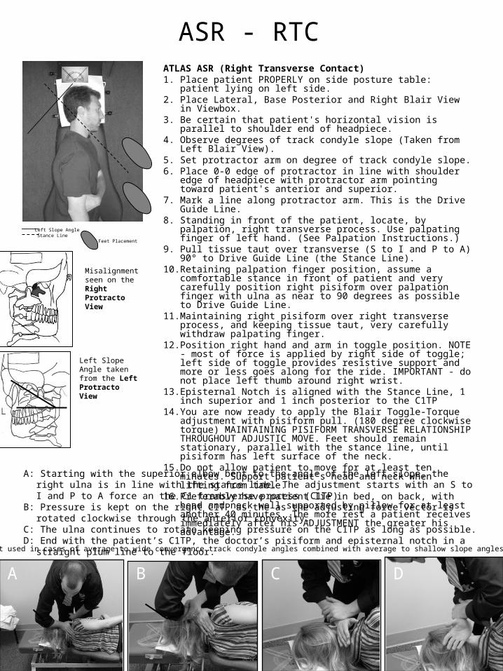
ASR - RTCATLAS ASR (Right Transverse Contact) 1. Place patient PROPERLY on side posture table: patient lying on left
side. 2. Place Lateral, Base Posterior and Right Blair View in Viewbox. 3. Be certain that patient's horizontal vision is parallel to shoulder end of
headpiece. 4. Observe degrees of track condyle slope (Taken from Left Blair View).5. Set protractor arm on degree of track condyle slope. 6. Place 0-0 edge of protractor in line with shoulder edge of headpiece
with protractor arm pointing toward patient's anterior and superior. 7. Mark a line along protractor arm. This is the Drive Guide Line. 8. Standing in front of the patient, locate, by palpation, right transverse
process. Use palpating finger of left hand. (See Palpation Instructions.) 9. Pull tissue taut over transverse (S to I and P to A) 90° to Drive Guide
Line (the Stance Line). 10. Retaining palpation finger position, assume a comfortable stance in
front of patient and very carefully position right pisiform over palpation finger with ulna as near to 90 degrees as possible to Drive Guide Line.
11. Maintaining right pisiform over right transverse process, and keeping tissue taut, very carefully withdraw palpating finger.
12. Position right hand and arm in toggle position. NOTE - most of force is applied by right side of toggle; left side of toggle provides resistive support and more or less goes along for the ride. IMPORTANT - do not place left thumb around right wrist.
13. Episternal Notch is aligned with the Stance Line, 1 inch superior and 1 inch posterior to the C1TP
14. You are now ready to apply the Blair Toggle-Torque adjustment with pisiform pull. (180 degree clockwise torque) MAINTAINING PISIFORM TRANSVERSE RELATIONSHIP THROUGHOUT ADJUSTIC MOVE. Feet should remain stationary, parallel with the stance line, until pisiform has left surface of the neck.
15. Do not allow patient to move for at least ten minutes. Support patient's head and neck when lifting from table.
16. Preferably have patient lie in bed, on back, with head and neck well supported by pillow for at least another 40 minutes. The more rest a patient receives immediately after his ADJUSTMENT the greater his advantage.
Left Slope AngleStance Line
Misalignment seen on the Right Protracto View
Left Slope Angle taken from the Left Protracto View
A: Starting with the superior elbow bent to the angle of the left slope, the right ulna is in line with the stance line. The adjustment starts with an S to I and P to A force an the C1 transverse process (C1TP)
B: Pressure is kept on the right C1TP as the as the adjusting force vector is rotated clockwise through the anterior convexity.
C: The ulna continues to rotate keeping pressure on the C1TP as long as possible.D: End with the patient’s C1TP, the doctor’s pisiform and episternal notch in a straight plum line to the floor.
A B C D
Note: Best used in cases of average to wide convergence track condyle angles combined with average to shallow slope angles
Feet Placement

ASL - LTCATLAS ASL (Left Transverse Contact) 1. Place patient PROPERLY on side posture table: patient lying on right
side. 2. Place Lateral, Base Posterior and Left Blair View in Viewbox. 3. Be certain that patient's horizontal vision is parallel to shoulder end of
headpiece. 4. Observe degrees of track condyle slope (Taken from Right Blair View).5. Set protractor arm on degree of track condyle slope. 6. Place 0-0 edge of protractor in line with shoulder edge of headpiece
with protractor arm pointing toward patient's anterior and superior. 7. Mark a line along protractor arm. This is the Drive Guide Line. 8. Standing in front of the patient, locate, by palpation, left transverse
process. Use palpating finger of right hand. (See Palpation Instructions.)
9. Pull tissue taut over transverse (S to I and P to A) 90° to Drive Guide Line (the Stance Line).
10. Retaining palpation finger position, assume a comfortable stance in front of patient and very carefully position left pisiform over palpation finger with ulna as near to 90 degrees as possible to Drive Guide Line.
11. Maintaining left pisiform over left transverse process, and keeping tissue taut, very carefully withdraw palpating finger.
12. Position left hand and arm in toggle position. NOTE - most of force is applied by left side of toggle; right side of toggle provides resistive support and more or less goes along for the ride. IMPORTANT - do not place left thumb around right wrist.
13. Episternal Notch is aligned with the Stance Line, 1 inch superior and 1 inch posterior to the C1TP
14. You are now ready to apply the Blair Toggle-Torque adjustment with pisiform pull. (180 degree counter-clockwise torque) MAINTAINING PISIFORM TRANSVERSE RELATIONSHIP THROUGHOUT ADJUSTIC MOVE. Feet should remain stationary, parallel with the stance line, until pisiform has left surface of the neck.
15. Do not allow patient to move for at least ten minutes. Support patient's head and neck when lifting from table.
16. Preferably have patient lie in bed, on back, with head and neck well supported by pillow for at least another 40 minutes. The more rest a patient receives immediately after his ADJUSTMENT the greater his advantage.
Right Slope AngleStance Line
Misalignment seen on the Left Protracto View
Right Slope Angle taken from the Right Protracto View
A: Starting with the superior elbow bent to the angle of the right slope, the left ulna is in line with the stance line. The adjustment starts with an S to I and P to A force an the C1 transverse process (C1TP)
B: Pressure is kept on the right C1TP as the as the adjusting force vector is rotated counter-clockwise through the anterior convexity.
C: The ulna continues to rotate keeping pressure on the C1TP as long as possible.D: End with the patient’s C1TP, the doctor’s pisiform and episternal notch in a straight plum line to the floor.
A
B C D
Note: Best used in cases of average to wide convergence track condyle angles combined with average to shallow slope angles
Feet Placement
A

A: Standing behind the patient, feet in a straight away stance and parallel to the right convexity, bring tissue pull and contact hand P to A and S to I. Contacting the posterior-superior C1TP.
B: Set the carpal angle from the right convergence angle found on the Base Posterior. The carpal line is the flat surface of the doctor’s posterior wrist, along the carpal bones.
C: The stabilization hand is brought in, wrapping the thumb around the wrist and antecubital fossas are straight across from each other.
D: End with the a shallow, no torque adjustment. Antecubital fossas are kept straight across from each other.
PIR – RTC (RPA)ATLAS PIR (Right Transverse Contact) (Right Sub-Arch)1. Place patient PROPERLY on side posture table: patient lying on left side.
Sagittal plane of patient's head should be parallel to transverse surface of headpiece. Patient's visual plane should be 90 degrees to long axis of adjusting table.
2. Place Lateral, Base Posterior, and Left Blair View in Viewbox. 3. Set protractor arm on degree measurement of posterior aspect of right
condyle convexity. 4. Place 0-0 line parallel to shoulder edge of headpiece with protractor arm
pointing P to A and S to I in relation with patient. Mark a line along protractor arm. This is the Stance Line.
5. Change protractor arm setting (or use another protractor) to the degree of right condyle convergence. (Take from Base Posterior View).
6. Stand behind patient with feet parallel to stance line, hips and shoulders 90 degrees to stance line. Use a straight away stance. Retain this position.
7. Locate, by palpation, the posterior-superior aspect of the right transverse process and hold with palpating finger of right hand.
8. Have assistant hold protractor on a level, just superior to the patient's head with protractor arm pointing to A to P and I to S (toward ceiling) and with 0-0 line parallel to stance line and floor until contact and adjustic set-up is completed.
9. Pulling tissue taut, take contact with left pisiform, carpal line parallel to protractor arm (as discussed in class), with hips, shoulders and ulna 90 degrees to stance line; feet parallel to stance line. Very carefully withdraw palpation finger. Snug contact of left pisiform over posterior superior aspect of right transverse should now exist.
10. Place right part of toggle (thumb around wrist) for the stabilization arm. 11. Line of drive is from posterior-superior to anterior-inferior. USE NO
TORQUE! IMPORTANT! USE NO TORQUE! 12. Do not allow patient to move for at least ten minutes. Support patient's head
and neck when lifting from table. 13. Preferably have patient lie in bed, on back, with head and neck well
supported by pillow for at least another 40 minutes. The more rest a patient receives immediately after his ADJUSTMENT the greater his advantage.
14. Posterior Arch contact is midway between the right tp and the posterior tubercle. Note-the feet will be wider apart.
Stance Line
Misalignment seen on the Left Protracto ViewRight Convexity Angle taken from the Left Protracto View
Right Convergence Angle taken from the Base Posterior View
A B
C
D
Feet Placement
Carpal Line
L

A: Standing behind the patient, feet in a straight away stance and parallel to the left convexity, bring tissue pull and contact hand P to A and S to I. Contacting the posterior-superior C1TP.
B: Set the carpal angle from the Left Convergence angle found on the Base Posterior. The carpal line is the flat surface of the doctor’s posterior wrist, along the carpal bones.
C: The stabilization hand is brought in, wrapping the thumb around the wrist and antecubital fossas are straight across from each other.
D: End with the a shallow, no torque adjustment. Antecubital fossas are kept straight across from each other.
PIL – LTC (LPA)ATLAS PIL (Left Transverse Contact) (Left Sub-Arch)1. Place patient PROPERLY on side posture table: patient lying on right side.
Sagittal plane of patient's head should be parallel to transverse surface of headpiece. Patient's visual plane should be 90 degrees to long axis of adjusting table.
2. Place Lateral, Base Posterior, and Right Blair View in Viewbox. 3. Set protractor arm on degree measurement of posterior aspect of left
condyle convexity. 4. Place 0-0 line parallel to shoulder edge of headpiece with protractor arm
pointing P to A and S to I in relation with patient. Mark a line along protractor arm. This is the Stance Line.
5. Change protractor arm setting (or use another protractor) to the degree of left condyle convergence. (Take from Base Posterior View).
6. Stand behind patient with feet parallel to stance line, hips and shoulders 90 degrees to stance line. Use a straight away stance. Retain this position.
7. Locate, by palpation, the posterior-superior aspect of the left transverse process and hold with palpating finger of left hand.
8. Have assistant hold protractor on a level, just superior to the patient's head with protractor arm pointing to A to P and I to S (toward ceiling) and with 0-0 line parallel to stance line and floor until contact and adjustic set-up is completed.
9. Pulling tissue taut, take contact with right pisiform, carpal line parallel to protractor arm (as discussed in class), with hips, shoulders and ulna 90 degrees to stance line; feet parallel to stance line. Very carefully withdraw palpation finger. Snug contact of right pisiform over posterior superior aspect of left transverse should now exist.
10. Place left part of toggle (thumb around wrist) for the stabilization arm. 11. Line of drive is from posterior-superior to anterior-inferior. USE NO
TORQUE! IMPORTANT! USE NO TORQUE! 12. Do not allow patient to move for at least ten minutes. Support patient's head
and neck when lifting from table. 13. Preferably have patient lie in bed, on back, with head and neck well
supported by pillow for at least another 40 minutes. The more rest a patient receives immediately after his ADJUSTMENT the greater his advantage.
14. Posterior Arch contact is on the midpoint between the left tp and the posterior tubercle. Note-feet should be wider apart.
Stance Line
Misalignment seen on the Right Protracto ViewLeft Convexity Angle taken from the Right Protracto View
Left Convergence Angle taken from the Base Posterior View
A
B
C
D
Feet Placement
Carpal Line
L

Axis Listings
ARS – Right Lamina Contact (RLC)
ALS- Left Lamina Contact (LLC)
PRI- Left Lamina Contact (LLC)
PRI – Right Spinous Contact (RSP)
PLI - Right Lamina Contact (LLC)
PLI – Left Spinous Contact (LSC)
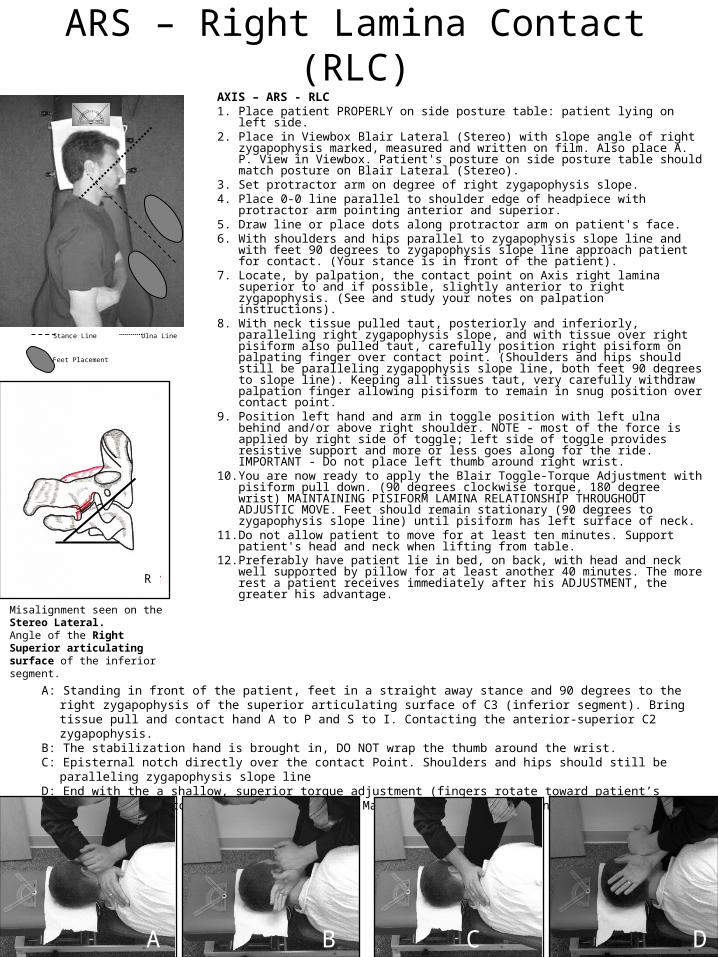
A: Standing in front of the patient, feet in a straight away stance and 90 degrees to the right zygapophysis of the superior articulating surface of C3 (inferior segment). Bring tissue pull and contact hand A to P and S to I. Contacting the anterior-superior C2 zygapophysis.
B: The stabilization hand is brought in, DO NOT wrap the thumb around the wrist.C: Episternal notch directly over the contact Point. Shoulders and hips should still be paralleling zygapophysis slope lineD: End with the a shallow, superior torque adjustment (fingers rotate toward patient’s head). 90o ulna torque, 180o wrist
torque. Maintain elbow bend on contact hand throughout thrust..
ARS – Right Lamina Contact (RLC)AXIS – ARS - RLC 1. Place patient PROPERLY on side posture table: patient lying on left side. 2. Place in Viewbox Blair Lateral (Stereo) with slope angle of right zygapophysis
marked, measured and written on film. Also place A. P. View in Viewbox. Patient's posture on side posture table should match posture on Blair Lateral (Stereo).
3. Set protractor arm on degree of right zygapophysis slope. 4. Place 0-0 line parallel to shoulder edge of headpiece with protractor arm
pointing anterior and superior. 5. Draw line or place dots along protractor arm on patient's face. 6. With shoulders and hips parallel to zygapophysis slope line and with feet 90
degrees to zygapophysis slope line approach patient for contact. (Your stance is in front of the patient).
7. Locate, by palpation, the contact point on Axis right lamina superior to and if possible, slightly anterior to right zygapophysis. (See and study your notes on palpation instructions).
8. With neck tissue pulled taut, posteriorly and inferiorly, paralleling right zygapophysis slope, and with tissue over right pisiform also pulled taut, carefully position right pisiform on palpating finger over contact point. (Shoulders and hips should still be paralleling zygapophysis slope line, both feet 90 degrees to slope line). Keeping all tissues taut, very carefully withdraw palpation finger allowing pisiform to remain in snug position over contact point.
9. Position left hand and arm in toggle position with left ulna behind and/or above right shoulder. NOTE - most of the force is applied by right side of toggle; left side of toggle provides resistive support and more or less goes along for the ride. IMPORTANT - Do not place left thumb around right wrist.
10. You are now ready to apply the Blair Toggle-Torque Adjustment with pisiform pull down. (90 degrees clockwise torque, 180 degree wrist) MAINTAINING PISIFORM LAMINA RELATIONSHIP THROUGHOUT ADJUSTIC MOVE. Feet should remain stationary (90 degrees to zygapophysis slope line) until pisiform has left surface of neck.
11. Do not allow patient to move for at least ten minutes. Support patient's head and neck when lifting from table.
12. Preferably have patient lie in bed, on back, with head and neck well supported by pillow for at least another 40 minutes. The more rest a patient receives immediately after his ADJUSTMENT, the greater his advantage.
Stance Line
Misalignment seen on the Stereo Lateral.Angle of the Right Superior articulating surface of the inferior segment.
A B C D
Feet Placement
Ulna Line
A B C D
R
A: Standing in front of the patient, feet in a straight away stance and parallel to the right zygapophysis of the superior articulating surface of C3 (inferior segment). Bring tissue pull and contact hand A to P and S to I. Contacting the anterior-superior C2 zygapophysis.
B: The stabilization hand is brought in, DO NOT wrap the thumb around the wrist.C: Episternal notch directly over the contact Point. Shoulders and hips should still be paralleling zygapophysis slope lineD: End with the a shallow, superior torque adjustment (fingers rotate toward patient’s head). 90o ulna torque, 180o wrist
torque.
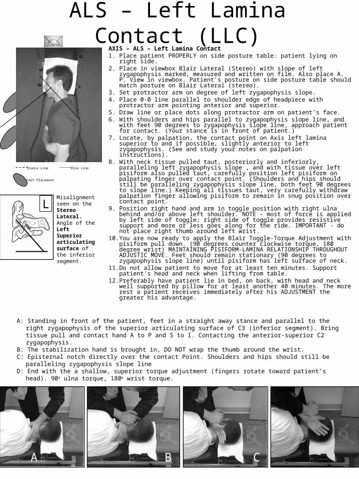
ALS – Left Lamina Contact (LLC)AXIS – ALS – Left Lamina Contact 1. Place patient PROPERLY on side posture table: patient lying on right side. 2. Place in viewbox Blair Lateral (Stereo) with slope of left zygapophysis marked,
measured and written on film. Also place A. P. View in viewbox. Patient's posture on side posture table should match posture on Blair Lateral (stereo).
3. Set protractor arm on degree of left zygapophysis slope. 4. Place 0-0 line parallel to shoulder edge of headpiece with protractor arm
pointing anterior and superior. 5. Draw line or place dots along protractor arm on patient's face. 6. With shoulders and hips parallel to zygapophysis slope line, and with feet 90
degrees to zygapophysis slope line, approach patient for contact. (Your stance is in front of patient.)
7. Locate, by palpation, the contact point on Axis left lamina superior to and if possible, slightly anterior to left zygapophysis. (See and study your notes on palpation instructions).
8. With neck tissue pulled taut, posteriorly and inferiorly, paralleling left zygapophysis slope , and with tissue over left pisiform also pulled taut, carefully position left pisiform on palpating finger over contact point. (Shoulders and hips should still be paralleling zygapophysis slope line, both feet 90 degrees to slope line.) Keeping all tissues taut, very carefully withdraw palpation finger allowing pisiform to remain in snug position over contact point.
9. Position right hand and arm in toggle position with right ulna behind and/or above left shoulder. NOTE - most of force is applied by left side of toggle; right side of toggle provides resistive support and more or less goes along for the ride. IMPORTANT - do not place right thumb around left wrist.
10. You are now ready to apply the Blair Toggle-Torque Adjustment with pisiform pull down. (90 degrees counter clockwise torque, 180 degree wrist) MAINTAINING PISIFORM-LAMINA RELATIONSHIP THROUGHOUT ADJUSTIC MOVE. Feet should remain stationary (90 degrees to zygapophysis slope line) until pisiform has left surface of neck.
11. Do not allow patient to move for at least ten minutes. Support patient's head and neck when lifting from table.
12. Preferably have patient lie in bed, on back, with head and neck well supported by pillow for at least another 40 minutes. The more rest a patient receives immediately after his ADJUSTMENT the greater his advantage.
Stance Line
Misalignment seen on the Stereo Lateral. Angle of the Left Superior articulating surface of the inferior segment.
A B C D
Feet Placement
Ulna Line
L
A B C D
A: Stance line is parallel to the left zygapophysis of the superior articulating surface of the inferior segment.B: Standing behind the patient, feet in a straight away stance line. Bring tissue pull and contact hand P to A and I to S.
Contacting the posterior C2 lamina. Contact elbow should be below pisiform.. C: The stabilization hand is brought in, wrap the thumb around the wrist. Stabilization ulna points upward to the ceiling,
contact ulna parallel to the stance line. Shoulders and hips are perpendicular to zygapophysis slope line (stance line).D: End with the a 180o inferior torque (fingers rotate toward patient’s feet), using shallow, P-A, I-S and R-L torque
adjustment.
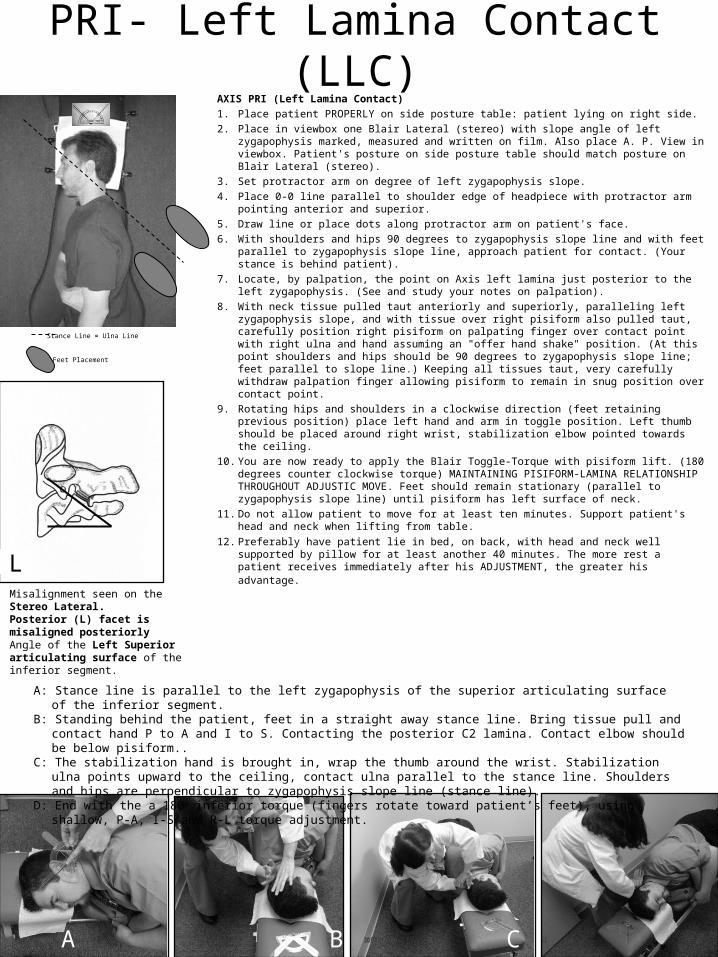
PRI- Left Lamina Contact (LLC)AXIS PRI (Left Lamina Contact)
1. Place patient PROPERLY on side posture table: patient lying on right side.
2. Place in viewbox one Blair Lateral (stereo) with slope angle of left zygapophysis marked, measured and written on film. Also place A. P. View in viewbox. Patient's posture on side posture table should match posture on Blair Lateral (stereo).
3. Set protractor arm on degree of left zygapophysis slope.
4. Place 0-0 line parallel to shoulder edge of headpiece with protractor arm pointing anterior and superior.
5. Draw line or place dots along protractor arm on patient's face.
6. With shoulders and hips 90 degrees to zygapophysis slope line and with feet parallel to zygapophysis slope line, approach patient for contact. (Your stance is behind patient).
7. Locate, by palpation, the point on Axis left lamina just posterior to the left zygapophysis. (See and study your notes on palpation).
8. With neck tissue pulled taut anteriorly and superiorly, paralleling left zygapophysis slope, and with tissue over right pisiform also pulled taut, carefully position right pisiform on palpating finger over contact point with right ulna and hand assuming an "offer hand shake" position. (At this point shoulders and hips should be 90 degrees to zygapophysis slope line; feet parallel to slope line.) Keeping all tissues taut, very carefully withdraw palpation finger allowing pisiform to remain in snug position over contact point.
9. Rotating hips and shoulders in a clockwise direction (feet retaining previous position) place left hand and arm in toggle position. Left thumb should be placed around right wrist, stabilization elbow pointed towards the ceiling.
10. You are now ready to apply the Blair Toggle-Torque with pisiform lift. (180 degrees counter clockwise torque) MAINTAINING PISIFORM-LAMINA RELATIONSHIP THROUGHOUT ADJUSTIC MOVE. Feet should remain stationary (parallel to zygapophysis slope line) until pisiform has left surface of neck.
11. Do not allow patient to move for at least ten minutes. Support patient's head and neck when lifting from table.
12. Preferably have patient lie in bed, on back, with head and neck well supported by pillow for at least another 40 minutes. The more rest a patient receives immediately after his ADJUSTMENT, the greater his advantage.
Stance Line = Ulna Line
Misalignment seen on the Stereo Lateral.Posterior (L) facet is misaligned posteriorlyAngle of the Left Superior articulating surface of the inferior segment.
Feet Placement
L
A B C D

A: Stance line is parallel to the right zygapophysis of the superior articulating surface of the inferior segment.B: Standing behind the patient, feet in a straight away stance line. Bring tissue pull and contact hand P to A and I to S.
Contacting the posterior C2 lamina. C: The stabilization hand is brought in, wrap the thumb around the wrist. Stabilization ulna points upward to the ceiling,
contact ulna parallel to the stance line. Shoulders and hips are perpendicular to zygapophysis slope line (stance line).D: End with the a 180o inferior torque (fingers rotate toward patient’s feet), using shallow, P-A, I-S and R-L torque
adjustment.
PLI- Right Lamina Contact (RLC)AXIS PLI (Right Lamina Contact) 1. Place patient PROPERLY on side posture table: patient lying on left side. 2. Place in viewbox one Blair Lateral (stereo) with slope angle of right
zygapophysis marked, measured and written on film. Also place A. P. View in viewbox. Patient's posture on side posture table should match posture on Blair Lateral (stereo).
3. Set protractor arm on degree of right zygapophysis slope. 4. Place 0-0 line parallel to shoulder edge of headpiece with protractor arm
pointing anterior and superior. 5. Draw line or place dots along protractor arm on patient's face. 6. With shoulders and hips 90 degrees to zygapophysis slope line and with feet
parallel to zygapophysis slope line, approach patient for contact. (Your stance is behind patient).
7. Locate, by palpation, the point on axis right lamina just posterior to the right zygapophysis. (See and study your notes on palpation).
8. With neck tissue pulled taut anteriorly and superiorly, paralleling right zygapophysis slope, and with tissue over left pisiform also pulled taut, carefully position left pisiform on palpating finger over contact point with left ulna and hand assuming an "offer hand shake" position. (At this point shoulders and hips should be 90 degrees to zygapophysis slope line: feet parallel to slope line). Keeping all tissues taut, very carefully withdraw palpation finger allowing pisiform to remain in snug position over contact point, with the elow blow the pisiform..
9. Rotating hips and shoulders in a counter clockwise direction (feet retaining previous position) place right hand and arm in toggle position. Right thumb should be placed around left wrist, right elbow pointed towards ceiling..
10. You are now ready to apply the Blair Toggle-Torque with pisiform lift. (180 degrees clockwise torque). MAINTAINING PISIFORM-LAMINA RELATIONSHIP THROUGHOUT ADJUSTIC MOVE. Feet should remain stationary (parallel to zygapophysis slope line) until pisiform has left surface of neck.
11. Do not allow patient to move for at least ten minutes. Support patient's head and neck when lifting from table.
12. Preferably have patient lie in bed, on back, with head and neck well supported by pillow for at least another 40 minutes. The more rest a patient receives immediately after his ADJUSTMENT, the greater his advantage.
Stance Line = Ulna Line
Misalignment seen on the Stereo Lateral.Anterior (R) facet is misaligned posteriorlyAngle of the Right Superior articulating surface of the inferior segment.
A B C D
Feet Placement
L
A B C D

A: Stance line is parallel to the table. Using the superior articulating surface of the inferior left segment for the ulna line.B: Standing behind the patient. Bring tissue pull and contact hand P to A and I to S along the ulna line. Contacting the
right posterior-inferior C2 spinous. C: The stabilization hand is brought in, wrap the thumb around the wrist. Stabilization ulna points upward to the ceiling,
contact ulna parallel to the stance line. Shoulders and are perpendicular to zygapophysis slope line (stance line).D: End with the a 180o inferior torque (fingers rotate toward patient’s feet), using shallow, P-A, I-S and R-L torque
adjustment.
PRI- Right Spinous Contact (RSC)AXIS PRI (Right Spinous Contact)
1. Place patient PROPERLY on side posture table; patient lying on left side. 2. Place in viewbox Blair Lateral Stereo with slope angle of left zygapophysis marked,
measured and written on film. Place A-P view in viewbox. Patient's posture on the table should match the posture on the Blair Lateral Stereo.
3. Set protractor arm on degree measurement of left zygapophysis slope. 4. Place 0-0 line parallel to shoulder edge of headpiece with protractor arm pointing
anterior and superior in relation with the patient. Draw line or place dots along protractor arm on patient's face.
5. With shoulders and hips 90 degrees to table and with feet parallel to the table, left foot anterior and right foot posterior, stance line is parallel to the table (stance behind the patient).
6. Locate, by palpation, the contact point on the right aspect of the axis spinous process. Contact will be the posterior inferior right lateral aspect of the axis spinous (see study notes on palpation).
7. With neck tissue pulled taut anteriorly and superiorly, with the left ulna paralleling the left zygapophysis slope and with the tissue over the spinous also pulled taut, carefully position your left pisiform on palpating finger over contact point (shoulders and hips should still be 90 degrees to the table). The ulna should be parallel to the left slope angle and your feet are parallel to the table. Keeping all the tissue taut, very carefully withdraw palpation finger, allowing pisiform to remain in snug position over contact point.
8. Position left hand and arm in toggle position with the left ulna posterior and parallel to left slope angle. Note: most of force is applied with the left side of the toggle. Right side of toggle provides resistive support and more or less goes along for the ride.
9. You are now ready to apply the Blair Toggle Torque Adjustment with pisiform lift, using 180 degrees of clockwise torque, maintaining pisiform right spinous relationship throughout the adjustic move. Feet should remain stationary and parallel to the adjusting table until pisiform has left the surface of the neck, using 180 degrees of clockwise torque.
10. Do not allow the patient to move for at least ten minutes. Support the patient's head and neck when lifting from the table.
11. Preferably have patient lie in bed, on back, with head and neck well supported by pillow for at least another 40 minutes. The more rest a patient receives immediately after his ADJUSTMENT, the greater his advantage.
Ulna Line
Misalignment seen on the Stereo Lateral.Posterior (L) facet is misaligned posteriorly.Angle of the Left Superior articulating surface of the inferior segment.
Feet Placement
L
A B C D

A: Stance line is parallel to the table. Using the superior articulating surface of the inferior right segment for the ulna line.B: Standing behind the patient. Bring tissue pull and contact hand P to A and I to S along the ulna line. Contacting the
right posterior-inferior C2 spinous. C: The stabilization hand is brought in, wrap the thumb around the wrist. Stabilization ulna points upward to the ceiling,
contact ulna parallel to the stance line. Shoulders and are perpendicular to zygapophysis slope line (stance line).D: End with the a 180o inferior torque (fingers rotate toward patient’s feet), using shallow, P-A, I-S and L-R torque
adjustment.
PLI- Left Spinous Contact (LSC)AXIS PLI (Left Spinous Contact)
1. Place patient PROPERLY on side posture table; patient lying on right side. 2. Place in viewbox Blair Lateral Stereo with slope angle of right zygapophysis
marked, measured and written on film. Place A-P view in viewbox. Patient's posture on the table should match the posture on the Blair Lateral Stereo.
3. Set protractor arm on degree measurement of right zygapophysis slope. 4. Place 0-0 line parallel to shoulder edge of headpiece with protractor arm pointing
anterior and superior in relation with the patient. Draw line or place dots along protractor arm on patient's face.
5. With shoulders and hips 90 degrees to table and with feet parallel to the table, right foot anterior and left foot posterior, stance line is parallel to the table (stance is behind the patient).
6. Locate, by palpation, the contact point on the left aspect of the axis spinous process. Contact will be the posterior inferior left lateral aspect of the axis spinous (see study notes on palpation).
7. With neck tissue pulled taut anteriorly and superiorly, with the right ulna paralleling the right zygapophysis slope and with the tissue over the spinous also pulled taut, carefully position your right pisiform on palpating finger over contact point (shoulders and hips should still be 90 degrees to the table). The ulna should be parallel to the right slope angle and your feet are parallel to the table. Keeping all the tissue taut, very carefully withdraw palpation finger, allowing pisiform to remain in snug position over contact point.
8. Position right hand and arm in toggle position with the right ulna posterior and parallel to right slope angle. Note: most of force is applied with the right side of the toggle. Left side of toggle provides resistive support and more or less goes along for the ride.
9. You are now ready to apply the Blair Toggle Torque Adjustment with pisiform lift, using 180 degrees of counter clockwise torque, maintaining pisiform left spinous relationship throughout the adjustic move. Feet should remain stationary and parallel to the adjusting table until pisiform has left the surface of the neck.
10. Do not allow the patient to move for at least ten minutes. Support the patient's head and neck when lifting from the table.
11. Preferably have patient lie in bed, on back, with head and neck well supported by pillow for at least another 40 minutes. The more rest a patient receives immediately after his ADJUSTMENT, the greater his advantage.
Ulna Line
Misalignment seen on the Stereo Lateral.Anterior (R) facet is misaligned posteriorlyAngle of the Right Superior articulating surface of the inferior segment.
A B C D
Feet Placement
R
A B C D

Atlas (C1) Opposite side of
Laterality Contact
ASR - LSA
ASL - RSA
ASR – LTC
ASL – RTC
PIR – LTC (LSA)
PIL – RTC (RSA)
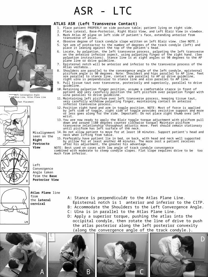
ASR - LTCATLAS ASR (Left Transverse Contact)
1. Place patient PROPERLY on side posture table; patient lying on right side. 2. Place Lateral, Base-Posterior, Right Blair View, and Left Blair View in viewbox. 3. Mark Atlas AP plane on left side of patient's face, extending anterior from transverse of atlas. 4. Observe degree of track condyle slope written on left Blair view. (See NOTE.) 5. Set arm of protractor to the number of degrees of the track condyle (left) and place it leaning
against the top of the patient's head. 6. Locate, by palpation, the left transverse process, palpating the left transverse on the anterior
inferior aspect, using palpating finger of the right hand. (See palpation instructions). Stance line is at right angles or 90 degrees to the AP plane line or drive guideline.
7. Episternal notch will be anterior and inferior to the transverse process of the Atlas vertebra.. 8. Shoulders are parallel to the convergence angle of the left condyle, episternal pisiform angle
is 90 degrees. Note: Shoulders and hips parallel to AP line, feet are parallel to stance line, contact arm parallel to AP or drive guideline, right ulna is perpendicular to stance line and also parallel to AP line.
9. Pull tissue taut over transverse, posteriorly and superiorly, parallel to drive guideline. 10. Retaining palpation finger position, assume a comfortable stance in front of patient and very
carefully position the left pisiform over palpation finger with ulna parallel to drive guideline. 11. Maintaining left pisiform over left transverse process, keeping tissue taut, very carefully
withdraw palpating finger, maintaining contact on anterior inferior transverse process. 12. Position right hand and arm in toggle position. NOTE: Most of force is applied by left side of
toggle, right side of toggle provides resistive support and more or less goes along for the ride. Important: Do not place right thumb over left wrist.
13. You are now ready to apply the Blair toggle torque adjustment with pisiform pull down and around (180 degrees counter clockwise torque) Maintain pisiform transverse relationship throughout adjustic move. Feet should remain stationary until pisiform has left surface of the neck.
14. Do not allow patient to move for at least 10 minutes. Support patient's head and neck when lifting from table.
15. Preferably have patient lie in bed, on back, with head and neck well supported by pillow for at least another 40 minutes. The more rest a patient receives after his adjustment, the greater his advantage.
NOTE: Best used on cases with low angle of track condyle convergence combined with moderate to steep condyle slopes. Flat slope requires drive to be too much from inferior.
Left Convergence AngleStance Line, Atlas Plane Line
Misalignment seen on the Right Protracto View
Left Convergence Angle taken from the Base Posterior View
A: Stance is perpendicular to the Atlas Plane Line. Episternal notch is 1” anterior and inferior to the C1TP.
B: Accommodate the Shoulders to the Left Convergence Angle.C: Ulna is in parallel to the Atlas Plane Line. D: Apply a superior torque, pushing the atlas into the occipital condyle, then
rotate the line of drive to push the atlas posterior along the left posterior convexity (along the convergence angle of the track condyle.).
B C
Feet Placement
Atlas Plane line fromthe lateral cervical
A
D
R

ASL - RTCATLAS ASL (Right Transverse Contact)
1. Place patient PROPERLY on side posture table; patient lying on left side. 2. Place Lateral, Base-Posterior, Right Blair View, and Left Blair View in viewbox. 3. Mark AP plane line of Atlas on patient's face, extending anterior from transverse of
atlas. 4. Observe degree of track condyle slope written on right Blair view. (See NOTE.) 5. Set arm of protractor to the number of degrees of the track condyle (right) and place
it leaning against the top of the patient's head. 6. Locate, by palpation, the right transverse process, palpating the right transverse on
the anterior inferior aspect, using palpating finger of the left hand. (See palpation instructions). Stance line is at right angles or 90 degrees to the AP plane line or drive guideline.
7. Episternal notch will be anterior and inferior to the transverse process of the Atlas vertebra..
8. Shoulders are parallel to the convergence angle of the right condyle, Episternal pisiform angle is 90 degrees. Note: Shoulders and hips parallel to AP line, feet are parallel to stance line, contact arm parallel to AP or drive guideline, left ulna is perpendicular to stance line and also parallel to AP line.
9. Pull tissue taut over transverse, posteriorly and superiorly, parallel to drive guideline. 10.Retaining palpation finger position, assume a comfortable stance in front of patient
and very carefully position the right pisiform over palpation finger with ulna parallel to drive guideline.
11.Maintaining right pisiform over right transverse process, keeping tissue taut, very carefully withdraw palpating finger, maintaining contact on anterior inferior transverse process.
12.Position left hand and arm in toggle position. NOTE: Most of force is applied by right side of toggle, left side of toggle provides resistive support and more or less goes along for the ride. Important: Do not place left thumb over right wrist.
13.You are now ready to apply the Blair toggle torque adjustment with pisiform pull down and around (180 degrees clockwise torque) Maintain pisiform transverse relationship throughout adjustic move. Feet should remain stationary until pisiform has left surface of the neck.
14.Do not allow patient to move for at least 10 minutes. Support patient's head and neck when lifting from table.
15.Preferably have patient lie in bed, on back, with head and neck well supported by pillow for at least another 40 minutes. The more rest a patient receives after his adjustment, the greater his advantage.
NOTE: Best used on cases with low angle of track condyle convergence combined with moderate to steep condyle slopes. Flat slope requires drive to be too much from inferior.
Left Convergence AngleStance Line, Atlas Plane Line
Misalignment seen on the Left Protracto View
Right Convergence Angle taken from the Base Posterior View
A: Stance is perpendicular to the Atlas Plane Line. Episternal notch is 1” anterior and inferior to the C1TP.
B: Accommodate the Shoulders to the Right Convergence Angle.C: Ulna is in parallel to the Atlas Plane Line. D: Apply a superior torque, pushing the atlas into the occipital condyle, then rotate the
line of drive to push the atlas posterior along the right posterior convexity (along the convergence angle of the track condyle).
B C
Feet Placement
Atlas Plane line fromthe lateral cervical
A
D
R

ASR - LSAATLAS ASR (Left Sub - Arch Contact) 1. Place patient PROPERLY on side posture table; patient lying on right side. 2. Place Lateral, Base-Posterior, Right Blair View, and Left Blair View in viewbox. 3. Mark Atlas AP plane on left side of patient's face, extending anterior from
transverse of atlas. 4. Observe degree of track condyle slope written on left Blair view. (SEE NOTE.) 5. Set protractor arm on degree of the left track condyle slope angle. 6. Standing in front of patient set angle arm protractor at 90 degrees to left slope
angle. 7. AP plane line becomes drive guideline with shoulders parallel to drive guide line.
Nail (left) arm parallel to drive guideline (ulna pisiform line). Hammer (right) arm 90 degrees to nail arm with arm anterior to patient's shoulder and episternal-pisiform angle at 90 degrees to slope angle.
8. Have assistant hold protractor on a level, just superior to the patient's head, with protractor arm pointing to posterior and superior (toward ceiling) and with 0-0 line parallel to stance line, keeping the bubble of the level centered until contact and adjustic set-up is completed.
9. Make contact posterior to mastoid on the inferior aspect of the subarch on side of track condyle (left) posterior to groove of second spinal nerve. Contact is posterior to mastoid subarch, just above lamina of axis. Line of drive is inferior to superior on the subarch.
10. Line of drive is inferior to superior with 90 degrees of superior torque with the left arm.
11. Maintaining left pisiform over left subarch contact, keeping tissue taut, very carefully withdraw palpating finger.
12. Position right hand and arm in toggle position with right ulna parallel to stance line.. NOTE: Most of force is applied by left side of toggle, right side of toggle provides guidance and resistive support and more or less goes along for the ride. Important: Do not place right thumb around left wrist.
13. You are now ready to apply the Blair toggle torque adjustment with pisiform pull down and around (180 degrees counter clockwise torque) Maintain pisiform subarch contact relationship throughout adjustic move. Feet should remain stationary until pisiform has left surface of the neck.
14. Do not allow patient to move for at least 10 minutes. Support patient's head and neck when lifting from table.
15. Preferably have patient lie in bed, on back, with head and neck well supported by pillow for at least another 40 minutes. The more rest a patient receives after his adjustment, the greater his advantage.
NOTE!! Subarch contact is preferred over transverse contact on A-S side opposite listings, on cases with low angle of track condyle convergence, combined with moderate to steep condyle slope. ~ —DO NOT use subarch contact with cleft posterior arch of Atlas!!
Subarch contact on side opposite is very effective and is often the best method of adjustment to "untorque" the "torqued subluxation" and achieve the "unlockment".
Atlas Plane Line AngleStance Line, perpendicular to APL
Misalignment seen on the Right Protracto View
Left Slope Angle taken from the Left Protracto View
A: Stance is perpendicular to the Atlas Plane Line. B: Accommodate the Sternum parallel with the left Slope, with the perpendicular arm pointing to
the episternal notch.C: Olecranon process is below the pisiform, parallel to the stance line parallel to the stance line.D: Apply a superior torque, pushing the atlas into the occipital condyle, then rotate the line of
drive to push the atlas posterior along the left posterior convexity. Stabilization arm gives the power to the adjustment, contact arm applies the torque.
C D
Feet Placement
A B
Atlas Plane line fromthe lateral cervical

ASL - RSAATLAS ASL (Right Sub - Arch Contact)
1. Place patient PROPERLY on side posture table; patient lying on left side.
2. Place Lateral, Base-Posterior, Right Blair View, and Left Blair View in viewbox.
3. Mark Atlas AP plane line on right side of patient's face, extending anterior from transverse of atlas.
4. Observe degree of track condyle slope written on right Blair view. (SEE NOTE.)
5. Set protractor arm on degree of the right track condyle slope angle.
6. Standing in front of patient set angle arm protractor at 90 degrees to right slope angle.
7. AP plane line becomes drive guideline with shoulders parallel to drive guide line. Nail (right) arm parallel to drive guideline (ulna pisiform line). Hammer (left) arm 90 degrees to nail arm with arm anterior to patient's shoulder and episternal-pisiform angle at 90 degrees to slope angle.
8. Have assistant hold protractor on a level, just superior to the patient's head, with protractor arm pointing to posterior and superior (toward ceiling) and with 0-0 line parallel to stance line, keeping the bubble of the level centered until contact and adjustic set-up is completed.
9. Make contact posterior to mastoid on the inferior aspect of the subarch on side of track condyle (right) posterior to groove of second spinal nerve. Contact is posterior to mastoid subarch, just above lamina of axis. Line of drive is inferior to superior on the subarch.
10.Line of drive is inferior to superior with 180 degrees of superior torque with the right arm.
11.Maintaining right pisiform over right subarch contact, keeping tissue taut, very carefully withdraw palpating finger.
12.Position left hand and arm in toggle position with left ulna parallel to stance line. NOTE: Most of force is applied by right side of toggle, left side of toggle provides guidance and resistive support and more or less goes along for the ride. Important: Do not place left thumb around right wrist.
13.You are now ready to apply the Blair toggle torque adjustment with pisiform pull down and around (180 degrees clockwise torque) Maintain pisiform-subarch contact relationship throughout adjustic move. Feet should remain stationary until pisiform has left surface of the neck.
14.Do not allow patient to move for at least 10 minutes. Support patient's head and neck when lifting from table.
15.Preferably have patient lie on BED, on back, with head and neck well supported by pillow for at least another 40 minutes. The more rest a patient receives after his adjustment, the greater his advantage.
NOTE!! Subarch contact is preferred over transverse contact on A-S side opposite listings, on cases with low angle of track condyle convergence, combined with moderate to steep condyle slope. — —DO NOT use subarch contact with cleft posterior arch of Atlas!! Subarch contact on side opposite is very effective and is often the best method of adjustment to "untorque" the "torqued subluxation" and achieve the "unlockment".
Atlas Plane Line AngleStance Line, perpendicular to APL
Misalignment seen on the Left Protracto View
Right Slope Angle taken from the Right Protracto View
A: Stance is perpendicular to the Atlas Plane Line. B: Accommodate the Sternum parallel with the right Slope, with the perpendicular arm pointing to the
episternal notch.C: Olecranon process is below the pisiform, parallel to the stance line parallel to the stance line.D: Apply a superior torque, pushing the atlas into the occipital condyle, then rotate the line of drive to push
the atlas posterior along the left posterior convexity. Stabilization arm gives the power to the adjustment, contact arm applies the torque.
C D
Feet Placement
A B
Atlas Plane line fromthe lateral cervical
R

A: Standing behind the patient, feet in a scissored stance and parallel to the right slope.
B: Accommodate the shoulders parallel to the left convergence angle, perpendicular arm pointing to the episternal notch.
C: The contact ulna is perpendicular to the atlas plane line. D: End with the a shallow, superior torque adjustment pushing the atlas
against the occipital condyle, parallel to the right slope. Rotate the drive line to push the atlas up the left anterior convexity.
PIL – RTC ATLAS PIL (Right Transverse Contact)
1. Place patient PROPERLY on side posture table; patient lying on left side. Sagittal plane of patient's head should be parallel to transverse surface of headpiece. Patient's visual plane should be 90 degrees to long axis of adjusting table.
2. Place Lateral, Base-Posterior, Right Blair View, and Left Blair View in viewbox. 3. Set protractor arm to the slope angle of the right condyle. (Taken from the right
Blair View). 4. Place 0-0 line parallel to shoulder edge of headpiece with protractor arm pointing
anterior and superior in relation with patient. Mark a line along protractor arm or place protractor above patient's head so that a line extending through the right transverse process would be a continuous line if such a line were drawn.
5. Set next protractor arm setting to degree of left condyle convergence. (Taken from Base-Posterior view).
6. Stand behind patient with feet parallel to stance line, which is parallel to the right slope angle. Hips and shoulders 90 degrees to stance line. Retain this position.
7. Locate, by palpation, the posterior-inferior aspect of the right transverse process, and hold with the palpation finger of the right hand.
8. Have assistant hold protractor on a level, just superior to the patient's head, with protractor arm pointing to anterior and superior (toward ceiling) and 0-0 line parallel to stance line, keeping the bubble of the level centered until contact and adjustic set-up is completed. In this instance the left convergence angle taken from Base-Posterior, as stated above, is the angle utilized for this contact and adjustment.
9. Pulling tissue taut, take contact with left pisiform. Episternal notch pisiform angle should be 90 degrees to left convergence angle, with hips, shoulders and ulna 90 degrees to stance line; feet parallel to stance line. Very carefully withdraw palpation finger. Snug contact of left pisiform over posterior inferior aspect of the right transverse should now exist.
10. Place right part of toggle. Do not place right thumb around wrist. NOTE: Most of the force is applied with the left side of the toggle, right side of toggle provides resistive support and more or less goes along for the ride.
11. You are now ready to apply the Blair toggle torque adjustment with pisiform pull down and around. (180 degrees counter clockwise torque). Maintain pisiform transverse relationship throughout the adjustic move. Feet should remain stationary until pisiform has left surface of neck. Line of drive is from posterior-inferior to anterior-superior, utilizing the Blair toggle torque adjustment with pisiform pull down.
12. Do not allow patient to move for at least 10 minutes. Support patient's head and neck when lifting from table.
13. Preferably have patient lie in bed, on back, with head and neck well supported by pillow for at least another 40 minutes. The more rest a patient receives after his adjustment, the greater his advantage.
Stance Line
Misalignment seen on the Right Protracto View
Left Convergence Angle taken from the Base Posterior View
A B C D
Feet Placement
Right Slope Angle
L
Right Slope from the Right Protracto View

A: Standing behind the patient, feet in a scissored stance and parallel to the Left slope.
B: Accommodate the shoulders parallel to the right convergence angle, perpendicular arm pointing to the episternal notch.
C: The contact ulna is perpendicular to the atlas plane line. D: End with the a shallow, superior torque adjustment pushing the atlas
against the occipital condyle, parallel to the left slope. Rotate the drive line to push the atlas up the right anterior convexity.
PIR – LTC ATLAS PIR (Left Transverse Contact)
1. Place patient PROPERLY on side posture table; patient lying on right side. Sagittal plane of patient's head should be parallel to transverse surface of headpiece. Patient's visual plane should be 90 degrees to long axis of adjusting table.
2. Place Lateral, Base-Posterior, Right Blair View, and Left Blair View in viewbox. 3. Set protractor arm to the slope angle of the left condyle. (Taken from the Left Blair
View). 4. Place 0-0 line parallel to shoulder edge of headpiece with protractor arm pointing
anterior and superior in relation with patient. Mark a line along protractor arm or place protractor above patient's head so that a line extending through the left transverse process would be a continuous line if such a line were drawn.
5. Set next protractor arm setting to degree of right condyle convergence. (Taken from Base-Posterior view).
6. Stand behind patient with feet parallel to stance line, which is parallel to the left slope angle. Hips and shoulders 90 degrees to stance line. Retain this position.
7. Locate, by palpation, the posterior-inferior aspect of the left transverse process, and hold with the palpation finger of the left hand.
8. Have assistant hold protractor on a level, just superior to the patient's head, with protractor arm pointing to anterior and superior (toward ceiling) and 0-0 line parallel to stance line, keeping the bubble of the level centered until contact and adjustic set-up is completed. In this instance the right convergence angle taken from Base-Posterior, as stated above, is the angle utilized for this contact and adjustment.
9. Pulling tissue taut, take contact with right pisiform. Episternal notch pisiform angle should be 90 degrees to right convergence angle, with hips, shoulders and ulna 90 degrees to stance line; feet parallel to stance line. Very carefully withdraw palpation finger. Snug contact of right pisiform over posterior inferior aspect of the left transverse should now exist.
10. Place left part of toggle. Do not place left thumb around wrist. NOTE: Most of the force is applied with the right side of the toggle, left side of toggle provides resistive support and more or less goes along for the ride.
11. You are now ready to apply the Blair toggle torque adjustment with pisiform pull down and around. (180 degrees clockwise torque). Maintain pisiform transverse relationship throughout the adjustic move. Feet should remain stationary until pisiform has left surface of neck. Line of drive is from posterior-inferior to anterior-superior, utilizing the Blair toggle torque adjustment with pisiform pull down.
12. Do not allow patient to move for at least 10 minutes. Support patient's head and neck when lifting from table.
13. Preferably have patient lie in bed, on back, with head and neck well supported by pillow for at least another 40 minutes. The more rest a patient receives after his adjustment, the greater his advantage.
Stance Line
Misalignment seen on the Left Protracto View
Right Convergence Angle taken from the Base Posterior View
A B C D
Feet Placement
Left Slope Angle
L
Left Slope from the Left Protracto View

Atlas (C1) Double Listings
ASR-ASL – RTC
ASL-ASR – LTC
PIR-PIL – RTC
PIL-PIR – LTC
ASR-PIR – RTC
ASL-PIL – LTC

ASR-ASL - RTCATLAS ASR-ASL (Right Transverse Contact)
1. Place patient PROPERLY on side posture table; patient lying on left side. 2. Place Lateral, Base-Posterior, and Right Blair View and Left Blair View in Viewbox. 3. Be certain that patient's horizontal vision is parallel to shoulder end of headpiece. 4. Observe degree of left track condyle slope angle (taken from Left Blair View-ASR) 5. Set protractor arm on degree of left track condyle slope angle. 6. Set protractor arm on degree of right condyle convergence angle. (ASL) 7. Place 0-0 edge of protractor in line with shoulder edge of headpiece with protractor arm pointing
toward patient's anterior and superior, (left slope angle-ASR) 8. Mark a line along protractor arm. This is the Drive Guide Line. 9. Place protractor at right convergence angle, just superior to the patient's head, with protractor arm
pointing posterior, and the 0-0 line parallel to the floor keeping the bubble of the level centered until contact and adjusting set up is completed.
10.Locate, by palpation, right transverse process. Use palpating finger of left hand. (See Palpation Instructions)
11.Pull tissue taut over transverse (inferiorly and anteriorly) 90 degrees to Drive Guide Line. 12.Retaining palpation finger position, assume a comfortable stance in front of the patient and very
carefully position the right pisiform over palpation finger with ulna as near to 90 degrees as possible to Drive Guide Line.
13.ASR Track condyle slope angle (left) is the Drive Guide Line, and taking into consideration the convergence of the right condyle, the episternal notch-pisiform angle must be 90 degrees to the convergence angle of the right track condyle for the ASL correction. This will place your line of drive for the second half of the adjustment on the AXIS of torque of the right track condyle.
14.Shoulders are parallel to the convergence angle of the right condyle. The episternal notch pisiform angle at 90 degrees to the right convergence angle. Note: Shoulders and hips are parallel to left slope angle or Drive Guide Line, and feet are parallel to stance line. Recheck: Right ulna is as near as possible to 90 degrees of the Drive Guide Line.
15.Maintain right pisiform over right transverse process, and keeping tissue taut, very carefully withdraw palpating finger.
16.Position left hand and arm in toggle position. NOTE: Most of force is applied by right side of toggle; left side of toggle provides resistive support and
more or less goes along for the ride. IMPORTANT: Do not place left thumb around right wrist. You are now ready to apply the Blair Toggle-Torque adjustment with pisiform pull. (180 degree
clockwise torque) MAINTAINING PISIFORM-TRANSVERSE RELATIONSHIP THROUGHOUT ADJUSTIC MOVE. Feet should remain stationary until pisiform has left surface of the neck. First half of the adjustment will be reduction and correction of the ASR listing as the ulna follows the arc parallel to the stance line until it reaches drive guide line of the left slope angle. Second half of the adjustment will be correction of the ASL right transverse contact until ulna is parallel with the floor, parallel to your stance line.
Do not allow patient to move for at least 10 minutes. Support patient's head and neck when lifting from table.
Preferably have patient lie in bed, on back, with neck and head will supported by pillow for at least another 40 minutes. The more rest a patient receives after his adjustment the greater his advantage.
Left Slope AngleStance Line
Misalignment seen on the Right and left Protracto Views
Left Slope Angle taken from the Left Protracto View
A: Stance line is parallel with the left slope. Accommodate the shoulders to the right convergence angle.B: Contact arm in line with the Stance line. Start adjustment by correcting the ASR as instructed in the ASR-RTC.C: When the contact arm reaches the level of the APL, the shoulders should now be parallel with the right convergence
angle and begin the ASL-RTC adjustment.D: Contact ulna drops so that the olecranon process is below the pisiform and push the atlas up the right convexity
A B C D
Feet Placement
Right Convergence Angle from the Base Posterior R

ASL-ASR - LTCATLAS ASR-ASL (Right Transverse Contact)
1. Place patient PROPERLY on side posture table; patient lying on right side. 2. Place Lateral, Base-Posterior, and Right Blair View and Left Blair View in Viewbox. 3. Be certain that patient's horizontal vision is parallel to shoulder end of headpiece. 4. Observe degree of right track condyle slope angle (taken from Right Blair View-ASL) 5. Set protractor arm on degree of right track condyle slope angle. 6. Set protractor arm on degree of left condyle convergence angle. (ASR) 7. Place 0-0 edge of protractor in line with shoulder edge of headpiece with protractor arm pointing
toward patient's anterior and superior, (right slope angle-ASL) 8. Mark a line along protractor arm. This is the Drive Guide Line. 9. Place protractor at left convergence angle, just superior to the patient's head, with protractor arm
pointing posterior, and the 0-0 line parallel to the floor keeping the bubble of the level centered until contact and adjusting set up is completed.
10. Locate, by palpation, left transverse process. Use palpating finger of right hand. (See Palpation Instructions)
11. Pull tissue taut over transverse (inferiorly and anteriorly) 90 degrees to Drive Guide Line. 12. Retaining palpation finger position, assume a comfortable stance in front of the patient and very
carefully position the left pisiform over palpation finger with ulna as near to 90 degrees as possible to Drive Guide Line.
13. ASL Track condyle slope angle (right) Is the Drive Guide Line, and taking into consideration the convergence of the left condyle, the episternal notch-pisiform angle must be 90 degrees to the convergence angle of the left track condyle for the ASR correction. This will place your line of drive for the second half of the adjustment on the AXIS of torque of the left track condyle.
14. Shoulders are parallel to the convergence angle of the left condyle. The episternal notch pisiform angle at 90 degrees to the left convergence angle. Note: Shoulders and hips are parallel to right slope angle or Drive Guide Line, and feet are parallel to stance line. Recheck: Left ulna is as near as possible to 90 degrees of the Drive Guide Line.
15. Maintain right pisiform over right transverse process, and keeping tissue taut, very carefully withdraw palpating finger.
16. Position right hand and arm in toggle position. NOTE: Most of force is applied by left side of toggle; right side of toggle provides resistive support
and more or less goes along for the ride. IMPORTANT: Do not place right thumb around left wrist. You are now ready to apply the Blair Toggle-Torque adjustment with pisiform pull. (180 degree
counterclockwise torque) MAINTAINING PISIFORM-TRANSVERSE RELATIONSHIP THROUGHOUT ADJUSTIC MOVE. Feet should remain stationary until pisiform has left surface of the neck. First half of the adjustment will be reduction and correction of the ASL listing as the ulna follows the arc parallel to the stance line until it reaches drive guide line of the right slope angle. Second half of the adjustment will be correction of the ASR left transverse contact until ulna is parallel with the floor, parallel to your stance line.
Do not allow patient to move for at least 10 minutes. Support patient's head and neck when lifting from table.
Preferably have patient lie in bed, on back, with neck and head will supported by pillow for at least another 40 minutes. The more rest a patient receives after his adjustment the greater his advantage.
Left Slope AngleStance Line
Misalignment seen on the Left & Right Protracto Views
Right Slope Angle taken from the Left Protracto View
A: Stance line is parallel with the right slope. Accommodate the shoulders to the left convergence angle.B: Contact arm in line with the stance line. Start adjustment by correcting the ASL as instructed in the ASL-LTC.C: When the contact arm reaches the level of the APL, the shoulders should now be parallel with the left convergence
angle and begin the ASR-LTC adjustment.D: Contact ulna drops so that the olecranon process is below the pisiform and push the atlas up the left convexity
A B C D
Feet Placement
Left Convergence Angle from the Base Posterior R

ASR-PIR - RTCATLAS ASR-PIR (Right Transverse Contact)
1. Place patient PROPERLY on side posture table; patient lying on left side. 2. Place Lateral, Base-Posterior, and Right Blair View and Left Blair View in Viewbox. 3. Be certain that patient's horizontal vision is parallel to shoulder end of headpiece. 4. "Observe degree of left track condyle slope angle (taken from Left Blair View-ASR) 5. Set protractor arm on degree of left track condyle slope angle.6. Set protractor arm on degree measurement of posterior aspect of right condyle convexity
taken from left Blair view. 7. Place 0-0 line parallel to shoulder edge of headpiece with protractor arm pointing
posterior and superior in relation to patient. Mark a line along protractor arm. This is the stance line standing in front of patient with feet parallel to the right track condyle convexity angle.
8. Locate, by palpation .right transverse process. Use palpating finger of left hand. (See Palpation Instructions)
9. Pull tissue taut over transverse (Inferiorly and anteriorly).10. 10. Retaining palpation finger position, assume a comfortable stance in front of the
patient and very carefully position the right pisiform over palpation finger with ulna as near to 180 degrees as possible to stance line.
11. Maintaining right pisiform over right transverse process and keeping tissue taut, very carefully withdraw palpating finger.
12. Position left hand and arm in toggle position. NOTE: Most of force is applied by right side of toggle; left side of toggle provides resistive support and more or less goes along for the ride. IMPORTANT: Do not place left thumb around right wrist.
13. You are now ready to apply the Blair Toggle-Torque adjustment with pisiform pull. (180 degree plus clockwise torque) MAINTAINING PISIFORM-TRANSVERSE RELATIONSHIP THROUGHOUT ADJUSTIC MOVE. Feet should remain stationary until pisiform has left surface of the neck.
14. The sequence of this adjustment: first portion of adjustment - correction starts with posterior-inferior right portion (PIR) until ulna is 90 degrees to the left slope angle. Second portion of the adjustment starts when ulna is 90 degrees to left slope angle, the balance of the adjustment is correction of the ASR which is achieved from 90 degrees to slope angle to completion of the Blair Toggle-Torque adjustment.
15. Do not allow patient to move for at least 10 minutes. Support patient's head and neck when lifting from table.
16. Preferably have patient lie In bed, on back, with neck and head will supported by pillow for at least another 40 minutes. The more rest a patient receives after his adjustment, the greater his advantage.
NOTE: Atlas ASR-PIR adjustment is most effective with moderate to shallow slope angles on the right condyle and with moderate convexity of the right condyle. In this adjustment shoulders and hips are 90 degrees to stance line. Feet are parallel to stance line, episternal notch is one inch beyond transverse process.
Note: if the PIR is the major, increase elbow bend of contact ulna. If ASR is the major, decrease elbow bend.
Right Convexity AngleStance Line
ASR Misalignment seen on the Right & Protracto View
Left Slope Angle for the stance line; Right Convexity for the Ulna placement, taken from the Left Protracto View
A: Starting with the stance line and superior (contact) elbow bent to the angle of the left slope, use the “swimmer’s move” to line up the ulna with the right posterior convexity.
B: The adjustment starts with an S to I and P to A force an the C1 transverse process (C1TP). Pressure is kept on the right C1TP as the as the adjusting force vector is rotated, with superior torque.
C: The ulna continues to rotate keeping pressure on the C1TP as the ASR-RTC correction is made.D: End with the patient’s C1TP, the doctor’s pisiform and episternal notch in a straight plum line to the floor.
A B C D
Feet Placement
Convexity
Slope
PIR seen on the left protracto view

ASL-PIL - LTCATLAS ASL-PIL (Left Transverse Contact)
1. Place patient PROPERLY on side posture table; patient lying on right side. 2. Place Lateral, Base-Posterior, and Right Blair View and Left Blair View in Viewbox. 3. Be certain that patient's horizontal vision is parallel to shoulder end of headpiece. 4. Observe degree of right track condyle slope angle (taken from Right Blair View-ASL) 5. Set protractor arm on degree of right track condyle slope angle. 6. Set protractor arm on degree measurement of posterior aspect of left condyle convexity
taken from right Blair view. 7. Place 0-0 line parallel to shoulder edge of headpiece with protractor arm pointing
posterior and superior in relation to patient. Mark a line along protractor arm. This is the stance line standing in front of patient with feet parallel to the left track condyle convexity angle.
8. Locate, by palpation, lt transverse process. Use palpating finger of right hand. (See Palpation Instructions)
9. Pull tissue taut over transverse (inferiorly and anteriorly). 10. Retaining palpation finger position, assume a comfortable stance in front of the patient
and very carefully position the left pisiform over palpation finger with ulna as near to 180 degrees as possible to stance line.
11. Maintaining left pisiform over left transverse process and keeping tissue taut, very carefully withdraw palpating finger.
12. Position right hand and arm in toggle position. NOTE: Most of force is applied by left side of toggle; right side of toggle provides resistive support and more or less goes along for the ride. IMPORTANT: Do not place right thumb around left wrist.
13. You are now ready to apply the Blair Toggle-Torque adjustment with pisiform pull. (180 degree plus counterclockwise torque) MAINTAINING PISIFORM-TRANSVERSE RELATIONSHIP THROUGHOUT ADJUSTIC MOVE. Feet should remain stationary until pisiform has left surface of the neck.
14. The sequence of this adjustment: first portion of adjustment - correction starts with posterior-inferior left portion (PIL) until ulna is 90 degrees to the right slope angle. Second portion of the adjustment starts when ulna is 90 degrees to right slope angle, the balance of the adjustment is correction of the ASL which is achieved from 90 degrees to slope angle to completion of the Blair Toggle-Torque adjustment.
15. Do not allow patient to move for at least 10 minutes. Support patient's head and neck when lifting from table.
16. Preferably have patient lie in bed, on back, with neck and head will supported by pillow for at least another 40 minutes. The more rest a patient receives after his adjustment, the greater his advantage.
NOTE: Atlas ASL-PIL adjustment is most effective with moderate to shallow slope angles on the left condyle and with moderate convexity of the left condyle. In this adjustment shoulders and hips are 90 degrees to stance line. Feet are parallel to stance line, episternal notch is one inch beyond transverse process.
Right Convexity AngleStance Line
ASL Misalignment seen on the Left Protracto View
Right Slope Angle for the stance line; Left Convexity for the Ulna placement, taken from the Left Protracto View
A: Starting with the stance line and superior (contact) elbow bent to the angle of the right slope, use the “swimmer’s move” to line up the ulna with the left posterior convexity.
B: The adjustment starts with an S to I and P to A force an the C1 transverse process (C1TP). Pressure is kept on the right C1TP as the as the adjusting force vector is rotated, with superior torque.
C: The ulna continues to rotate keeping pressure on the C1TP as the ASL-LTC correction is made.D: End with the patient’s C1TP, the doctor’s pisiform and episternal notch in a straight plum line to the floor.
A B C D
Feet Placement
Convexity
Slope
PIL seen on the Right protracto view

A: Standing behind the patient, feet in a straight away stance and parallel to the right convexity, bring tissue pull and contact hand P to A and S to I. Contacting the posterior-superior C1TP.
B: Set the carpal angle from the right convergence angle found on the Base Posterior. The carpal line is the flat surface of the doctor’s posterior wrist, along the carpal bones.
C: The stabilization hand is brought in, do not wrapping the thumb around the wrist and antecubital fossas are straight across from each other.
D: End with the a shallow, 180 degree superior torque adjustment.
PIR-PIL – RTC (RPA) ATLAS PIR-PIL (Right Transverse Contact) (Right Sub-Arch)
1. Place patient PROPERLY on side posture table; patient lying on left side. Saggital plane of patient's head should be parallel to transverse surface of headpiece. Patient's visual plane should be 90 degrees to long axis of adjusting table.
2. Place Lateral, Base-Posterior, Right Blair View, and Left Blair View in viewbox. 3. Set protractor arm oh degree measurement of posterior aspect of right condyle
convexity. (Taken from the left Blair View) 4. Place 0-0 line parallel to shoulder edge of headpiece with protractor arm pointing
anterior and inferior in relation with patient. Mark a line along protractor arm. This is the stance line.
5. Change protractor arm setting to degree of right condyle convergence. (taken from Base-Posterior view)
6. Stand behind patient with feet parallel to stance line, hips and shoulders 90 degrees to stance line. Retain this position.
7. Locate, by palpation, and contact the posterior-superior aspect of the right transverse process (or the posterior-superior aspect of the right posterior arch of Atlas, posterior to the groove for the first spinal nerve and vertebral artery (avoid contact with the area of the vertebral artery and nerve)). Hold with palpating finger of the right hand.
8. Have assistant hold protractor on a level Just superior to the patient's head, with protractor arm pointing to posterior and superior (toward ceiling) and with 0-0 line parallel to stance line, keeping the bubble of the level centered until contact and adjustic set-up is completed.
9. Pulling tissue taut, take contact with left pisiform, carpal line parallel to protractor arm (as discussed in class), with hips, shoulders and ulna 90 degrees to stance line; feet parallel to stance line. Very carefully withdraw palpation finger. Snug contact of left pisiform over posterior superior aspect of the right transverse should now exist.
10. Place right part of toggle. Do not place thumb around wrist. 11. Line of drive is from posterior-superior to anterior-inferior. NOTE: In this
adjustment use 180 degrees counterclockwise torque. First part of the adjustment will be to correct the PIR, second portion of the adjustment will be to correct the PIL
12. Do not allow patient to move for at least ten minutes. Support patient's head and neck when lifting from table.
13. Preferably have patient lie in bed, on back with neck and head well supported by pillow for at least another 40 minutes. The more rest a patient receives immediately after his ADJUSTMENT, the greater his advantage.
Stance Line
Misalignment s seen on the Left & right Protracto Views
Right Convexity Angle taken from the Left Protracto View
Right Convergence Angle taken from the Base Posterior View
A B C
D
Feet Placement
Carpal Line
L

A: Standing behind the patient, feet in a straight away stance and parallel to the left convexity, bring tissue pull and contact hand P to A and S to I. Contacting the posterior-superior C1TP.
B: Set the carpal angle from the Left convergence angle found on the Base Posterior. The carpal line is the flat surface of the doctor’s posterior wrist, along the carpal bones.
C: The stabilization hand is brought in, do not wrapping the thumb around the wrist and antecubital fossas are straight across from each other.
D: End with the a shallow, 180 degree superior torque adjustment.
PIL-PIR – LTC (LPA)ATLAS PIL-PIR (Left Transverse Contact) (Left Sub-Arch)
1. Place patient PROPERLY on side posture table; patient lying on right side. Saggital plane of patient's head should be parallel to transverse surface of headpiece. Patient's visual plane should be 90 degrees to long axis of adjusting table.
2. Place Lateral, Base-Posterior, Right Blair View, and Left Blair View in Viewbox. 3. Set protractor arm on degree measurement of posterior aspect of left condyle
convexity. (Taken from the right Blair View) 4. Place 0-0 line parallel to shoulder edge of headpiece with protractor arm pointing 5. anterior and inferior in relation with patient. Mark a line along protractor arm . This
is the stance line. 6. Change protractor arm setting to degree of left condyle convergence. (taken from
Base-Posterior view) 7. Stand behind patient with feet parallel to stance line, hips and shoulders 90
degrees to stance line. Retain this position. 8. Locate, by palpation, and contact the posterior-superior aspect of the left
transverse process (or the posterior-superior aspect of the left posterior arch) . Hold with palpating finger of the left hand.
9. Have assistant hold protractor on a level, just superior to the patient's head, with protractor arm pointing to posterior and superior (toward ceiling) and with 0-0 line parallel to stance line, keeping the bubble of the level centered until contact and adjustic set-up is completed.
10. Pulling tissue taut, take contact with right pisiform, carpal line parallel to protractor arm (as discussed in class), with hips, shoulders and ulna 90 degrees to stance line; feet parallel to stance line. Very carefully withdraw palpation finger. Snug contact of right pisiform over posterior superior aspect of the left transverse should now exist.
11. P lace left part of toggle. Do not place thumb around wrist. 12. Line of drive is from posterior-superior to anterior-inferior. NOTE: In this
adjustment use 180 degrees clockwise torque. First part of the adjustment will be to correct the PIL, second portion of the adjustment will be to correct the PIR.
13. Do not allow patient to move for at least ten minutes. Support patient's head and neck when lifting from table.
14. Preferably have patient lie in bed, on back with neck and head well supported by pillow for at least another 40 minutes. The more rest a patient receives immediately after his ADJUSTMENT, the greater his advantage.
Stance Line
Misalignment s seen on the Right & Left Protracto View
Left Convexity Angle taken from the Right Protracto View
Left Convergence Angle taken from the Base Posterior View
A
B
C
D
Feet Placement
Carpal Line
L
AxisLower CervicalDouble Listings
• ARS-PRI – RLC
• ALS-PLI – LLC
• PRI-PLI – PSC
• PLI-PRI - PSC

A: Standing behind of the patient, feet are parallel with the table. B: Using the slope angle of the right superior articulation surface of the inferior segment (for C2, measure the
C3 superior articulation surface) tissue pull A-P and S-I, down the C2-3 articulation following the direction of correction. Contacting the Axis right lamina, midway between spinous and right zygapophysis with the chiropractor’s inferior hand (NOTE: Only Blair adjustment given with the inferior hand).
C: The stabilization hand is brought in, DO NOT wrap the thumb around the wrist. Using a superior torque, the first part of the adjustment pulls the segment A-P, correcting the ARS.
D: The second half of the adjustment is to correct the PRI by pushing R-L and P-A on the right spinous. End with the a shallow, superior torque adjustment 180o torque.
ARS-PRI – Right Lamina Contact (RLC)
AXIS – ARS-PRI (Right Lamina Contact) 1. Place patient PROPERLY on side posture table,- patient lying on left side. 2. Place in viewbox Blair lateral stereo with slope angles of both right and left
zygapophysis marked; measured and written on film. Also place AP view in viewbox, patient's posture on side posture table should match posture on Blair lateral stereos.
3. Set protractor arm on degree of right zygapophysis slope. 4. Place 0-0 line parallel to shoulder edge of headpiece with protractor arm
pointing anterior and superior. 5. Draw line or place dots along protractor arm on patient's face. 6. With shoulders and hips 90 degrees to table and with feet parallel to the table, left
foot anterior and right foot posterior, stance line is parallel to the table (stance is behind the patient). When contact is made, shoulder will be moved to a position parallel to the zygapophysis line of the right and left slope angles.
7. Locate by palpation the contact point on the Axis right lamina, midway between spinous and right zygapophysis (see and study notes on palpation instruction).
8. With neck tissue pulled taut, posteriorly and inferiorly, paralleling the right zygapophysis slope and with the tissue over right pisiform (shoulders and hips should now be paralleling the zygapophysis slope line and both feet are parallel to table). Keeping all tissue taut, very carefully withdraw palpating finger, allowing pisiform to remain in snug position over contact point.
9. Position left hand and arm in toggle position with right ulna behind and above the right shoulder. NOTE: Most of force is applied by right side of toggle, left side provides resistive support and more or less goes along for the ride. IMPORTANT: Do not place left thumb around right wrist. NOTE: First half of adjustment is to correct ARS on the right zygapophysis slope angle. Second half of the adjustment is to correct PRI on left zygapophysis slope angle.
10. You are now ready to apply the Blair Toggle-Torque adjustment with pisiform pull down (180 degrees clockwise torque) maintaining pisiform lamina relationship throughout the adjustic move. Feet should remain stationary, parallel to the table until pisiform has left surface of the neck.
11. Do not allow patient to move for at least 10 minutes. Support patient's head and neck when lifting from table.
12. Preferably have patient lie In bed, on back, with neck and head well supported by pillow for at least another 40 minutes. The more rest a patient receives after his adjustment, the greater his advantage.
Stance Line
Misalignment s seen on the Stereo Lateral.Angle of the Right Superior articulating surface of the inferior segment.
Feet Placement
Ulna Line
A B C D
L
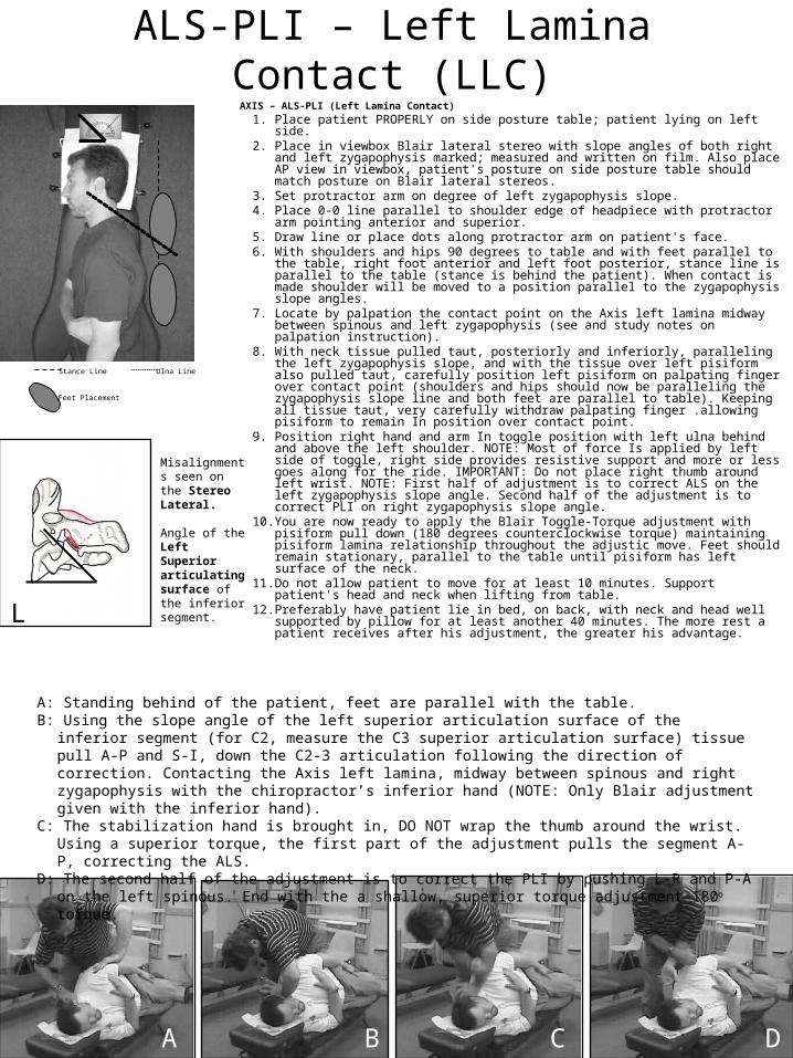
A: Standing behind of the patient, feet are parallel with the table. B: Using the slope angle of the left superior articulation surface of the inferior segment (for C2, measure the C3
superior articulation surface) tissue pull A-P and S-I, down the C2-3 articulation following the direction of correction. Contacting the Axis left lamina, midway between spinous and right zygapophysis with the chiropractor’s inferior hand (NOTE: Only Blair adjustment given with the inferior hand).
C: The stabilization hand is brought in, DO NOT wrap the thumb around the wrist. Using a superior torque, the first part of the adjustment pulls the segment A-P, correcting the ALS.
D: The second half of the adjustment is to correct the PLI by pushing L-R and P-A on the left spinous. End with the a shallow, superior torque adjustment 180o torque.
ALS-PLI – Left Lamina Contact (LLC)AXIS – ALS-PLI (Left Lamina Contact)
1. Place patient PROPERLY on side posture table; patient lying on left side. 2. Place in viewbox Blair lateral stereo with slope angles of both right and left
zygapophysis marked; measured and written on film. Also place AP view in viewbox, patient's posture on side posture table should match posture on Blair lateral stereos.
3. Set protractor arm on degree of left zygapophysis slope. 4. Place 0-0 line parallel to shoulder edge of headpiece with protractor arm
pointing anterior and superior. 5. Draw line or place dots along protractor arm on patient's face. 6. With shoulders and hips 90 degrees to table and with feet parallel to the
table, right foot anterior and left foot posterior, stance line is parallel to the table (stance is behind the patient). When contact is made shoulder will be moved to a position parallel to the zygapophysis slope angles.
7. Locate by palpation the contact point on the Axis left lamina midway between spinous and left zygapophysis (see and study notes on palpation instruction).
8. With neck tissue pulled taut, posteriorly and inferiorly, paralleling the left zygapophysis slope, and with the tissue over left pisiform also pulled taut, carefully position left pisiform on palpating finger over contact point (shoulders and hips should now be paralleling the zygapophysis slope line and both feet are parallel to table). Keeping all tissue taut, very carefully withdraw palpating finger .allowing pisiform to remain In position over contact point.
9. Position right hand and arm In toggle position with left ulna behind and above the left shoulder. NOTE: Most of force Is applied by left side of toggle, right side provides resistive support and more or less goes along for the ride. IMPORTANT: Do not place right thumb around left wrist. NOTE: First half of adjustment is to correct ALS on the left zygapophysis slope angle. Second half of the adjustment is to correct PLI on right zygapophysis slope angle.
10. You are now ready to apply the Blair Toggle-Torque adjustment with pisiform pull down (180 degrees counterclockwise torque) maintaining pisiform lamina relationship throughout the adjustic move. Feet should remain stationary, parallel to the table until pisiform has left surface of the neck.
11. Do not allow patient to move for at least 10 minutes. Support patient's head and neck when lifting from table.
12. Preferably have patient lie in bed, on back, with neck and head well supported by pillow for at least another 40 minutes. The more rest a patient receives after his adjustment, the greater his advantage.
Stance Line
Misalignments seen on the Stereo Lateral.
Angle of the Left Superior articulating surface of the inferior segment.
B
Feet Placement
Ulna Line
A C D
L

A: Stance line is parallel to the zygapophysis of the superior articulating surface for the inferior segment of the larger misalignment.B: Standing behind the patient, feet in a straight away stance line. Bring tissue pull and contact hand P to A and I to S. Contacting
the posterior C2 lamina. (See NOTE above for the specific contact point)C: Tissue pull P-A, I-S to the posterior spinous. The stabilization hand is brought in, wrap the thumb around the wrist. Contact ulna
parallel to the stance line. D: End with the a 180o inferior torque (fingers rotate toward patient’s feet).
PRI-PLI – Posterior Spinous Contact (PSC)AXIS PRI (Left Lamina Contact)
NOTE: Contact in a double PI Axis If both articular misalignments are equal, then a direct posterior inferior spinous contact may be made and may be made from either side. (Example is Double PI Axis Posterior Spinous Contact with patient lying on right side.) If the Right facet articular misalignment is larger, contact the Left side of the posterior spinous and use the Right facet angle for the stance line. If the Left facet articular misalignment is larger, contact the Right side of the posterior spinous and use the Left facet angle for the stance line
1. Example: Place patient properly on side posture table with patient lying on appropriate side. Example on drawing - right side.
2. Place in viewbox Blair Lateral Stereo with slope angle of both right and left zygapophysis marked, measured and written on film. Place A-P view in viewbox. Patient's posture on the table should match the posture on the Blair lateral Stereos.
3. Set protractor arm on proper degree of zygapophysis slope angle (See NOTE above). 4. 4. Place 0-0 line parallel to shoulder edge of headpiece with protractor arm pointing anterior and
superior in relation with patient. 5. Draw line or place dots along protractor arm on patient's face. 6. Example on drawing - patient lying on right side. With shoulders and hips 90 degrees to the
table and with feet parallel to the table, right foot anterior and left foot posterior, stance line is parallel to the table. (Stance is behind the patient). When contact is made, shoulder will be moved to a position parallel to the zygapophysis line of the slope angle.
7. Locate, by palpation, the contact point on the posterior inferior aspect of the axis spinous process. Contact will be posterior inferior of the posterior inferior aspect of the axis spinous for equal misalignments on the right and left facets. (see study notes on palpation). For a larger misalignment on the left than the right, the patient will lie on the left side and contact is made on the right posterior spinous. For a larger misalignment on the right than the left, the patient will lie on the right side and contact is made on the left posterior spinous.
8. With neck tissue pulled taut anteriorly and superiorly, and with the right ulna paralleling the zygapophysis slope and with the tissue over the spinous also pulled taut, carefully position your right pisiform on palpating finger over contact point (shoulders and hips should still be parallel to the zygapophysis slope). The right ulna (in the example given) should be parallel to the zygapophysis slope angle and your feet are parallel to the table. Keeping all tissue taut, very carefully withdraw palpating finger allowing pisiform to remain in snug position over contact point.
9. Position right hand and arm in toggle position with right ulna posterior and parallel to the zygapophysis slope angle as listed above. Note: most of force is applied with the right side of the toggle. Left side of toggle provides resistive support and more or less goes along for the ride.
10. You are now ready to apply the Blair Toggle Torque Adjustment with pisiform lift using 180 degrees of counterclockwise torque maintaining pisiform left spinous relationship throughout the adjustic move. Feet should remain stationary and parallel to the adjusting table until pisiform has left the surface of the neck.
11. Do not allow patient to move for at least ten minutes. Support patient's head and neck when lifting from table.
12. Preferably have patient lie in bed, on back, with neck and head well supported by pillow for at least another 40 minutes. The more rest a patient receives immediately after his ADJUSTMENT, the greater his advantage.
Stance Line Ulna Line
Misalignments seen on the Stereo Lateral.Posterior (L) facet is misaligned posteriorly
Angle of the Left Superior articulating surface of the inferior segment.
Feet Placement
L
A B C D