Embed Size (px)
Citation preview
1Toor S, et al. Br J Ophthalmol 2017;0:1–7. doi:10.1136/bjophthalmol-2017-310282
AbstrActbackground/aims To investigate the presence of asymmetrical accommodation in hyperopic anisometropic amblyopia.Methods Accommodation in each eye and binocular vergence were measured simultaneously using a PlusoptiX SO4 photorefractor in 26 children aged 4–8 years with hyperopic anisometropic amblyopia and 13 controls (group age-matched) while they viewed a detailed target moving in depth.results Without spectacles, only 5 (19%) anisometropes demonstrated symmetrical accommodation (within the 95% CI of the mean gain of the sound eye of the anisometropic group), whereas 21 (81%) demonstrated asymmetrical accommodation. Of those, 15 (58%) showed aniso-accommodation and 6 (23%) demonstrated ’anti-accommodation’ (greater accommodation for distance than for near). In those with anti-accommodation, the response gain in the sound eye was (0.93±0.20) while that of the amblyopic eye showed a negative accommodation gain of (−0.44±0.23). Anti-accommodation resolved with spectacles. Vergence gains were typical in those with symmetrical and asymmetrical accommodation.conclusion The majority of hyperopic anisometropic amblyopes demonstrated non-consensual asymmetrical accommodation. Approximately one in four demonstrated anti-accommodation.
IntroductIonEvidence suggests that accommodation is symmet-rical in each eye,1–5 so in anisometropia, the least ametropic eye determines the amount of accommo-dation, with the amblyopic eye ‘lagging behind’.6–10 However, some report that subtle asymmetrical accommodation can occur in typical, young adults,11 12 demonstrating that there is a mechanism to drive different responses in each eye separately.
Asymmetrical accommodation, however, has rarely been considered clinically. Although reduced accommodation has been reported in amblyopic eyes (overview in von Noorden and Campos p. 26013), accommodation in clinical and research settings is generally tested monocularly, and so asymmetrical accommodation would have gone undetected. Reduced accommodation in the amblyopic eye could be ascribed to reduced visual acuity (VA) or sensory loss over the central retinal region7 8 due to monocular contrast deprivation in anisometropia.14
A case study from our lab, where vergence and binocular accommodation were assessed simulta-neously and continuously, reported a child with
hyperopic anisometropic amblyopia, who demon-strated an extreme example of asymmetrical accom-modation.15 Without their spectacle correction, the sound eye accommodated appropriately for target distance but the amblyopic eye repeatedly ‘anti-ac-commodated’ (accommodation in the wrong direc-tion for the change in target distance), showing a greater accommodation response in the distance than at near.
The aim of this study was to determine whether the presence of asymmetrical accommodation was more widespread in hyperopic anisometropic amblyopia.
MethodsThe prospective study adhered to the Declara-tion of Helsinki and obtained both University and National Health Service ethics approval. Informed consent was obtained from parents and age-appro-priate assent from the children.
ParticipantsHyperopic anisometropic amblyopes, aged 4–8 years, were recruited from an Orthoptic department. All had been assessed with cycloplegic retinoscopy, fundus and media check and an orthoptic examina-tion. All had been prescribed fully corrected specta-cles, worn full time for at least 6 weeks. Amblyopia was defined by the VA in either eye. All had VA in the amblyopic eye worse than 0.2 logarithm of the minimum angle of resolution (logMAR), VA in the non-amblyopic eye of at least 0.2 logMAR, with >0.1 logMAR interocular difference.
A control group with a mean age matched to the patient group was recruited from the University of Reading typically developing Child Development Group database. All had VA ≥0.2 logMAR in each eye, with no more than 0.1 logMAR interocular difference.
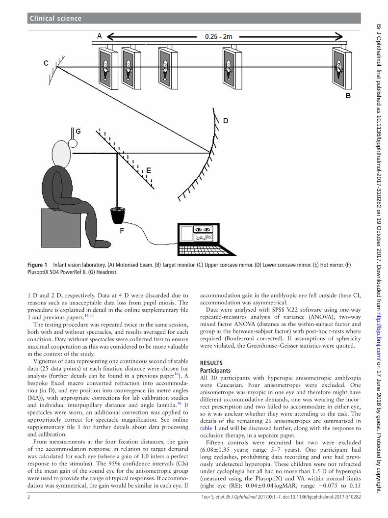
Laboratory testingA PlusoptiX SO4 photorefractor in PowerRef II mode made simultaneous and continuous refrac-tion and eye position recordings in both eyes at 25 Hz. The target was a detailed cartoon picture of a clown’s face subtending 3.15° at 2 m, which contained detailed elements down to 1 screen pixel but were easily identifiable even with reduced VA. Instructions were minimal, children were simply asked to ‘watch the clown’. The target was presented via a mirror arrangement (figure 1). Measurements were taken at five fixation distances in a pseu-do-random order (0.33, 2, 0.25, 1 and 0.5 m), representing demands of 3 dioptres (D), 0.5 D, 4 D,
Clinical science
Asymmetrical accommodation in hyperopic anisometropic amblyopiaSonia Toor,1 Anna M Horwood,2,3 Patricia Riddell2
to cite: Toor S, Horwood AM, Riddell P. Br J Ophthalmol Published Online First: [please include Day Month Year]. doi:10.1136/bjophthalmol-2017-310282
► Additional material is published online only. To view please visit the journal online (http:// dx. doi. org/ 10. 1136/ bjophthalmol- 2017- 310282).
1Academic Unit of Ophthalmology and Orthoptics, University of Sheffield, Sheffield, UK2Infant Vision Laboratory, School of Psychology & Clinical Language Sciences, University of Reading, Reading, UK3Orthoptic Department, Royal Berkshire Hospital, Reading, UK
correspondence toDr Sonia Toor, Academic Unit of Ophthalmology and Orthoptics, Faculty of Medicine, Dentistry and Health, University of Sheffield, Beech Hill Road, Sheffield, S10 2RX, UK; sonia. toor@ sheffield. ac. uk
Received 2 February 2017Revised 4 August 2017Accepted 20 August 2017
BJO Online First, published on October 19, 2017 as 10.1136/bjophthalmol-2017-310282
Copyright Article author (or their employer) 2017. Produced by BMJ Publishing Group Ltd under licence.
on 17 June 2018 by guest. Protected by copyright.
http://bjo.bmj.com
/B
r J Ophthalm
ol: first published as 10.1136/bjophthalmol-2017-310282 on 19 O
ctober 2017. Dow
nloaded from
2 Toor S, et al. Br J Ophthalmol 2017;0:1–7. doi:10.1136/bjophthalmol-2017-310282
clinical science
1 D and 2 D, respectively. Data at 4 D were discarded due to reasons such as unacceptable data loss from pupil miosis. The procedure is explained in detail in the online supplementary file 1 and previous papers.16 17
The testing procedure was repeated twice in the same session, both with and without spectacles, and results averaged for each condition. Data without spectacles were collected first to ensure maximal cooperation as this was considered to be more valuable in the context of the study.
Vignettes of data representing one continuous second of stable data (25 data points) at each fixation distance were chosen for analysis (further details can be found in a previous paper16). A bespoke Excel macro converted refraction into accommoda-tion (in D), and eye position into convergence (in metre angles (MA)), with appropriate corrections for lab calibration studies and individual interpupillary distance and angle lambda.16 If spectacles were worn, an additional correction was applied to appropriately correct for spectacle magnification. See online supplementary file 1 for further details about data processing and calibration.
From measurements at the four fixation distances, the gain of the accommodation response in relation to target demand was calculated for each eye (where a gain of 1.0 infers a perfect response to the stimulus). The 95% confidence intervals (CIs) of the mean gain of the sound eye for the anisometropic group were used to provide the range of typical responses. If accommo-dation was symmetrical, the gain would be similar in each eye. If
accommodation gain in the amblyopic eye fell outside these CI, accommodation was asymmetrical.
Data were analysed with SPSS V.22 software using one-way repeated-measures analysis of variance (ANOVA), two-way mixed factor ANOVA (distance as the within-subject factor and group as the between-subject factor) with post-hoc t-tests where required (Bonferroni corrected). If assumptions of sphericity were violated, the Greenhouse–Geisser statistics were quoted.
resuLtsParticipantsAll 30 participants with hyperopic anisometropic amblyopia were Caucasian. Four anisometropes were excluded. One anisometrope was myopic in one eye and therefore might have different accommodative demands, one was wearing the incor-rect prescription and two failed to accommodate in either eye, so it was unclear whether they were attending to the task. The details of the remaining 26 anisometropes are summarised in table 1 and will be discussed further, along with the response to occlusion therapy, in a separate paper.
Fifteen controls were recruited but two were excluded (6.08±0.35 years; range 5–7 years). One participant had long eyelashes, prohibiting data recording and one had previ-ously undetected hyperopia. These children were not refracted under cycloplegia but all had no more than 1.5 D of hyperopia (measured using the PlusoptiX) and VA within normal limits (right eye (RE): 0.04±0.04 logMAR, range −0.075 to 0.15
Figure 1 Infant vision laboratory. (A) Motorised beam. (B) Target monitor. (C) Upper concave mirror. (D) Lower concave mirror. (E) Hot mirror. (F) PlusoptiX SO4 PowerRef II. (G) Headrest.
on 17 June 2018 by guest. Protected by copyright.
http://bjo.bmj.com
/B
r J Ophthalm
ol: first published as 10.1136/bjophthalmol-2017-310282 on 19 O
ctober 2017. Dow
nloaded from
3Toor S, et al. Br J Ophthalmol 2017;0:1–7. doi:10.1136/bjophthalmol-2017-310282
clinical science
logMAR; left eye (LE): 0.03±0.04 logMAR, range −0.05 to 0.125 logMAR).
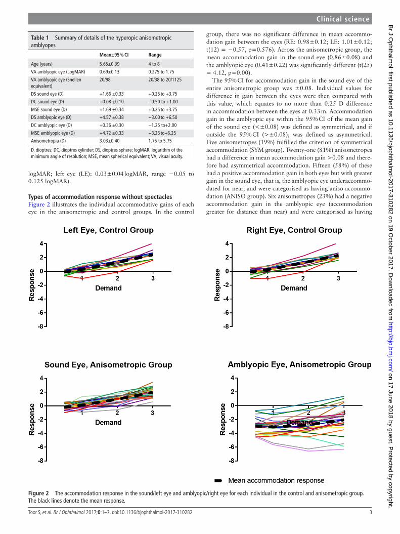
types of accommodation response without spectaclesFigure 2 illustrates the individual accommodative gains of each eye in the anisometropic and control groups. In the control
group, there was no significant difference in mean accommo-dation gain between the eyes (RE: 0.98±0.12; LE: 1.01±0.12; t(12) = −0.57, p=0.576). Across the anisometropic group, the mean accommodation gain in the sound eye (0.86±0.08) and the amblyopic eye (0.41±0.22) was significantly different (t(25) = 4.12, p=0.00).
The 95% CI for accommodation gain in the sound eye of the entire anisometropic group was ±0.08. Individual values for difference in gain between the eyes were then compared with this value, which equates to no more than 0.25 D difference in accommodation between the eyes at 0.33 m. Accommodation gain in the amblyopic eye within the 95% CI of the mean gain of the sound eye (<±0.08) was defined as symmetrical, and if outside the 95% CI (>±0.08), was defined as asymmetrical. Five anisometropes (19%) fulfilled the criterion of symmetrical accommodation (SYM group). Twenty-one (81%) anisometropes had a difference in mean accommodation gain >0.08 and there-fore had asymmetrical accommodation. Fifteen (58%) of these had a positive accommodation gain in both eyes but with greater gain in the sound eye, that is, the amblyopic eye underaccommo-dated for near, and were categorised as having aniso-accommo-dation (ANISO group). Six anisometropes (23%) had a negative accommodation gain in the amblyopic eye (accommodation greater for distance than near) and were categorised as having
table 1 Summary of details of the hyperopic anisometropic amblyopes
Mean±95% cI range
Age (years) 5.65±0.39 4 to 8
VA amblyopic eye (LogMAR) 0.69±0.13 0.275 to 1.75
VA amblyopic eye (Snellen equivalent)
20/98 20/38 to 20/1125
DS sound eye (D) +1.66 ±0.33 +0.25 to +3.75
DC sound eye (D) +0.08 ±0.10 −0.50 to +1.00
MSE sound eye (D) +1.69 ±0.34 +0.25 to +3.75
DS amblyopic eye (D) +4.57 ±0.38 +3.00 to +6.50
DC amblyopic eye (D) +0.36 ±0.30 −1.25 to+2.00
MSE amblyopic eye (D) +4.72 ±0.33 +3.25 to+6.25
Anisometropia (D) 3.03±0.40 1.75 to 5.75
D, dioptres; DC, dioptres cylinder; DS, dioptres sphere; logMAR, logarithm of the minimum angle of resolution; MSE, mean spherical equivalent; VA, visual acuity.
Figure 2 The accommodation response in the sound/left eye and amblyopic/right eye for each individual in the control and anisometropic group. The black lines denote the mean response.
on 17 June 2018 by guest. Protected by copyright.
http://bjo.bmj.com
/B
r J Ophthalm
ol: first published as 10.1136/bjophthalmol-2017-310282 on 19 O
ctober 2017. Dow
nloaded from
4 Toor S, et al. Br J Ophthalmol 2017;0:1–7. doi:10.1136/bjophthalmol-2017-310282
clinical science
anti-accommodation (ANTI group). This method of defining the type of accommodation response was used to investigate the control group. Five controls (38%) demonstrated symmet-rical accommodation and eight demonstrated aniso-accommo-dation (62%). No controls had anti-accommodation. Those controls with aniso-accommodation had a mean difference in gain between the eyes of 0.04 (±0.15), which was significantly lower than the difference of 0.23 (±0.14) found in the ANISO group (t(21) = −2.374, p=0.027). An example patient from
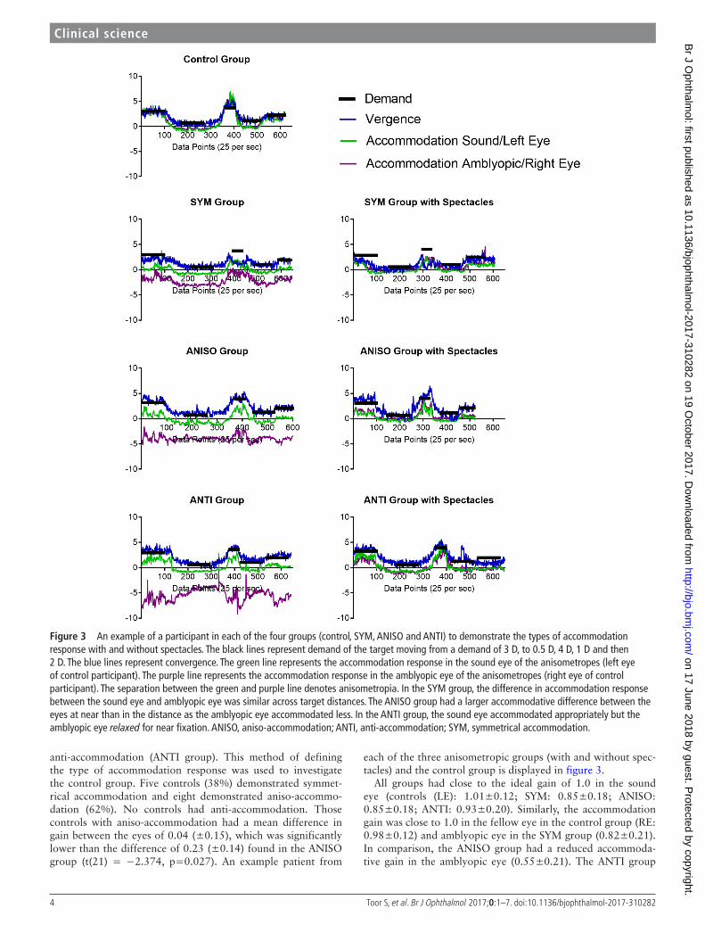
each of the three anisometropic groups (with and without spec-tacles) and the control group is displayed in figure 3.
All groups had close to the ideal gain of 1.0 in the sound eye (controls (LE): 1.01±0.12; SYM: 0.85±0.18; ANISO: 0.85±0.18; ANTI: 0.93±0.20). Similarly, the accommodation gain was close to 1.0 in the fellow eye in the control group (RE: 0.98±0.12) and amblyopic eye in the SYM group (0.82±0.21). In comparison, the ANISO group had a reduced accommoda-tive gain in the amblyopic eye (0.55±0.21). The ANTI group
Figure 3 An example of a participant in each of the four groups (control, SYM, ANISO and ANTI) to demonstrate the types of accommodation response with and without spectacles. The black lines represent demand of the target moving from a demand of 3 D, to 0.5 D, 4 D, 1 D and then 2 D. The blue lines represent convergence. The green line represents the accommodation response in the sound eye of the anisometropes (left eye of control participant). The purple line represents the accommodation response in the amblyopic eye of the anisometropes (right eye of control participant). The separation between the green and purple line denotes anisometropia. In the SYM group, the difference in accommodation response between the sound eye and amblyopic eye was similar across target distances. The ANISO group had a larger accommodative difference between the eyes at near than in the distance as the amblyopic eye accommodated less. In the ANTI group, the sound eye accommodated appropriately but the amblyopic eye relaxed for near fixation. ANISO, aniso-accommodation; ANTI, anti-accommodation; SYM, symmetrical accommodation.
on 17 June 2018 by guest. Protected by copyright.
http://bjo.bmj.com
/B
r J Ophthalm
ol: first published as 10.1136/bjophthalmol-2017-310282 on 19 O
ctober 2017. Dow
nloaded from
5Toor S, et al. Br J Ophthalmol 2017;0:1–7. doi:10.1136/bjophthalmol-2017-310282
clinical science
showed a negative accommodative gain in the amblyopic eye (−0.44±0.23).
An ANOVA revealed no significant difference in the accom-modative gain between the sound eye of the three anisometropic groups and the control group (LE) (F(3,35) = 1.42, p=0.253). For the amblyopic eye (RE in the control group), there was a significant main effect between the four groups (F(3,35) = 27.41, p<0.001). The accommodative gain in the amblyopic eye of the ANTI group was significantly different to each of the other groups (all p<0.001).
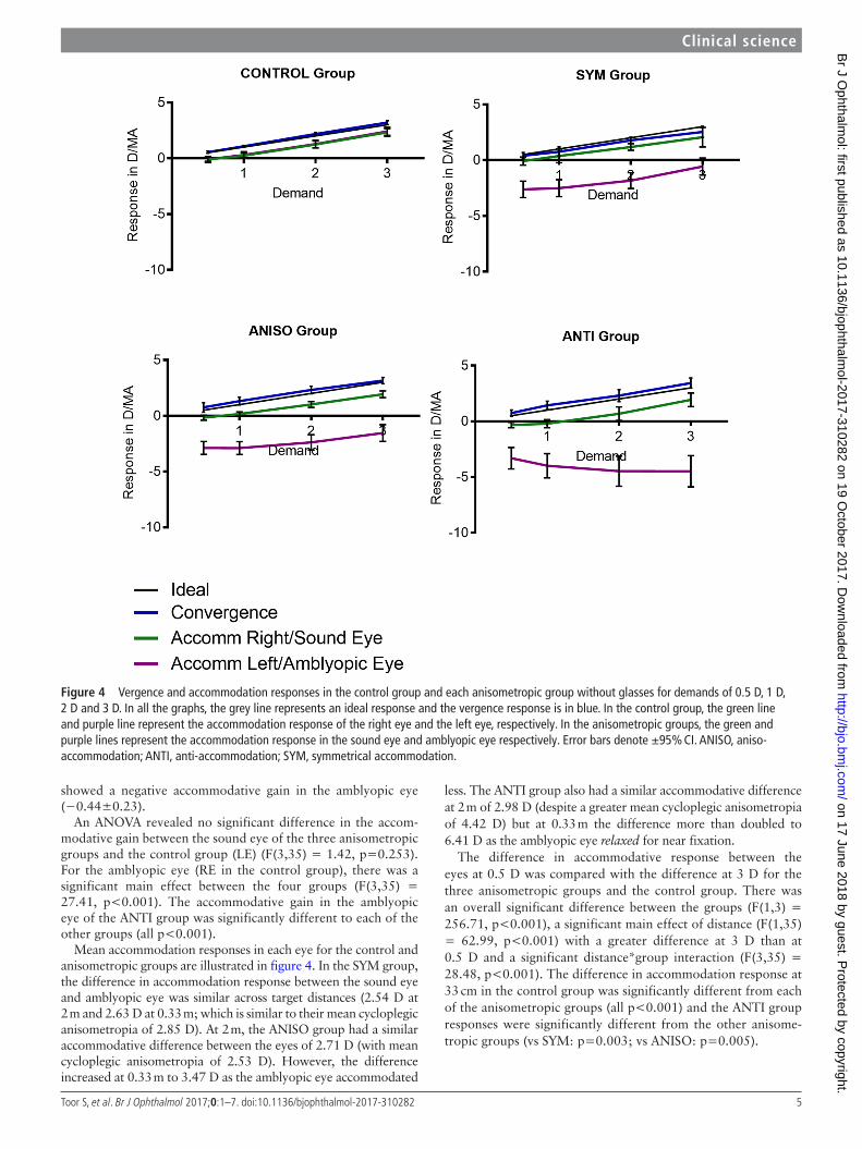
Mean accommodation responses in each eye for the control and anisometropic groups are illustrated in figure 4. In the SYM group, the difference in accommodation response between the sound eye and amblyopic eye was similar across target distances (2.54 D at 2 m and 2.63 D at 0.33 m; which is similar to their mean cycloplegic anisometropia of 2.85 D). At 2 m, the ANISO group had a similar accommodative difference between the eyes of 2.71 D (with mean cycloplegic anisometropia of 2.53 D). However, the difference increased at 0.33 m to 3.47 D as the amblyopic eye accommodated
less. The ANTI group also had a similar accommodative difference at 2 m of 2.98 D (despite a greater mean cycloplegic anisometropia of 4.42 D) but at 0.33 m the difference more than doubled to 6.41 D as the amblyopic eye relaxed for near fixation.
The difference in accommodative response between the eyes at 0.5 D was compared with the difference at 3 D for the three anisometropic groups and the control group. There was an overall significant difference between the groups (F(1,3) = 256.71, p<0.001), a significant main effect of distance (F(1,35) = 62.99, p<0.001) with a greater difference at 3 D than at 0.5 D and a significant distance*group interaction (F(3,35) = 28.48, p<0.001). The difference in accommodation response at 33 cm in the control group was significantly different from each of the anisometropic groups (all p<0.001) and the ANTI group responses were significantly different from the other anisome-tropic groups (vs SYM: p=0.003; vs ANISO: p=0.005).
Figure 4 Vergence and accommodation responses in the control group and each anisometropic group without glasses for demands of 0.5 D, 1 D, 2 D and 3 D. In all the graphs, the grey line represents an ideal response and the vergence response is in blue. In the control group, the green line and purple line represent the accommodation response of the right eye and the left eye, respectively. In the anisometropic groups, the green and purple lines represent the accommodation response in the sound eye and amblyopic eye respectively. Error bars denote ±95% CI. ANISO, aniso-accommodation; ANTI, anti-accommodation; SYM, symmetrical accommodation.
on 17 June 2018 by guest. Protected by copyright.
http://bjo.bmj.com
/B
r J Ophthalm
ol: first published as 10.1136/bjophthalmol-2017-310282 on 19 O
ctober 2017. Dow
nloaded from
6 Toor S, et al. Br J Ophthalmol 2017;0:1–7. doi:10.1136/bjophthalmol-2017-310282
clinical science
Accommodation response with spectaclesWith spectacles, the 95% CI of the sound eye of the entire anisometropic group was 0.09. On comparison of the ambly-opic eye to this value, 8 anisometropes (32%) had symmetrical accommodation, 17 (68%) had aniso-accommodation and no anisometropes demonstrated anti-accommodation (no data were collected from one child) but still demonstrated some aniso-ac-commodation. Figure 3 illustrates the effect of wearing specta-cles for each patient example in each group.
VergenceVergence gains were typical16 18 (control: 1.07±0.07; SYM: 0.88±0.24: ANISO 0.97±0.11; ANTI: 1.04±0.10) with no significant difference between the four groups (F(3,35) = 1.71, p=0.183). Therefore, both eyes were fixating the target and any difference in refraction cannot be ascribed to off-axis errors.
dIscussIonThe majority of the hyperopic anisometropic amblyopes had asymmetrical accommodation. Fifty-eight per cent had aniso-ac-commodation, with greater accommodative lag in the amblyopic eye at near. More interestingly, 23% of anisometropes demon-strated anti-accommodation. The sound eye accommodated appropriately when viewing a near target but the amblyopic eye accommodated in the opposite direction with a greater accom-modation response at distance than at near. This finding indicates that the child with anti-accommodation reported by Horwood and Riddell15 is not a unique case. Only 19% of anisometropes were found to have symmetrical accommodation, contradicting previous literature.6–10
It is very possible that other researchers and clinicians have overlooked the existence of asymmetrical accommodation, as objective accommodation is usually measured monocularly, even under binocular conditions. There is an assumption that testing one eye reflects the response of both eyes. Any reduced accom-modation response in the amblyopic eye, as discussed by von Noorden and Campos,13 could be ascribed to reduced VA in a fixing amblyopic eye, driving weak accommodation in both eyes.
Some studies have tried to induce aniso-accommodation but results have been negative, weak or fleeting.1–3 11 12 The natu-rally occurring, long-term abnormal input of developmental anisometropia is a more extreme visual experience than is possible to induce experimentally and may enable such responses to develop.
Although the ANISO group continued to underaccommodate somewhat for near, it was dramatic that the anti-accommodation resolved with spectacle correction. This suggests that anti-ac-commodation is not hardwired, but more driven by visual input and subject to short-term variation.
Our data only allow us to speculate on possible mechanisms. It is difficult to account for anti-accommodation with the current models in which both eyes are driven by a single accommodative signal. It becomes easier to explain this condition if accommoda-tion is driven independently. The anti-accommodation might be explained by a misinterpretation of blur cues in the amblyopic eye. An alternate explanation is that the anti-accommodation is the result of an active strategy that avoids conflict between a clear image in the sound eye and a less clear image in the amblyopic eye. For distant targets, where accommodative demand is low for both eyes, some accommodative effort could be made in the amblyopic eye to compensate for the anisometropia. On viewing a near target, however, while the blur signal to the sound eye would result in appropriate accommodation to clear the image,
the necessary accommodative effort required to both accommo-date for near and overcome the hyperopia might be too great for the amblyopic eye to compensate. Rather than partially accom-modating, this might result in total relaxation of the amblyopic eye therefore producing anti-accommodation. Full correction of the anisometropic blur with spectacles would reduce the accom-modative effort required by the amblyopic eye and hence make it possible for the amblyopic eye to accommodate.
The ability to make simultaneous measurements of accommodation in each eye and confirm on-axis refrac-tion by measuring simultaneous vergence allowed us to find behaviour, which may have been missed by other methods. The study has some limitations but they are unlikely to signifi-cantly affect the results. We were unable to make individual calibrations of refraction in these children. There is evidence that group means are acceptable for studies such as these,19 but individual responses and gains may be more variable than the mean data suggest. The limited linear operating range of the photorefractor may have caused further inaccuracies in refrac-tion measurements as calculations become non-linear towards these limits. However, our calibration studies on older chil-dren and adults and those published by others19 suggest that the PlusoptiX photorefractor is more likely to underestimate refraction, than overestimate it at these limits. This suggests that anti-accommodation would be even more marked than reported in this paper.
The majority of the control group also demonstrated aniso-accommodation to some extent. This is unlikely to be due to calibration error as the calibration factor should not differ in either eye. The results suggest that subtle aniso-ac-commodation in normals might be more common than previ-ously thought.1–3 11 12 We did not refract the controls under cycloplegia, but during lab testing we determine the maximum hyperopic refraction found at any time in the session, which correlates extremely well with cycloplegic refraction.
This is a small-scale study with the possible consequence that some of the statistical analysis may have been underpow-ered. The finding of any significant differences even in this relatively small group suggests that anti-accommodation in hyperopic anisometropic amblyopia is genuine and worthy of further study. Our findings provide clear evidence that accommodation in not necessarily a consensual response and provides further support that children should be wearing their full cycloplegic prescription to avoid aniso-accommodation and anti-accommodation.
concLusIonThe majority of children with hyperopic anisometropic ambly-opia have asymmetrical, rather than symmetrical, accommo-dation without spectacles, refuting previous suggestions in the literature.1–5 10 The majority of these children have aniso-accom-modation but 23% anti-accommodate. This suggests that there must be a mechanism by which it is possible to drive accommo-dation in each eye independently, even if this is rarely necessary in the general population.
contributors All authors made substantial contributions to the research. AH was the principal investigator. ST collected the data and drafted the manuscript. All authors contributed to the design, analysis and interpretation of the data, and in the revising of the manuscript.
Funding This research was supported by a UK Medical Research Council Clinical Scientist Fellowship awarded to AH (G0802809).
competing interests None declared.
Patient consent Obtained.
on 17 June 2018 by guest. Protected by copyright.
http://bjo.bmj.com
/B
r J Ophthalm
ol: first published as 10.1136/bjophthalmol-2017-310282 on 19 O
ctober 2017. Dow
nloaded from
7Toor S, et al. Br J Ophthalmol 2017;0:1–7. doi:10.1136/bjophthalmol-2017-310282
clinical science
ethics approval National Health Service.
Provenance and peer review Not commissioned; externally peer reviewed.
data sharing statement Data reported in this article are openly available from Zenodo at http:// dx. doi. org/ 10. 5281/ zenodo. 996512
open Access This is an Open Access article distributed in accordance with the terms of the Creative Commons Attribution (CC BY 4.0) license, which permits others to distribute, remix, adapt and build upon this work, for commercial use, provided the original work is properly cited. See: http:// creativecommons. org/ licenses/ by/ 4. 0/
© Article author(s) (or their employer(s) unless otherwise stated in the text of the article) 2017. All rights reserved. No commercial use is permitted unless otherwise expressly granted.
RefeRenCes 1 Koh LH, Charman WN. Accommodative responses to anisoaccommodative targets.
Ophthalmic Physiol Opt 1998;18:254–62. 2 Bharadwaj SR, Candy TR. The effect of lens-induced anisometropia on accommodation
and vergence during human visual development. Invest Ophthalmol Vis Sci 2011;52:3595–603.
3 Flitcroft DI, Judge SJ, Morley JW. Binocular interactions in accommodation control: effects of anisometropic stimuli. J Neurosci 1992;12:188–203.
4 Ball EA. A study of consensual accommodation. Am J Optom Arch Am Acad Optom 1952;29:561–74.
5 Campbell FW. Correlation of accommodation between the two eyes. J Opt Soc Am 1960;50:738.
6 Rutstein RP. Contemporary issues in amblyopia treatment. Optometry 2005;76:570–8. 7 Ciuffreda KJ, Hokoda SC, Hung GK, et al. Accommodative stimulus/response function
in human amblyopia. Doc Ophthalmol 1984;56:303–26.
8 Ciuffreda KJ, Rumpf D. Contrast and accommodation in amblyopia. Vision Res 1985;25:1445–57.
9 Li CH, Chen PL, Chen JT, et al. Different corrections of hypermetropic errors in the successful treatment of hypermetropic amblyopia in children 3 to 7 years of age. Am J Ophthalmol 2009;147:357–63.
10 Manh V, Chen AM, Tarczy-Hornoch K, et al. Accommodative performance of children with unilateral amblyopia. Invest Ophthalmol Vis Sci 2015;56:1193–207.
11 Marran L, Schor CM. Lens induced aniso-accommodation. Vision Res 1998;38:3601–19.
12 Benavente-Perez A, Nour A, Troilo D. The accommodative response under lens-imposed anisometropia in humans [abstract]. Invest Ophthalmol Vis Sci 2010;51:P3932.
13 von Noorden GK, Campos EC. Binocular vision and ocular motility: theory and management of strabismus. 6th edn. Missouri, USA: Mosby Inc, 2002:260.
14 Bradley A, Freeman RD. Contrast sensitivity in anisometropic amblyopia. Invest Ophthalmol Vis Sci 1981;21:467–76.
15 Horwood AM, Riddell PM. Independent and reciprocal accommodation in anisometropic amblyopia. J Aapos 2010;14:447–9.
16 Horwood AM, Riddell PM. The use of cues to convergence and accommodation in naïve, uninstructed participants. Vision Res 2008;48:1613–24.
17 Horwood AM, Riddell PM. Developmental changes in the balance of disparity, blur, and looming/proximity cues to drive ocular alignment and focus. Perception 2013;42:693–715.
18 Horwood AM, Riddell PM. A novel experimental method for measuring vergence and accommodation responses to the main near visual cues in typical and atypical groups. Strabismus 2009;17:9–15.
19 Bharadwaj SR, Sravani NG, Little JA, et al. Empirical variability in the calibration of slope-based eccentric photorefraction. J Opt Soc Am A Opt Image Sci Vis 2013;30:923–31.
on 17 June 2018 by guest. Protected by copyright.
http://bjo.bmj.com
/B
r J Ophthalm
ol: first published as 10.1136/bjophthalmol-2017-310282 on 19 O
ctober 2017. Dow
nloaded from


![SHACKEL Psychological LEFT Fci~~~~ci]-SYSTEM G..bjo.bmj.com/content/bjophthalmol/44/2/89.full.pdf · B. SHACKEL Psychological ResearchLaboratory, ... Therefore the apparatus and method](https://img.pdfslide.us/doc/110x75/5b32acca7f8b9adf6c8c4c5a/shackel-psychological-left-fcici-system-gbjobmjcomcontentbjophthalmol44289fullpdf.jpg)