Embed Size (px)
Citation preview
BRIEF REPORT
Asymmetric posterior reversible encephalopathy syndromein patient with hyperplastic anterior choroidal artery
Andrea Romano • Pugliese Silvia • Pierallini Alberto •
Francesca Tavanti • Giuliano Sette • Sara La Starza •
Luigi Maria Fantozzi • Alessandro Bozzao
Received: 1 November 2010 / Accepted: 20 December 2010 / Published online: 5 January 2011
� The Author(s) 2010. This article is published with open access at Springerlink.com
Abstract We describe a case of asymmetric PRES due
to the presence of hyperplastic anterior choroidal artery
(AChA) in a man affected by sever hypertension. Posterior
reversible encephalopathy syndrome (PRES) has become
synonymous with a unique pattern of brain vasogenic edema
and predominates in the parietal and occipital regions,
accompanied by clinical neurological alterations. Sever
hypertension is a risk factor that exceeds the limits of brain
autoregulation, leading to breakthrough brain edema. In our
knowledge this is the first case reported in literature, in which
a similar vascular abnormality is linked to a PRES syndrome.
Keywords Anterior choroidal artery � PRES � Vasogenic
edema � Hypertension � Headache
Introduction
Posterior reversible encephalopathy syndrome (PRES)
relates to a transient pattern of brain edema mostly involving
the parietal and occipital regions. Severe hypertension
may lead to PRES when overcoming brain autoregulation
with subsequent breakthrough brain edema. MR imaging
demonstrates reversible and symmetric focal hemispheric
edema. We describe a case of asymmetric PRES associated
with the presence of a hyperplastic anterior choroidal artery
(AChA) in a man affected by severe hypertension.
Case report
A 58-year man, with a history of mild hypertension, was
referred to the emergency room for a sudden onset of
visual disturbance, difficulty in speech, headache and
mental confusion. Clinical examination revealed increased
blood pressure (220 mmHg), mild anemia (HGB, 10.5 g/dl)
and hyper-cholesterolemia (250 mg/dl) on routine blood
work.
Neurologic examination revealed left homonymous lat-
eral hemianopia, mild difficulty in searching words, mild
pronation and downward drift of the right arm.
Electro-encephalography was positive for left temporo-
occipital alterations.
CT revealed bilateral cortical–subcortical patchy hypo-
dense foci localized in the parietal-occipital lobes, asym-
metric for left predominance without enhancement after
i.v. injection. Multiple hypodense confluent areas localized
in the supratentorial white matter as a result of chronic
ischemic cerebral disease were evident as well.
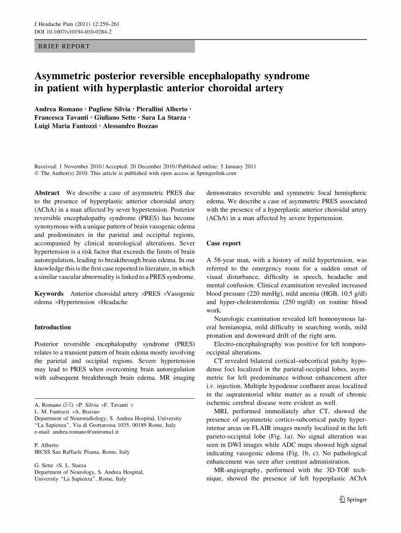
MRI, performed immediately after CT, showed the
presence of asymmetric cortico-subcortical patchy hyper-
intense areas on FLAIR images mostly localized in the left
parieto-occipital lobe (Fig. 1a). No signal alteration was
seen in DWI images while ADC maps showed high signal
indicating vasogenic edema (Fig. 1b, c). No pathological
enhancement was seen after contrast administration.
MR-angiography, performed with the 3D-TOF tech-
nique, showed the presence of left hyperplastic AChA
A. Romano (&) � P. Silvia � F. Tavanti �L. M. Fantozzi � A. Bozzao
Department of Neuroradiology, S. Andrea Hospital, University
‘‘La Sapienza’’, Via di Grottarossa 1035, 00189 Rome, Italy
e-mail: [email protected]
P. Alberto
IRCSS San Raffaele Pisana, Rome, Italy
G. Sette � S. L. Starza
Department of Neurology, S. Andrea Hospital,
University ‘‘La Sapienza’’, Rome, Italy
123
J Headache Pain (2011) 12:259–261
DOI 10.1007/s10194-010-0284-2
partially supplying the distribution of the ipsilateral pos-
terior cerebral artery (PCA) (Fig. 1f–h).
Because of the suspicion of acute hypertensive
encephalopathy, he was treated with mannitol (100 mL
6/day), potassium canrenoate (50 mg 1cp/day), furosemid
(25 mg 1cp/day).
Seven days after admission, symptoms resolved, blood
pressure was 140/90 mmHg.
Eleven days after admission, the patient was discharged
with complete resolution of the symptoms, stable reduction
of the blood pressure (115/70 mmHg) and a therapy
composed by potassium canrenoate (50 mg 1cp/day),
ramipril (10 mg 1cp/day), doxazosin mesylate (2 mg 1 cp/
day), amlodipine besylate(10 mg 1 cp/day) and acetylsal-
icylic acid (100 mg 1cp/day).
MRI follow-up performed 3 months later demonstrated
complete resolution of the signal alterations (Fig. 1d, e).
Discussion
The AChA is a branch of the internal carotid artery running
between the temporal lobe and the brain stem and reaching
the choroid plexus in the temporal ventricular horn. Along
its course, the AChA supplies several territories, in par-
ticular optic tract, posterior limb of internal capsule and
pulvinar. Several anomalies in course and caliber have
been described. In particular, enlargement of the AChA has
been reported with cerebral angiography in many patho-
logical cases [1]. In up to 1% of subjects, the AChA is
hyperplastic and supplies all, or a portion, of the PCA
territory. Takahashi et al. [2] described 25 hyperplastic
arteries supplying part of the territory of distribution of the
PCA. The hyperplastic arteries were further classified into
four subtypes according to the distribution area and course
of the vessel. In type 3 an anomalous artery supplies the
parieto-occipital and calcarine arteries [3], as observed in
our patient. This is a result of anastomotic channels
development, hemodynamically favored, between AChA
and PCA or PCOMA that determined variable degrees of
hypoplasia of the PCA [3].
In our patient the hyperplastic AChA supplied the terri-
tory affected by T2 signal alteration, suggesting a potential
role of the vascular anomaly in its genesis.
PRES is an acute rapidly evolving clinical condition
characterized by headache, nausea and vomiting,
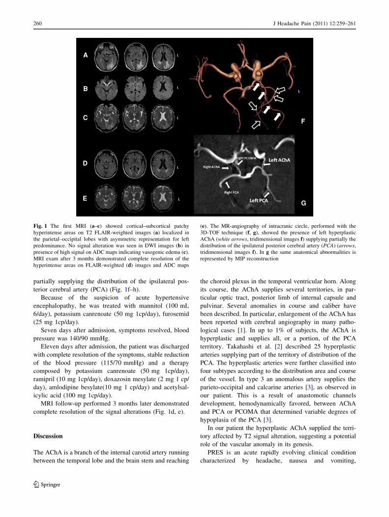
Fig. 1 The first MRI (a–c) showed cortical–subcortical patchy
hyperintense areas on T2 FLAIR-weighted images (a) localized in
the parietal–occipital lobes with asymmetric representation for left
predominance. No signal alteration was seen in DWI images (b) in
presence of high signal on ADC maps indicating vasogenic edema (c).
MRI exam after 3 months demonstrated complete resolution of the
hyperintense areas on FLAIR-weighted (d) images and ADC maps
(e). The MR-angiography of intracranic circle, performed with the
3D-TOF technique (f, g), showed the presence of left hyperplastic
AChA (white arrows, tridimensional images f) supplying partially the
distribution of the ipsilateral posterior cerebral artery (PCA) (arrows,
tridimensional images f). In g the same anatomical abnormalities is
represented by MIP reconstruction
260 J Headache Pain (2011) 12:259–261
123
abnormalities of visual perception, mental status abnor-
malities, seizure and focal neurological signs associated
with transient radiological brain anomalies. Different
conditions might be responsible (eclampsia, hypertensive
encephalopathy, renal disease with hypertension, neuro-
toxicity of cyclosporine A or other immunosuppressive
drugs and bone marrow transplantation) [4–7]. Other rare
pathologic conditions, such as intracranial hypotension, are
recently discussed [8, 9].
The presence of hypertension and the breakthrough of
cerebral autoregulation are retained as the most common
causes of PRES.
As mentioned radiological findings are usually revers-
ible [5, 7]. MR diffusion-weighted imaging usually con-
firms the presence of vasogenic edema with high values of
ADC.
In our patient, many findings were compatible with
PRES. Clinical onset and symptoms (headache with nau-
sea, visual disorders, altered mental status and seizure)
were evident in acute phase and completely resolved at
discharge. In our patient, the choice of using anti-hyper-
tensive care was due to the difficulty of a fast reduction of
blood pressure, avoiding other complications [10].
Lesion distribution in MRI was characteristic of PRES
as well, although clearly asymmetric. Common involve-
ment of atypical brain location other than parietal or
occipital regions is known [11]. The authors in this study
assessed that partial or asymmetric PRES was most com-
monly recognized in patients who have had organ trans-
plantation and eclampsia with severe hypertension or
normal blood pressure. Severe hypertension, renal failure,
immunosuppression could be also associated with asym-
metric PRES [12]. Variable expression of the PRES pat-
terns could be related to differences in arterial anatomy, as
demonstrated in our case, preexisting vascular disease or
regional hemispheric involvement in a clinical toxic pro-
cess [11].
Acute severe hypertension with transient loss of auto-
regulation system (sympathetic innervations) and sub-
sequent vasogenic edema [7, 11] seems the cause of PRES
in our patient. Posterior distribution of brain alterations has
been attributed to the sympathetic innervations of the
cerebral vessels. This distribution has an antero-posterior
gradient with a relatively reduced innervation of the pos-
terior cerebral arterial circulation [13], and therefore, dur-
ing acute elevation of blood pressure, posterior brain
regions may be particularly susceptible to breakthrough of
autoregulation. The presence of a ‘‘double’’ vascular dis-
trict (AChA and PCA) in the same vascular territory during
acute hypertensive attack might be the cause of ‘‘blood
hyper-influx syndrome’’ leading to asymmetric vasogenic
edema.
Although the presence of hyperplastic AChA is well
known [2], to our knowledge this is the first case reported
in literature in which such a vascular abnormality was
linked to PRES syndrome.
Conflict of interest None.
Open Access This article is distributed under the terms of the
Creative Commons Attribution License which permits any use, dis-
tribution and reproduction in any medium, provided the original
author(s) and source are credited.
References
1. Wollschlaeger G, Wollschlaeger P, Meyer PG et al (1969)
Widening or hyperplasia of the anterior choroidal artery. Radi-
ology 93:1079–1083
2. Takahashi S, Suga T, Kawata Y et al (1990) Anterior choroidal
artery: angiographic analysis of variant and anomalies. AJNR Am
J Neuroradiol 11:719–729
3. Antonietti LCL, Glastonbury CM, Adler F et al (2006) Hyper-
plastic anterior choroidal artery identified using magnetic reso-
nance angiography: a report of two cases. Cerebrovasc Dis
22:450–452
4. Finocchi V, Bozzao A, Bonamini M et al (2005) Magnetic res-
onance imaging in posterior reversible encephalopathy syndrome:
report of three cases and review of literature. Arch Gynecol
Obstet 271:79–85
5. Byrom F (1954) The pathogenesis of hypertensive encephalopa-
thy and its relation to the malignant phase of hypertension.
Lancet 2:201–211
6. Schwartz RB, Mulkern RV, Gudbjartsson H et al (1998) Diffu-
sion imaging in hypertensive encephalopathy: clues to patho-
genesis. AJNR Am J Neuroradiol 19:859–862
7. Sibai BM, Spinnato JA, Watson DL et al (1985) Eclampsia. IV.
Neurological findings and future outcome. Am J Obstet Gynecol
152:184–192
8. Pugliese S, Finocchi V, Borgia ML et al (2010) Intracranial
hypotension and PRES: case report. J Headache Pain 11:437–440
9. Yang Y, Wang JM, Zhou HW et al (2010) Intracranial hypo-
tension and PRES. J Headache Pain 11:447
10. Striano P, Striano S, Tortora F et al (2005) Clinical spectrum and
critical care management of posterior reversible encephalopathy
syndrome (PRES). Med Sci Monit 11:549–553
11. Bartynski WS, Boardman JF (2007) Distinct imaging patterns and
lesion distribution in posterior reversible encephalopathy syn-
drome. AJNR Am J Neuroradiol 28:1320–1327
12. Fugate JE, Claassen DO, Cloft HJ et al (2010) Posterior revers-
ible encephalopathy syndrome: associated clinical and radiologic
findings. Mayo Clin Proc 85:427–432
13. Chen CJ, Barkovich AJ (1998) A hyperplastic anterior choroidal
artery with double persistent anastomotic channels. AJNR Am J
Neuroradiol 19:1758–1760
J Headache Pain (2011) 12:259–261 261
123


![Unilateral Choroidal Osteoma with Choroidal Neovascularization...Surgical evacuation of the choroidal neovascular membrane has been reported [12] but the visual outcome was not favorable](https://img.pdfslide.us/doc/110x75/6053732923e31173be575e28/unilateral-choroidal-osteoma-with-choroidal-neovascularization-surgical-evacuation.jpg)