Embed Size (px)
Citation preview

The American Journal of Pathology, Vol. 189, No. 1, January 2019
ajp.amjpathol.org
TUMORIGENESIS AND NEOPLASTIC PROGRESSION
Astroprincin (FAM171A1, C10orf38)
A Regulator of Human Cell Shape and Invasive GrowthTiina Rasila,* Olga Saavalainen,* Hesham Attalla,* Petri Lankila,* Caj Haglund,yz Erkki Hölttä,* and Leif C. Andersson*
From the Department of Pathology* and the Research Programs Unit,y Translational Cancer Biology, University of Helsinki, Helsinki; and HUSLAB,z
Helsinki University Hospital, Helsinki, Finland
Accepted for publication
C
T
h
September 10, 2018.
Address correspondence toLeif C. Andersson, M.D., Ph.D.,Department of Pathology,University of Helsinki, Haart-maninkatu 3 (PB 21), 00014Helsinki, Finland. E-mail: [email protected].
opyright ª 2019 American Society for Inve
his is an open access article under the CC B
ttps://doi.org/10.1016/j.ajpath.2018.09.006
Our group originally found and cloned cDNA for a 98-kDa type 1 transmembrane glycoprotein of unknownfunction. Because of its abundant expression in astrocytes, it was called the protein astroprincin (APCN).Two thirds of the evolutionarily conserved protein is intracytoplasmic, whereas the extracellular domaincarries two N-glycosidic side chains. APCN is physiologically expressed in placental trophoblasts, skeletaland hearth muscle, and kidney and pancreas. Overexpression of APCN (cDNA) in various cell lines inducedsprouting of slender projections, whereas knockdown of APCN expression by siRNA caused disappearanceof actin stress fibers. Immunohistochemical staining of human cancers for endogenous APCN showedelevated expression in invasive tumor cells compared with intratumoral cells. Human melanoma cells (SK-MEL-28) transfected with APCN cDNA acquired the ability of invasive growth in semisolid medium(Matrigel) not seen with control cells. A conserved carboxyterminal stretch of 21 amino acids was found tobe essential for APCN to induce cell sprouting and invasive growth. Yeast two-hybrid screening revealedseveral interactive partners, of which ornithine decarboxylase antizyme-1, NEEP21 (NSG1), and ADAM10were validated by coimmunoprecipitation. This is the first functional description of APCN. These datashow that APCN regulates the dynamics of the actin cytoskeletal and, thereby, the cell shape and invasivegrowth potential of tumor cells. (Am J Pathol 2019, 189: 177e189; https://doi.org/10.1016/j.ajpath.2018.09.006)
Supported by The Academy of Finland grant 265620 (L.C.A.), TheSigrid Jusélius Foundation (L.C.A. and C.H.), The Magnus EhrnroothFoundation (L.C.A.), Finska Läkaresällskapet (L.C.A.), and Liv och Hälsa(L.C.A.).
T.R. and O.S. contributed equally to this work.Disclosures: None declared.
We originally obtained evidence for a previously unchar-acterized protein, when a human brain expression librarywas screened with a polyclonal antibody made in sheepagainst human brain homogenate. The full-length cDNAwas cloned and its mRNA was initially deposited on July11, 2004, in GenBank (https://www.ncbi.nlm.nih.gov/nuccore; accession number AY683003.1). A peptideantibody to the protein was generated, and a highexpression was found in brain astrocytes byimmunohistochemistry; hence, the protein was namedastroprincin (APCN).
With the clone mapping and sequencing of humanchromosome 10, the APCN gene was identified and thecoding sequence was annotated as C10orf38, subsequentlycalled FAM171A1.1 On the basis of the coding sequence,APCN was found to be a protein of 98-kDa size containing890 amino acids (https://www.ncbi.nlm.nih.gov; accessionnumber NP_001010924.1).
stigative Pathology. Published by Elsevier Inc
Y-NC-ND license (http://creativecommons.org
There is scanty previous information on APCN in theliterature. APCN/FAM171A1 has occurred in differentsettings of screening for gene expression or proteomics.Simmen et al2 investigated the impact of the transcriptionregulator Krüppel-like factor 9 in human HEC-1-A endo-metrial carcinoma cells. Among the genes showingenhanced mRNA expression on overexpression of Krüppel-like factor 9 was C10orf38, annotated as putativemembraneeassociated protein. Liao et al3 searched forproteins carrying ZU5 motifs and found the presence ofextracellular ZU5-like domains in the FAM171 proteins.
.
/licenses/by-nc-nd/4.0).

Rasila et al
The ZU5 domains are versatile protein-protein interactionmodules mediating, for example, bridging between ankyrinand b-spectrin.4
Prunotto et al5 performed proteomic analysis of podocyteexomeeenriched fractions from human urine and foundFAM171A1 among the 1195 identified proteins. In astudy by temporal proteomics during nerve growthfactoreinduced neural outgrowth in SH-SY5Y neuroblas-toma cells, FAM171A1 was among the 1923 proteinsshowing transiently up-regulated expression.6
St-Denis et al7 used complementary affinity purificationand proximity-based interaction proteomics to screen forinteractomes of 140 human proteins with phosphatasecatalytic domains. Among the 1335 identified proteins,FAM171A1 was found to interact with the protein tyrosinephosphatase receptor type F or leukocyte antigen-relatedtyrosine phosphatase.7 This transmembrane tyrosine phos-phatase has been functionally linked to adherent junctionsbetween epithelial cells and involved in regulation ofb-catenin signaling. Huttlin et al8 investigated HEK293Tcells by affinity purificationemass spectrometry prote-omics and found evidence for interaction betweenFAM171A1 and procadherin g subfamily B1, a calcium-dependent cell adhesion protein that has been implicatedin the establishment and maintenance of brain neuronalconnections.9
In a genome-wide transcriptome analysis of humanepidermal melanocytes, Haltaufderhyde and Oancea10 founda 12-fold up-regulated expression of FAM171A1 in lightlypigmented melanocytes compared with darkly pigmentedcells. Santuario-Facio et al11 analyzed genetic signatures ofhigh-grade breast cancer and found FAM171A1 among thenine tumor-associated genes displaying overexpressedexpression in triple-negative aggressive tumors.
There is, however, no previous information about themolecular function(s) of APCN/FAM171A1. This studyreports the first functional characterization of the APCNprotein. It shows that APCN is an evolutionarily conserved98-kDa transmembrane type I glycoprotein expressed invarious normal and malignant cells. Data demonstrate thatAPCN is involved in regulation of the cytoskeletaldynamics and, thereby, the cell shape and invasive growthof tumor cells.
Materials and Methods
Bioinformatics
Online databases and tools (ExPASy, https://www.expasy.org, last accessed December 12, 2016; Swiss Institute ofBioinformatics, Lausanne, Switzerland) were used toidentify known motifs from the APCN sequence. Online-alignment services Basic Local Alignment Search Tool(BLAST; http://blast.ncbi.nlm.nih.gov/Blast.cgi, last accessedFebruary 5, 2018), Multiple Alignment using Fast FourierTransform (MAFFT; https://mafft.cbrc.jp/alignment/server,
178
last accessed October 10, 2017), and ClustalW2 (http://www.ebi.ac.uk/Tools/clustalw2/index.html, last accessed October10, 2017) were used to compare APCN sequences betweendifferent species.To determine the tissue transcription profile of APCN, a
radioactive probe was prepared for the region, spanningnucleotides 86 to 417 of the open reading frame and hy-bridized to a Multiple Tissue Expression Array and MultipleTissue Northern Blot (Clontech Laboratories, Palo Alto,CA), as described previously.12
In Situ Hybridization
Tissue sections from early (sixth week of gestation)placenta were hybridized with antisense and sense com-plementary RNA probes corresponding to the region 808 to1237 of APCN cDNA. The complementary RNA probeswere labeled with digoxigenin-UTP by in vitro transcrip-tion with T7 polymerase using a DIG RNA Labelling Kit(Roche Diagnostics GmbH, Mannheim, Germany), and200 ng of the probe was used for each hybridization. AVentana Discovery Staining Platform was used for auto-mated hybridization. Detection of the DIG label was per-formed with monoclonal biotinylated anti-digoxinantibody (Jackson Immuno Research Laboratories Inc.,West Grove, PA) at 1:2000 dilution using a BlueMap kit(Ventana Medical Systems Inc., Tucson, AZ). The slideswere counterstained with Nucleofast Red (Ventana Medi-cal Systems Inc.).
Preparation of Peptide Antibodies to APCN in Rabbits
A synthetic peptide (SVTSHGRPEAPGTKELM) corre-sponding to amino acids 378 to 394 of APCN wassynthesized on a four-branch lysine core as multiple antigenpresentation peptide (MAP4) with Applied Biosystems(Foster City, CA) 433A automated peptide synthesizer usingFmoc chemistry. The peptide was purified by reverse-phasehigh-performance liquid chromatography and verified bymatrix-assisted laser adsorption time-of-flight spectroscopy.Two female rabbits were immunized with 400 mg of the
peptide polymer in Freund’s complete adjuvant. After 4weeks, three booster injections with 200 mg peptide polymerin Freud’s incomplete adjuvant were given with 3-weekintervals; and 10 days after the last immunization, blood wascollected and the sera were isolated. The antibody wasproduced at the Viikki Laboratory Animal Center, Univer-sity of Helsinki (Helsinki, Finland). All animals werehandled in strict accordance with good animal practice, asdefined by the relevant Finnish animal welfare bodies, andthe European Communities Council directive (86/609/EEC).
Immunostaining
Formalin-fixed, paraffin-embedded anonymous tissue spec-imens were collected from the archives of the HUSLAB and
ajp.amjpathol.org - The American Journal of Pathology

Astroprincin Regulates Tumor Invasion
the Department of Pathology, Haartman Institute, Universityof Helsinki, in accordance with the Finnish legislation andlocal ethical guidelines. Sections (4 mm thick) were depar-affinized in xylene and rehydrated. Antigen was retrievedby microwaving in 10 mmol/L citric acid monohydrate for3 � 5 minutes at 650 W. Endogenous peroxidase activitywas blocked by treatment with 0.5% H2O2. The slides wereincubated overnight in a refrigerator at 4�C with the primaryantibody in phosphate-buffered saline (PBS) containing0.5% normal human serum. The same procedure was usedfor negative controls, except that the incubation overnighttook place in PBS diluent without antibody. The reactionwas visualized with 3-amino-9-ethylcarbazole (Vectastain;Vector Laboratories, Burlingame, CA). Immunohistochem-ical staining of some sections was performed in anAutostainer 480 (Lab Vision Corp., Fremont, CA) by theDako REAL EnVision Detection System, Peroxidase/DABþ, Rabbit/Mouse (Dako, Glostrup, Denmark).
Cells grown on coverslips were rinsed once with coldPBS and fixed in ice-cold methanol for 10 minutes at 4�C.Alternatively, cells were fixed with 3.5% paraformaldehydefor 15 minutes at room temperature and permeabilized with0.02% IGEPAL CO-630 (Sigma Aldrich, St. Louis, MO) for20 minutes at room temperature. Blocking was done withhuman serum diluted 1:10 in 1� PBS or with BackgroundBlocker (Enzo Life Sciences, Farmingdale, NY) for 30 mi-nutes at room temperature. Cells were first incubated withprimary antibody for 1 hour, washed three times with 1�PBS, and then incubated with species-appropriate secondaryantibodies conjugated with either Alexa Fluor-488 or AlexaFluor-555 (both at 1:1000 dilution; Thermo Fisher Scienti-fic, Waltham, MA) or with fluorescein isothiocyanate goatanti-mouse (at 1:50 dilution; Dako) or tetramethylrhod-amine (TRITC) swine anti-rabbit (at 1:30 dilution; Dako).
Wheat germ agglutinineTRITC (20 mg/mL; EY Labo-ratories Inc., San Mateo, CA) was used to visualize cellsurface membrane. Wheat germ agglutinineTRITC wasadded to cells grown on coverslips after initial rinsing,incubated for 30 minutes at 4�C in the dark, and fixed.
Cell Cultures and Transient Transfections
COS-7, HEK293, MCF-7, NIH3T3, and SK-MEL-28 celllines were obtained from ATCC (Manassas, VA). TheU373MG astrocytoma line was kindly provided by Prof.Bengt Westermark (University of Uppsala, Uppsala,Sweden), and SK-MEL-103 (NRAS Q61R) and SK-MEL-147 (NRAS Q61R) both originated from Dr. AlanHoughton (Memorial Sloan-Kettering Cancer Center, NewYork, NY) and were kindly provided by Dr. Maria Soengas(Spanish National Cancer Research Center, Madrid,Spain). The cell lines, except for COS-7, were grown inRPMI 1640 medium supplemented with 10% (v/v) fetalbovine serum (Sigma-Aldrich), 1 mmol/L L-glutamine, 50mg/mL penicillin, and 50 mg/mL streptomycin at 37�C in ahumidified atmosphere of 5% CO2 in air. COS-7 cells were
The American Journal of Pathology - ajp.amjpathol.org
grown under similar conditions in Dulbecco’s modifiedEagle’s medium.
Cells were transfected using Lipofectamine 2000 trans-fection reagent (Invitrogen, Carlsbad, CA) or withFuGENE6 transfection reagent (Roche DiagnosticsGmbH), according to the manufacturer’s directions. Lip-ofectamine was also used when cells were transfected with6-carboxyfluoresceinelabelled siRNA against FAM171A1(Sigma-Aldrich) and control siRNA (Ambion; ThermoFisher Scientific). Lines of SK-MEL-28 cells stabilityoverexpressing APCN or pcDNA3 empty vector wereselected by cultivation in the presence of 1000 mg/mLG418.
RNA Interference
SK-MEL-147 and U373MG cells were seeded on coverslips(18 mm) at 30,000 cells per well on a 12-well scale or100,000 cells per 6-well scale 24 hours before siRNAtransfection. Cells were transfected with 20 nmol/L APCNsiRNA 6-FAM (sense, 50-CUGAUGAGUGGAGUCCAUU[dT][dT][6FAM]-30; antisense, 50-AAUGGACUCCACU-CAUCAG-30) or 20 nmol/L MISSION siRNA FluorescentUniversal Negative Control Number 1, 6-FAM (Sigma-Aldrich). Cells were transfected using MISSION siRNATransfection Reagent (Sigma-Aldrich), according to manu-facturer’s protocol.
RT-PCR, Real-Time Quantitative PCR, and Primers
To examine the transcription of the APCN gene in cells,total RNA was extracted using the TRI REAGENTeRNA/DNA/Protein isolation reagent (Molecular Research Center,Inc., Cincinnati, OH), according to the manufacturer’s in-structions. Total RNA (1 mg) was reverse transcribed withthe High Capacity RNA-to-cDNA Kit (Applied Biosystems,Foster City, CA), and cDNA was used as a template forquantitative real-time PCR analysis using a Maxima SYBRGreen/ROX qPCR Master Mix (Thermo Fisher Scientific)and a LightCycler II instrument (Roche DiagnosticsGmbH). Primers used for real-time analysis includedthe following: APCN, 50-TTACCACGTATCACACG-GTG-30(forward) and 50-TTTGGAACTCTCCAGTCCTG-30(reverse); and glyceraldehyde-3-phosphate dehydroge-nase, 50-GGTGAAGGTCGGAGTCAAC-30(forward) and50-CAAATGAGCCCCAGCCTTC-30(reverse).
Antibodies
The following antibodies were used in this study (includingdilutions/amounts used for immunofluorescence, immuno-histochemistry, Western blot analysis, or immunoprecipita-tion): MAP346 (1:70, immunofluorescence; 1:400,immunohistochemistry; 1:200, Western blot analysis),FLAG (F3165; Sigma-Aldrich; 10 mg/mL, immunofluores-cence; 3 mg/mL, Western blot analysis; 5 or 7.5 mg,
179

Rasila et al
immunoprecipitation), Myc (562; MBL International,Woburn, MA; 1:3000, Western blot analysis; and M4439;Sigma-Aldrich; 1:7000 Western blot analysis, 7.5 mgimmunoprecipitation), NSG1 (HPA035775; Sigma-Aldrich;1:100 Western blot analysis; and bs-840R; Bioss Anti-bodies, Woburn, MA), green fluorescent protein (632375;Clontech Laboratories, Mountain View, CA; 3 mg/mLimmunofluorescence), ADAM10 (MAB1427; R&D Sys-tems; 1:50 immunohistochemistry; and sc-48400; SantaCruz Biotechnology, Dallas, TX; 1:20 immunofluores-cence), and antizyme-1 (AZ-1; HPA009291; Sigma-Aldrich; 1:2000 Western blot analysis).
Imaging
Microphotographs were taken with an Olympus BX51microscope (Olympus Corp., Shinjuku, Japan) equippedwith a Nikon Digital Sight DS-U1 camera system (Nikon
Table 1 The List of Oligonucleotides Used in This Study
Application Name Sequ
flAPCN flAPCN-For 50-AflAPCN-Rev 50-A
cytAPCN cytAPCN-For 50-AflAPCN-Rev 50-A
tcAPCN tcAPCN-For 50-TflAPCN-Rev 50-A
tmAPCN flAPCN-For 50-AtmAPCN-Rev 50-A
T1-APCN flAPCN-For 50-AT1-APCN-Rev 50-A
T2-APCN T2-APCN-For 50-AflAPCN-Rev 50-A
T3-APCN flAPCN-For 50-AT3-APCN-Rev 50-A
D1-APCN flAPCN-For 50-AD1-APCN-Rev 50-GD1-APCN-For 50-GflAPCN-Rev 50-A
D3-APCN flAPCN-For 50-AD3-APCN-Rev 50-CD3-APCN-For 50-AflAPCN-Rev 50-A
APCNS136A S136A_APCN_for 50-CS136A_APCN_rev 50-G
APCNN159A N159A_APCN_for 50-CN159A_APCN_rev 50-G
APCNN190A N190A_APCN_for 50-GN190A_APCN_rev 50-G
APCNN194A N194A_APCN_for 50-GN194A_APCN_rev 50-G
APCNGFP flAPCN-For 50-AflAPCN-GFP-Rev 50-A
NEEP21 NEEP21-For 50-ANEEP21-Rev 50-A
NEEP21 NEEP21-For2 50-ANEEP21-Rev2 50-A
180
Corp., Shinjuku, Japan), a Zeiss Axioplan 2 Imagingfluorescence microscope (Carl Zeiss AG, Oberkoschen,Germany), and a Leica confocal laser-scanning microscope(Leica TCS SP2; Leica Microsystem GmbH, Wetzlar,Germany).
Yeast Two-Hybrid Screening
Yeast two-hybrid screening was performed using theMatchmaker GAL4 Two-Hybrid System 3 (Clontech Lab-oratories, Palo Alto, CA). The full-length APCN cDNA wascloned in frame with the GAL4 DNA-binding domain ofpGBKT7 vector and then used as a bait construct. The baitconstruct was then transformed into yeast strain AH109.The yeast strain Y187 was pretransformed with a humanplacenta Matchmaker cDNA library (Clontech Laboratories,Palo Alto, CA) in the pACT2 vector.
ence
AAAAGCTTATGAGCAGGTCCGCGACGCTGCTGC-30
AAGGATCCTTTAATGTTAAACGCCATCAGG-30
AAAAGCTTATGTATTATTGCAGGAGGAAGTGC-30
AAGGATCCTTTAATGTTAAACGCCATCAGG-30
AAAAGCTTATGCACACGGTGTTTCTTTTGG-30
AAGGATCCTTTAATGTTAAACGCCATCAGG-30
AAAAGCTTATGAGCAGGTCCGCGACGCTGCTGC-30
AAGGATCCAAGGAGACACAGCAAAACCAAAAG-30
AAAAGCTTATGAGCAGGTCCGCGACGCTGCTGC-30
AAGGATCCGAGAGACTCCGACATGGATGCGTTCTGAGG-30
AAAAGCTTATGCCTGAGAACACCAGCTACAGTGACC-30
AAGGATCCTTTAATGTTAAACGCCATCAGG-30
AAAAGCTTATGAGCAGGTCCGCGACGCTGCTGC-30
AAGGATCCTCCTTGGTCATCATCATCATCGTCTCCC-30
AAAAGCTTATGAGCAGGTCCGCGACGCTGCTGC-30
TACTTTCCTGTAAACGCTGTCCATCATACATTCAGTGGGTC-30
ACAGCGTTTACAGGAAAGTACTG-30
AAGGATCCTTTAATGTTAAACGCCATCAGG-30
AAAAGCTTATGAGCAGGTCCGCGACGCTGCTGC-30
CGGGCTGATTTTGGTTCATTCATCATACATTCAGTGGGTCTG-30
ATGAACCAAAATCAGCCCGG-30
AAGGATCCTTTAATGTTAAACGCCATCAGG-30
GTCCAAATAGTAGCAGGATTCCAAGGTGCC-30
GCACCTTGGAATCCTGCTACTATTTGGACG-30
TGAGGTTGCCTGAGGCCACCAGCTACAGTGAC-30
TCACTGTAGCTGGTGGCCTCAGGCAACCTCGA-30
GATTAGACGGAGCTGGAACAGGAAACAGC-30
CTGTTTCCTGTTCCAGCTCCGTCTAATCC-30
GAAATGGAACAGGAGCCAGCACCAGGCATGAC-30
TCATGCCTGGTGCTGGCTCCTGTTCCATTTCC-30
AAAAGCTTATGAGCAGGTCCGCGACGCTGCTGC-30
AAGGATCCAATTTAATGTTAAACGCCATCAGGGG-30
AAAAGCTTATGGTGAAGTTGGGGAACAATTTCGCAG-30
AAGGATCCAGCTGACTTCTCAGCCGCTTCAG-30
AAAAGAATTCATGGTGAAGTTGGGGAACAATTTCGCAGAG-30
AAAAGTCGACCTAAGCTGACTTCTCAGCCGCTTCAG-30
ajp.amjpathol.org - The American Journal of Pathology
Astroprincin Regulates Tumor Invasion
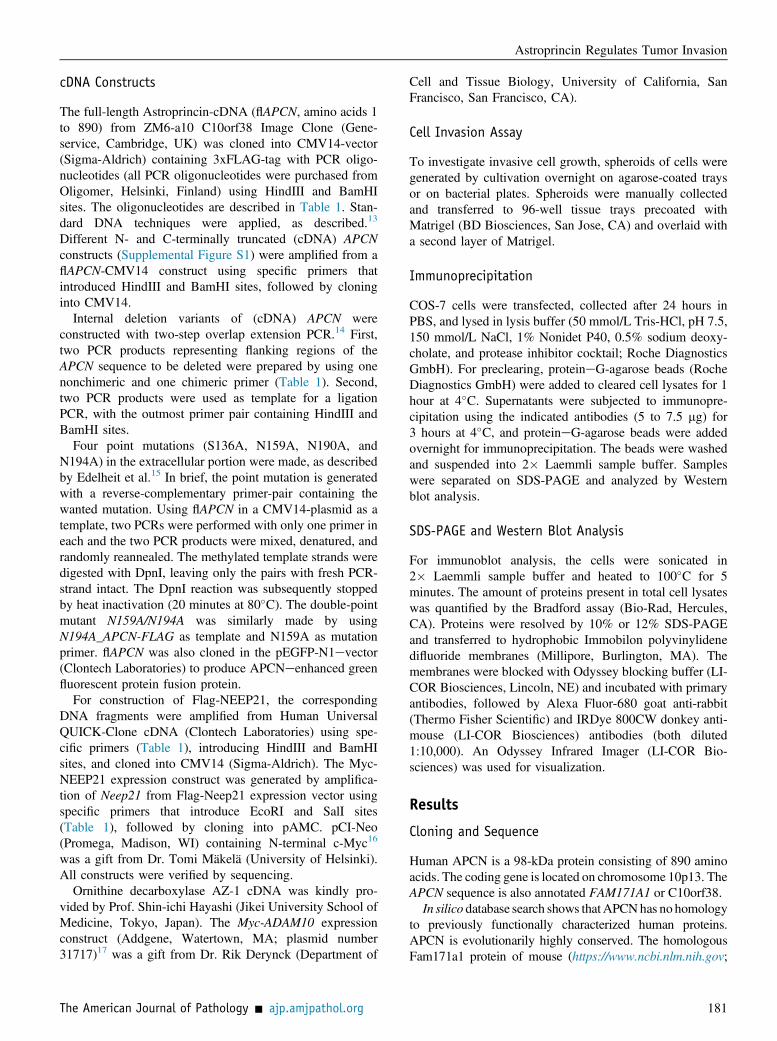
cDNA Constructs
The full-length Astroprincin-cDNA (flAPCN, amino acids 1to 890) from ZM6-a10 C10orf38 Image Clone (Gene-service, Cambridge, UK) was cloned into CMV14-vector(Sigma-Aldrich) containing 3xFLAG-tag with PCR oligo-nucleotides (all PCR oligonucleotides were purchased fromOligomer, Helsinki, Finland) using HindIII and BamHIsites. The oligonucleotides are described in Table 1. Stan-dard DNA techniques were applied, as described.13
Different N- and C-terminally truncated (cDNA) APCNconstructs (Supplemental Figure S1) were amplified from aflAPCN-CMV14 construct using specific primers thatintroduced HindIII and BamHI sites, followed by cloninginto CMV14.
Internal deletion variants of (cDNA) APCN wereconstructed with two-step overlap extension PCR.14 First,two PCR products representing flanking regions of theAPCN sequence to be deleted were prepared by using onenonchimeric and one chimeric primer (Table 1). Second,two PCR products were used as template for a ligationPCR, with the outmost primer pair containing HindIII andBamHI sites.
Four point mutations (S136A, N159A, N190A, andN194A) in the extracellular portion were made, as describedby Edelheit et al.15 In brief, the point mutation is generatedwith a reverse-complementary primer-pair containing thewanted mutation. Using flAPCN in a CMV14-plasmid as atemplate, two PCRs were performed with only one primer ineach and the two PCR products were mixed, denatured, andrandomly reannealed. The methylated template strands weredigested with DpnI, leaving only the pairs with fresh PCR-strand intact. The DpnI reaction was subsequently stoppedby heat inactivation (20 minutes at 80�C). The double-pointmutant N159A/N194A was similarly made by usingN194A_APCN-FLAG as template and N159A as mutationprimer. flAPCN was also cloned in the pEGFP-N1evector(Clontech Laboratories) to produce APCNeenhanced greenfluorescent protein fusion protein.
For construction of Flag-NEEP21, the correspondingDNA fragments were amplified from Human UniversalQUICK-Clone cDNA (Clontech Laboratories) using spe-cific primers (Table 1), introducing HindIII and BamHIsites, and cloned into CMV14 (Sigma-Aldrich). The Myc-NEEP21 expression construct was generated by amplifica-tion of Neep21 from Flag-Neep21 expression vector usingspecific primers that introduce EcoRI and SalI sites(Table 1), followed by cloning into pAMC. pCI-Neo(Promega, Madison, WI) containing N-terminal c-Myc16
was a gift from Dr. Tomi Mäkelä (University of Helsinki).All constructs were verified by sequencing.
Ornithine decarboxylase AZ-1 cDNA was kindly pro-vided by Prof. Shin-ichi Hayashi (Jikei University School ofMedicine, Tokyo, Japan). The Myc-ADAM10 expressionconstruct (Addgene, Watertown, MA; plasmid number31717)17 was a gift from Dr. Rik Derynck (Department of
The American Journal of Pathology - ajp.amjpathol.org
Cell and Tissue Biology, University of California, SanFrancisco, San Francisco, CA).
Cell Invasion Assay
To investigate invasive cell growth, spheroids of cells weregenerated by cultivation overnight on agarose-coated traysor on bacterial plates. Spheroids were manually collectedand transferred to 96-well tissue trays precoated withMatrigel (BD Biosciences, San Jose, CA) and overlaid witha second layer of Matrigel.
Immunoprecipitation
COS-7 cells were transfected, collected after 24 hours inPBS, and lysed in lysis buffer (50 mmol/L Tris-HCl, pH 7.5,150 mmol/L NaCl, 1% Nonidet P40, 0.5% sodium deoxy-cholate, and protease inhibitor cocktail; Roche DiagnosticsGmbH). For preclearing, proteineG-agarose beads (RocheDiagnostics GmbH) were added to cleared cell lysates for 1hour at 4�C. Supernatants were subjected to immunopre-cipitation using the indicated antibodies (5 to 7.5 mg) for3 hours at 4�C, and proteineG-agarose beads were addedovernight for immunoprecipitation. The beads were washedand suspended into 2� Laemmli sample buffer. Sampleswere separated on SDS-PAGE and analyzed by Westernblot analysis.
SDS-PAGE and Western Blot Analysis
For immunoblot analysis, the cells were sonicated in2� Laemmli sample buffer and heated to 100�C for 5minutes. The amount of proteins present in total cell lysateswas quantified by the Bradford assay (Bio-Rad, Hercules,CA). Proteins were resolved by 10% or 12% SDS-PAGEand transferred to hydrophobic Immobilon polyvinylidenedifluoride membranes (Millipore, Burlington, MA). Themembranes were blocked with Odyssey blocking buffer (LI-COR Biosciences, Lincoln, NE) and incubated with primaryantibodies, followed by Alexa Fluor-680 goat anti-rabbit(Thermo Fisher Scientific) and IRDye 800CW donkey anti-mouse (LI-COR Biosciences) antibodies (both diluted1:10,000). An Odyssey Infrared Imager (LI-COR Bio-sciences) was used for visualization.
Results
Cloning and Sequence
Human APCN is a 98-kDa protein consisting of 890 aminoacids. The coding gene is located on chromosome 10p13. TheAPCN sequence is also annotated FAM171A1 or C10orf38.
In silico database search shows thatAPCNhas no homologyto previously functionally characterized human proteins.APCN is evolutionarily highly conserved. The homologousFam171a1 protein of mouse (https://www.ncbi.nlm.nih.gov;
181

Homo sapiensHomo sapiensMus musculusMus musculusGallus gallusGallus gallusXenopus laevisXenopus laevisDanio rerioDanio rerio
HomoHomoMusMusGallusGallusXenopusXenopusDanioDanio
HomoHomoMusMusGallusGallusXenopusXenopusDanioDanio
HomoHomoMusMusGallusGallusXenopusXenopusDanioDanio
HomoHomoMusMusGallusGallusXenopusXenopusDanioDanio
HomoHomoMusMusGallusGallusXenopusXenopusDanioDanio
HomoHomoMusMusGallusGallusXenopusXenopusDanioDanio
HomoHomoMusMusGallusGallusXenopusXenopusDanioDanio
Figure 1 Top panels: Alignment of APCN protein sequences in human (Homo sapiens), mouse (Mus musculus), chicken (Gallus gallus), African clawed frog(Xenopus laevis), and zebra fish (Danio rerio). Conserved regions are marked in yellow. The transmembrane region is marked in red. Sequence missing in aquaticspecies (amphibian and fish) is marked in green. Bottom panel: Organization and nucleotide lengths of the APCN exons. The transmembrane region (red) isencoded by exon 7, and the intracellular portion is encoded almost exclusively by exon 8. C, carboxyterminal end of the protein; N, amino terminal end of theprotein.
Rasila et al
accession number NP_001074630) shows 92% of chicken(accession number XP_418631) 81% of frog (https://www.ncbi.nlm.nih.gov/protein/NP_001088656; accession numberNP_001088656) and 67% of zebra fish (accession numberXP_001335931); 48% identifies with several large conservedregions in common with human APCN (Figure 1).
The Molecular Structure of APCN
Different online tools (ExPASy website) were used topredict the APCN protein structure. These suggested thatAPCN is a type I transmembrane protein with the
182
hydrophobic amino acids from 304 to 324 in the trans-membrane region. The orientation of APCN was confirmedby expression of an APCNegreen fluorescent proteincDNA construct in COS-7 cells that revealed intracellularlocalization of green fluorescent proteinelabeled C-termi-nus (Supplemental Figure S2, AeF). The extracellular partof APCN is encoded by the seven short exons and containsseveral evolutionarily conserved stretches. The regionimmediately outside and inside the transmembrane stretch,encoded by exon 7, is virtually identical from zebra fish tohuman, suggesting some important function. The longintracytoplasmic portion, representing approximately two
ajp.amjpathol.org - The American Journal of Pathology

Figure 2 APCN is N-glycosylated. A: Western blot analysis with antibodies to FLAG of lysates from COS-7 cells transfected with (cDNA) flAPCN-Flag andcultured with (lane 1) and without (lane 2) tunicamycin. B: Point mutations showing that N159 and N194 are the major glycosylation sites. Western blotanalysis of lysates from COS-7 cells transfected with the following cDNA constructs: flAPCN-Flag (lane 1), APCN-Flag with mutation of the putativeglycosaminoglycan-binding site (VSGF; lane 2), APCN-Flag mutated at N159A (lane 3), APCN-Flag mutated at N194A (lane 4), and double-mutated N159A/N194A APCN-Flag (lane 5). MW, molecular weight.
Astroprincin Regulates Tumor Invasion
thirds of the protein, is encoded almost exclusively by thelarge exon 8 and contains several evolutionarily conservedregions (Figure 1).
A database search revealed a putative glycosaminoglycan-binding site (VSGF) and three potential N-glycosylation sites(N159, N190, and N194) in the extracellular portion as well asseveral putative phosphorylation sites throughout the sequence.
APCN Is Glycosylated
The size of the APCN apoprotein is 98 kDa. Western blotanalysis with antibodies to FLAG of lysates from COS-7 andMCF-7 cells transfected with (cDNA) APCN-flag revealed amajor, slightly diffuse band of 130 kDa apparent molecularweight (MW). Western blot analysis of lysates from trans-fected cells treated with tunicamycin yielded a band ofapproximately 100 to 115 kDa (Figure 2A). This reduction inapparent MW is indicative of N-linked glycosylation. Theexperiment was performed in MCF-7 and COS-7 cells (datanot shown), with identical results in both cell lines.
To map the glycosylation pattern, the three predictedN-glycosylation sites were subjected to N-A point mutations(N159A, N190A, and N194A). N190A_APCN gave aWestern blot analysis result identical to intact flAPCN.N159A_APCN and N194_APCN yielded reduced expres-sion of the higher MW band with accentuation of the 100- to115-kDa band. A double-mutant N159/194A-APCN lead todisappearance of the high MW band (Figure 2B). Mutationof the putative glycosaminoglycan binding site (S136A) did
The American Journal of Pathology - ajp.amjpathol.org
not change the apparent MW of APCN (Figure 2B). Thesefindings indicated that N159 and N194 are the N-glyco-sylation sites. The glycan structures of the N-glycoside sidechains remain to be determined.
Tissue Expression of APCN
A radiolabeled fragment of the cDNA, corresponding to 86to 417 bp of the coding region, was used as a probe toinvestigate the tissue distribution of APCN mRNA. Dotblot analysis revealed expression in various tissues(Supplemental Figure S3A and Supplemental Table S1).High expression levels were observed in the different partsof the brain. Abundant expression of APCN message wasalso seen in fetal tissues, especially in the brain, heart, andkidney (Supplemental Figure S3B).
To localize the tissue distribution of APCN proteinexpression, an antibody was raised in rabbits to a syntheticpeptide representing amino acids 378 to 394 (calledMAP346). The specificity of MAP346 was validated bydouble immunofluorescence (Supplemental Figure S2,GeI). Because brain tissue showed strong signals inNorthern blot analysis, sections from normal human brainwere stained by immunohistochemistry. An intense reticularstaining was seen in glia limitans in a distributioncorresponding to the abundant astrocyte processes and inpyramidal neurons (Figure 3A). Because of the robustappearance of the protein in astrocytes (Figure 3B), theprotein was called astroprincin.
Figure 3 Immunohistochemistry showing APCNexpression in glia limitans of human brain and in pyra-midal neurons (A) and astrocytes (B). Scale bars: 100 mm(A); 20 mm (B).
183
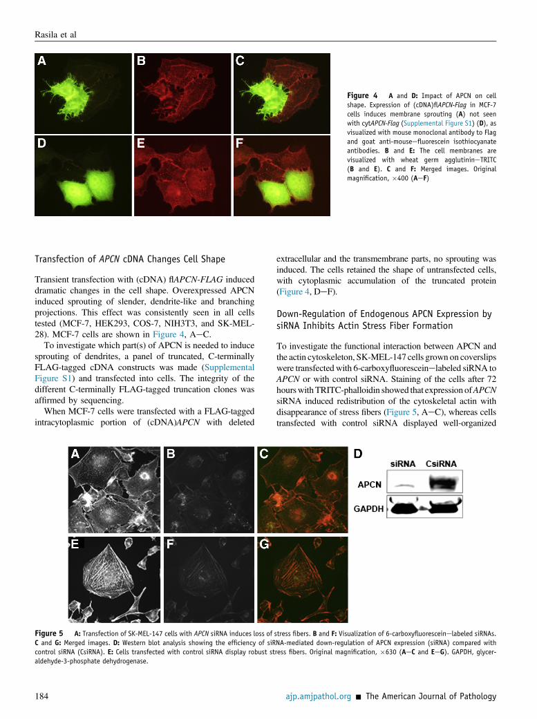
Figure 4 A and D: Impact of APCN on cellshape. Expression of (cDNA)flAPCN-Flag in MCF-7cells induces membrane sprouting (A) not seenwith cytAPCN-Flag (Supplemental Figure S1) (D), asvisualized with mouse monoclonal antibody to Flagand goat anti-mouseefluorescein isothiocyanateantibodies. B and E: The cell membranes arevisualized with wheat germ agglutinineTRITC(B and E). C and F: Merged images. Originalmagnification, �400 (AeF)
Rasila et al
Transfection of APCN cDNA Changes Cell Shape
Transient transfection with (cDNA) flAPCN-FLAG induceddramatic changes in the cell shape. Overexpressed APCNinduced sprouting of slender, dendrite-like and branchingprojections. This effect was consistently seen in all cellstested (MCF-7, HEK293, COS-7, NIH3T3, and SK-MEL-28). MCF-7 cells are shown in Figure 4, AeC.
To investigate which part(s) of APCN is needed to inducesprouting of dendrites, a panel of truncated, C-terminallyFLAG-tagged cDNA constructs was made (SupplementalFigure S1) and transfected into cells. The integrity of thedifferent C-terminally FLAG-tagged truncation clones wasaffirmed by sequencing.
When MCF-7 cells were transfected with a FLAG-taggedintracytoplasmic portion of (cDNA)APCN with deleted
Figure 5 A: Transfection of SK-MEL-147 cells with APCN siRNA induces loss ofC and G: Merged images. D: Western blot analysis showing the efficiency of siRcontrol siRNA (CsiRNA). E: Cells transfected with control siRNA display robust staldehyde-3-phosphate dehydrogenase.
184
extracellular and the transmembrane parts, no sprouting wasinduced. The cells retained the shape of untransfected cells,with cytoplasmic accumulation of the truncated protein(Figure 4, DeF).
Down-Regulation of Endogenous APCN Expression bysiRNA Inhibits Actin Stress Fiber Formation
To investigate the functional interaction between APCN andthe actin cytoskeleton, SK-MEL-147 cells grown on coverslipswere transfected with 6-carboxyfluoresceinelabeled siRNA toAPCN or with control siRNA. Staining of the cells after 72hourswithTRITC-phalloidin showed that expression ofAPCNsiRNA induced redistribution of the cytoskeletal actin withdisappearance of stress fibers (Figure 5, AeC), whereas cellstransfected with control siRNA displayed well-organized
stress fibers. B and F: Visualization of 6-carboxyfluoresceinelabeled siRNAs.NA-mediated down-regulation of APCN expression (siRNA) compared withress fibers. Original magnification, �630 (AeC and EeG). GAPDH, glycer-
ajp.amjpathol.org - The American Journal of Pathology

Figure 6 A: Relative expression levels of endogenous APCN mRNA, measured by real-time quantitative PCR in the melanoma cell lines SK-MEL-28, SK-MEL-103, and SK-MEL-147. BeD: SK-MEL-28 does not invade Matrigel (B), like SK-MEL-103 (C) and SK-MEL-147 (D) cells. EeJ: Transfection of SK-MEL-28 cells withthe empty vector (control; E and H), with (cDNA)flAPCN (F and I), or with (cDNA)APCN lacking the 21 C-terminal amino acids (G and J) showed that APCNinduced sprouting (F) and ability to invade Matrigel (I) and that the C-terminal 21 amino acids are needed for the APCN activity (G and J). Originalmagnification: �100 (BeD and HeJ); �200 (EeG). GAPDH, glyceraldehyde-3-phosphate dehydrogenase.
Astroprincin Regulates Tumor Invasion
stress fibers (Figure 5, DeG). APCN siRNA transfection ofU393MG astrocytoma cells also induced disappearance ofstressfibers and accumulationof actin in donut-like protrusionsat the cell edges (Supplemental Figure S4).
The Conserved Carboxyterminal 21 Amino Acids ofAPCN Are Needed for Cell Sprouting and InvasiveGrowth
Analysis by real-time quantitative PCR and Western blotanalysis of human melanoma cell lines revealed higherexpression of endogenous APCN transcript and protein inSK-MEL-103 and in SK-MEL-147 than in SK-MEL-28melanoma cells (Figure 6A). SK-MEL-28 cells did not
The American Journal of Pathology - ajp.amjpathol.org
invade Matrigel (Figure 6B), whereas SK-MEL-103(Figure 6C) and SK-Mel-147 (Figure 6D) cells displayedinvasive growth in Matrigel. Overexpression of full-length (cDNA)APCN in SK-MEL-28 cells inducedsprouting of slender projections and ability to growinvasively in Matrigel (Figure 6, F and I), not seen withSK-MEL-28 cells transfected with the empty vector(Figure 6, E and H).
To investigate which part(s) of the APCN molecule wasof importance for induction of invasive cell growth, mutatedconstructs of (cDNA)APCN were expressed in SK-MEL-28cells. Expression of (cDNA)T3-APCN (SupplementalFigure S1) with deletion of the ultimate 21 carbox-yterminal amino acids (869 to 890) in SK-MEL-28 cells did
185
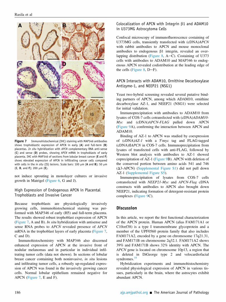
Figure 7 Immunohistochemical (IHC) staining with MAP346 antibodiesshows trophoblastic expression of APCN in early (A) and full-term (B)placentas. In situ hybridization with APCN complementary RNA anti-sense(C) and sense (D) probes, showing APCN mRNA in trophoblasts of earlyplacenta. IHC with MAP346 of sections from lobular breast cancer (E and F)shows elevated expression of APCN in infiltrating cancer cells comparedwith cells in the in situ (IS) lesions. Scale bars: 100 mm (A and B); 50 mm(C, D, and F); 200 mm (E).
Rasila et al
not induce sprouting in monolayer cultures or invasivegrowth in Matrigel (Figure 6, G and J).
High Expression of Endogenous APCN in PlacentalTrophoblasts and Invasive Cancer
Because trophoblasts are physiologically invasivelygrowing cells, immunohistochemical staining was per-formed with MAP346 of early (H5) and full-term placenta.The results showed robust trophoblast expression of APCN(Figure 7, A and B). In situ hybridization with antisense andsense RNA probes to APCN revealed presence of APCNmRNA in the trophoblast layers of early placenta (Figure 7,C and D).
Immunohistochemistry with MAP346 also discernedenhanced expression of APCN at the invasive front ofnodular melanomas and in particular in individual infil-trating tumor cells (data not shown). In sections of lobularbreast cancer containing both noninvasive, in situ lesionsand infiltrating tumor cells, a robustly up-regulated expres-sion of APCN was found in the invasively growing cancercells. Normal lobular epithelium remained negative forAPCN (Figure 7, E and F).
186
Colocalization of APCN with Integrin b1 and ADAM10in U373MG Astrocytoma Cells
Confocal microscopy of immunofluorescence costaining ofU373MG cells, transiently transfected with (cDNA)APCNwith rabbit antibodies to APCN and mouse monoclonalantibodies to endogenous b1 integrin, revealed an over-lapping distribution (Figure 8, AeC). Costaining of U373cells with antibodies to ADAM10 and MAP346 to endog-enous APCN revealed codistribution at the leading edge ofthe cells (Figure 8, DeF).
APCN Interacts with ADAM10, Ornithine DecarboxylaseAntizyme-1, and NEEP21 (NSG1)
Yeast two-hybrid screening revealed several putative bind-ing partners of APCN, among which ADAM10, ornithinedecarboxylase AZ-1, and NEEP21 (NSG1) were selectedfor initial validation.Immunoprecipitation with antibodies to ADAM10 from
lysates of COS-7 cells cotransfected with (cDNA)ADAM10-Myc and (cDNA)APCN-FLAG pulled down APCN(Figure 9A), confirming the interaction between APCN andADAM10.Binding of AZ-1 to APCN was studied by coexpression
of (cDNA)AZ-1 with a 50myc tag and FLAG-tagged(cDNA)flAPCN in COS-7 cells. Immunoprecipitation fromlysates of transfected cells with anti-FLAG, followed byWestern blot analysis with antibodies to AZ-1 showedcoprecipitation of AZ-1 (Figure 9B). APCN with deletion ofthe conserved portion between amino acids 541 and 746(D3-APCN) (Supplemental Figure S1) did not pull downAZ-1 (Supplemental Figure S5).Immunoprecipitation of lysates from COS-7 cells
cotransfected with NEEP21-Myc and APCN-Flag cDNAconstructs with antibodies to APCN also brought downNEEP21, indicating formation of detergent-resistant proteincomplexes (Figure 9C).
Discussion
In this article, we report the first functional characterizationof the APCN protein. Human APCN (alias FAM171A1 orC10orf38) is a type I transmembrane glycoprotein and amember of the UPF0560 protein family that also includesFAM171A2, encoded by a gene on chromosome 17q21.31,and FAM171B on chromosome 2q32.1. FAM171A2 shows39% and FAM171B shows 32% identity with APCN. TheAPCN gene is located on chromosome 10p13, a region thatis deleted in DiGeorge type 2 and velocardiofacialsyndromes.18
Hybridization experiments and immunohistochemistryrevealed physiological expression of APCN in various tis-sues, particularly in the brain, where the astrocytes exhibitabundant APCN.
ajp.amjpathol.org - The American Journal of Pathology

Figure 8 A and B: Confocal microscopy ofU373MG cells transiently transfected with (cDNA)APCN-Flag and costained with monoclonal anti-bodies to human b1-integrin (A) and with anti-FLAG (B). C: Merged image. D and E: Staining ofU373MG astrocytoma cells with antibodies toADAM10 (D) and with MAP346 to endogenousAPCN (E). F: Merged image. Original magnification:�400 (AeC); �630 (DeF).
Astroprincin Regulates Tumor Invasion
Yeast two-hybrid screening indicated ornithine decar-boxylase (ODC) AZ-1 binding to APCN. This was validatedby coimmunoprecipitation, and the binding site was local-ized to a domain between amino acids 541 and 746,including the region between amino acids 541 and 569 thatis highly conserved in the three members of the FAM171proteins and in Fam171a1 proteins of different species. Theactivity of ODC, the rate-limiting enzyme of polyaminesynthesis, is intimately coupled to cell activation,transformation, and proliferation.19
ODC translocates to the plasma membrane during cellactivation.20 ODC activity is required for microvascularsprouting and remodeling of the actin cytoskeleton inendothelial cells.21 Moreover, ODC activity or local poly-amine synthesis regulates the activity and traffic of RhoA,which is a main regulator of the dynamics of the actincytoskeleton.22 A sizeable portion of cellular ODC is
Figure 9 Interaction of APCN with ADAM10 (A), antizyme-1 (AZ-1; B), and NEEof lysates from COS-7 cells transiently cotransfected with (cDNA)ADAM10 and (cDNand (cDNA) NEEP21-Flag and (cDNA)flAPCN (C). IP, immunoprecipitation.
The American Journal of Pathology - ajp.amjpathol.org
sequestered in catalytically inactive form to AZ-1 and getsreleased and activated by competitive binding of antizymeinhibitor. The interaction between AZ-1 and APCN mayprovide a mechanism by which ODC is targeted to themembrane, where the activity is of importance for cyto-skeletal reorganization occurring during formation ofcellular sprouts and acquisition of invasive phenotype.
Overexpression of full-length (cDNA)APCN inducedoutgrowth of slender and frequently branched extensions inboth epithelial and mesenchymal cell lines. This was notseen with (cDNA)APCN lacking the extracellular portion.These finding suggested the APCN is involved in signalingbetween the cytoskeleton and the external environment.
A role for APCN in the maintenance of an organized actincytoskeleton is further supported by these findings, showingthat down-regulation of endogenous APCN by siRNAinterference provoked disappearance of organized actin
P21 (NSG1) (C), shown by coimmunoprecipitation and Western blot analysisA)flAPCN-Flag (A), (cDNA) AZ-1 with a 50myc tag and (cDNA) flAPCN-Flag (B),
187

Rasila et al
stress fibers. Our attempts to delete endogenous APCNexpression by clustered regularly interspaced short palin-dromic repeats (CRISPR)/Cas9 technology failed becauseviable cells could not be selected with total knockout of bothalleles. Occasional polykaryotic cells with >20 nuclei percell were found (data not shown). This suggests that totalabsence of endogenous APCN causes disturbance in theactin dynamics needed for cytokinesis.
APCN is not known to mediate cell adhesion, but byconfocal microscopy, a colocalization with integrin b1 wasobserved. Whether there is a direct interaction betweenintegrin b1 and APCN or whether the observed colocali-zation is attributable to formation of a larger complex withother proteins remains to be investigated.
ADAM10, like APCN, is a transmembrane type Iglycoprotein with a complex N-glycoside side chain. Theshort intracytoplasmic portion contains two SH3 motifs anda consensus binding site for calmodulin. It is a member oftransmembrane zinc-dependent metalloproteinase orsheddase acting as amyloid precursor protein, cleaving a-secretase. More than 20 membrane-bound proteins havebeen identified as ADAM10 substrates, including Notch,proeepidermal growth factor, ErbB2, E-cadherin, CD44,and inflammatory cytokines.23 ADAM10 is also essentialfor migration of neuronal precursors during embryonic brainmorphogenesis.24 Double immunofluorescence showedcolocalization of endogenous ADAM10 and APCN in theleading edge of U373MG astrocytoma cells. It is temptingto speculate that interaction between ADAM10 and APCNis of relevance for brain morphogenesis and in particular forthe arborization of astrocytes. Elevated expression ofADAM10 has also been reported to enhance growth andmetastatic dissemination of neoplasms, including mela-nomas, breast cancer, and liver cancers.25,26 Release ofNLGN3, which promotes glioma growth and differentiation,was recently shown to be mediated by ADAM10.27
There is limited information in the literature about aneuron-enriched endosomal protein of 21 kDa, NSG1(NEEP21). NEEP21 has been found functionally involvedin regulation of endosomal vesicle trafficking and mem-brane receptor recycling.28 NEEP21 is involved in sorting ofthe neuron-glial adhesion molecule L1/NgCAM thatregulates outgrowth of neurites.29 Elevated expression ofL1/NgCAM, on the other hand, has been found in a varietyof cancers, where it correlates with aggressive behavior ofthe neoplasm.30
By expression of deletion mutants of APCN cDNA, it wasfound that the 21 C-terminal amino acids are required forinduction of invasive growth and sprouting of SK-MEL-28melanoma cells. This sequence is also present in the twoother members of the FAM171 protein family and isconserved in APCN of different species. A motif searchrevealed domains of homology in MAP1A, a structuralprotein that is involved in cross-bridging between microtu-bules and other cytoskeletal elements.31,32 These includeEphexin-2, which is a guanine nucleotide exchange factor
188
for RhoA GTPase; and pleckstrin homology domain-interacting protein, which is involved in regulation of cellmorphology and cytoskeletal organization.33,34 Moreover,BCAS1, which is amplified in a variety of cancers andassociates with more aggressive tumor types, also carries ahomologous motif. We are presently investigating themolecular details of the interactions of the N-terminaldomain of APCN.BLAST searches of the conserved extracellular regions of
APCN revealed a neural cell adhesion moleculeehomologous motif in the domain between amino acids 85and 113. NCAM mediates neuron-neuron adhesion and isinvolved in neurite outgrowths. A region present in APCN137 to 160 is also found in Septin5, which is encoded by agene located on 22q11.2, a chromosomal region that isdeleted in diseases, including DiGeorge and veloc-ardiofacial syndromes.18
Taken together, we report the first functional character-ization of APCN. Further investigations are required tounravel the molecular details of APCN, but the emergingpicture is an evolutionarily highly conserved type I trans-membrane glycoprotein that is involved in the regulation ofthe cytoskeletal dynamics and, thereby, the cell shape andinvasive growth behavior of tumor cells.
Acknowledgments
We thank Tiiu Arumäe for technical help, Prof. BengtWestermark (University of Uppsala) for providing theU373MG astrocytoma line, Dr. Maria Soengas (SpanishNational Cancer Research Center) for providing SK-MEL-103 and SK-MEL-147 (both originating from Dr. AlanHoughton, Memorial Sloan-Kettering Cancer Center), Dr.Tomi Mäkelä (University of Helsinki) for proving pCI-Neocontaining N-terminal c-Myc, Prof. Shin-ichi Hayashi (JikeiUniversity School of Medicine) for providing antizyme-1cDNA, and Dr. Rik Derynck (University of California, SanFrancisco) for providing the Myc-ADAM10 expressionconstruct (Addgene plasmid number 31717).
Supplemental Data
Supplemental material for this article can be found athttps://doi.org/10.1016/j.ajpath.2018.09.006.
References
1. Deloukas P, Earthrowl ME, Grafham DV, Rubenfield M, French L,Steward CA, et al: The DNA sequence and comparative analysis ofhuman chromosome 10. Nature 2004, 429:375e381
2. Simmen FA, Su Y, Xiao R, Zeng Z, Simmen RC: The Kruppel-likefactor 9 (KLF9) network in HEC-1-A endometrial carcinoma cellssuggests the carcinogenic potential of dys-regulated KLF9 expression.Reprod Biol Endocrinol 2008, 6:41
ajp.amjpathol.org - The American Journal of Pathology

Astroprincin Regulates Tumor Invasion
3. Liao Y, Pei J, Cheng H, Grishin NV: An ancient autoproteolyticdomain found in GAIN, ZU5 and nucleoporin98. J Mol Biol 2014,426:3935e3945
4. Ipsaro JJ, Huang L, Gutierrez L, MacDonald RI: Molecular epitopes ofthe ankyrin-spectrin interaction. Biochemistry 2008, 47:7452e7464
5. Prunotto M, Farina A, Lane L, Pernin A, Schifferli J, Hochstrasser DF,Lescuyer P, Moll S: Proteomic analysis of podocyte exosome-enrichedfraction from normal human urine. J Proteomics 2013, 82:193e229
6. Emdal KB, Pedersen AK, Bekker-Jensen DB, Tsafou KP, Horn H,Lindner S, Schulte JH, Eggert A, Jensen LJ, Francavilla C, Olsen JV:Temporal proteomics of NGF-TrkA signaling identifies an inhibitoryrole for the E3 ligase Cbl-b in neuroblastoma cell differentiation. SciSignal 2015, 8:ra40
7. St-Denis N, Gupta GD, Lin ZY, Gonzalez-Badillo B, Veri AO,Knight JDR, Rajendran D, Couzens AL, Currie KW, Tkach JM,Cheung SWT, Pelletier L, Gingras AC: Phenotypic and interactionprofiling of the human phosphatases identifies diverse mitotic regula-tors. Cell Rep 2016, 17:2488e2501
8. Huttlin EL, Bruckner RJ, Paulo JA, Cannon JR, Ting L, Baltier K,Colby G, Gebreab F, Gygi MP, Parzen H, Szpyt J, Tam S, Zarraga G,Pontano-Vaites L, Swarup S, White AE, Schweppe DK, Rad R,Erickson BK, Obar RA, Guruharsha KG, Li K, Artavanis-Tsakonas S, Gygi SP, Harper JW: Architecture of the humaninteractome defines protein communities and disease networks. Na-ture 2017, 545:505e509
9. Gayet O, Labella V, Henderson CE, Kallenbach S: The b1 isoform ofprotocadherin-gamma (Pcdhgamma) interacts with the microtubule-destabilizing protein SCG10. FEBS Lett 2004, 578:175e179
10. Haltaufderhyde KD, Oancea E: Genome-wide transcriptome analysisof human epidermal melanocytes. Genomics 2014, 104:482e489
11. Santuario-Facio SK, Cardona-Huerta S, Perez-Paramo YX,Trevino V, Hernandez-Cabrera F, Rojas-Martinez A, Uscanga-Perales G, Martinez-Rodriguez JL, Martinez-Jacobo L, Padilla-Rivas G, Munoz-Maldonado G, Gonzalez-Guerrero JF, Valero-Gomez J, Vazquez-Guerrero AL, Martinez-Rodriguez HG, Barboza-Quintana A, Barboza-Quintana O, Garza-Guajardo R, Ortiz-Lopez R: A new gene expression signature for triple negative breastcancer using frozen fresh tissue before neoadjuvant chemotherapy.Mol Med 2017, 23:101e111
12. Pitkänen LT, Heiskala M, Andersson LC: Expression of a novelhuman ornithine decarboxylase-like protein in the central nervoussystem and testes. Biochem Biophys Res Commun 2001, 287:1051e1057
13. Sambrook J, Russell DW: Molecular Cloning: A Laboratory Manual.Cold Spring Harbor. NY, Cold Spring Harbor Laboratory Press, 2001
14. Lee J, Lee HJ, Shin MK, Ryu WS: Versatile PCR-mediated insertionor deletion mutagenesis. Biotechniques 2004, 36:398e400
15. Edelheit O, Hanukoglu A, Hanukoglu I: Simple and efficient site-directed mutagenesis using two single-primer reactions in parallel togenerate mutants for protein structure-function studies. BMC Bio-technol 2009, 9:61
16. Tiainen M, Ylikorkala A, Mäkelä TP: Growth suppression by Lkb1 ismediated by a G(1) cell cycle arrest. Proc Natl Acad Sci U S A 1999,96:9248e9251
17. Liu C, Xu P, Lamouille S, Xu J, Derynck R: TACE-mediated ecto-domain shedding of the type I TGF-beta receptor downregulates TGF-beta signaling. Mol Cell 2009, 35:26e36
The American Journal of Pathology - ajp.amjpathol.org
18. Daw SC, Taylor C, Kraman M, Call K, Mao J, Schuffenhauer S,Meitinger T, Lipson T, Goodship J, Scambler P: A common region of10p deleted in DiGeorge and velocardiofacial syndromes. Nat Genet1996, 13:458e460
19. Kahana C: Regulation of cellular polyamine levels and cellular pro-liferation by antizyme and antizyme inhibitor. Essays Biochem 2009,46:47e61
20. Heiskala M, Zhang J, Hayashi S, Hölttä E, Andersson LC: Trans-location of ornithine decarboxylase to the surface membrane duringcell activation and transformation. EMBO J 1999, 18:1214e1222
21. Kucharzewska P, Welch JE, Svensson KJ, Belting M: Ornithinedecarboxylase and extracellular polyamines regulate microvascularsprouting and actin cytoskeleton dynamics in endothelial cells. ExpCell Res 2010, 316:2683e2691
22. Mäkitie LT, Kanerva K, Andersson LC: Ornithine decarboxylaseregulates the activity and localization of rhoA via polyamination. ExpCell Res 2009, 315:1008e1014
23. White JM: ADAMs: modulators of cell-cell and cell-matrix in-teractions. Curr Opin Cell Biol 2003, 15:598e606
24. Jorissen E, Prox J, Bernreuther C, Weber S, Schwanbeck R,Serneels L, Snellinx A, Craessaerts K, Thathiah A, Tesseur I,Bartsch U, Weskamp G, Blobel CP, Glatzel M, De Strooper B,Saftig P: The disintegrin/metalloproteinase ADAM10 is essentialfor the establishment of the brain cortex. J Neurosci 2010, 30:4833e4844
25. Mochizuki S, Okada Y: ADAMs in cancer cell proliferation and pro-gression. Cancer Sci 2007, 98:621e628
26. Murphy G: The ADAMs: signalling scissors in the tumour microen-vironment. Nat Rev Cancer 2008, 8:929e941
27. Venkatesh HS, Tam LT, Woo PJ, Lennon J, Nagaraja S, Gillespie SM,Ni J, Duveau DY, Morris PJ, Zhao JJ, Thomas CJ, Monje M: Tar-geting neuronal activity-regulated neuroligin-3 dependency in high-grade glioma. Nature 2017, 549:533e537
28. Muthusamy N, Chen YJ, Yin DM, Mei L, Bergson C: Complementaryroles of the neuron-enriched endosomal proteins NEEP21 and calcyonin neuronal vesicle trafficking. J Neurochem 2015, 132:20e31
29. Yap CC, Wisco D, Kujala P, Lasiecka ZM, Cannon JT, Chang MC,Hirling H, Klumperman J, Winckler B: The somatodendritic endo-somal regulator NEEP21 facilitates axonal targeting of L1/NgCAM. JCell Biol 2008, 180:827e842
30. Altevogt P, Doberstein K, Fogel M: L1CAM in human cancer. Int JCancer 2016, 138:1565e1576
31. Fink JK, Jones SM, Esposito C, Wilkowski J: Human microtubule-associated protein 1a (MAP1A) gene: genomic organization, cDNAsequence, and developmental- and tissue-specific expression. Geno-mics 1996, 35:577e585
32. Halpain S, Dehmelt L: The MAP1 family of microtubule-associatedproteins. Genome Biol 2006, 7:224
33. Lajoie-Mazenc I, Tovar D, Penary M, Lortal B, Allart S, Favard C,Brihoum M, Pradines A, Favre G: MAP1A light chain-2 interacts withGTP-RhoB to control epidermal growth factor (EGF)-dependent EGFreceptor signaling. J Biol Chem 2008, 283:4155e4164
34. Fuhrmann-Stroissnigg H, Noiges R, Descovich L, Fischer I,Albrecht DE, Nothias F, Froehner SC, Propst F: The light chains ofmicrotubule-associated proteins MAP1A and MAP1B interact withalpha1-syntrophin in the central and peripheral nervous system. PLoSOne 2012, 7:e49722
189