Embed Size (px)
Citation preview

Astrocytes Secrete Exosomes Enriched with ProapoptoticCeramide and Prostate Apoptosis Response 4 (PAR-4)POTENTIAL MECHANISM OF APOPTOSIS INDUCTION IN ALZHEIMER DISEASE (AD)*□S
Received for publication, January 7, 2012, and in revised form, April 19, 2012 Published, JBC Papers in Press, April 24, 2012, DOI 10.1074/jbc.M112.340513
Guanghu Wang‡1, Michael Dinkins‡1, Qian He‡, Gu Zhu‡, Christophe Poirier§, Andrew Campbell¶,Margot Mayer-Proschel¶, and Erhard Bieberich‡2
From the ‡Program in Developmental Neurobiology, Institute of Molecular Medicine and Genetics, Medical College of Georgia,Georgia Health Sciences University, Augusta, Georgia 30912, the §Vascular Biology Center, Georgia Health Sciences University,Augusta, Georgia 30912, and the ¶Department of Biomedical Genetics, University of Rochester Medical Center, University ofRochester, Rochester, New York 14642
Background: In AD, amyloid protein is associated with neurodegeneration, which may involve amyloid effects onastrocytes.Results: In astrocytes, amyloid peptide triggers secretion of proapoptotic exosomes (“apoxosomes”) that are associated withceramide and PAR-4.Conclusion: Activation of nSMase2 and expression of PAR-4 is critical for the secretion of apoxosomes and glial apoptosis.Significance: Apoxosomes may contribute to glial apoptosis, and therefore, neurodegeneration in AD.
Amyloid protein is well known to induce neuronal cell death,whereas only little is known about its effect on astrocytes. Wefound that amyloid peptides activated caspase 3 and inducedapoptosis in primary cultured astrocytes, which was preventedby caspase 3 inhibition. Apoptosis was also prevented byshRNA-mediated down-regulation of PAR-4, a protein sensitiz-ing cells to the sphingolipid ceramide. Consistent with a poten-tially proapoptotic effect of PAR-4 and ceramide, astrocytes sur-rounding amyloid plaques in brain sections of the 5xFADmouse(and Alzheimer disease patient brain) showed caspase 3 activa-tion and were apoptotic when co-expressing PAR-4 and cer-amide. Apoptosis was not observed in astrocytes with deficientneutral sphingomyelinase 2 (nSMase2), indicating that ceramidegenerated by nSMase2 is critical for amyloid-induced apoptosis.Antibodies against PAR-4 and ceramide prevented amyloid-in-duced apoptosis in vitro and in vivo, suggesting that apoptosiswas mediated by exogenous PAR-4 and ceramide, potentiallyassociated with secreted lipid vesicles. This was confirmed bythe analysis of lipid vesicles from conditioned medium showingthat amyloid peptide induced the secretion of PAR-4 and C18ceramide-enriched exosomes. Exosomes were not secreted bynSMase2-deficient astrocytes, indicating that ceramide gener-ated by nSMase2 is critical for exosome secretion. Consistentwith the ceramide composition in amyloid-induced exosomes,exogenously added C18 ceramide restored PAR-4-containingexosome secretion in nSMase2-deficient astrocytes. Moreover,isolated PAR-4/ceramide-enriched exosomes were taken up by
astrocytes and induced apoptosis in the absence of amyloid pep-tide. Taken together, we report a novel mechanism of apoptosisinduction by PAR-4/ceramide-enriched exosomes, which maycritically contribute to Alzheimer disease.
Alzheimer disease (AD)3 is a devastating neurodegenerativedisease associatedwith the buildup of protein aggregateswithinand surrounding neurons in the hippocampus and cortex (1).Two forms of protein aggregates, Tau protein fibrillary tanglesand amyloid � (A�) protein plaques, are known to induce neu-ronal malfunction and apoptosis (2). The protein buildup islinked to a variety of mutations, among which mutations of�-secretase, an enzyme processing the amyloid precursor pro-tein (APP), lead to the most severe forms of AD (3). Aberrantprocessing of APP results in the secretion, aggregation, andplaque formation of neurotoxic peptides such as the A� pep-tides A�1–40, A�1–42, and A�25–35 (4).Although most research has focused on the effect of Tau
protein tangles and A� plaques on neuronal function and sur-vival, it is nowwell established that astrocytes also play a role inAD pathology (5–14). In the CNS, astrocytes are indispensablefor the support of neurons. However, astrocytes can also adoptan activated state where they start to migrate and proliferateand induce inflammatory and proapoptotic cell signaling path-ways (8, 10, 15, 16). Astrocytic reactivation has been observed inmanyneurodegenerative diseases, includingAD, and is thoughtto occur as a response to oxidative stress and cytokine release(8, 10–12, 16–18). As a consequence, astrocytes not only fail tosupport neurons, but also generate a toxic environment that isdetrimental to neurons and astrocytes themselves. Astrocytic
* This work was supported, in whole or in part, by National Institutes of HealthGrant 1R01AG034389 (to E. B.). G. W. is supported by awards from GeorgiaHealth Sciences University and American Heart Association.
□S This article contains supplemental Figs. S1–S6.1 Both authors contributed equally to this work.2 To whom correspondence should be addressed: Program in Developmental
Neurobiology, Institute of Molecular Medicine and Genetics, Medical Col-lege of Georgia/Georgia Health Sciences University, 1120 15th St., Rm.CA4012, Augusta, GA 30912. Tel.: 706-721-9113; Fax: 706-721-8685; E-mail:[email protected].
3 The abbreviations used are: AD, Alzheimer disease; FAD, familial AD; A�,amyloid �; APP, amyloid precursor protein; nSMase2, neutral sphingomy-elinase 2; GFAP, glial fibrillary acidic protein; Z, benzyloxycarbonyl; FMK,fluoromethyl ketone; UC, ultracentrifugation.
THE JOURNAL OF BIOLOGICAL CHEMISTRY VOL. 287, NO. 25, pp. 21384 –21395, June 15, 2012© 2012 by The American Society for Biochemistry and Molecular Biology, Inc. Published in the U.S.A.
21384 JOURNAL OF BIOLOGICAL CHEMISTRY VOLUME 287 • NUMBER 25 • JUNE 15, 2012
by guest on March 4, 2020
http://ww
w.jbc.org/
Dow
nloaded from

dysfunction combined with cell death has been described tooccur in AD; glial apoptosis has been found to be correlatedwith the number of senile plaques, although a clear associationof apoptosis with labeling of A� protein has not been shown yet(19). Activation of caspases has been suggested to contribute toastrocyte damage, but the mechanism by which astrocytesmight die in AD remains unclear (20).In this study, we propose a novel mechanism that sensitizes
astrocytes to amyloid-induced cell death via concurrent up-regulation of PAR-4 and ceramide. We also demonstrate thatthe level of proapoptotic ceramide in astrocytes is regulated byneutral sphingomyelinase 2 (nSMase2). Further, we show forthe first time that astrocytic apoptosis is associated with thesecretion of PAR-4/ceramide-containing lipid vesicles (exo-somes) that can induce cell death even in cells not exposed toamyloid protein. Therefore, our finding identifies a novelmechanism of apoptosis induction by exosomes that aresecreted by amyloid protein-exposed astrocytes.
EXPERIMENTAL PROCEDURES
Materials—Human brain tissue fromAD patients was a gen-erous gift fromDr. DavidHill, Georgia Health Sciences Univer-sity, Augusta, GA. The 5XFADmouse was purchased fromTheJackson Laboratory (Bar Harbor, ME). Amyloid peptides werefromAnaSpec (Fremont, CA) and rPeptide (Bogart, GA). Anti-ceramide antibodywas generated in our laboratory (rabbit IgG)or obtained from Glycobiotech (mouse IgM; Kuekels, Ger-many). Anti-PAR-4, PKC� (atypical PKC), CD9, and anti-GFAPantibodies were from Santa Cruz Biotechnology (Santa Cruz,CA). Monoclonal anti-TSG101 mouse IgG was from Abcam(Cambridge, MA). Monoclonal anti-amyloid 4G8 IgG wasobtained from Millipore (Billerica, MA). The anti-activatedcaspase 3 antibody was obtained from Cell Signaling Technol-ogy (Danvers, MA). Fluorophore-conjugated secondary anti-bodies were from Jackson ImmunoResearch Laboratories(West Grove, PA). Magnetic beads coated with anti-rabbit IgGwere obtained from Miltenyi (Auburn, CA) and New EnglandBiolabs (Ipswich, MA). Ceramide was obtained from AvantiPolar Lipids (Alabaster, AL). The ExoQuick exosome purifica-tion kit and anti-Alix1 rabbit IgGwere fromSystemBiosciencesSBI (Mountain View, CA). Vybrant CM-DiI and the Click-iTCy5 TUNEL assay were purchased from Invitrogen. The Pro-teoStat amyloid plaque detection kit was from Enzo Life Sci-ences (Farmingdale, NY). All reagents were of analytical gradeor higher.Cultivation of Primary Astrocytes and Incubation with Amy-
loid Peptide—Cortical primary astrocytes were prepared frompostnatal day 1–3 pups following an established protocol (21).The proportion of astrocytes was quantified by immunocyto-chemistry using an antibody against GFAP. A typical culturewas found to consist of more than 95% of GFAP� cells. Theastrocytes were incubated with amyloid peptide and otherreagents in phenol red and serum-free medium. When fro/fropupswere used, genotypingwas performed twice using tail- andastrocyte-derivedDNA following standard protocols. Fibrillaryamyloid peptide 25–35 and 1–42 was prepared using a pub-lished protocol (4, 22).
Preparation and Characterization of Exosomes—A fractionof exosomes secreted from astrocytes was prepared by ultra-centrifugation as previously published (23, 24). In brief, astro-cyte-conditioned medium was first centrifuged for 15 min at1,000� g to remove cells and large cell debris. The supernatantwas then centrifuged for 30min at 20,000� g, and the resultingsupernatant passed through a 0.22-�m filter to remove residualcell debris. Exosomes were then pelleted by ultracentrifugationfor 2 h at 150,000 � g, and the pellet was resuspended in 100 �lof phosphate-buffered saline (PBS) for exposure to cells, sphin-golipidomic analysis, immunoblotting, or magnetic activatedcell sorting using anti-ceramide or anti-CD9 antibodies. Highlypurified exosomes were also prepared using ExoQuick, a com-mercially available exosome purification kit (25).LipidAnalysis—Lipidswere extracted fromcells and pelleted
vesicles using the Folch extraction method with chloroform/methanol (2:1) as previously described (21). Different ceramidespecies were quantified in the sphingolipidomics (LC-MS/MS)analysis core facility at the Medical University of South Caro-lina, Charleston, SC (under the supervision of Dr. Jacek Bie-lawski). The lipid concentration was normalized to proteinand lipid phosphate when analyzing organic cell extracts and tolipid phosphate when analyzing vesicle extracts.RNAi Knockdown of PAR-4—The shRNAwas designed using
a commercially available program of the manufacturer (Invit-rogen). Regions of the 5�-untranslated, coding, and 3�-untrans-lated region were chosen, and the oligonucleotides weresubcloned into the pFUG-GFP vector. Packing of the lentivirususing 293T cells and transduction of astrocytes with the virusfollowed a standard protocol. The shRNA clone (3-5) with theforward sequence 5�-ctagcctgtgattcactgcgttgtttcaagagaacaacg-cagtgaatcacaggcttt-3�and the reverse sequence 5�-tcgaaagcc-tgtgattcactgcgttgttctctggaaacaacgcagtgaatca-3� showed successfulknockdown of PAR-4 in astrocytes as determined by immuno-blotting.Injection of Amyloid Peptide—Two �g of A�25–35 was
mixed with 20% of HiLyte Fluor 488-labeled peptide (AnaSpec)and injected into the hippocampus of wild type (C57BL/6)mouse brain (12 days old) with or without adding 1 �g of anti-ceramide or anti-PAR-4 rabbit IgG (5 �l total). Brains werefixed with 4% paraformaldehyde in PBS 48 h after injection forcryosectioning and immunostaining.Immunocytochemical and Histochemical Analysis—Astro-
cytes or frozen brain sections were fixed with 4% paraformal-dehyde in PBS and then permeabilized by incubation with 0.2%Triton X-100 in PBS for 5 min at room temperature. Theimmunostaining of fixed cells or brain sections followed proce-dures described previously (21). Cell nuclei were stained with 2�g/ml Hoechst 33258 in PBS for 30 min at room temperature.Amyloid plaques were stained with amyloid-specific antibod-ies, thioflavin S, or ProteoStat following the manufacturers’protocols. Confocal fluorescence microscopy was performedwith an LSM510 confocal scanningmicroscope. LSM510Meta3.2 software was used for image acquisition from confocalmicroscopy. Adobe PhotoshopCS2 softwarewas used for back-ground reduction, pseudo-colorizing, and overlaying of pseu-do-colorized grayscale images. Images obtainedwith secondaryantibody only were used as negative controls representing the
Astrocyte-derived Exosomes Induce Apoptosis
JUNE 15, 2012 • VOLUME 287 • NUMBER 25 JOURNAL OF BIOLOGICAL CHEMISTRY 21385
by guest on March 4, 2020
http://ww
w.jbc.org/
Dow
nloaded from

background intensity in a particular laser channel. Antigen-specific immunostaining was quantified by counting cells thatshowed signals 2-fold or more above background fluorescence.Statistical Analysis—Counting of cells was performed by three
individuals in a blinded assay using at least five fields on each sec-tionor slide fromfour independent sampleswith at least 50 cells ineach field. Means and S.D. were determined for counts of singlesignals, and the two-signal distributions were analyzed using atwo-tailed, equal variance Student’s t test inMicrosoft Excel 2007.p � 0.05 was considered statistically significant.
RESULTS
Amyloid Peptide Induces Apoptosis in Astrocytes in Vitrothrough a PAR-4-dependent Mechanism—We performed invitro studies to test whether exposure to the amyloid peptideinduced cell death in astrocytes. Primary astrocyteswere preparedfrom the cortex of wild type mice, expanded in serum-containingmedium, and then incubated in serum-free medium with variousconcentrations of the 25–35 and 1–42 amyloid peptides. Thesetwo peptide fragments of the amyloid protein are widely used inAD studies and have been shown to induce neuronal apoptosisusing a concentration in the 10–50 �M range (26).
Immunocytochemical staining of ceramide and PAR-4showed that treatmentwith the amyloid peptidesA�25–35 andA�1–42 induced apoptosis in astrocytes concurrent with theco-expression of PAR-4 and ceramide (Fig. 1A, bottom panel,see supplemental Figs. S1 and S2 for GFAP immunostainingand phase contrast images). Apoptosis was not detected whenthe reverse peptide 35–25 was used as the control (Fig. 1A, toppanel, see supplemental Figs. S1 and S2 for GFAP immuno-staining and phase contrast images). A quantitative evaluationof TUNEL- orHoechst-stained (condensed) nuclei showed that40 �M A�25–35 induced apoptosis in more than 30% of theastrocytes (Fig. 1B). A similar rate of apoptosis was found forA�1–42. Immunoblotting showed that caspase 3 was signifi-cantly activated when cells were incubated with A�25–35 (Fig.1C). Accordingly, amyloid peptide-induced apoptosis was pre-ventedwhen astrocyteswere incubatedwith the cell-permeablepan-caspase inhibitor (Z-VAD-FMK) (Fig. 1B).To determine whether expression of PAR-4 was critical for
amyloid-induced apoptosis, we knocked down PAR-4 usinglentivirus-transduced shRNA. Immunoblotting showed thatthe shRNA (3-5) reduced the expression of PAR-4 to less than20% of the wild type level (supplemental Fig. S3A, lane 4).
FIGURE 1. Amyloid peptide induces apoptosis in primary cultures of astrocytes, which is prevented by inhibition of caspases. A, incubation of wild typeastrocytes with 40 �M A�25–35 or inverse control peptide A�35–25 for 24 h. Apoptotic cells were stained with TUNEL. B, quantification of apoptotic cells. Notethat co-incubation with the cell-permeable pan-caspase inhibitor Z-VAD-FMK prevents A�25–35-induced apoptosis. n � 5; p � 0.01. *, comparison of control(0 �M) with 10 �M A�; **, comparison of 10 �M A� with 20 �M A�; ***, comparison of 20 �M A� with 40 �M A�. C, immunoblotting for cleaved (active) caspase3 using protein from A�25–35-treated and control astrocytes.
Astrocyte-derived Exosomes Induce Apoptosis
21386 JOURNAL OF BIOLOGICAL CHEMISTRY VOLUME 287 • NUMBER 25 • JUNE 15, 2012
by guest on March 4, 2020
http://ww
w.jbc.org/
Dow
nloaded from

Down-regulation of PAR-4 was concurrent with up-regulationof Bcl-2, an antiapoptotic protein whose expression is sup-pressed by PAR-4 (27, 28), which confirmed that the knock-downwas specific for PAR-4. Immunocytochemistry for PAR-4showed that in shRNA-expressing cells (GFP-labeled), the levelof amyloid-induced apoptosis (40 �M A�25–35) was less than5% (supplemental Fig. S3B, bottom panel). The proportion ofapoptotic cells transfected with the lentivirus vector control(30% of theGFP-labeled cells, supplemental Fig. S3B, top panel)was comparable with that of amyloid-incubated untransfectedcells (Fig. 1B). These results confirmed that PAR-4 is a criticalfactor mediating amyloid-induced apoptosis in astrocytes.PAR-4 and Ceramide Are Co-expressed in Apoptotic Astro-
cytes Surrounding Amyloid Plaques—In previous studies, wehave shown that ceramide is elevated in hippocampal tissue fromnewborn presenilin 1 (PS1) knock-in mice, a mouse model for
early-onset familial AD (21). A more detailed analysis suggestedthat ceramide induced apoptosis in PAR-4-expressing astrocytes(21). As our new results showed that PAR-4 was critical for amy-loid-induced apoptosis in vitro, we testedwhether PAR-4 and cer-amide were co-expressed in astrocytes in vivo by performingimmunohistochemical analysis with hippocampal tissue sectionsderived from the 5XFADmouse, anADmodel withmassive amy-loid plaque formation, andADpatient brain. The presence of cer-amide was confirmed by using two different anti-ceramide anti-bodies (rabbit IgG andmouse IgM).Fig. 2A shows that in the 5XFAD mouse, GFAP� cells sur-
rounding amyloid plaques showed activation of caspase 3.About 20% of the plaques were in contact with GFAP�/TUNEL� cells, indicating that the environment of amyloidplaques induced activation of caspase 3-mediated apoptosis inglial cells (Fig. 2B). GFAP� astrocytes that were not in contact
FIGURE 2. Astrocytes in contact with amyloid co-express ceramide and PAR-4, activate caspase 3, and undergo apoptosis in brain tissue from 5XFADmouse. A–D, in cryosections of the 5XFAD mouse (8 months of age, cortex), GFAP� astrocytes that are in contact with amyloid plaques show activation ofcaspase 3 (A, antibody against cleaved caspase 3), undergo apoptosis (B), and show co-expression of PAR-4 and ceramide (C and D).
Astrocyte-derived Exosomes Induce Apoptosis
JUNE 15, 2012 • VOLUME 287 • NUMBER 25 JOURNAL OF BIOLOGICAL CHEMISTRY 21387
by guest on March 4, 2020
http://ww
w.jbc.org/
Dow
nloaded from
with amyloid did not show TUNEL labeling, confirming thatapoptosis was associated with the presence of plaques. At least80% of the GFAP� apoptotic cells were also PAR-4� and cer-amide�, indicating that apoptosis of astrocytes was associatedwith co-expression of PAR-4 and ceramide (Fig. 2,C andD, seesupplemental Fig. S6A for differential interference contrastimages). These results were consistent with immunohisto-chemical analysis of AD patient brain sections (supplementalFig. S4), suggesting that amyloid induces apoptosis in astrocytesco-expressing PAR-4 and ceramide.CeramideGenerated by nSMase2 Is Critical for Amyloid Pep-
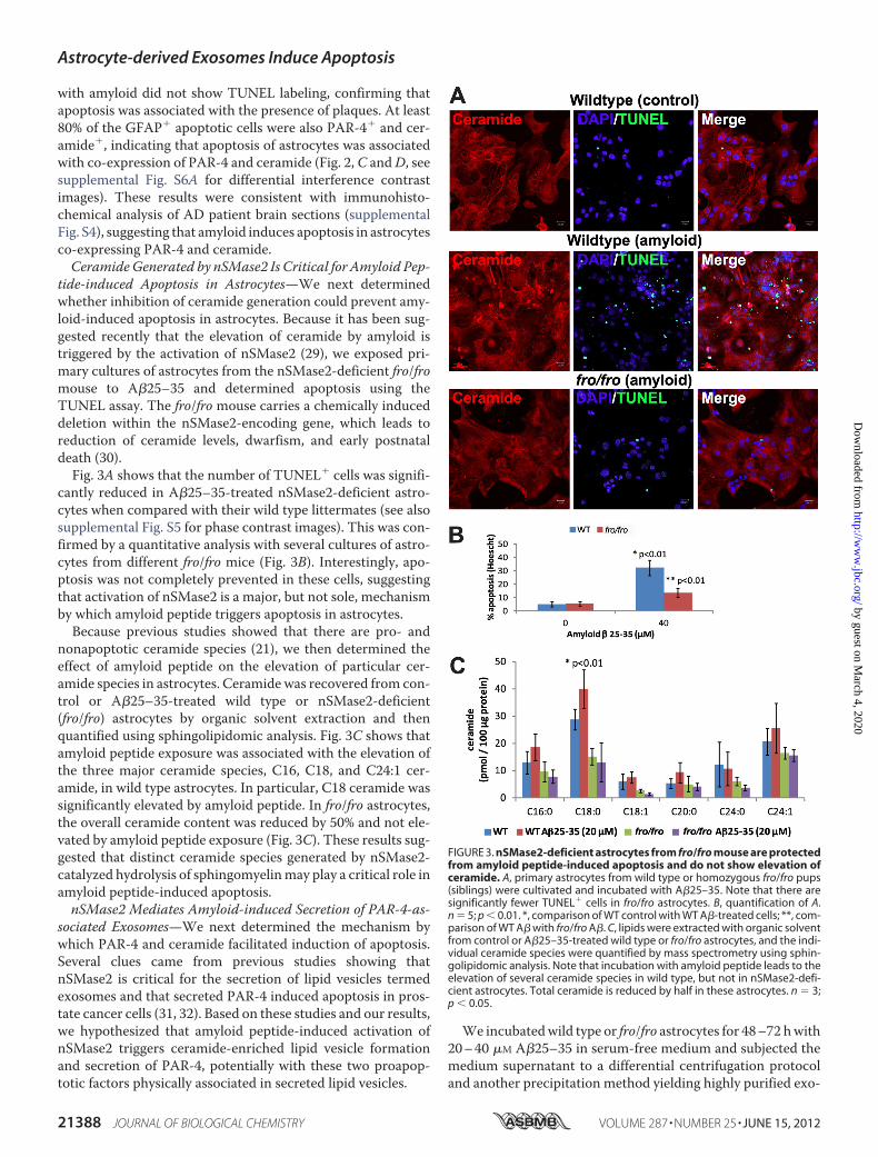
tide-induced Apoptosis in Astrocytes—We next determinedwhether inhibition of ceramide generation could prevent amy-loid-induced apoptosis in astrocytes. Because it has been sug-gested recently that the elevation of ceramide by amyloid istriggered by the activation of nSMase2 (29), we exposed pri-mary cultures of astrocytes from the nSMase2-deficient fro/fromouse to A�25–35 and determined apoptosis using theTUNEL assay. The fro/fro mouse carries a chemically induceddeletion within the nSMase2-encoding gene, which leads toreduction of ceramide levels, dwarfism, and early postnataldeath (30).Fig. 3A shows that the number of TUNEL� cells was signifi-
cantly reduced in A�25–35-treated nSMase2-deficient astro-cytes when compared with their wild type littermates (see alsosupplemental Fig. S5 for phase contrast images). This was con-firmed by a quantitative analysis with several cultures of astro-cytes from different fro/fro mice (Fig. 3B). Interestingly, apo-ptosis was not completely prevented in these cells, suggestingthat activation of nSMase2 is a major, but not sole, mechanismby which amyloid peptide triggers apoptosis in astrocytes.Because previous studies showed that there are pro- and
nonapoptotic ceramide species (21), we then determined theeffect of amyloid peptide on the elevation of particular cer-amide species in astrocytes. Ceramide was recovered from con-trol or A�25–35-treated wild type or nSMase2-deficient(fro/fro) astrocytes by organic solvent extraction and thenquantified using sphingolipidomic analysis. Fig. 3C shows thatamyloid peptide exposure was associated with the elevation ofthe three major ceramide species, C16, C18, and C24:1 cer-amide, in wild type astrocytes. In particular, C18 ceramide wassignificantly elevated by amyloid peptide. In fro/fro astrocytes,the overall ceramide content was reduced by 50% and not ele-vated by amyloid peptide exposure (Fig. 3C). These results sug-gested that distinct ceramide species generated by nSMase2-catalyzed hydrolysis of sphingomyelinmay play a critical role inamyloid peptide-induced apoptosis.nSMase2 Mediates Amyloid-induced Secretion of PAR-4-as-
sociated Exosomes—We next determined the mechanism bywhich PAR-4 and ceramide facilitated induction of apoptosis.Several clues came from previous studies showing thatnSMase2 is critical for the secretion of lipid vesicles termedexosomes and that secreted PAR-4 induced apoptosis in pros-tate cancer cells (31, 32). Based on these studies and our results,we hypothesized that amyloid peptide-induced activation ofnSMase2 triggers ceramide-enriched lipid vesicle formationand secretion of PAR-4, potentially with these two proapop-totic factors physically associated in secreted lipid vesicles.
We incubatedwild type or fro/fro astrocytes for 48–72 hwith20–40 �M A�25–35 in serum-free medium and subjected themedium supernatant to a differential centrifugation protocoland another precipitation method yielding highly purified exo-
FIGURE 3. nSMase2-deficient astrocytes from fro/fro mouse are protectedfrom amyloid peptide-induced apoptosis and do not show elevation ofceramide. A, primary astrocytes from wild type or homozygous fro/fro pups(siblings) were cultivated and incubated with A�25–35. Note that there aresignificantly fewer TUNEL� cells in fro/fro astrocytes. B, quantification of A.n � 5; p � 0.01. *, comparison of WT control with WT A�-treated cells; **, com-parison of WT A� with fro/fro A�. C, lipids were extracted with organic solventfrom control or A�25–35-treated wild type or fro/fro astrocytes, and the indi-vidual ceramide species were quantified by mass spectrometry using sphin-golipidomic analysis. Note that incubation with amyloid peptide leads to theelevation of several ceramide species in wild type, but not in nSMase2-defi-cient astrocytes. Total ceramide is reduced by half in these astrocytes. n � 3;p � 0.05.
Astrocyte-derived Exosomes Induce Apoptosis
21388 JOURNAL OF BIOLOGICAL CHEMISTRY VOLUME 287 • NUMBER 25 • JUNE 15, 2012
by guest on March 4, 2020
http://ww
w.jbc.org/
Dow
nloaded from

somes, ExoQuick, to harvest exosomes released to the medium(24, 25). Fig. 4A shows that PAR-4 was enriched in the mediumwhen wild type astrocytes were incubated with A�25–35 (lane3). There were only trace amounts of PAR-4 in the mediumfrom astrocytes treated with the inactive inverse peptideA�35–25 used as the control (lane 2), suggesting that the ele-vation of PAR-4 in the medium was due to the incubation withthe active amyloid peptide. The absence of staining forGAPDHconfirmed that PAR-4 was not passively released to themedium from dying cells or cell debris, but actively secreted.Thiswas consistentwith the observation that PAR-4was recov-ered from an ultracentrifugation pellet, which also containedthe exosome marker protein TSG101 (lane 6). A more specificisolation method using the ExoQuick system demonstratedthat PAR-4 was associated with exosomes, which was con-firmed by staining for the exosome markers TSG101 and Alix1(Fig. 4B). The observation that the amount of TSG101 or Alix1was concurrently increased with that of PAR-4 suggested thatamyloid peptide elevated the amount of PAR-4-associated exo-
somes. Consistent with this assumption, the exosome fractioncontained atypical PKC, a protein kinase we have shown toform a complex with ceramide and PAR-4 (33–36). In contrastto wild type cells, astrocytes cultivated from nSMase2-deficientfro/fro brain did not secrete PAR-4 or ceramide when exposedto A�25–35 (Fig. 4C, lanes 3 and 4). These results suggestedthat nSMase2 was instrumental for the amyloid peptide-in-duced secretion of PAR-4-associated exosomes.Next, we tested whether ceramide was critical for the secre-
tion of exosomal PAR-4 from amyloid peptide-exposed astro-cytes. fro/fro astrocytes were treatedwith an equimolarmixtureof exogenous C16 andC18 ceramide (2�M each), 40�MA�25–35, or a combination of ceramide and amyloid peptide. Thesetwo ceramide species were chosen because they were elevatedby amyloid peptide (Fig. 3C). They have also been shown to betaken up by cells when added to the medium (34, 37–43). Fig.4D shows that incubation with ceramide or A�25–35 moder-ately elevated secretion of proteins that were stained for PAR-4,but of lower molecular masses (33 or 14 kDa). However, when
FIGURE 4. Amyloid peptide triggers secretion of PAR-4 and ceramide in lipid vesicles that induce apoptosis in astrocytes. A, wild type astrocytes wereincubated with 20 �M A�25–35 for 48 h in serum-free medium, and secreted vesicles were harvested from the medium by ultracentrifugation (UC). Lane 1, celllysate; lane 2, medium control (A�35–25); lane 3, A�25–35-treated astrocytes; lane 4, marker proteins (not stained); lane 5, UC pellet from control astrocytes; lane6, A�-treated astrocytes. B, exosomes harvested by centrifugation with the ExoQuick reagent. Lane 1, control (A�35–25); lane 2, A�25–35-treated astrocytes.Alix, Alix1. C, UC pellets from wild type and nSMase2-deficient fro/fro astrocytes. Lane 1, cell lysate; lane 2, marker proteins (not stained); lane 3, A�35–25-treatedfro/fro astrocytes; lane 4, A�25–35-treated fro/fro; lanes 5 and 6, UC pellets from wild type astrocytes. D, medium from fro/fro astrocytes treated with C16 andC18 ceramide (Cer) with or without A�25–35. Lane 1, cell lysate; lane 2, control: lane 3, C16 � C18 ceramide; lane 4, amyloid peptide; lane 5, amyloid peptide �ceramide. E, as described for D, but exosomes were isolated using ExoQuick reagent and a combination of A�25–35 with either C16 (lane 5) or C18 ceramide(lane 6). F, co-immunoprecipitation (IP) assay using lipid vesicles resuspended from the UC pellet of medium conditioned by A�25–35-treated wild typeastrocytes. Vesicles were subjected to magnetic activated cell sorting, and the flow-through fraction was analyzed by immunoblotting. G, sphingolipidomicanalysis of lipid vesicles in the UC pellet wild type astrocytes without or with A�25–35 incubation. n � 2; p � 0.05 (*); p � 0.01 (**).
Astrocyte-derived Exosomes Induce Apoptosis
JUNE 15, 2012 • VOLUME 287 • NUMBER 25 JOURNAL OF BIOLOGICAL CHEMISTRY 21389
by guest on March 4, 2020
http://ww
w.jbc.org/
Dow
nloaded from

combined, exogenous ceramide and amyloid peptide dramati-cally increased secretion of full-length PAR-4 (Fig. 4D, lane 5),which was associated with exosomes (Fig. 4E, lanes 5 and 6).Interestingly, when combined with A�25–35, C18 ceramidewas �4-fold more potent in triggering the secretion of PAR-4-associated exosomes (Fig. 4E, lane 6) than C16 ceramide (lane5, see supplemental Fig. S5 for phase contrast images). Thisresult was consistent with the observation that C18 ceramidewas most elevated in amyloid peptide-treated wild type astro-cytes (Fig. 3C) and suggested that this particular ceramide spe-cies was critical for the amyloid-induced secretion of exosomesand their association with PAR-4.PAR-4-associated Exosomes Are Enriched with C18 and
C24:1 Ceramide—To determine whether ceramide was specif-ically enriched in PAR-4-associated exosomes, we further ana-lyzed the vesicular pellet obtained by ultracentrifugation of themedium from control and amyloid peptide-treated astrocytes.We incubatedmedium-derived vesicles fromA�25–35-treatedwild type astrocytes with magnetic beads that were coated withanti-ceramide rabbit IgG, anti-CD9 rabbit IgG, or nonspecificcontrol IgG. The beadswere then removed usingmagnetic acti-vated cell sorting, and the flow-through was analyzed for thepresence of PAR-4. Fig. 4F shows that anti-ceramide IgG andanti-CD9 IgG, but not control IgG, depleted the vesicle fractionof PAR-4, suggesting that PAR-4 is associated with ceramide-containing vesicles secreted to the medium. The presence ofCD9 confirmed that the PAR-4/ceramide-containing vesicleswere exosomes, consistent with the results of the ExoQuickpurification procedure (Fig. 4B).
We then analyzed the exosomes for their content of cer-amide. A mass spectrometric (sphingolipidomics) analysisshowed that the lipid vesicles fromA�25–35-treated astrocyteswere enriched with C18 and C24:1 ceramide and that C18 cer-amide was the predominant ceramide species (Fig. 4G). Thisresult was consistent with the observation that amyloid peptideinduced elevation of this ceramide species in wild type astro-cytes (Fig. 3C), concurrent with increased secretion of exo-somes (Fig. 4B). It was also consistent with the result that exog-enous C18 ceramide could specifically restore the secretion ofPAR-4-associated exosomes in amyloid peptide-treated fro/froastrocytes (Fig. 4E), and therefore, provided further evidencefor the assumption that this ceramide species is instrumentalfor the secretion of exosomes that are associated with PAR-4.PAR-4/Ceramide Exosomes Induce Apoptosis in the Absence
of Amyloid Peptide—So far, we have shown that amyloid pep-tide induced secretion of exosomes associated with PAR-4 andceramide. We next tested whether PAR-4/ceramide-contain-ing exosomes induced apoptosis when astrocytes were exposedto them in the absence of amyloid peptide. To detect astrocytesthat bound and took up exosomes, we labeled exosomes fluo-rescently by loading fro/fro orwild type astrocytes withVybrantCM-DiI. This red fluorescent dye is taken up by living cells andpermanently stains cellular membranes. We then washed offexcess dye in the medium and incubated cells with 20 �M
A�25–35 or A�1–42. After 36 h of incubation, Vybrant CM-DiI was clearly visible in a perinuclear compartment and vesi-cles in processes and at the cell surface (Fig. 5A, arrows, seesupplemental Fig. S6C for single channel images).
FIGURE 5. Exosomes isolated from medium of amyloid peptide-treated astrocytes induce apoptosis. Wild type astrocytes were loaded with VybrantCM-DiI and then incubated with fresh medium supplemented with 20 �M A�25–35. A, red fluorescence is visible in a perinuclear compartment and vesiclesalong processes and at the cell membrane (arrows). Bar � 5 �m. B, exosomes were resuspended and added to wild type astrocytes. Only astrocytes taking upexosomes (red fluorescent, arrows) round up and show condensed nuclei (Hoechst dye, blue). Bar � 10 �m. C, confocal laser scanning immunofluorescencemicrocopy of exosome-treated astrocytes using TUNEL staining and anti-ceramide rabbit IgG (Cy5, pseudo-colored in blue). Bar � 20 �m. D, quantification ofapoptosis induced by exosomes from A�25–35-treated wild type astrocytes. n � 3, p � 0.01. E, quantification of apoptosis induced by A�25–35 in the presenceof anti-ceramide (Anti-Cer) or PAR-4 IgG (1 �g/ml). n � 5; p � 0.01. Con, control. *, comparison control IgG with amyloid-treated control IgG; **, comparison anticeramide IgG with amyloid-treated anti ceramide IgG; ***, comparison anti PAR-4 IgG with amyloid-treated anti PAR-4 lgG.
Astrocyte-derived Exosomes Induce Apoptosis
21390 JOURNAL OF BIOLOGICAL CHEMISTRY VOLUME 287 • NUMBER 25 • JUNE 15, 2012
by guest on March 4, 2020
http://ww
w.jbc.org/
Dow
nloaded from

Themedium from theVybrant CM-DiI-loaded and amyloid-treated astrocytes was harvested, and exosomes were isolatedby ultracentrifugation. The exosomes were resuspended andthen added to wild type astrocytes in the absence of amyloidpeptide. Fig. 5B shows that astrocytes, which bound and tookup exosomes (red fluorescent, arrows), rounded up and showedcondensed nuclei (blue, arrows), two typical characteristics ofapoptosis (see supplemental Fig. S6D for single channel imagesand phase contrast). Apoptosis was confirmed using TUNELstaining (Fig. 5C). Apoptotic cells that have taken up exosomesstrongly stained for ceramide, suggesting that ceramide wassupplied to the cells via exosomes, or alternatively, that thesevesicles triggered a process of endogenous ceramide genera-tion, ultimately leading to apoptosis. A quantitative analysisconfirmed that only the resuspended pellet from mediumderived fromwild type astrocytes treated with amyloid peptide,but not from wild type astrocytes without amyloid or fro/froastrocytes with or without amyloid, induced apoptosis (Fig.5D). Taken together these results suggested that amyloid pep-tide triggers the co-distribution and secretion of PAR-4 andceramide in exosomes that induce apoptosis in astrocytes.Evidence that the generation and secretion of ceramide-en-
riched exosomes is a critical step in the induction of apoptosiswas obtained by treating cells with antibodies against ceramide.It has been shown that an anti-ceramide antibody preventedcell death induced by extracellular ceramide in lung emphy-sema, although this previous study did not investigate whetherceramide was presented in the form of lipid vesicles or exo-somes (44). Consistent with these results, anti-ceramide IgG,but not nonspecific rabbit IgG used as a control, significantlyreduced A�25–35-induced apoptosis by blocking extracel-lular ceramide (Fig. 5E). In addition, our results confirmedthat ceramide-enriched exosomes are critical for the induc-tion of apoptosis by amyloid peptide. A PAR-4 antibody wasonly partially effective in preventing apoptosis, suggestingthat exosomes enclosing PAR-4 rather than PAR-4 pre-sented at the vesicle surface were responsible for the induc-tion of apoptosis.The protective effects of anti-ceramide and anti-PAR-4 anti-
bodies were also tested in vivo by injecting fluorescently labeledA�25–35 intomouse brain, with orwithout adding anti-ceramideor anti-PAR-4 IgG. TUNEL assays and immunohistochemicalanalysis for GFAP showed that the addition of the antibodies pre-vented glial apoptosis induced by the injected amyloid peptide(Fig. 6). Consistent with the assumption that amyloid protein orpeptide leads to activation of caspase 3 and eventually inducesapoptosis of astrocytes, many apoptotic cells were found withinthe population of reactive astrocytes migrating toward the lesionsite containing the amyloid peptide (Fig. 6A, arrows). Used as thecontrol, the contralateral site did not show glial activation or apo-ptotic cells (Fig. 6B). Co-injection of amyloid peptide with anti-ceramide or anti-PAR-4 antibodies prevented glial apoptosis,althoughmigration of reactive astrocytes to the lesion sitewas stillobserved (Fig. 6,C andD). These results were consistent with thein vitrodata (Fig. 5E) and suggested that also in vivo, glial apoptosisis induced by amyloid peptide, which is mediated by exogenousceramide and PAR-4, potentially associated with exosomes.
Finally, to test whether PAR-4 and ceramide were co-distrib-uted with exosomes in cells, we performed immunocytochem-istry for PAR-4, ceramide, and TSG101 in control and amyloidpeptide-treated astrocytes. In control astrocytes, immuno-staining of PAR-4, ceramide, and TSG101 showed only littleoverlap (Fig. 7A). Upon treatment with amyloid peptide,PAR-4, ceramide, and TSG101 were redistributed to the peri-nuclear region and to numerous vesicles in processes (Fig. 7B,see supplemental Fig. S6B for phase contrast images). At highermagnification (Fig. 7C), co-distribution at the cell surface ofastrocytic processes appeared in vesicle- or bleb-like structuressimilar to the structures stainedwithVybrantCM-DiI (Fig. 5A).When astrocytes were treated with Vybrant CM-DiI-labeledexosomes, attached vesicles were co-localized with ceramide(Fig. 7D and fluorescence profile in Fig. 7E), suggesting thatastrocytic processes are sites for the dynamic exchange of cer-amide- and PAR-4-associated exosomes.
DISCUSSION
The present study reveals a novel mechanism by which cer-amide- and PAR-4-enriched exosomes can induce apoptosis in
FIGURE 6. Injection of amyloid peptide into mouse brain induces apopto-sis in hippocampal astrocytes, which is prevented by anti-ceramide oranti-PAR-4 antibodies. Coronal cryosections and confocal immunofluores-cence microscopy using an antibody against GFAP (Cy5) and a Cy3 Click-iTTUNEL assay (arrows point at apoptotic cells) show that apoptosis is detect-able in �5% of Hoechst-stained cells in the hippocampus in an area of 100�m surrounding the injected amyloid peptide. About 70% of these cells areGFAP� (n � 2). There are no TUNEL� cells detectable when anti-ceramide oranti-PAR-4 antibodies are co-injected with the amyloid peptide. A, A�-in-jected; ipsilateral; B, A�-injected, contralateral; C, anti-ceramide antibody-co-injected, ipsilateral; D, anti-PAR-4 antibody-co-injected, ipsilateral.
Astrocyte-derived Exosomes Induce Apoptosis
JUNE 15, 2012 • VOLUME 287 • NUMBER 25 JOURNAL OF BIOLOGICAL CHEMISTRY 21391
by guest on March 4, 2020
http://ww
w.jbc.org/
Dow
nloaded from

astrocytes and potentially other cell types. Given the role ofthese exosomes in apoptosis, we propose that they will bereferred to as apoxosomes. Further, our results strongly suggestthat nSMase2 is critical for the enrichment of ceramide inapoxosomes. Ceramide may contribute to apoxosome forma-tion and secretion or induction of apoptosis in astrocytes whenpresented in the form of apoxosomes.We previously showed that the expression of PAR-4 and the
simultaneous elevation of ceramide induce apoptosis in neuralprogenitor cells (45, 46). Ceramide is a sphingolipid that is gen-erated by the sphingomyelinase-catalyzed hydrolysis of sphin-gomyelin and is known to induce apoptosis in cells that aresensitive to it. We have shown that elevation of PAR-4 sensi-tizes neural progenitor cells toward ceramide (33, 45–47). Fur-ther, we found that long chain ceramide induces apoptosis inastrocytes from the Psen1 ADmouse, whereas wild type astro-cytes appear to be resistant to ceramide (21).We suggested thatthe ceramide sensitivity of Psen1-derived astrocytes resulted
from the elevated expression of PAR-4, which was not found inwild type astrocytes.As most AD patients do not carry the PSEN1 mutation, the
effect of amyloid on wild type astrocytes is likely to be of signifi-cance for the etiology of AD. Our new results demonstrate thatexposure to the amyloid peptide triggers the secretion of PAR-4 inceramide-associatedvesicles fromwild typeastrocytes.Consistentwith our previous studies, ceramide-induced apoptosis in astro-cytes was critically dependent on the expression of PAR-4.More-over, AD patient brain sections and sections from the 5XFADmouse showed co-expression of PAR-4 and ceramide concurrentwith glial apoptosis when in cells coming into contact with amy-loidplaques.The fact that glial apoptosis isnot frequently reportedin AD (although it has been reported in Ref. 19) can be explainedby the rapid clearance of apoptotic cells.Observations in the 5XFAD mouse are particularly interest-
ing in regard to the fact that the knock-in of a transgene withmutations in PSEN1 and APP is Thy1-dependent, and there-
FIGURE 7. PAR-4 and ceramide are associated with exosomes secreted by amyloid peptide-treated astrocytes. A, control cells not treated with amyloiddo not show co-distribution of PAR-4, ceramide, and TSG101. Bar � 2 �m. B, A�25–35 treatment induces co-distribution of PAR-4, ceramide, and TSG101, whichis predominantly found in the perinuclear region and bleb-like membrane structures on the surface of astrocytic processes (arrow). Bar � 1 �m. C–E, incubationof astrocytes with Vybrant CM-DiI-labeled exosomes shows co-distribution with TSG101, ceramide, and PAR-4 (profile analysis of pixel intensity in E). Bar � 3�m.
Astrocyte-derived Exosomes Induce Apoptosis
21392 JOURNAL OF BIOLOGICAL CHEMISTRY VOLUME 287 • NUMBER 25 • JUNE 15, 2012
by guest on March 4, 2020
http://ww
w.jbc.org/
Dow
nloaded from
fore, themutated amyloid protein is only expressed in neurons.Nevertheless, the plaque size is severalfold larger than thatof the plaque-generating neurons, suggesting that amyloidbyproducts do not immediately self-intoxicate and kill the neu-rons, but affect other cells aswell. This assumption is supportedby our data showing that injected amyloid peptide led to acuteapoptosis in reactive astrocytes. Thus, the effects of neuron-derived amyloid peptide on astrocytes would lead to glial andneuronal dysfunction, and eventually, neuronal degeneration.The predominant amyloid peptide in human AD is the frag-ment composed of amino acids 1–42 of APP (4). Within thisfragment, a peptide composed of residues 25–35 (A�25–35) isconsidered the toxic peptide inducing apoptosis in AD. There-fore, we performed most of the experiments with A�25–35,which induced apoptosis at the same level as the 1–42 peptide.Our results also suggest that ceramide elevated by amyloid
peptide is a key factor in the events observed in our studies. In aprevious study, incubation of astrocytes with amyloid peptideshowed intracellular elevation of ceramide in vitro, althoughthe ceramide species were not identified, and ceramide secre-tion or glial apoptosis was not reported (29). To our knowledge,we provide for the first time evidence that amyloid-inducedceramide elevation is associated with glial cell death. Our databased on sphingolipidomic analysis show that C18 ceramide ismost elevated, consistent with the composition of ceramidessecreted in apoxosomes to the medium of amyloid-treatedastrocytes. We found that there is a remarkable correlationbetween the ceramide species affected by nSMase2 deficiencyand those secreted to themedium by amyloid-treated wild typeastrocytes. The residual apoptosis in nSMase2-deficient astro-cytes suggests that a portion of proapoptotic ceramide mayhave been generated and secreted by an alternative process.Several enzymatic reactions have been described to generateceramide, and the role of these reactions in the elevation orsecretion of ceramide and the induction of apoptosis in astro-cytes will be further investigated in our future studies.Apart from its role in the induction of apoptosis, nSMase2-
deficient astrocytes did not secrete apoxosomes, a result inagreement with previous findings showing that ceramide gen-eration by nSMase2 is required for exosome formation (32).Exogenously added C18 ceramide restored the formation andsecretion of PAR-4-associated exosomes in amyloid peptide-treated nSMase2-deficient astrocytes. This result is remarkablein that it clearly shows the significance of a particular ceramidespecies for exosome formation. It is also in accordance withstudies from our laboratory as well as other laboratories show-ing that exogenousC16 orC18 ceramide is taken up by cells andthat specific ceramide vesicles bind to PAR-4 via associationwith atypical PKC (33, 36–39).Ceramidemay have a dual function by triggering apoxosome
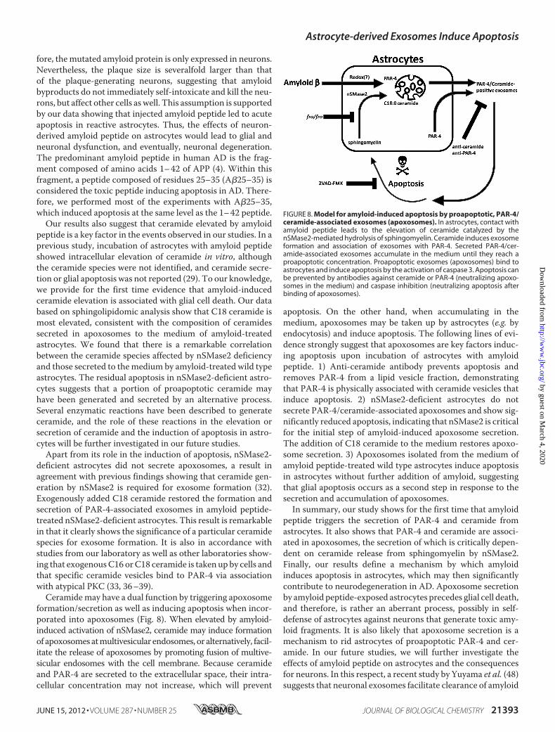
formation/secretion as well as inducing apoptosis when incor-porated into apoxosomes (Fig. 8). When elevated by amyloid-induced activation of nSMase2, ceramide may induce formationof apoxosomesatmultivesicular endosomes, or alternatively, facil-itate the release of apoxosomes by promoting fusion of multive-sicular endosomes with the cell membrane. Because ceramideand PAR-4 are secreted to the extracellular space, their intra-cellular concentration may not increase, which will prevent
apoptosis. On the other hand, when accumulating in themedium, apoxosomes may be taken up by astrocytes (e.g. byendocytosis) and induce apoptosis. The following lines of evi-dence strongly suggest that apoxosomes are key factors induc-ing apoptosis upon incubation of astrocytes with amyloidpeptide. 1) Anti-ceramide antibody prevents apoptosis andremoves PAR-4 from a lipid vesicle fraction, demonstratingthat PAR-4 is physically associated with ceramide vesicles thatinduce apoptosis. 2) nSMase2-deficient astrocytes do notsecrete PAR-4/ceramide-associated apoxosomes and show sig-nificantly reduced apoptosis, indicating that nSMase2 is criticalfor the initial step of amyloid-induced apoxosome secretion.The addition of C18 ceramide to the medium restores apoxo-some secretion. 3) Apoxosomes isolated from the medium ofamyloid peptide-treated wild type astrocytes induce apoptosisin astrocytes without further addition of amyloid, suggestingthat glial apoptosis occurs as a second step in response to thesecretion and accumulation of apoxosomes.In summary, our study shows for the first time that amyloid
peptide triggers the secretion of PAR-4 and ceramide fromastrocytes. It also shows that PAR-4 and ceramide are associ-ated in apoxosomes, the secretion of which is critically depen-dent on ceramide release from sphingomyelin by nSMase2.Finally, our results define a mechanism by which amyloidinduces apoptosis in astrocytes, which may then significantlycontribute to neurodegeneration in AD. Apoxosome secretionby amyloid peptide-exposed astrocytes precedes glial cell death,and therefore, is rather an aberrant process, possibly in self-defense of astrocytes against neurons that generate toxic amy-loid fragments. It is also likely that apoxosome secretion is amechanism to rid astrocytes of proapoptotic PAR-4 and cer-amide. In our future studies, we will further investigate theeffects of amyloid peptide on astrocytes and the consequencesfor neurons. In this respect, a recent study by Yuyama et al. (48)suggests that neuronal exosomes facilitate clearance of amyloid
FIGURE 8. Model for amyloid-induced apoptosis by proapoptotic, PAR-4/ceramide-associated exosomes (apoxosomes). In astrocytes, contact withamyloid peptide leads to the elevation of ceramide catalyzed by thenSMase2-mediated hydrolysis of sphingomyelin. Ceramide induces exosomeformation and association of exosomes with PAR-4. Secreted PAR-4/cer-amide-associated exosomes accumulate in the medium until they reach aproapoptotic concentration. Proapoptotic exosomes (apoxosomes) bind toastrocytes and induce apoptosis by the activation of caspase 3. Apoptosis canbe prevented by antibodies against ceramide or PAR-4 (neutralizing apoxo-somes in the medium) and caspase inhibition (neutralizing apoptosis afterbinding of apoxosomes).
Astrocyte-derived Exosomes Induce Apoptosis
JUNE 15, 2012 • VOLUME 287 • NUMBER 25 JOURNAL OF BIOLOGICAL CHEMISTRY 21393
by guest on March 4, 2020
http://ww
w.jbc.org/
Dow
nloaded from

peptide by microglia, a function depending on the activation ofnSMase2 in neurons. It remains to be investigatedwhether glialexosomes participate in this process, and therefore, have addi-tional functions distinct from induction of apoptosis. We willalso determine whether blocking of ceramide generation or theproapoptotic effect of PAR-4/ceramide-associated apoxo-somes is a possible treatment option for AD.
Acknowledgments—We are grateful to Dr. Mark Noble (University ofRochester, Rochester, NY) for stimulating scientific discussions andediting of the manuscript. We thank the imaging core facility at theGeorgia Health Sciences University, Augusta, GA (under the supervi-sion of Drs. Paul and Ana McNeil) for support. Support by the Insti-tute of Molecular Medicine and Genetics (under the directorship ofDr. Lin Mei) at the Georgia Health Sciences University is alsoacknowledged.
REFERENCES1. Goedert,M., and Spillantini, M. G. (2006) A century of Alzheimer disease.
Science 314, 777–7812. Ittner, L.M., andGötz, J. (2011) Amyloid-� andTau: a toxic pas de deux in
Alzheimer disease. Nat. Rev. Neurosci. 12, 65–723. Czech, C., Tremp, G., and Pradier, L. (2000) Presenilins and Alzheimer
disease: biological functions and pathogenicmechanisms.Prog.Neurobiol.60, 363–384
4. Millucci, L., Ghezzi, L., Bernardini, G., and Santucci, A. (2010) Conforma-tions and biological activities of amyloid-� peptide 25–35. Curr. ProteinPept. Sci. 11, 54–67
5. Verkhratsky, A., Olabarria,M., Noristani, H.N., Yeh, C. Y., and Rodriguez,J. J. (2010) Astrocytes in Alzheimer disease. Neurotherapeutics 7,399–412
6. Mattson, M. P., Pedersen,W. A., Duan,W., Culmsee, C., and Camandola,S. (1999) Cellular and molecular mechanisms underlying perturbed en-ergy metabolism and neuronal degeneration in Alzheimer and Parkinsondiseases. Ann. N.Y. Acad. Sci. 893, 154–175
7. Perez, J. L., Carrero, I., Gonzalo, P., Arevalo-Serrano, J., Sanz-Anquela,J. M., Ortega, J., Rodriguez, M., and Gonzalo-Ruiz, A. (2010) Soluble olig-omeric forms of �-amyloid (A�) peptide stimulate A� production viaastrogliosis in the rat brain. Exp. Neurol. 223, 410–421
8. Agostinho, P., Cunha, R. A., and Oliveira, C. (2010) Neuroinflammation,oxidative stress, and the pathogenesis of Alzheimer disease. Curr. Pharm.Des. 16, 2766–2778
9. Farfara, D., Lifshitz, V., and Frenkel, D. (2008) Neuroprotective and neu-rotoxic properties of glial cells in the pathogenesis of Alzheimer disease.J. Cell Mol. Med. 12, 762–780
10. Li, C., Zhao, R., Gao, K., Wei, Z., Yin, M. Y., Lau, L. T., Chui, D., and HoiYu, A. C. (2011) Astrocytes: implications for neuroinflammatory patho-genesis of Alzheimer disease. Curr. Alzheimer Res. 8, 67–80
11. Rodríguez, J. J., Olabarria, M., Chvatal, A., and Verkhratsky, A. (2009)Astroglia in dementia and Alzheimer disease. Cell Death Differ. 16,378–385
12. Meda, L., Baron, P., and Scarlato, G. (2001) Glial activation in Alzheimerdisease: the role of A� and its associated proteins. Neurobiol. Aging 22,885–893
13. Nagele, R. G., Wegiel, J., Venkataraman, V., Imaki, H., Wang, K. C., andWegiel, J. (2004) Contribution of glial cells to the development of amyloidplaques in Alzheimer disease. Neurobiol. Aging 25, 663–674
14. Allaman, I., Bélanger, M., and Magistretti, P. J. (2011) Astrocyte-neuronmetabolic relationships: for better and for worse. Trends Neurosci. 34,76–87
15. Fuller, S., Steele, M., and Münch, G. (2010) Activated astroglia duringchronic inflammation in Alzheimer disease: do they neglect their neuro-supportive roles?Mutat Res. 690, 40–49
16. Lee, Y. J., Han, S. B., Nam, S. Y., Oh, K. W., and Hong, J. T. (2010) Inflam-
mation and Alzheimer disease. Arch. Pharm. Res. 33, 1539–155617. Croisier, E., and Graeber, M. B. (2006) Glial degeneration and reactive
gliosis in �-synucleinopathies: the emerging concept of primary gliode-generation. Acta Neuropathol. 112, 517–530
18. Van Eldik, L. J., Thompson, W. L., Ralay Ranaivo, H., Behanna, H. A., andMartin Watterson, D. (2007) Glia proinflammatory cytokine up-regula-tion as a therapeutic target for neurodegenerative diseases: function-based and target-based discovery approaches. Int. Rev. Neurobiol. 82,277–296
19. Kobayashi, K., Hayashi, M., Nakano, H., Fukutani, Y., Sasaki, K.,Shimazaki, M., and Koshino, Y. (2002) Apoptosis of astrocytes with en-hanced lysosomal activity and oligodendrocytes in white matter lesions inAlzheimer disease. Neuropathol. Appl. Neurobiol. 28, 238–251
20. Mouser, P. E., Head, E., Ha, K. H., and Rohn, T. T. (2006) Caspase-medi-ated cleavage of glial fibrillary acidic protein within degenerating astro-cytes of the Alzheimer disease brain. Am. J. Pathol. 168, 936–946
21. Wang, G., Silva, J., Dasgupta, S., and Bieberich, E. (2008) Long-chain cer-amide is elevated in presenilin 1 (PS1M146V) mouse brain and inducesapoptosis in PS1 astrocytes. Glia 56, 449–456
22. Millucci, L., Raggiaschi, R., Franceschini, D., Terstappen, G., and Santucci,A. (2009) Rapid aggregation and assembly in aqueous solution of A�
(25–35) peptide. J. Biosci. 34, 293–30323. Fauré, J., Lachenal, G., Court, M., Hirrlinger, J., Chatellard-Causse, C.,
Blot, B., Grange, J., Schoehn, G., Goldberg, Y., Boyer, V., Kirchhoff, F.,Raposo, G., Garin, J., and Sadoul, R. (2006) Exosomes are released bycultured cortical neurons.Mol. Cell Neurosci. 31, 642–648
24. Guescini,M., Genedani, S., Stocchi, V., andAgnati, L. F. (2010) Astrocytesand glioblastoma cells release exosomes carrying mtDNA. J. NeuralTransm. 117, 1–4
25. Taylor, D. D., Zacharias, W., and Gercel-Taylor, C. (2011) Exosome iso-lation for proteomic analyses and RNA profiling.Methods Mol. Biol. 728,235–246
26. Caraci, F., Chisari, M., Frasca, G., Canonico, P. L., Battaglia, A., Calafiore,M., Battaglia, G., Bosco, P., Nicoletti, F., Copani, A., and Sortino, M. A.(2005) Nicergoline, a drug used for age-dependent cognitive impairment,protects cultured neurons against �-amyloid toxicity. Brain Res. 1047,30–37
27. Mattson, M. P., Duan,W., Chan, S. L., and Camandola, S. (1999) Par-4: anemerging pivotal player in neuronal apoptosis and neurodegenerative dis-orders. J. Mol. Neurosci. 13, 17–30
28. Cheema, S. K.,Mishra, S. K., Rangnekar, V.M., Tari, A.M., Kumar, R., andLopez-Berestein, G. (2003) Par-4 transcriptionally regulates Bcl-2 througha WT1-binding site on the bcl-2 promoter. J. Biol. Chem. 278,19995–20005
29. Jana, A., and Pahan, K. (2010) Fibrillar amyloid-�-activated human astro-glia kill primary human neurons via neutral sphingomyelinase: implica-tions for Alzheimer disease. J. Neurosci. 30, 12676–12689
30. Aubin, I., Adams, C. P., Opsahl, S., Septier, D., Bishop, C. E., Auge, N.,Salvayre, R., Negre-Salvayre, A., Goldberg,M., Guénet, J. L., and Poirier, C.(2005) A deletion in the gene encoding sphingomyelin phosphodiesterase3 (Smpd3) results in osteogenesis and dentinogenesis imperfecta in themouse. Nat. Genet. 37, 803–805
31. Burikhanov, R., Zhao, Y., Goswami, A., Qiu, S., Schwarze, S. R., and Rang-nekar, V. M. (2009) The tumor suppressor Par-4 activates an extrinsicpathway for apoptosis. Cell 138, 377–388
32. Trajkovic, K., Hsu, C., Chiantia, S., Rajendran, L., Wenzel, D., Wieland, F.,Schwille, P., Brügger, B., and Simons, M. (2008) Ceramide triggers bud-ding of exosome vesicles into multivesicular endosomes. Science 319,1244–1247
33. Wang, G., Silva, J., Krishnamurthy, K., Tran, E., Condie, B. G., and Bie-berich, E. (2005) Direct binding to ceramide activates protein kinase C�
before the formation of a proapoptotic complex with PAR-4 in differenti-ating stem cells. J. Biol. Chem. 280, 26415–26424
34. Wang, G., Krishnamurthy, K., Umapathy, N. S., Verin, A. D., and Bie-berich, E. (2009) The carboxyl-terminal domain of atypical protein kinaseC� binds to ceramide and regulates junction formation in epithelial cells.J. Biol. Chem. 284, 14469–14475
35. Bieberich, E., Kawaguchi, T., and Yu, R. K. (2000) N-Acylated serinol is a
Astrocyte-derived Exosomes Induce Apoptosis
21394 JOURNAL OF BIOLOGICAL CHEMISTRY VOLUME 287 • NUMBER 25 • JUNE 15, 2012
by guest on March 4, 2020
http://ww
w.jbc.org/
Dow
nloaded from
novel ceramide mimic inducing apoptosis in neuroblastoma cells. J. Biol.Chem. 275, 177–181
36. Bieberich, E. (2011) Lipid vesicle-mediated affinity chromatography usingmagnetic activated cell sorting (LIMACS): a novel method to analyze pro-tein-lipid interaction. J. Vis. Exp. 26, pii: 265
37. Gamen, S., Hanson, D. A., Kaspar, A., Naval, J., Krensky, A. M., and Anel,A. (1998) Granulysin-induced apoptosis. I. Involvement of at least twodistinct pathways. J. Immunol. 161, 1758–1764
38. Herget, T., Esdar, C., Oehrlein, S. A., Heinrich, M., Schütze, S., Maelicke,A., and van Echten-Deckert, G. (2000) Production of ceramides causesapoptosis during early neural differentiation in vitro. J. Biol. Chem. 275,30344–30354
39. Paris, F., Grassmé, H., Cremesti, A., Zager, J., Fong, Y., Haimovitz-Fried-man, A., Fuks, Z., Gulbins, E., and Kolesnick, R. (2001) Natural ceramidereverses Fas resistance of acid sphingomyelinase�/� hepatocytes. J. Biol.Chem. 276, 8297–8305
40. López-Montero, I., Rodriguez, N., Cribier, S., Pohl, A., Vélez, M., andDevaux, P. F. (2005) Rapid transbilayer movement of ceramides in phos-pholipid vesicles and in human erythrocytes. J. Biol. Chem. 280,25811–25819
41. Fukunaga, T., Nagahama,M., Hatsuzawa, K., Tani, K., Yamamoto, A., andTagaya, M. (2000) Implication of sphingolipid metabolism in the stabilityof the Golgi apparatus. J. Cell Sci. 113, 3299–3307
42. Wang, G., Krishnamurthy, K., Chiang, Y. W., Dasgupta, S., and Bieberich,
E. (2008) Regulation of neural progenitor cell motility by ceramide andpotential implications for mouse brain development. J. Neurochem. 106,718–733
43. Wang, G., Krishnamurthy, K., and Bieberich, E. (2009) Regulation of pri-mary cilia formation by ceramide. J. Lipid Res. 50, 2103–2110
44. Petrache, I., Natarajan, V., Zhen, L., Medler, T. R., Richter, A. T., Cho, C.,Hubbard, W. C., Berdyshev, E. V., and Tuder, R. M. (2005) Ceramideup-regulation causes pulmonary cell apoptosis and emphysema-like dis-ease in mice. Nat. Med. 11, 491–498
45. Bieberich, E., MacKinnon, S., Silva, J., Noggle, S., and Condie, B. G. (2003)Regulation of cell death in mitotic neural progenitor cells by asymmetricdistribution of prostate apoptosis response 4 (PAR-4) and simultaneouselevation of endogenous ceramide. J. Cell Biol. 162, 469–479
46. Bieberich, E., Silva, J., Wang, G., Krishnamurthy, K., and Condie, B. G.(2004) Selective apoptosis of pluripotent mouse and human stem cells bynovel ceramide analogues prevents teratoma formation and enriches forneural precursors in ES cell-derived neural transplants. J. Cell Biol. 167,723–734
47. Bieberich, E. (2011) There is more to a lipid than just being a fat: sphingo-lipid-guided differentiation of oligodendroglial lineage from embryonicstem cells. Neurochem. Res. 36, 1601–1611
48. Yuyama, K., Sun, H., Mitsutake, S., and Igarashi, Y. (2012) Sphingolipid-modulated exosome secretion promotes the clearance of amyloid-� bymicroglia. J. Biol. Chem. 287, 10977–10989
Astrocyte-derived Exosomes Induce Apoptosis
JUNE 15, 2012 • VOLUME 287 • NUMBER 25 JOURNAL OF BIOLOGICAL CHEMISTRY 21395
by guest on March 4, 2020
http://ww
w.jbc.org/
Dow
nloaded from

Campbell, Margot Mayer-Proschel and Erhard BieberichGuanghu Wang, Michael Dinkins, Qian He, Gu Zhu, Christophe Poirier, Andrew
INDUCTION IN ALZHEIMER DISEASE (AD)Apoptosis Response 4 (PAR-4): POTENTIAL MECHANISM OF APOPTOSIS
Astrocytes Secrete Exosomes Enriched with Proapoptotic Ceramide and Prostate
doi: 10.1074/jbc.M112.340513 originally published online April 24, 20122012, 287:21384-21395.J. Biol. Chem.
10.1074/jbc.M112.340513Access the most updated version of this article at doi:
Alerts:
When a correction for this article is posted•
When this article is cited•
to choose from all of JBC's e-mail alertsClick here
Supplemental material:
http://www.jbc.org/content/suppl/2012/04/24/M112.340513.DC1
http://www.jbc.org/content/287/25/21384.full.html#ref-list-1
This article cites 47 references, 16 of which can be accessed free at
by guest on March 4, 2020
http://ww
w.jbc.org/
Dow
nloaded from