Embed Size (px)
Citation preview
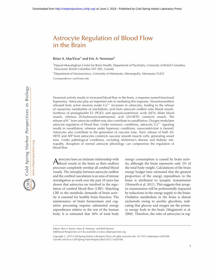
Astrocyte Regulation of Blood Flowin the Brain
Brian A. MacVicar1 and Eric A. Newman2
1Djavad Mowafaghian Centre for Brain Health, Department of Psychiatry, University of British Columbia,Vancouver, British Columbia V6T 2B5, Canada
2Department of Neuroscience, University of Minnesota, Minneapolis, Minnesota 55455
Correspondence: [email protected]
Neuronal activity results in increased blood flow in the brain, a response named functionalhyperemia. Astrocytes play an important role in mediating this response. Neurotransmittersreleased from active neurons evoke Ca2þ increases in astrocytes, leading to the releaseof vasoactive metabolites of arachidonic acid from astrocyte endfeet onto blood vessels.Synthesis of prostaglandin E2 (PGE2) and epoxyeicosatrienoic acids (EETs) dilate bloodvessels, whereas 20-hydroxyeicosatetraenoic acid (20-HETE) constricts vessels. Therelease of Kþ from astrocyte endfeet may also contribute to vasodilation. Oxygen modulatesastrocyte regulation of blood flow. Under normoxic conditions, astrocytic Ca2þ signalingresults in vasodilation, whereas under hyperoxic conditions, vasoconstriction is favored.Astrocytes also contribute to the generation of vascular tone. Tonic release of both 20-HETE and ATP from astrocytes constricts vascular smooth muscle cells, generating vesseltone. Under pathological conditions, including Alzheimer’s disease and diabetic reti-nopathy, disruption of normal astrocyte physiology can compromise the regulation ofblood flow.
Astrocytes have an intimate relationship withblood vessels in the brain as their endfoot
processes completely envelop all cerebral bloodvessels. The interplay between astrocyte endfeetand the cerebral vasculature is an area of intenseinvestigation as work over the past 10 years hasshown that astrocytes are involved in the regu-lation of cerebral blood flow (CBF). MatchingCBF to the metabolic demands of brain activ-ity is essential for healthy brain function. Themaintenance of brain homeostasis and cog-nitive processing requires substantial energyexpenditures relative to the rest of the humanbody. It is estimated that 20% of total body
energy consumption is caused by brain activ-ity, although the brain represents only 2% ofthe total body weight. Calculations of the brainenergy budget have estimated that the greatestproportion of the energy expenditure in thebrain is attributed to synaptic transmission(Howarth et al. 2012). This suggests that synap-tic transmission will be preferentially impactedby reductions in the energy supply to the brain.Oxidative metabolism in the brain is almostexclusively owing to aerobic glycolysis, indi-cating that glucose and oxygen are the prima-ry energy fuels in the brain (Magistretti et al.2000). Therefore, the roles of astrocytes in reg-
Editors: Ben A. Barres, Marc R. Freeman, and Beth Stevens
Additional Perspectives on Glia available at www.cshperspectives.org
Copyright # 2015 Cold Spring Harbor Laboratory Press; all rights reserved; doi: 10.1101/cshperspect.a020388
Cite this article as Cold Spring Harb Perspect Biol 2015;7:a020388
1
on June 2, 2018 - Published by Cold Spring Harbor Laboratory Press http://cshperspectives.cshlp.org/Downloaded from

ulating CBF is likely to be of fundamental im-portance in providing appropriate and consis-tent energy supply to support brain function.
There are two important levels at which CBFis regulated. Basal CBF, which is remarkablyhigher than the rest of the body (Magistretti etal. 2000), is regulated so that the brain receivesan adequate supply of blood at all times. Auto-regulatory mechanisms ensure that CBF doesnot vary in the face of changes in systemic bloodpressure. CBF is also regulated in response tobrain activity. It has long been recognized, sincethe classic papers of Mosso (1880) and Roy andSherrington (1890), that the brain has an intrin-sic ability to rapidly regulate its own blood sup-ply in response to local energy requirements,a response named functional hyperemia. Thishomeostatic response is important for provid-ing increased delivery of glucose and oxygen attimes of intense activity and metabolic demand.
HISTORICAL OVERVIEW OF FUNCTIONALHYPEREMIA
Functional hyperemia was first described in the1880s by Angelo Mosso (1880), who observedchanges in brain volume in patients with skulldefects, allowing direct observation of the cor-tical surface. Mosso found that sensory stimuliproduced an increase in brain volume, repre-senting increased cortical blood flow. A decadelater, Roy and Sherrington (1890) showed thatstimulation of sensory nerves in dogs producedincreases in cortical blood flow. They speculatedthat “the chemical products of cerebral metab-olism can cause variations of the caliber of thecerebral vessels: that in this reaction the brainpossesses an intrinsic mechanism by which itsvascular supply can be varied locally in corre-spondence with local variations of functionalactivity.”
In the late 1800s, around the same time thatfunctional hyperemia was first observed, astro-cyte morphology was described by Virchow(1858), Golgi (1894), Ramon y Cajal (1995)(first published in 1897), and others. They ob-served that astrocytes contacted both blood ves-sels, which are enveloped by astrocyte endfeet,and neurons (Fig. 1). Thus, astrocytes are ide-
ally positioned to mediate neurovascular cou-pling, relaying signals from neurons to bloodvessels. The association of astrocytes with bloodvessels led Ramon y Cajal to suggest that thesecells might regulate blood flow in the brain.Ramon y Cajal wrote, “The perivascular neuro-glial cells live only in the proximity of the cap-illaries of the gray matter, to which they sendone or more thick appendages inserted in theouter side of the endothelium. Each capillarygives insertion to thousands of these pseudo-pods (endfeet), which diverge in all directions.The object of such elements is to evoke, bycontraction of the aforementioned appendices,local dilations of the vessels” (Ramon y Cajal1895). Ramon y Cajal’s suggestion that astro-cytes regulate blood flow, albeit by an incorrectmechanism, was prescient.
More recently, astrocyte regulation of bloodflow has been investigated directly. Paulson andNewman (1987) proposed that astrocytes me-diate functional hyperemia by a Kþ siphoningmechanism, releasing Kþ onto blood vesselsfrom their endfeet in response to neuronal ac-tivity. Later, Zonta et al. (2003) suggested thatneurovascular coupling is mediated by the re-lease of prostaglandins from astrocytes. Theyshowed that prostaglandin E2 (PGE2) release,resulting from increases in glial Ca2þ, evokesvessel dilation.
Since the publication of Zonta et al. (2003),the role of astrocytes in mediating functionalhyperemia has been studied intensely. Severalmechanisms of glial control of CBF have beenproposed and tested. These mechanisms are dis-cussed in the following sections.
NEUROVASCULAR COUPLING
It was originally hypothesized that the controlof CBF flow was mediated by a negative feed-back mechanism, whereby the generation ofmetabolites from active neurons (such as CO2)were the signals that resulted in increased CBF(Roy and Sherrington 1890). However, it is nowrecognized that brain activity itself can increaseCBF. Indeed, CBF increases to such an extentthat more oxygen is provided to active brainregions than is consumed. Recordings of tissue
B.A. MacVicar and E.A. Newman
2 Cite this article as Cold Spring Harb Perspect Biol 2015;7:a020388
on June 2, 2018 - Published by Cold Spring Harbor Laboratory Press http://cshperspectives.cshlp.org/Downloaded from

pO2 levels during activation of cortical synapticactivity show that pO2 increases rapidly abovebasal levels during and after repetitive synapticactivity (Offenhauser et al. 2005; Devor et al.2011). This oversupply of oxygen is the basisof the BOLD (blood oxygen level–dependent)effect in fMRI (functional magnetic resonanceimaging), whereby increased levels of blood oxy-
gen are detected by the change in the magneticproperties of hemoglobin as the proportion ofoxygenated hemoglobin increases and the de-oxygenated form is decreased. It is now recog-nized that synaptic and brain activity itself leadsto enhanced CBF and that astrocytes are keyplayers in mediating neurovascular coupling,the transduction of brain activity to alterations
Figure 1. Drawing of brain astrocytes by Santiago Ramon y Cajal. (A,B) Astrocytes, the darker cells in thedrawing, contact both neurons, (C,D) the lighter cells, and (F) a blood vessel. As suggested by Ramon y Cajal,astrocytes are ideally situated to mediate signaling from neurons to blood vessels and to increase cerebral bloodflow (CBF) in response to neuronal activity.
Astrocyte Regulation of Blood Flow in the Brain
Cite this article as Cold Spring Harb Perspect Biol 2015;7:a020388 3
on June 2, 2018 - Published by Cold Spring Harbor Laboratory Press http://cshperspectives.cshlp.org/Downloaded from

in CBF. Although direct signaling from neuronsto blood vessels contributes to the regulation ofCBF (Attwell et al. 2010), astrocytes play an im-portant role in mediating neurovascular cou-pling (Fig. 2).
MECHANISMS OF ASTROCYTE-MEDIATEDNEUROVASCULAR COUPLING
Arachidonic Acid–Mediated NeurovascularCoupling
Work over the past decade has shown that neu-rovascular coupling is mediated, to a significantdegree, by a Ca2þ-dependent astrocytic mecha-nism. The link between astrocyte Ca2þ and vas-cular changes was best shown by uncaging ofCa2þ in astrocytes in brain slices, retinal ex-plants, and in vivo. These experiments showed
unequivocally that Ca2þ transients in astrocytescan induce dilations and constrictions in adja-cent arterioles. Mulligan and MacVicar (2004)showed that arterioles display constrictions asopposed to the dilations, reported by Zonta etal. (2003), when Ca2þ transients were evokedwith two-photon Ca2þ uncaging in endfeet ad-jacent to arterioles. The arteriole responses wereblocked by inhibiting phospholipase A2, theenzyme that liberates arachidonic acid (AA)from membrane lipids, and by inhibiting theconversion of AA to the vasoconstrictive lipid,20-hydroxyeicosatetraenoic acid (20-HETE). InCa2þ uncaging experiments in vivo, Takano etal. (2006) showed that arteriole dilations in thecortex were triggered by astrocyte Ca2þ tran-sients via a mechanism sensitive to the inhibi-tion of cylclooxygenase-1 (COX-1), one of theenzymes that synthesizes PGE2 from AA. Ta-
Synapse
Glutamate
NMDARnNOS
O2 and glucose
NO NO
cGMP
En
do
thel
ium
Dilate
Constrict
20-HETE
NO
PGAAPLA2
Ca2+
Neuron
AA
PLA2
EETEET
PG
K+PGgk(Ca)
Ca2+
AA
AA
Arteriolesmoothmuscle
Lumen
mGluRAstrocyte
O2 andglucose
Figure 2. Summary of signaling pathways that mediate neurovascular coupling in the brain. Synaptically releasedglutamate acts on N-methyl-D-aspartate receptors (NMDARs) in neurons to increase [Ca2þ]i, causing neuronalnitric oxide synthase (nNOS) to release nitric oxide (NO), which activates smooth muscle guanylate cyclase.Raised [Ca2þ]i may also (dashed line) generate arachidonic acid (AA) from phospholipase A2 (PLA2), which isconverted to prostaglandins (PG) that dilate vessels. Glutamate also raises [Ca2þ]i in astrocytes by activatingmetabotropic glutamate receptors (mGluR), generating arachidonic acid, and three types of AA metabolites:prostaglandins and EETs in astrocytes, which dilate vessels, and 20-HETE in smooth muscle, which constrictsvessels. An increase of [Ca2þ]i in astrocyte endfeet may also activate Ca2þ-gated Kþ channels (gk(Ca), alternativeabbreviation, BK), releasing Kþ, which dilates vessels. (From Attwell et al. 2010; reprinted, with permission,from the authors.)
B.A. MacVicar and E.A. Newman
4 Cite this article as Cold Spring Harb Perspect Biol 2015;7:a020388
on June 2, 2018 - Published by Cold Spring Harbor Laboratory Press http://cshperspectives.cshlp.org/Downloaded from

kano et al. also showed that COX-1 was ex-pressed in the endfeet of cortical astrocytes,supporting the role for PGE2 synthesis by astro-cytes. These results suggested that PGE2 is re-leased from astrocytes to cause arteriole dila-tions similar to the mechanism proposed byZonta et al. (2003) (Fig. 3). The first hints ofthe complexity of astrocyte vascular couplingcame from work in the retina explant by Meteaand Newman (2006). They showed that, in thesame preparation, Ca2þ uncaging in astrocytesand Muller cells (the principal retinal glial cells)could trigger both arteriole constrictions via20-HETE synthesis and dilations via the forma-tion of other vasoactive lipids and epoxyeicosa-
trienoic acids (EETs), that are also synthesizedfrom AA. Subsequent work by Mishra et al.(2011) showed that astrocyte-evoked vasodila-tion in the retina is mediated by synthesis ofboth EETs and PGE2.
Part of the explanation for the divergentobservations that both dilations and constric-tions could be triggered by astrocyte Ca2þ sig-naling came from the work of Gordon et al.(2008). In this study of brain slices, the inves-tigators reported that the polarity of the vascu-lar response to astrocyte Ca2þ transients wasdependent on the level of pO2 in the super-fusion solution. Arteriole constrictions thatwere evoked by Ca2þ uncaging in astrocytes in
Vessel lumen
Smooth muscle cell
Astrocyte endfoot
Constriction
Ca2+ Ca2+
Ca2+Ca2+
AAAA
Glycolysis
20-HETE
Adenosine Adenosine
Glycolysis
PGE2
Lactate
PGE2COXPLA2
Low O2High O2
PLA2
CYP4A
Dilatation
PGT
Figure 3. Summary of the modulation of arachidonic acid metabolite-mediated neurovascular coupling bytissue pO2 and lactate. Intracellular Ca2þ transients activate PLA2 to liberate arachidonic acid (AA) from plasmamembrane lipids. In high pO2, AA is converted to 20-HETE, which increases intracellular Ca2þ in smoothmuscle cells, causing vasoconstriction (left side). Extracellular adenosine that can inhibit Ca2þ entry intosmooth muscle cells is also lower in high tissue pO2, further promoting vasoconstriction. In lower pO2 (rightside), the vasodilation pathway dominates and AA is converted to PGE2 by COX-1 and is released by diffusion.Extracellular PGE2 is cleared from the extracellular space by the prostaglandin transporter (PGT), which isexpressed in astrocytes and neurons. When pO2 decreases, glycolysis is enhanced and extracellular levels oflactate increase. Extracellular lactate attenuates PGE2 uptake by PGT leading to higher extracellular PGE2 andenhanced relaxation of smooth muscle tone and vasodilation. In addition, higher levels of extracellular aden-osine reduces smooth muscle cell constriction via A2A activation. (From Gordon et al. 2011; reprinted, withpermission, from the authors.)
Astrocyte Regulation of Blood Flow in the Brain
Cite this article as Cold Spring Harb Perspect Biol 2015;7:a020388 5
on June 2, 2018 - Published by Cold Spring Harbor Laboratory Press http://cshperspectives.cshlp.org/Downloaded from

high O2 (95%) solutions were reversed to arte-riole dilations in more physiological (20%) so-lutions. Oxygen modulation of neurovascularcoupling occurred because low pO2 increasedextracellular lactate concentrations, which, inturn, reduced the uptake and clearance of PGE2by the prostaglandin transporter. In high pO2,the conversion of AA to 20-HETE predomi-nates, leading to constriction, whereas, in lowpO2, the PGE2 dilation pathway is dominant(Fig. 3). In addition, the increased adenosinetone in low pO2 solutions reduced the vaso-constriction. A similar switch from dilation toconstriction was observed in the retina explantwhen tissue pO2 was altered from a low O2 levelto a high level (Mishra et al. 2011). However,this O2-modulatory effect on retinal blood flowwas not observed in vivo when animals weremade hyperoxic. The results of these experi-ments indicated that the polarity of astrocytemodulation of CBF may reflect the metabolicstate of the brain tissue and may be modifiedby the level of the extracellular lactate concen-tration. It is intriguing that a link between themagnitude of CBF changes and lactate levels hasbeen observed in awake human subjects in anfMRI study (Mintun et al. 2004), in which anincreased lactate/pyruvate ratio was observedto augment blood flow in the physiologicallyactivated human brain. In addition, a nuclearmagnetic resonance (NMR) spectroscopy studyshowed a remarkably strong correlation be-tween functional hyperemia and brain lactatelevels (Lin et al. 2010).
The dilation or constriction of arteriole di-ameter as a result of Ca2þ transients in astro-cyte endfeet has been replicated in numerouslaboratories over the past 10 years. It is clearthat Ca2þ-dependent activation of PLA2 in as-trocytes is an important initial step in the for-mation of vasoactive lipids that are synthesizedfrom AA and can triggereitherarteriole dilationsor constrictions. The reliance on PLA2 as thecritical first step in this pathway has been sup-ported by studies in PLA2 knockout transgenicmice, in which the ability of astrocyte Ca2þ sig-nals to modify CBF is lost when the Ca2þ-depen-dent form, cPLA2, is not expressed (He et al.2012). However, controversies remain concern-
ing when Ca2þ signals are generated in astrocytesby synaptic activity and whether these Ca2þ sig-nals precede the functional hyperemia responsein vivo. Among the points of contention are:
† Glutamate mGluR5 receptors can mediateastrocyte Ca2þ signaling in juvenile but notin adult mice (Sun et al. 2013) and rats (Duf-fy and MacVicar 1995). It is not clear whichreceptors generate astrocytic Ca2þ signals inadults.
† Several neurotransmitters, besides glutamate,evoke Ca2þ signaling in astrocytes (Schipkeand Kettenmann 2004) and could also leadto the modulation of CBF. However, releaseof these neurotransmitters is not consistent-ly associated with neuronal activity as is glu-tamatergic signaling.
† It is well established that neuronal activ-ity results in Ca2þ signaling in astrocytes invivo. What is not clear, however, is how con-sistent this astrocytic Ca2þ signaling is andwhether it precedes changes in CBF. Somestudies have shown rapid Ca2þ signaling inastrocytes that precedes arteriole dilation(Winship et al. 2007; Lind et al. 2013). Otherstudies show that Ca2þ signaling occurs infew astrocytes and develops slowly after va-sodilation has begun (Nizar et al. 2013;Bonder and McCarthy 2014; Paukert et al.2014). These divergent results leave openthe question of whether rapid functional hy-peremia responses are mediated by a Ca2þ-dependent astrocyte mechanism.
Potassium-Mediated Neurovascular Coupling
Neurovascular coupling may also be mediatedby the glial release of Kþ onto blood vessels.When the extracellular Kþ concentration([Kþ]o) is increased from a resting level of�5 mM to �15 mM, blood vessels dilate. Potas-sium-induced vasodilation is mediated byan increase in the conductance of inwardly rec-tifying Kþ channels on vascular smooth mus-cle cells (Filosa et al. 2006; Haddy et al. 2006)and by activation of the Naþ-Kþ ATPase on thesmooth muscle cells (Bunger et al. 1976; Haddy
B.A. MacVicar and E.A. Newman
6 Cite this article as Cold Spring Harb Perspect Biol 2015;7:a020388
on June 2, 2018 - Published by Cold Spring Harbor Laboratory Press http://cshperspectives.cshlp.org/Downloaded from

1983), both of which result in hyperpolarizationand relaxation of the muscle cells. However,larger Kþ increases, above �15 mM, depolarizevascular smooth muscle cells, resulting in vaso-constriction (Girouard et al. 2010). However,[Kþ]o in the brain normally reaches �12 mM
only during intense seizure activity and hasnot been observed to exceed 15 mM except dur-ing pathological processes, such as spreadingdepression or stroke (Vyskocil et al. 1972; Som-jen 2001).
Active neurons release Kþ into the extracel-lular space, resulting in an increase in [Kþ]o.Light stimulation produces slow, transient[Kþ]o increases of �1 mM in the cat visual cor-tex (Singer and Lux 1975; Connors et al. 1979)and in the synaptic layers of the cat and frogretina (Dick et al. 1985; Karwoski et al. 1985).Higher [Kþ]o elevations are evoked by directelectrical stimulation of afferent pathways andby induction of seizure activity in the brain(Heinemann and Lux 1977).
Paulson and Newman (1987) proposed thatneurovascular coupling is mediated by a feed-forward glial cell Kþ siphoning mechanism(Newman et al. 1984). According to this model,the diffusion of Kþ through the extracellularspace from neurons to blood vessels is enhancedby a Kþ current flow through astrocytes. The[Kþ]o increase as a result of neuronal activitygenerates an influx of Kþ into astrocytes andresults in astrocyte depolarization. Depolariza-tion, in turn, induces a Kþ efflux from cell end-feet, which have a high density of Kþ channels(Newman 1984, 1986). Because astrocyte end-feet terminate on blood vessels, the Kþ effluxoccurs directly onto the vessels. Computer sim-ulations of Kþ dynamics (Paulson and Newman1987) show that glial Kþ siphoning results in aKþ increase at the vessel that is larger and morerapid than would occur solely by Kþ diffusionthrough the extracellular space. Potassium si-phoned from astrocytes onto blood vesselscould mediate neurovascular coupling.
More recently, the glial Kþ siphoning hy-pothesis of neurovascular coupling was testedin the retina (Metea et al. 2007). The results donot support the hypothesis. Glial cells were de-polarized by current injection through patch
pipettes. This depolarization should evoke Kþ
efflux from the cell endfeet and result in vasodi-lation. Although vessels dilated in response tobath-applied increases in [Kþ]o, current injec-tion did not induce vasodilation. In a second testof the Kþ siphoning hypothesis, light-evokedvasodilation was assessed in mice in whichKir4.1 Kþ channels were genetically knockedout. Kir4.1 is the principal Kþ channel in retinalglial cells (Kofuji et al. 2000) and Kþ siphoningfluxes should be nearly abolished in Kir4.1knockout animals. However, light-evoked vaso-dilation was as large in the knockout animalsas in wild-type controls. The experiments showthat glial cell Kþ siphoning does not contributesignificantly to neurovascular coupling in theretina.
Although neurovascular coupling may notbe mediated by Kþ siphoning, it could be me-diated by a related Kþ mechanism. Astrocytesexpress big potassium (BK) Ca2þ-activated Kþ
channels as well as Kir channels (Price et al.2002). When neuronal activity evokes Ca2þ in-creases in astrocytes, BK Kþ channels will open(Filosa et al. 2006). BK channels are also mod-ulated by arachidonic acid metabolites andCa2þ-dependent increases in EETs will openthe channels (Gebremedhin et al. 2003; Dunnand Nelson 2010). BK channel opening will re-sult in an efflux of Kþ from glial cell endfeetonto blood vessels, which could lead to vesseldilation. Genetic and pharmacological block ofBK channels results in a reduction of whiskerstimulation-evoked blood flow increases in thecortex, supporting this Kþ-glial BK channel hy-pothesis of neurovascular coupling (Filosa et al.2006; Girouard et al. 2010).
REGULATION OF BLOOD FLOWBY CAPILLARIES
Within the brain vascular network, regulationof blood flow occurs at the level of arterioles,with neuronal activity leading to arteriole dila-tion and increased blood flow. Recent workhas shown, however, that capillaries can active-ly regulate blood flow as well. Sensory stimula-tion leads to active dilation of brain capillaries(Chaigneau et al. 2003; Lacar et al. 2012; Hall
Astrocyte Regulation of Blood Flow in the Brain
Cite this article as Cold Spring Harb Perspect Biol 2015;7:a020388 7
on June 2, 2018 - Published by Cold Spring Harbor Laboratory Press http://cshperspectives.cshlp.org/Downloaded from

et al. 2014), which could play an important rolein regulating blood flow as a significant frac-tion of total vascular resistance likely residesin these small vessels (Blinder et al. 2013; Hallet al. 2014). Active regulation of blood flow bycapillaries has also been shown in the retina,where flickering light stimulation results in theselective dilation of specific capillaries (Korn-field and Newman 2014).
Active control of capillary diameter is me-diated by pericytes and their processes, whichsurround capillaries. Pericytes, the only cellsassociated with these vessels that express con-tractile proteins (Nehls and Drenckhahn 1991;Hughes and Chan-Ling 2004; Hamilton et al.2010), can actively dilate and constrict in re-sponse to vasoactive agents (Schonfelder et al.1998; Peppiatt et al. 2006; Puro 2007; Hamiltonet al. 2010). Hall et al. (2014) have shown thatpericytes in the somatosensory cortex activelyrelax in response to whisker-pad stimulation.The resulting capillary dilation precedes the di-lation of upstream arterioles, demonstratingthat pericytes play an important role in control-ling blood flow.
There are still many unanswered questionsconcerning how neurovascular signaling mech-anisms control pericyte tone. It is possible thatsome of the same glial and neuronal signalingmechanisms that control the tone of smoothmuscles cells surrounding arterioles also mod-ify pericyte tone. For example, Hall et al. (2014)showed that glutamate-induced pericyte relax-ation is mediated by activation of PGE2 re-ceptors and by nitric oxide (NO) inhibition ofthe synthesis of the vasoconstrictor 20-HETE.These are two of the same astrocyte-mediatedsignaling mechanisms that control the contrac-tile state of arteriole smooth muscle cells.
ASTROCYTE REGULATION OF VASCULARBASAL TONE
The supply of oxygen and nutrients to the brainis controlled by the basal tone of blood vessels,as well as by changes in tone that occur duringfunctional hyperemia. Basal vascular tone iscontrolled by a number of mechanisms. Theseinclude extrinsic innervation from the auto-
nomic nervous system, intrinsic innervationfrom subcortical neurons and cortical interneu-rons, and the local release of vasoactive agentsfrom vascular cells (Hamel 2006). A numberof vasoactive agents modulate cerebrovasculartone, including noradrenaline, released by sym-pathetic terminals and by locus coeruleus neu-rons (Bekar et al. 2012); serotonin, released byraphe nucleus neurons (Cohen et al. 1996); andneuropeptides, released from local interneurons(Cauli et al. 2004).
Vasoconstricting agents released from astro-cytes also contribute to the generation of vas-cular tone. As discussed above, Ca2þ signalingin astrocytes results in the production of thevasoconstrictor 20-HETE, as well as the vaso-dilators PGE2 and EETs. 20-HETE is a well-known vasoconstrictor of arteries and arterioles(Imig et al. 1996) and is thought to act by in-hibiting Ca2þ-activated Kþ channels and by ac-tivating voltage-gated Ca2þ channels on vascu-lar smooth muscle cells (Zou et al. 1996). Catcerebral arterial smooth muscle cells express cy-tochrome P450 4A enzymes and produce thevasoconstrictor 20-HETE, which enhances L-type Ca2þ currents (Gebremedhin et al. 1998).In addition, Cyp450 4A2, the enzyme that syn-thesizes 20-HETE in smooth muscle cells, isinhibited by NO, a potent vasodilator (Oyekanet al. 1999). These studies indicate that a balancebetween 20-HETE and NO levels is importantunder some circumstances for setting the basaltone of cerebral blood vessels (Gordon et al.2007; Metea and Newman 2007). This conclu-sion is supported by observations, in brain slicesand in vivo, that blocking NO release leads to avasoconstriction that is 20-HETE synthesis de-pendent (Zonta et al. 2003; Mulligan and Mac-Vicar 2004). The importance of this pathway insetting basal tone was shown in a recent studythat described a very long-lasting (2 h) decreasein CBF following spreading depression that wascaused by increased 20-HETE in the cerebralcortex (Fordsmann et al. 2013).
Astrocytes also generate vascular tone bythe tonic release of ATP (Kur and Newman2013). Hippocampal astrocytes tonically releaseATP, resulting in extracellular ATP levels of�10 mM (Pascual et al. 2005), whereas astro-
B.A. MacVicar and E.A. Newman
8 Cite this article as Cold Spring Harb Perspect Biol 2015;7:a020388
on June 2, 2018 - Published by Cold Spring Harbor Laboratory Press http://cshperspectives.cshlp.org/Downloaded from

cytes and Muller cells in the retina release ATP inresponse to cellular Ca2þ increases (Newman2001). Released ATP tonically constricts arteri-oles by activating P2X1 receptors on vascularsmooth muscle cells. Vascular tone is loweredwhen extracellular ATP levels are reduced byenzyme degradation, when P2X1 receptors areblocked with purinergic antagonists, and whenglial cells are selectively poisoned with the toxinfluorocitrate (Kur and Newman 2013).
ASTROCYTES AND FUNCTIONALHYPEREMIA IN PATHOLOGY
Alzheimer’s Disease
Alzheimer’s disease is characterized, classi-cally, by extracellular accumulation of amyloidplaques and intracellular protein inclusions,now known as neurofibrillary tangles (Alz-heimer 1907; Kosik et al. 1986; Goedert et al.1988; Graeber and Mehraein 1999). In addition,there are recent observations of physiologicaland phenotypic changes in the cells surround-ing plaques in mouse models of Alzheimer’sdisease. For example, spontaneous intercellularCa2þ waves and higher resting Ca2þ levels inastrocytes have been observed (Kuchibhotlaet al. 2009), in addition to abnormal synchro-nous neuronal hyperactivity and signs of pro-found microglia activation (Terry et al. 1991;Akiyama et al. 2000; Wyss-Coray 2006; Vennetiet al. 2008). These changes in astrocyte Ca2þ
signaling could generate inappropriate neuro-vascular changes as there are several vasoactivelipids, described in this review, that are generat-ed in response to Ca2þ signals in astrocytes.These alterations in both neuronal and astro-cyte Ca2þ signaling could synergistically leadto impaired vascular control and possibly leadto constrictions of arterioles and tissue hypoxia(Mulligan and MacVicar 2004; Gordon et al.2008; Attwell et al. 2010). In addition, the over-production of amyloid-b has been linked tothe disrupted vasoregulation that occurs as aresult of increased free-radical production viaNADPH oxidase (Iadecola 2004; Park et al.2005) that could be enhanced during transienthypoxia (Zhang et al. 2014). Disruptions in sig-
naling to the brain’s vascular network by glialcells, as well as neurons, could be importantin the development of Alzheimer’s dementiain addition to vascular dementia itself (Iadecola2013).
Diabetic Retinopathy
Functional hyperemia is also disrupted in dia-betic retinopathy. In healthy subjects, flickeringlight can dilate retinal arterioles by �7% (Polaket al. 2002; Garhofer et al. 2004a). This func-tional hyperemia response is substantially re-duced in patients with type 1 and type 2 dia-betes, in which flicker-evoked arteriole dilationis decreased by �60% (Fig. 4) (Garhofer et al.2004b; Nguyen et al. 2009; Pemp et al. 2009).This reduction in functional hyperemia is alsoseen in an animal model of type 1 diabetic ret-inopathy (Mishra and Newman 2010, 2012).Importantly, the reduction in functional hy-peremia, in both patients and in the animalmodel, is observed before the appearance ofovert clinical retinopathy, suggesting that theloss of neurovascular coupling may be a causalfactor in the development of retinal pathology(Mandecka et al. 2007; Mishra and Newman2010).
The loss of functional hyperemia in the di-abetic retina is likely caused by the disruptionof signaling from glial cells to blood vessels.Glial-evoked dilation of blood vessels in the ret-ina is reduced by NO (Metea and Newman2006), presumably by NO inhibition of EETsproduction. In early stages of diabetic retinop-athy, there is an up-regulation of inducible ni-tric oxide synthase (iNOS) (Du et al. 2002; Mi-shra and Newman 2010), leading to increasedNO levels in the retina (Kowluru et al. 2000) andto a reduction in glial-evoked vessel dilation(Mishra and Newman 2010). However, whenNO production is inhibited by selective iNOSblockers in diabetic animals, both glial-evokedand flicker-evoked vessel dilation is restored tocontrol levels (Fig. 4B,C) (Mishra and Newman2010, 2012). iNOS inhibitors may similarly re-store neurovascular coupling in diabetic pa-tients, and represents a possible therapy forslowing the progression of diabetic retinopathy.
Astrocyte Regulation of Blood Flow in the Brain
Cite this article as Cold Spring Harb Perspect Biol 2015;7:a020388 9
on June 2, 2018 - Published by Cold Spring Harbor Laboratory Press http://cshperspectives.cshlp.org/Downloaded from

11
Art
erie
s
AF
licke
r-in
duce
d va
sodi
lata
tion
Vei
ns
% Vasodilation
*10 9 8 7 6 5 4 3 2 1 0
Dia
bete
s pa
tient
sH
ealth
y co
ntro
ls
C12 10
8 6 4 2 0
*
Increase in diameter (%)
Con
trol
Dia
betic
Dia
betic
+ A
G IV
Dia
betic
+ A
G H
20
(6)
(6)
(3)
(7)
B
Con
trol
Dia
betic
Arteriole diameterD
iabe
tic
Ligh
t stim
ulat
ion
(15
sec)
5%di
latio
n
Dia
betic
AG
-IV
AG
-H20
Figu
re4.
Fli
cker
-in
du
ced
vaso
dil
atio
no
fre
tin
alb
loo
dve
ssel
sis
dep
ress
edin
dia
bet
icre
tin
op
ath
y.(A
)F
lick
er-i
nd
uce
dva
sod
ilat
ion
of
pri
mar
yar
teri
ole
san
dve
nu
les
isd
epre
ssed
inp
atie
nts
wit
hty
pe
1d
iab
etes
.(P
anel
Ais
fro
mP
emp
etal
.20
09;r
epri
nte
d,w
ith
per
mis
sio
n,f
rom
the
Ass
oci
atio
nfo
rR
esea
rch
inV
isio
nan
dO
ph
thal
mo
logy
#20
09.)
Ban
dC
show
that
the
dep
ress
ion
offl
icke
r-in
du
ced
vaso
dil
atio
nin
dia
bet
icra
tsis
reve
rsed
by
the
iNO
Sin
hib
ito
ram
ino
guan
idin
e(A
G).
(B)
Bo
thac
ute
AG
adm
inis
trat
ion
(AG
-IV
)an
dch
ron
icad
min
istr
atio
nin
dri
nki
ng
wat
er(A
G-H
2O
)re
vers
esth
elo
sso
ffl
icke
r-in
du
ced
vaso
dil
atio
nin
dia
bet
ican
imal
sin
vivo
.(C
)Su
mm
ary
of
resu
lts.
(Pan
els
Ban
dC
are
fro
mM
ish
raan
dN
ewm
an20
12;r
epri
nte
d,w
ith
per
mis
sio
n,f
rom
the
auth
ors
and
Fro
nti
ers
inN
euro
scie
nce
Cre
ativ
eC
om
mo
nat
trib
uti
on
lice
nse
.)
B.A. MacVicar and E.A. Newman
10 Cite this article as Cold Spring Harb Perspect Biol 2015;7:a020388
on June 2, 2018 - Published by Cold Spring Harbor Laboratory Press http://cshperspectives.cshlp.org/Downloaded from

CONCLUSION
The regulation of CBF is essential for properbrain function. Numerous mechanisms, bothneuronal and glial, contribute to blood flowregulation. Astrocytes contribute to the regu-lation of CBF in several ways. Calcium-depen-dent synthesis of metabolites of arachidonicacid by astrocytes modulates CBF. Synthesis ofPGE2 and EETs dilate blood vessels, whereas20-HETE constricts vessels. The release of Kþ
may also contribute to vasodilation. However,the precise role that astrocytes play in regulatingblood flow remains an open question, as theneurotransmitters that evoke astrocytic Ca2þ
signaling and the time course of the astrocyticCa2þ signals remain in dispute. The metabolicstate of the brain influences astrocytic control ofCBF, as pO2 levels determine whether vasodila-tion or constriction will dominate. Astrocytesalso contribute to the generation of vasculartone. Tonic release of both 20-HETE and ATPfrom astrocytes constrict vascular smooth mus-cle cells, generating vessel tone. Under patho-logical conditions, including Alzheimer’s dis-ease and diabetic retinopathy, disruption ofnormal astrocyte physiology can compromisethe regulation of blood flow.
ACKNOWLEDGMENTS
The authors’ work is supported by FondationLeducq of France (B.A.M. and E.A.N.), theCanadian Institutes of Health Research (CIHR)funding reference numbers 244825 and 245760and TCE-117869 in the framework of the ERA-NET NEURON (B.A.M.), a Canada ResearchChair (B.A.M.), and the National Institutes ofHealth of the United States (EY004077 andEY023216, E.A.N.).
REFERENCES
Akiyama H, Barger S, Barnum S, Bradt B, Bauer J, Cole GM,Cooper NR, Eikelenboom P, Emmerling M, Fiebich BL, etal. 2000. Inflammation and Alzheimer’s disease. Neuro-biol Aging 21: 383–421.
Alzheimer A. 1907. Uber eine eigenartige Erkrankung derHirnrinde [About a peculiar disease of the cerebral cor-tex]. Allg Z Psychiat 64: 146–148.
Attwell D, Buchan AM, Charpak S, Lauritzen M, MacVicarBA, Newman EA. 2010. Glial and neuronal control ofbrain blood flow. Nature 468: 232–243.
Bekar LK, Wei HS, Nedergaard M. 2012. The locus coeru-leus-norepinephrine network optimizes coupling of ce-rebral blood volume with oxygen demand. J Cereb BloodFlow Metab 32: 2135–2145.
Blinder P, Tsai PS, Kaufhold JP, Knutsen PM, Suhl H, Klein-feld D. 2013. The cortical angiome: An interconnectedvascular network with noncolumnar patterns of bloodflow. Nat Neurosci 16: 889–897.
Bonder DE, McCarthy KD. 2014. Astrocytic Gq-GPCR-linked IP3R-dependent Ca2þ signaling does not mediateneurovascular coupling in mouse visual cortex in vivo. JNeurosci 34: 13139–13150.
Bunger R, Haddy FJ, Querengasser A, Gerlach E. 1976. Stud-ies on potassium induced coronary dilation in the isolat-ed guinea pig heart. Pflugers Arch 363: 27–31.
Cauli B, Tong XK, Rancillac A, Serluca N, Lambolez B,Rossier J, Hamel E. 2004. Cortical GABA interneuronsin neurovascular coupling: Relays for subcortical vasoac-tive pathways. J Neurosci 24: 8940–8949.
Chaigneau E, Oheim M, Audinat E, Charpak S. 2003. Two-photon imaging of capillary blood flow in olfactory bulbglomeruli. Proc Natl Acad Sci 100: 13081–13086.
Cohen Z, Bonvento G, Lacombe P, Hamel E. 1996. Serotoninin the regulation of brain microcirculation. Prog Neuro-biol 50: 335–362.
Connors B, Dray A, Fox P, Hilmy M, Somjen G. 1979. LSD’seffect on neuron populations in visual cortex gauged bytransient responses of extracellular potassium evoked byoptical stimuli. Neurosci Lett 13: 147–150.
Devor A, Sakadzic S, Saisan PA, Yaseen MA, Roussakis E,Srinivasan VJ, Vinogradov SA, Rosen BR, Buxton RB,Dale AM, et al. 2011. “Overshoot” of O2 is required tomaintain baseline tissue oxygenation at locations distal toblood vessels. J Neurosci 31: 13676–13681.
Dick E, Miller RF, Bloomfield S. 1985. Extracellular Kþ ac-tivity changes related to electroretinogram components:II. Rabbit (E-type) retinas. J Gen Physiol 85: 911–931.
Du Y, Smith MA, Miller CM, Kern TS. 2002. Diabetes-in-duced nitrative stress in the retina, and correction byaminoguanidine. J Neurochem 80: 771–779.
Duffy S, MacVicar BA. 1995. Adrenergic calcium signallingin astrocyte networks within the hippocampal slice. JNeurosci 15: 5535–5550.
Dunn KM, Nelson MT. 2010. Potassium channels and neu-rovascular coupling. Circ J 74: 608–616.
Filosa JA, Bonev AD, Straub SV, Meredith AL, WilkersonMK, Aldrich RW, Nelson MT. 2006. Local potassium sig-naling couples neuronal activity to vasodilation in thebrain. Nat Neurosci 9: 1397–1403.
Fordsmann JC, Ko RWY, Choi HB, Thomsen K, Witgen BM,Mathiesen C, Lonstrup M, Piilgaard H, MacVicar BA,Lauritzen M. 2013. Increased 20-HETE synthesis ex-plains reduced cerebral blood flow but not impaired neu-rovascular coupling after cortical spreading depression inrat cerebral cortex. J Neurosci 33: 2562–2570.
Garhofer G, Zawinka C, Resch H, Huemer KH, Dorner GT,Schmetterer L. 2004a. Diffuse luminance flicker increases
Astrocyte Regulation of Blood Flow in the Brain
Cite this article as Cold Spring Harb Perspect Biol 2015;7:a020388 11
on June 2, 2018 - Published by Cold Spring Harbor Laboratory Press http://cshperspectives.cshlp.org/Downloaded from

blood flow in major retinal arteries and veins. Vision Res44: 833–838.
Garhofer G, Zawinka C, Resch H, Kothy P, Schmetterer L,Dorner GT. 2004b. Reduced response of retinal vesseldiameters to flicker stimulation in patients with diabetes.Br J Ophthalmol 88: 887–891.
Gebremedhin D, Lange AR, Narayanan J, Aebly MR, JacobsER, Harder DR. 1998. Cat cerebral arterial smooth mus-cle cells express cytochrome P450 4A2 enzyme and pro-duce the vasoconstrictor 20-HETE which enhances L-type Ca2þ current. J Physiol 507: 771–781.
Gebremedhin D, Yamaura K, Zhang C, Bylund J, KoehlerRC, Harder DR. 2003. Metabotropic glutamate receptoractivation enhances the activites of two types of Ca2þ-activated Kþ channels in rat hippocampal astrocytes. JNeurosci 23: 1678–1687.
Girouard H, Bonev AD, Hannah RM, Meredith A, AldrichRW, Nelson MT. 2010. Astrocytic endfoot Ca2þ and BKchannels determine both arteriolar dilation and constric-tion. Proc Natl Acad Sci 107: 3811–3816.
Goedert M, Wischik CM, Crowther RA, Walker JE, Klug A.1988. Cloning and sequencing of the cDNA encoding acore protein of the paired helical filament of Alzheimerdisease: Identification as the microtubule-associated pro-tein tau. Proc Natl Acad Sci 85: 4051–4055.
Golgi C. 1894. Untersuchungen uber den feineren bau deszentralen und peripherischen nervensystems [Studies onthe finer construction of the central and peripheral nervoussystem]. Gustav Fischer, Jena, Germany.
Gordon GRJ, Mulligan SJ, MacVicar BA. 2007. Astrocytecontrol of the cerebrovasculature. Glia 55: 1214–1221.
Gordon GRJ, Choi HB, Rungta RL, Ellis-Davies GCR, Mac-Vicar BA. 2008. Brain metabolism dictates the polarity ofastrocyte control over arterioles. Nature 456: 745–749.
Gordon GRJ, Howarth C, MacVicar BA. 2011. Bidirectionalcontrol of arteriole diameter by astrocytes. Exp Physiol 96:393–399.
Graeber MB, Mehraein P. 1999. Reanalysis of the first case ofAlzheimer’s disease. Eur Arch Psychiatry Clin Neurosci249: 10–13.
Haddy FJ. 1983. Potassium effects on contraction in arterialsmooth muscle mediated by Naþ, Kþ-ATPase. FASEB J42: 239–243.
Haddy FJ, Vanhoutte PM, Feletou M. 2006. Role of potas-sium in regulating blood flow and blood pressure. Am JPhysiol Regul Integr Comp Physiol 290: R546–R552.
Hall CN, Reynell C, Gesslein B, Hamilton NB, Mishra A,Sutherland BA, O’Farrell FM, Buchan AM, Lauritzen M,Attwell D. 2014. Capillary pericytes regulate cerebralblood flow in health and disease. Nature 508: 55–60.
Hamel E. 2006. Perivascular nerves and the regulation ofcerebrovascular tone. J Appl Physiol 100: 1059–1064.
Hamilton NB, Attwell D, Hall CN. 2010. Pericyte-mediatedregulation of capillary diameter: A component of neuro-vascular coupling in health and disease. Front Neuroenerg2: 5.
He L, Linden DJ, Sapirstein A. 2012. Astrocyte inositol tri-phosphate receptor type 2 and cytosolic phospholipaseA2a regulate arteriole responses in mouse neocorticalbrain slices. PLoS ONE 7: e42194.
Heinemann U, Lux HD. 1977. Ceiling of stimulus inducedrises in extracellular potassium concentration in the ce-rebral cortex of cat. Brain Res 120: 231–249.
Howarth C, Gleeson P, Attwell D. 2012. Updated energybudgets for neural computation in the neocortex andcerebellum. J Cereb Blood Flow Metab 32: 1222–1232.
Hughes S, Chan-Ling T. 2004. Characterization of smoothmuscle cell and pericyte differentiation in the rat retina invivo. Invest Ophthalmol Vis Sci 45: 2795–2806.
Iadecola C. 2004. Neurovascular regulation in the normalbrain and in Alzheimer’s disease. Nat Rev Neurosci 5:347–360.
Iadecola C. 2013. The pathobiology of vascular dementia.Neuron 80: 844–866.
Imig JD, Zou AP, Stec DE, Harder DR, Falck JR, Roman RJ.1996. Formation and actions of 20-hydroxyeicosatetrae-noic acid in rat renal arterioles. Am J Physiol 270: R217–R227.
Karwoski CJ, Newman EA, Shimazaki H, Proenza LM. 1985.Light-evoked increases in extracellular Kþ in the plexi-form layers of amphibian retinas. J Gen Physiol 86: 189–213.
Kofuji P, Ceelen PW, Zahs KR, Surbeck LW, Lester HA, New-man EA. 2000. Genetic inactivation of an inwardly recti-fying potassium channel (Kir4.1 subunit) in mice: Phe-notypic impact in retina. J Neurosci 20: 5733–5740.
Kornfield TE, Newman EA. 2014. Regulation of blood flowin the retinal trilaminar vascular network. J Neurosci 34:11504–11513.
Kosik KS, Joachim CL, Selkoe DJ. 1986. Microtubule-asso-ciated protein tau (t) is a major antigenic component ofpaired helical filaments in Alzheimer disease. Proc NatlAcad Sci 83: 4044–4048.
Kowluru RA, Engerman RL, Kern TS. 2000. Abnormalitiesof retinal metabolism in diabetes or experimental galac-tosemia VIII: Prevention by aminoguanidine. Curr EyeRes 21: 814–819.
Kuchibhotla KV, Lattarulo CR, Hyman BT, Bacskai BJ. 2009.Synchronous hyperactivity and intercellular calciumwaves in astrocytes in Alzheimer mice. Science 323:1211–1215.
Kur J, Newman EA. 2013. Purinergic control of vasculartone in the retina. J Physiol 592: 491–504.
Lacar B, Herman P, Hartman NW, Hyder F, Bordey A. 2012.S phase entry of neural progenitor cells correlates withincreased blood flow in the young subventricular zone.PLoS ONE 7: e31960.
Lin AL, Fox PT, Hardies J, Duong TQ, Gao JH. 2010. Non-linear coupling between cerebral blood flow, oxygen con-sumption, and ATP production in human visual cortex.Proc Natl Acad Sci 107: 8446–8451.
Lind BL, Brazhe AR, Jessen SB, Tan FCC, Lauritzen MJ.2013. Rapid stimulus-evoked astrocyte Ca2þ elevationsand hemodynamic responses in mouse somatosensorycortex in vivo. Proc Natl Acad Sci 110: E4678–E4687.
Magistretti PJ, Pellerin L, Martin JL. 2000. Brain energymetabolism: An integrated cellular perspective. In Psy-chopharmacology—4th generation of progress (ed. BloomFE, Kupfer DJ), Raven, New York.
Mandecka A, Dawczynski J, Blum M, Muller N, Kloos C,Wolf G, Vilser W, Hoyer H, Muller UA. 2007. Influence of
B.A. MacVicar and E.A. Newman
12 Cite this article as Cold Spring Harb Perspect Biol 2015;7:a020388
on June 2, 2018 - Published by Cold Spring Harbor Laboratory Press http://cshperspectives.cshlp.org/Downloaded from

flickering light on the retinal vessels in diabetic patients.Diabetes Care 30: 3048–3052.
Metea MR, Newman EA. 2006. Glial cells dilate and con-strict blood vessels: A mechanism of neurovascular cou-pling. J Neurosci 26: 2862–2870.
Metea MR, Newman EA. 2007. Signaling within the neuro-vascular unit in the retina. Exp Physiol 924: 635–640.
Metea MR, Kofuji P, Newman EA. 2007. Neurovascular cou-pling is not mediated by potassium siphoning from glialcells. J Neurosci 27: 2468–2471.
Mintun MA, Vlassenko AG, Rundle MM, Raichle ME. 2004.Increased lactate/pyruvate ratio augments blood flow inphysiologically activated human brain. Proc Natl Acad Sci101: 659–664.
Mishra A, Newman EA. 2010. Inhibition of inducible nitricoxide synthase reverses the loss of functional hyperemiain diabetic retinopathy. Glia 58: 1996–2004.
Mishra A, Newman EA. 2012. Aminoguanidine reverses theloss of functional hyperemia in a rat model of diabeticretinopathy. Front Neuroenerg 3: 10.
Mishra A, Hamid A, Newman EA. 2011. Oxygen modula-tion of neurovascular coupling in the retina. Proc NatlAcad Sci 108: 17827–17831.
Mosso A. 1880. Sulla circolazione del sangue nel cervellodell’uomo [On the circulation of blood in the humanbrain]. R Accad Lincei 5: 237–358.
Mulligan SJ, MacVicar BA. 2004. Calcium transients in as-trocyte endfeet cause cerebrovascular constrictions. Na-ture 431: 195–199.
Nehls V, Drenckhahn D. 1991. Heterogeneity of microvas-cular pericytes for smooth muscle typea-actin. J Cell Biol113: 147–154.
Newman EA. 1984. Regional specialization of retinal glialcell membrane. Nature 309: 155–157.
Newman EA. 1986. High potassium conductance in astro-cyte endfeet. Science 233: 453–454.
Newman EA. 2001. Propagation of intercellular calciumwaves in retinal astrocytes and Muller cells. J Neurosci21: 2215–2223.
Newman EA, Frambach DA, Odette LL. 1984. Control ofextracellular potassium levels by retinal glial cell Kþ si-phoning. Science 225: 1174–1175.
Nguyen TT, Kawasaki R, Wang JJ, Kreis AJ, Shaw J, Vilser W,Wong TY. 2009. Flicker light-induced retinal vasodilationin diabetes and diabetic retinopathy. Diabetes Care 32:2075–2080.
Nizar K, Uhlirova H, Tian P, Saisan PA, Cheng Q,Reznichenko L, Weldy KL, Steed TC, Sridhar VB, Mac-Donald CL, et al. 2013. In vivo stimulus-induced vasodi-lation occurs without IP3 receptor activation and mayprecede astrocytic calcium increase. J Neurosci 33:8411–8422.
Offenhauser N, Thomsen K, Caesar K, Lauritzen M. 2005.Activity-induced tissue oxygenation changes in rat cere-bellar cortex: Interplay of postsynaptic activation andblood flow. J Physiol 565: 279–294.
Oyekan AO, Youseff T, Fulton D, Quilley J, McGiff JC.1999. Renal cytochrome P450 v-hydroxylase and epox-ygenase activity are differentially modified by nitric oxideand sodium chloride. J Clin Invest 104: 1131–1137.
Park L, Anrather J, Zhou P, Frys K, Pitstick R, Younkin S,Carlson GA, Iadecola C. 2005. NADPH-oxidase-derivedreactive oxygen species mediate the cerebrovascular dys-function induced by the amyloidb peptide. J Neurosci 25:1769–1777.
Pascual O, Casper KB, Kubera C, Zhang J, Revilla-Sanchez R,Sul JY, Takano H, Moss SJ, McCarthy K, Haydon PG.2005. Astrocytic purinergic signaling coordinates synap-tic networks. Science 310: 113–116.
Paukert M, Agarwal A, Cha J, Doze VA, Kang JU, Bergles DE.2014. Norepinephrine controls astroglial responsivenessto local circuit activity. Neuron 82: 1263–1270.
Paulson OB, Newman EA. 1987. Does the release of potas-sium from astrocyte endfeet regulate cerebral blood flow?Science 237: 896–898.
Pemp B, Garhofer G, Weigert G, Karl K, Resch H, Wolzt M,Schmetterer L. 2009. Reduced retinal vessel responseto flicker stimulation but not to exogenous nitric oxidein type 1 diabetes. Invest Ophthalmol Vis Sci 50: 4029–4032.
Peppiatt CM, Howarth C, Mobbs P, Attwell D. 2006. Bidirec-tional control of CNS capillary diameter by pericytes.Nature 443: 700–704.
Polak K, Schmetterer L, Riva CE. 2002. Influence of flickerfrequency on flicker-induced changes of retinal vesseldiameter. Invest Ophthalmol Vis Sci 43: 2721–2726.
Price DL, Ludwig JW, Mi H, Schwarz TL, Ellisman MH.2002. Distribution of rSlo Ca2þ-activated Kþ channelsin rat astrocyte perivascular endfeet. Brain Res 956:183–193.
Puro DG. 2007. Physiology and pathobiology of the peri-cyte-containing retinal microvasculature: New develop-ments. Microcirculation 14: 1–10.
Ramon y Cajal S. 1895. Algunas conjeturas sobre el meca-nismo anatomico de la ideacion, asociacion y atencion[Some conjectures on the anatomical mechanism of ide-ation, association and attention]. Rev Med Cirugıa Prac-ticas 19: 497–508.
Ramon y Cajal S. 1995. Histology of the nervous system of manand vertebrates (trans. Swanson N, Swanson LW). OxfordUniversity Press, New York.
Roy CS, Sherrington CS. 1890. On the regulation of theblood-supply of the brain. J Physiol 11: 85–108.
Schipke CG, Kettenmann H. 2004. Astrocyte responses toneuronal activity. Glia 47: 226–232.
Schonfelder U, Hofer A, Paul M, Funk RH. 1998. In situobservation of living pericytes in rat retinal capillaries.Microvascu Res 56: 22–29.
Singer W, Lux HD. 1975. Extracellular potassium gradientsand visual receptive fields in the cat striate cortex. BrainRes 96: 378–383.
Somjen GG. 2001. Mechanisms of spreading depression andhypoxic spreading depression-like depolarization. Phys-iol Rev 81: 1065–1096.
Sun W, McConnell E, Pare JF, Xu Q, Chen M, Peng W, LovattD, Han X, Smith Y, Nedergaard M. 2013. Glutamate-dependent neuroglial calcium signaling differs betweenyoung and adult brain. Science 339: 197–200.
Takano T, Tian GF, Peng W, Lou N, Libionka W, Han X,Nedergaard M. 2006. Astrocyte-mediated control of ce-rebral blood flow. Nat Neurosci 9: 260–267.
Astrocyte Regulation of Blood Flow in the Brain
Cite this article as Cold Spring Harb Perspect Biol 2015;7:a020388 13
on June 2, 2018 - Published by Cold Spring Harbor Laboratory Press http://cshperspectives.cshlp.org/Downloaded from

Terry RD, Masliah E, Salmon DP, Butters N, DeTeresa R, HillR, Hansen LA, Katzman R. 1991. Physical basis of cogni-tive alterations in Alzheimer’s disease: Synapse loss is themajor correlate of cognitive impairment. Ann Neurol 30:572–580.
Venneti S, Wang G, Nguyen J, Wiley CA. 2008. The posi-tron emission tomography ligand DAA1106 bindswith high affinity to activated microglia in human neu-rological disorders. J Neuropathol Exp Neurol 67: 1001–1010.
Virchow RL. 1858. Cellular pathology as based upon physio-logical and pathological histology. John Churchill, Lon-don.
Vyskocil F, Kritz N, Bures J. 1972. Potassium-selective mi-croelectrodes used for measuring the extracellular brainpotassium during spreading depression and anoxic de-polarization in rats. Brain Res 39: 255–259.
Winship IR, Plaa N, Murphy TH. 2007. Rapid astrocytecalcium signals correlate with neuronal activity and onset
of the hemodynamic response in vivo. J Neurosci 27:6268–6272.
Wyss-Coray T. 2006. Inflammation in Alzheimer disease:Driving force, bystander or beneficial response? NatMed 12: 1005–1015.
Zhang J, Malik A, Choi HB, Ko RWY, Dissing-Olesen L,MacVicar BA. 2014. Microglial CR3 activation triggerslong-term synaptic depression in the hippocampus viaNADPH oxidase. Neuron 82: 195–207.
Zonta M, Angulo MC, Gobbo S, Rosengarten B, HossmannKA, Pozzan T, Carmignoto G. 2003. Neuron-to-astrocytesignaling is central to the dynamic control of brain mi-crocirculation. Nat Neurosci 6: 43–50.
Zou AP, Fleming JT, Falck JR, Jacobs ER, GebremedhinD, Harder DR, Roman RJ. 1996. 20-HETE is an endog-enous inhibitor of the large-conductance Ca2þ-activatedKþ channel in renal arterioles. Am J Physiol 270: R228–R237.
B.A. MacVicar and E.A. Newman
14 Cite this article as Cold Spring Harb Perspect Biol 2015;7:a020388
on June 2, 2018 - Published by Cold Spring Harbor Laboratory Press http://cshperspectives.cshlp.org/Downloaded from

March 27, 20152015; doi: 10.1101/cshperspect.a020388 originally published onlineCold Spring Harb Perspect Biol
Brian A. MacVicar and Eric A. Newman Astrocyte Regulation of Blood Flow in the Brain
Subject Collection Glia
MaintenanceThe Nodes of Ranvier: Molecular Assembly and
Matthew N. Rasband and Elior Peles
Oligodendrocyte Development and PlasticityDwight E. Bergles and William D. Richardson
Microglia in Health and DiseaseRichard M. Ransohoff and Joseph El Khoury Support
Oligodendrocytes: Myelination and Axonal
Mikael Simons and Klaus-Armin NaveThe Astrocyte: Powerhouse and Recycling Center
Bruno Weber and L. Felipe Barros Central Nervous System GliaDrosophila
Marc R. Freeman
Development and PlasticityMicroglia Function in Central Nervous System
Dorothy P. Schafer and Beth StevensSynapse: Adaptable, Multitasking Glial CellsPerisynaptic Schwann Cells at the Neuromuscular
Chien-Ping Ko and Richard Robitaille
the Central Nervous SystemOligodendrocyte Development and Myelination in Transcriptional and Epigenetic Regulation of
Ben Emery and Q. Richard Lu
and EliminationAstrocytes Control Synapse Formation, Function,
Won-Suk Chung, Nicola J. Allen and Cagla Eroglu
ControversiesOrigin of Microglia: Current Concepts and Past
Florent Ginhoux and Marco Prinz
Schwann Cell MyelinationJames L. Salzer
Remyelination−−Glia Disease and RepairRobin J.M. Franklin and Steven A. Goldman Repair
Schwann Cells: Development and Role in Nerve
LloydKristján R. Jessen, Rhona Mirsky and Alison C.
Astrocytes in Neurodegenerative DiseaseHemali Phatnani and Tom Maniatis
Perineurial GliaSarah Kucenas
http://cshperspectives.cshlp.org/cgi/collection/ For additional articles in this collection, see
Copyright © 2015 Cold Spring Harbor Laboratory Press; all rights reserved
on June 2, 2018 - Published by Cold Spring Harbor Laboratory Press http://cshperspectives.cshlp.org/Downloaded from