Embed Size (px)
Citation preview

AsthmaDr. Tara Husain
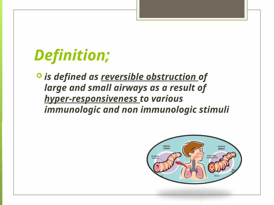
Definition; is defined as reversible obstruction
of large and small airways as a result of hyper-responsiveness to various immunologic and non immunologic stimuli

Risk factors Environment; inhaled allergens respiratory viral infections chemical and biologic air pollutants
Biological and genetic factors

EARLY CHILDHOOD RISK FACTORS FOR PERSISTENT ASTHMA
Parental Asthma History of other allergy Sever lower respiratory tract infection Wheezing apart from colds Male gender Low birth weight Exposure to tobacco smoke Reduced lung function at birth

Pathogenesis;
Infants are more susceptible to airway obstruction due to;
Smaller airway size. Lower elastic recoil of the lung. Decreased smooth muscle support
of the small airways. Relative mucus gland hyperplasia. Decreased collateral channels of
ventilation between the alveoli

Asthma triggers; viral infections Aeroallergens (animal dander, dust mites) Seasonal aeroallergens ( Pollens) tobacco smoke Air pollutants;( Ozone, Sulfur dioxide) Strong or noxious odors Cold , dry air Exercise Crying, laughter, hyperventilation

Aggravating factors in asthma Gastroesophageal reflux Rhinitis Sinusitis

Clinical Manifestations Intermittent dry coughing and expiratory
wheezing are the most common symptoms
shortness of breath and chest tightness symptoms can be worse at night Daytime symptoms, often linked with
physical activities symptoms can be subtle ; (limitation of
physical activities, general fatigue ,or sleep disturbance)
Lack of improvement with bronchodilator and corticosteroid is inconsistent with the diagnosis of asthma

Examination; expiratory wheezing and a prolonged
expiratory phase Decreased breath sounds in some of the
lung fields, commonly the right lower posterior lobe, are consistent with regional hypoventilation owing to airways obstruction.
Crackles and rhonchi can sometimes be heard, resulting from excess mucus production and inflammatory exudate in the airways
Differential diagnosis; Chronic rhinitis Sinusitis Adenoidal or tonsillar hypertrophy Laryngotracheobronchomalacia Laryngotracheobronchitis Vocal cord dysfunction Foreign body aspiration Viral bronchiolitis Gastroesophageal reflux Tuberculosis Heart failure

Diagnosis; Mainly clinical Pulmonary function test Radiography Skin test Radioallergosorbent (RASTA) test
Tidal volume
Is the lung volume representing the normal amount of air displaced between normal inhalation and exhalation with no extra effort applied
Pulmonary function test

Total lung capacity; is the maximum volume to which
the lung can be expanded with the greatest possible inspiratory effort;
TLC= VC +RV

Vital capacity; Is the maximum amount of air the
person can expel from the lung after first filling the lung to the maximum extent

Functional residual volume and residual volume;Functional residual capacity; is
the volume of air present in lung at the end of passive expiration
Residual volume; is the amount of air left in the lung after maximum exhalation

Spirometry objective measure of airflow limitation Measures the forced expiratory volume
in one second. usually feasible in children > 6 yr FEV1 is within 5% on 3 attempts, then
the highest FEV1 effort of the 3 is used Normal values are based on height,
gender, and ethnicity. typically decreases in asthmatics by >
15% after exercise challenge

Pulmonary function test; useful in evaluating an older
child’s functional status, although they rarely yield an etiological diagnosis
Abnormalities described in form of obstructive or restrictive lung disease

obstructive disease; there is low flow rate and increased residual volume RV and functional residual volume FRV
Restrictive disease; there is low vital capacity and total lung capacity with relative preservation of flow rates and FRV.
A significant increase in pulmonary function test after bronchodilator therapy indicate reactive airway disease
Chest X-ray;
often appear to be normal, aside of subtle nonspecific finding of hyperinflation and peribronchial thickening.
CXR is not routine in asthmatic patients

Indications of chest x-ray;1- First attack of bronchospasm.2- localized wheeze or other finding on osculation.3- High grade fever or toxic appearance.4- Sudden deterioration in patients condition despite treatment with signs suspecting pneumothorax.5- suspicion of foreign body.

Skin test;
There are 2 methods;1-Prick or puncture technique.2-Intradermal injection technique

Radioallergosorbent (RASTA) test;
This is an in vitro test to determine the presence of antigen specific IgE in serum of the patient.

asthma masqueraders
aspiration pneumonitis bronchiolitis obliterans cystic fibrosis allergic bronchopulmonary mycoses
(aspergillosis) ciliary dyskinesias immune deficiencies