Embed Size (px)
Citation preview

Assumptions:
1) Matter is composed of discrete particles (i.e. electrons, nucleus)2) Distance between particles >> particle size3) X-ray photons are small particles
Interact with body in binomial processPass through body with probability pInteract with body with probability 1-p (Absorption or scatter)
4) No scatter photons for now (i.e. receive photons at original energy or not at all.

The number of interactions (removals) number of x-ray photons and ∆x
∆N = -µN∆x
µ = linear attenuation coefficient (units cm-1)
N |∆x|N + ∆N
Id (x,y) = ∫ I0 () exp [ -∫ u (x,y,z,) dz] d
Integrate over and depth.
If a single energy I0() = I0 ( - o),
If homogeneous material, then µ (x,y,z, 0) = µ0
Id (x,y) = I0 e -µ0l
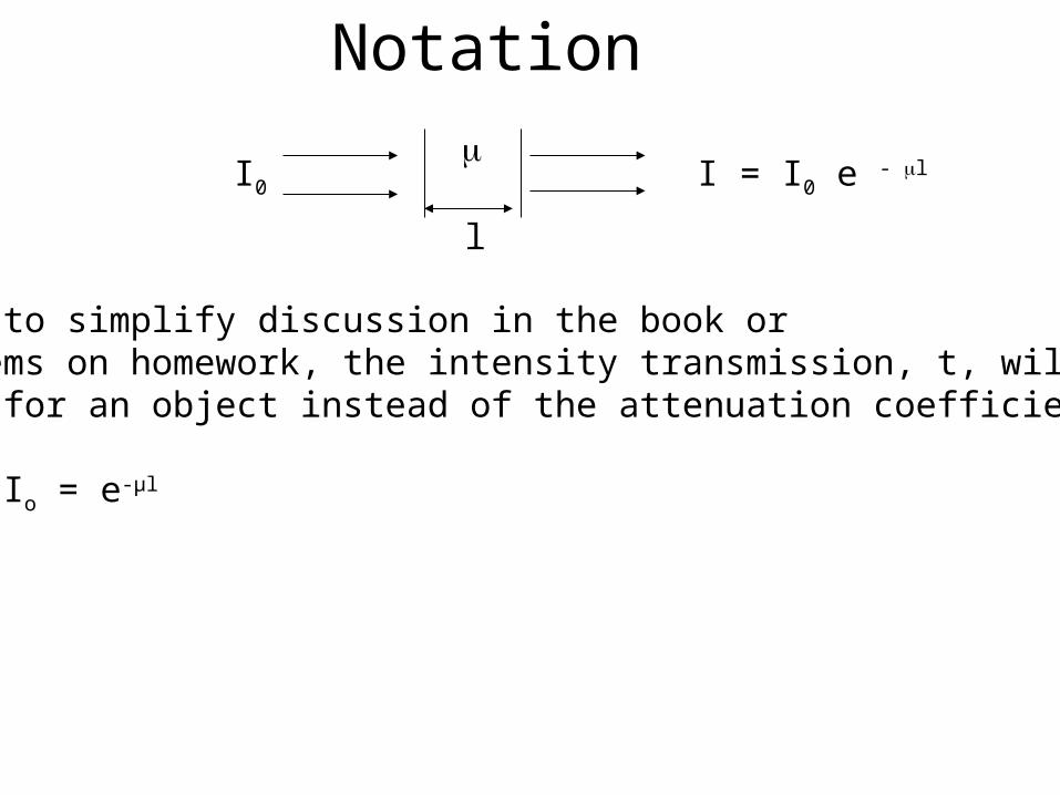
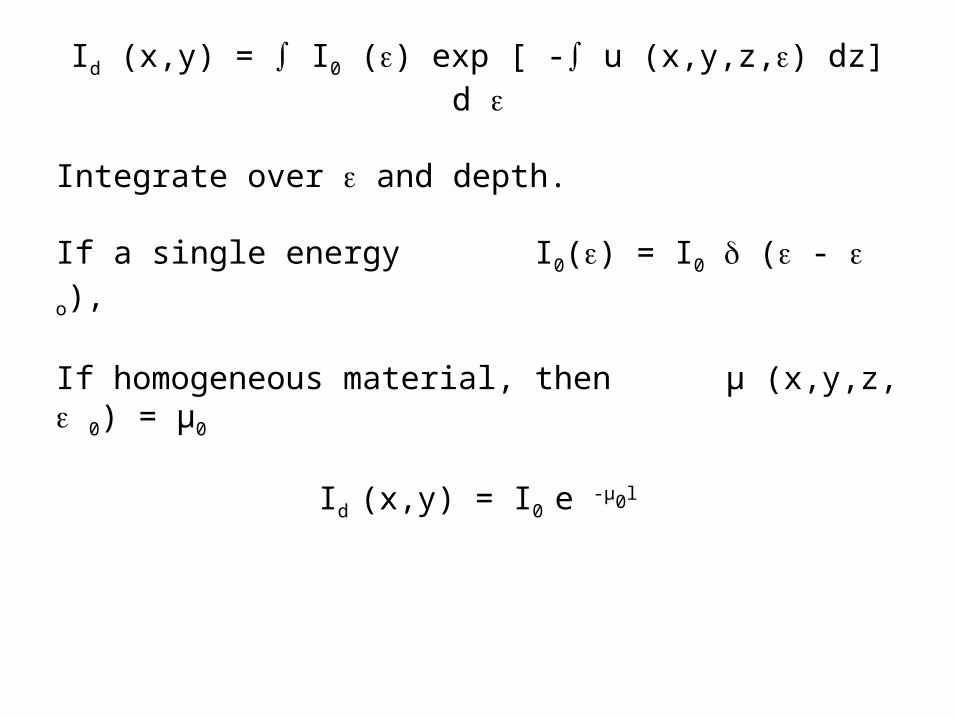
Often to simplify discussion in the book orproblems on homework, the intensity transmission, t, will be given for an object instead of the attenuation coefficient
t = I/Io = e-µl
I0
l
I = I0 e - l
Notation

Accelerate electrons towards anode. Three types of events can happen.
Typically Tungsten TargetHigh melting pointHigh atomic number
Andrew Webb, Introduction to Biomedical Imaging, 2003, Wiley-Interscience.
1. Collision events -> heat2. Photoelectric effect3. Braking of electron by
nucleus creates an x-ray (Bremstrahlung effect)

There are different interactions creating X-ray photons between the accelerated electrons and the target. Maximum energy is created when an electron gives all of its energy, 0 , to one photon. Or, the electron can produce n photons, each with energy 0/n. Or it can produce a number of events in between. Interestingly, this process creates a relatively uniform spectrum. Power output is proportional to 0 2
0 Photon energy spectrum
Intensity= nh
Thin Target X-ray Formation

Gun
X-rays
Thick Target X-ray Formation
We can model target as a series of thin targets. Electrons successively loses energy as they moves deeper into the target.
Each layer produces a flat energy spectrum with decreasingpeak energy level.
0
RelativeIntensity

Thick Target X-ray Formation
In the limit as the thin target planes get thinner, a wedge shaped energy profile is generated.
RelativeIntensity
0
Again, 0 is the energy of the accelerated electrons.

Lower energy photons are absorbed with aluminum to block radiation that will be absorbedby surface of body and won’t contribute to image.
The photoelectric effect(details coming in attenuation section) will create significant spikes of energy when accelerated electrons collide with tightly bound electrons, usually in the K shell.
Thick Target X-ray Formation
Andrew Webb, Introduction to Biomedical Imaging, 2003, Wiley-Interscience. (
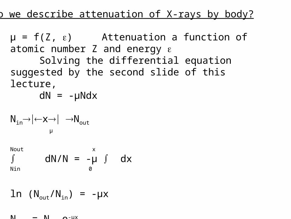
µ = f(Z, ) Attenuation a function of atomic number Z and energy
Solving the differential equation suggested by the second slide of this lecture,
dN = -µNdx
Ninx Nout
µ
Nout
x
∫ dN/N = -µ ∫ dxNin 0
ln (Nout/Nin) = -µx
Nout = Nin e-µx
How do we describe attenuation of X-rays by body?

If material attenuation varies in x, we can write attenuation as u(x)
Nout = Nin e -∫µ(x) dx
Io photons/cm2
(µ (x,y,z))Id (x,y) = I0 exp [ -∫ µ(x,y,z) dz]
Assume: perfectly collimated beam ( for now), perfect detector no loss of resolution
Actually recall that attenuation is also a function of energy , µ = µ(x,y,z, ). We will often assume a single energy source, I0
= I0(). After analyzing a single energy, we can add the effects of other energies by superposition.
Id (x,y)
Detector Plane

Diagnostic Range
50 keV < E < 150 keV ≈ 0.5%
Rotate anode to prevent melting
1. Current Units are in mA
2. Time Units · sec
What parameters do we have to play with?
3. Energy ( keV)

µ/p cm2/gm
We simply remultiply by the density to return to the linearattenuation coefficient. For example:t = e- (µ/p)pl
Mixture
µ/p = (µ1/p1) w1 + (µ2/p2) w2 + …
w0 = fraction weight of each element
Since mass is providing the attenuation, we will consider the linear attenuation coefficient, µ, as normalized to the density of the object first. This is termed the mass attenuation coefficient.

1. Coherent scatter or Rayleigh (Small significance)
2. Photoelectric absorption
3. Compton Scattering – Most serious significance

Coherent Scattering - Rayleigh
µ/p 1/2
••••
••
•
•
•
•
Coherent scattering varies over diagnostic energy range as:

log /
log ( Photon energy)K-edge
1p 3
Photoelectric effect varies over diagnostic energy range as:
Andrew Webb, Introduction to Biomedical Imaging, 2003, Wiley-Interscience.

Longest photoelectron range 0.03 cm
Fluorescent radiation example:Calcium 4 keV Too low to be of interest.
Quickly absorbedItems introduced to the body:
Ba, Iodine have K-lines close to region of diagnostic interest.
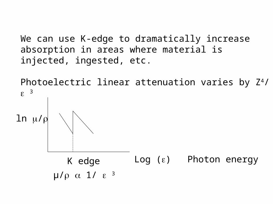
We can use K-edge to dramatically increase absorption in areas where material is injected, ingested, etc.
Photoelectric linear attenuation varies by Z4/ 3
ln /
Log () Photon energyK edge
µ/ 1/ 3

Interaction of photons and electrons produce scattered photons of reduced energy.
When will this be a problem?Is reduced energy a problem?Is change in direction a problem?
EOuterShellelectron
E’ photon
v Electron (“recoil”)

Satisfy Conservation of Energy and Momentum
1) (m-mo = electron mass : relativistic effects)
Conservation of Momentum
2)
3)
20 )( cmmEE
20 )/(1/ cvmm
)cos()cos( mvc
E
c
E
sinsin0 mvc
E

Energy of recoil or Compton electron
can be rewritten as h = 6.63 x 10⁻³⁴ Jsec eV = 1.62 x 10⁻¹⁹ J
mo = 9.31 x 10⁻³¹ kg
∆ = h/ moc (1 - cos ) = 0.0241 A0 (1 - cos )∆ at = π = 0.048 Angstroms
EEE
20
)cos1(1cmhv
hh
Energy of Compton photon
1
EE
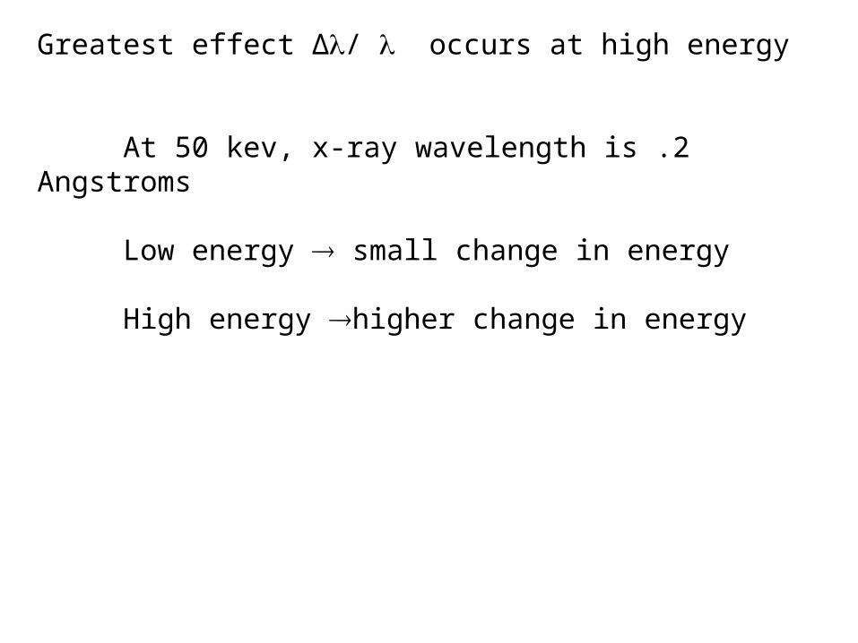
Greatest effect ∆/ occurs at high energy
At 50 kev, x-ray wavelength is .2 Angstroms
Low energy small change in energy
High energy higher change in energy

µ/ electron mass density
Unfortunately, almost all elements have electron mass density ≈ 3 x 10 23 electrons/gram
Hydrogen (exception) ≈ 6.0 x 10²³ electrons/gram
Mass attenuation coefficient (µ/) for Compton scattering isZ independent
Compton Linear Attenuation Coefficient µ p
Avg atomic number for Bone ~ 20Avg atomic number for body 7 or 8

Rayleigh, Compton, Photoelectric are independent sources of attenuation
t = I/I0 = e-µl = exp [ -(uR + up + uc)l]
µ () ≈ pNg { f() + CR (Z2/ 1.9) + Cp (Z3.8/ 3.2)} Compton Rayleigh Photoelectric
Ng electrons/gram ( electron mass density)So Ng is electrons/cm3
Ng = NA (Z/A) ≈ NA /2 (all but H) A = atomic mass
f() = 0.597 x 10⁻²⁴ exp [ -0.0028 (-30)]for 50 keV to 200 keV in keV

Attenuation Mechanisms
Curve on left shows how photoelectric effects dominates atlower energies and how Compton effect dominates at higher energies.Curve on right shows that mass attenuation coefficient varies little over100 kev. Ideally, we would image at lower energies to create contrast.
Andrew Webb, Introduction to Biomedical Imaging, 2003, Wiley-Interscience.

Photoelectric vs. Compton Effect
Macovski, Medical Imaging Systems, Prentice-Hall
The curve above shows that the Compton effect dominates at higher energy values as a function of atomic number.Ideally, we would like to use lower energies to use the higher contrast available with The photoelectric effect. Higher energies are needed however as the body gets thicker.