Embed Size (px)
Citation preview
●
w●
m8S●
aknpftS(ff●
db(wtrn9�a
A
mbCtOntEQNV(
OVD
0d
Associations of Birth Weight With Ocular Biometry,Refraction, and Glaucomatous Endophenotypes: The
Australian Twins Eye Study
CONG SUN, ANNE-LOUISE PONSONBY, SHAYNE A. BROWN, LISA S. KEARNS, JANE R. MACKINNON,JULIE M. BARBOUR, JONATHAN B. RUDDLE, ALEX W. HEWITT, MARGRET J. WRIGHT,
NICHOLAS G. MARTIN, TERENCE DWYER, AND DAVID A. MACKEY
mIb●
ssstb
FweooFdlatlscrhmartb
tmpmrbWt
PURPOSE: To examine the relationship of birth weightith ocular measures in a Caucasian twin population.DESIGN: Cross-sectional study of 1498 twins (308onozygotic and 441 dizygotic pairs) aged between 5 to0 years participating in the Australian Twins Eyetudy.METHODS: All participants underwent ophthalmic ex-
mination including bilateral cycloplegic autorefraction,eratometry, interpupillary distance (IPD), central cor-eal thickness, intraocular pressure (IOP), and retinalhotography. Birth weight and gestation were obtainedrom a self-administered questionnaire. A subset of thewins also participated in the Tasmanian Infant Healthtudy (288) and the Childhood Blood Pressure Study184), which collected data on birth parameters allowingor verification of data. Linear mixed models were usedor the main analysis.
RESULTS: Both the within-pair (�w 0.27, 95% confi-ence interval [CI] 0.15, 0.38 mm per kg increase inirth weight, P < .001) and between-pair associations�B 0.22, 95% CI 0.08, 0.35, P � .002) of birth weightith axial length were significant and of similar magni-
ude (difference in effect, P � .56), after adjusting forelevant confounders. In contrast, birth weight wasegatively associated with corneal curvature (�w �0.82,5% CI �1.09, �0.55 diopters per kg increase; �B
0.69, 95% CI �0.98, �0.41, both P < .001). Thesessociations remained significant within dizygotic and
Supplemental Material available at AJO.com.ccepted for publication Jun 19, 2010.From the Centre for Eye Research, University of Melbourne, Depart-ent of Ophthalmology, Royal Victorian Eye and Ear Hospital, Mel-
ourne, Australia (C.S., L.S.K., J.B.R., A.W.H., D.A.M.); Murdochhildren’s Research Institute, Melbourne, Australia (C.S., A.L.P., T.D.);
he Menzies Institute (A.L.P., S.A.B., T.D.), and the Department ofphthalmology (D.A.M.), Royal Hobart Hospital, University of Tasma-ia, Hobart, Australia; the Department of Ophthalmology, Royal Hospi-al for Sick Children, Glasgow, United Kingdom (J.R.M.); Launcestonye Institute and the Eye Hospital, Launceston, Australia (J.M.B.);ueensland Institute of Medical Research, Brisbane, Australia (M.J.W.,.G.M.); and the Lions Eye Institute, Centre for Ophthalmology andisual Science, University of Western Australia, Perth, Australia
D.A.M.).Inquiries to Professor David A. Mackey, Lions Eye Institute, Centre forphthalmology and Visual Science, University of Western Australia, 2
oerdun Street, Nedlands, Western Australia 6009, Australia; e-mail:[email protected]
© 2010 BY ELSEVIER INC. A002-9394/$36.00oi:10.1016/j.ajo.2010.06.028
onozygotic pairs. Refraction, anterior chamber depth,PD, IOP, and optic disc parameters are unrelated toirth weight.CONCLUSIONS: Consistent with previous studies in
ingleton children, lower birth weight is associated withhorter axial length and more curved corneas in this twintudy. This also adds new insights into the emmetropiza-ion process. (Am J Ophthalmol 2010;xx:xxx. © 2010y Elsevier Inc. All rights reserved.)
ETAL ORIGINS OF ADULT DISEASE HAS BEEN AN AREA
of active research in the past 2 decades.1,2 Manyepidemiologic studies have shown that lower birth
eight, shorter birth length, and smaller head circumfer-nce, regarded as markers for impaired intrauterine devel-pment, are associated with increased risk or earlier onsetf cardiovascular diseases, hypertension, and diabetes.1
urthermore, new research has produced preliminary evi-ence that intrauterine environment may also have long-asting impact on the development of ocular dimensionsnd retinal microcirculation. For example, recent popula-ion-based studies reported that smaller birth size such asow birth weight was associated with ocular traits such ashorter axial length,3,4 narrower vitreous chamber,3 moreurved corneas,3,4 larger cup-to-disc ratio,5 and narroweretinal arteriolar caliber.6 However, birth size seems toave little long-term effect on refractive error such asyopia.3,4 Importantly, these investigations suggest that
dverse intrauterine growth may confer increased futureisk of diseases specifically affecting the eye (eg, glaucoma-ous optic neuropathy)5 as well as other parts of theody.1,2
To date, limited population-based studies have exploredhe relationship between birth parameters and oculareasures.3,5 Moreover, it is still unknown whether any
otential associations between birth parameters and oculareasures are confounded by shared genes or shared envi-
onmental factors (eg, maternal nutrition) that affect bothirth size and the development of ocular parameters.ithin-pair analysis in a twin population offers an oppor-
unity to disentangle these effects,6,7 and we are unaware
f any previous study examining this.LL RIGHTS RESERVED. 1
rmlt
●
(darrpw(aa2Adu
rdAaP1
emi1wg
●
fdsolftwgwc
wTwc
aw�
●
toctcruJ
duoacfico
●
wps
2
The purpose of this report is therefore to investigate theelationship between birth weight and a range of oculareasures (ocular biometry, refraction, and glaucoma-re-
ated endophenotypes) in a large Caucasian twin popula-ion.
METHODS
STUDY POPULATION: The Australian Twins Eye StudyATES), involving 2235 twins and nontwin siblings, wasesigned to investigate the relative influence of geneticnd environmental factors on a variety of ocular traitselated to glaucoma. The study design and details of sampleecruitment are described elsewhere.8 In brief, the studyopulation comprising predominantly Caucasian twinsere ascertained from the Tasmanian Infant Health Study
TIHS) cohort,9 the Brisbane Adolescent Twin Study,6,10
nd the Australia Twin Registry;11 through media appeal;nd from a school-recruitment approach during the period000 to 2008. All twins and their nontwin siblings in theTES answered a standardized questionnaire providing
etails of sociodemographic and medical information andnderwent a thorough ophthalmic examination.A total of 288 children participating in the ATES were
ecruited from the TIHS, which examined sudden infanteath syndrome in infants during the years 1988 to 1995.9
mong these children, 184 multiplets (born between 1991nd 1993) also participated in the Childhood Bloodressure Study that examined cardiovascular diseases in999.12
Of the total population of 2235 twin individuals, wexcluded 269 nontwin siblings, triplets, and twins withissing zygosity data; 349 persons who had no birth weight
nformation; and an additional 119 single twins, leaving498 twin individuals with known zygosity and birtheight, comprising 308 monozygotic (MZ) and 441 dizy-otic (DZ) twin pairs for this analysis.
BIRTH PARAMETERS ASSESSMENT: We collected in-ormation on birth weight, birth order, and gestationuration for all participants in the ATES from a detailedelf-administered questionnaire. Two hundred eighty-eightf the twins also had data relating to birth weight, birthength, head circumference, and gestational age extractedrom medical birth records as part of their involvement inhe TIHS, details of which have been published else-here.6 Briefly, last menstrual period was used to estimateestational duration. These data provided a means byhich the self-reported birth parameters in the ATESould be validated.
Reliability assessment of 283 individuals with both birtheight and gestational age data in the ATES and theIHS showed very high agreement between the 2 studies,ith an intraclass correlation coefficient of 0.978 (95%
onfidence interval [CI], 0.972–0.982) for birth weight, tAMERICAN JOURNAL OF
nd 0.992 (0.990-0.994) for gestational age. Low birtheight was defined as �2500 g. Prematurity was defined as37 weeks gestation duration.13
MEASUREMENT OF OPTIC DISC PARAMETERS: Allwins and nontwin siblings had 10-degree stereoscopicptic disc–centered photographs taken (Nidek fundusamera 3-Dx/F; Nidek, Gamagori, Japan) after pupil dila-ion with tropicamide 1% or cyclopentolate 1% (forhildren). All fundus photographs were digitalized at highesolution (2102 � 1435 pixels, 2900 dpi, 36-bit color)sing a Nikon CoolScan scanner (Nikon Corp, Tokyo,apan).
Assessments of optic disc parameters, including opticisc, cup, and rim area, were performed by 2 trained graderssing a standardized grading program.14 Re-measurementf 50 randomly selected retinal photographs 3 monthspart showed high intragrader reproducibility, with intra-lass correlation coefficient (95% CI) of 0.94 (0.87-0.98)or optic disc area. The intergrader reliability was assessedn 73 randomly selected retinal images, and interclassorrelation coefficient (95% CI) was 0.75 (0.67–0.86) forptic disc area.
ZYGOSITY TESTING: Blood or mouth swab samplesere collected for DNA extraction in the ATES. Twinair zygosity was confirmed by up to 12 highly polymorphichort tandem repeats (STR), with an accuracy of more
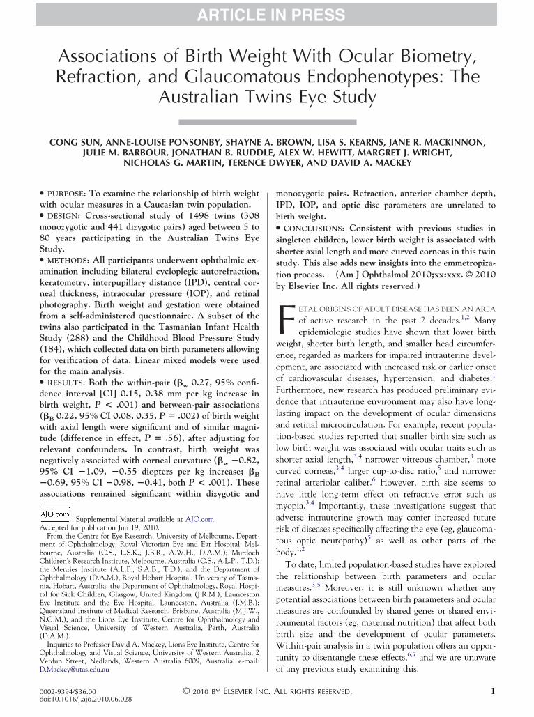
TABLE 1. Characteristics of Monozygotic and DizygoticTwin Pairsa
Characteristic
MZ Twins
(n � 616; 308
Pairs)
DZ Twins
(n � 882; 441
Pairs) P Valuec
Demographic factors
Age, yearsa 19.0 17.0 �.001
Male gender, % 31.2 46.4 �.001
Birth parameters
Birth weight, kg 2.4 2.5 �.001
Low birth weight, % 54.4 41.7 �.001
Small for gestational age, % 18.6 13.8 .16
Birth length, cma,b 47.0 47.0 .03
Head circumference, cma,b 33.0 33.0 .002
Prematurity, % 42.2 35.5 .004
Anthropometric measures
Height, m 1.6 1.7 .19
Body mass index, kg/m2 22.8 21.8 .57
DZ � dizygotic; MZ � monozygotic.aData show crude means (or median where indicated with %.bData available for only 50 MZ and 75 DZ twin pairs.cP � .05, represents statistically significant difference in
means or proportions, adjusted for age and gender, except for
age and male gender, and rank sum test for birth length and
head circumference.
han 99%.15
OPHTHALMOLOGY MONTH 2010
●
V
odt5FsssdsTptc((iGlc
cyrsauHc
●
ic
rIrcec0om
igoswlM
laTazCa
wlwwc
Iw
V
MEASUREMENT OF OCULAR BIOMETRY AND OTHER
ARIABLES: Participants underwent a comprehensivephthalmic examination.8 Postcycloplegic interpupillaryistance (IPD) measure, central keratometry, and refrac-ive errors for both eyes were measured using a Humphery-98 automatic refractor (Carl Zeiss Meditec, Inc, Miami,lorida, USA) after pupil dilation. To assess refractiontatus, spherical equivalents were calculated using thetandard formula of the algebraic sum of the value of thephere and half the cylinder value (sphere � 0.5 cylin-er).16 After topical anesthesia, bilateral intraocular pres-ure (IOP) was measured for each participant using theONO-PEN XL (Reichert Ophthalmic Instruments, De-ew, New York, USA). Measurements of central cornealhickness were obtained from the average reading of theentral cornea using the Pachymeter Tomey SP 2000Tomey Corp, Nagoya, Japan) or Pachmate DGH 55DGH Technology, Inc, Exton, Pennsylvania, USA). Thentraocular lens (IOL) Master (Carl Zeiss, Oberkochen,ermany) was used to obtain ocular biometry (axial
ength, anterior chamber depth, and horizontal and verti-al corneal curvature).
Data for current height and weight in the ATES wereollected from a self-administered questionnaire. Forounger twins with no height and weight data, we used 184eadings available in the concurrent child blood pressuretudy,17 which measured child height in centimeters using
wall-mounted measuring tape and weight in kilogramssing a digital scale (SECA, model 782 2321009; Vogel &alke, Hamburg, Germany). Body mass index (BMI) was
alculated as kg/m2.
STATISTICAL ANALYSIS: A range of ocular measuresncluding axial length, anterior chamber depth, corneal
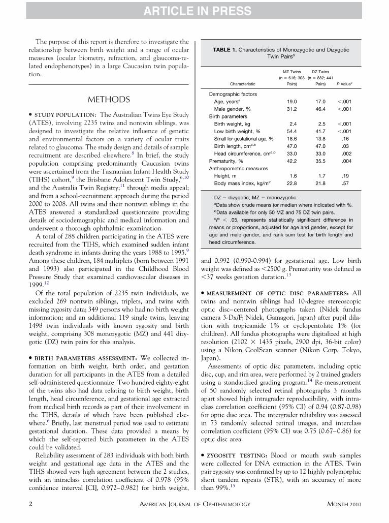
TABLE 2. . Correlation Between Ocular Outcom
Axial
Length
(mm)
Anterior
Chamber
Depth
(mm)
Corneal
Curvature
(diopters)
Axial length (mm) 1.00
Anterior chamber depth (mm) .41a 1.00
Corneal curvature (diopters) �0.57a .07 1.00
Central corneal thickness (�m) .06 �.02 �0.04
Refractive error (diopters) �0.16b �0.31a �0.43a
Mean intraocular pressure
(mm Hg)
0 0 .04
Optic disc area (mm2) .03 �0.18a �0.25a
Optic cup area (mm2) .12 �0.08 �0.20a
Area cup-to-disc ratio .13 �0.03 �0.14b
IPD measure (mm) .30a .04 �0.30a
IPD � interpupillary distance.aP �.001; b.05 � P �.001 (all other data, P �.05).
urvature, IPD measure (postcycloplegic), and glaucoma- S
BIRTH WEIGHT AND OCULAOL. XX, NO. X
elated endophenotypes such as central corneal thickness,OP, optic disc area, optic cup area, and optic cup-to-discatio were outcomes of interest and were all analyzed asontinuous variables. Refraction was measured as sphericalquivalent and was analyzed both as a continuous and aategorical trait (1 for spherical equivalent � median andfor those � median). Spearman correlations between eyeutcomes were obtained with Bonferonni adjustment forultiple comparisons.18
Standard linear regression was performed with “twins asndividuals,” initially controlling for age at examination andender.19 Logistic regression was performed for categoricalutcome such as refractive error. Multivariable linear regres-ion models were then constructed for those ocular outcomesith a significant association in the age-gender-adjusted
inear models (P � .05) (for more details, see Supplementalaterial at AJO.com).The main form of analysis was then conducted by using
inear mixed regression models, treating each twin pair ascluster to account for the correlated nature of the data.19
his model provided estimates of both within-pair (�w)nd between-pair (�B) associations. Stratified analysis byygosity was also performed using linear mixed models.ovariates included in the model were based on statistical
nd biological consideration of confounding.We also investigated the associations of birth weight
ith ocular biometry in preterm twins using multivariableinear regression, accounting for family structure. Finally,e investigated the potential interaction of risk factorsith gender. Where interactions were statistically signifi-ant (P � .01), stratified analyses were performed.
All analyses were performed for the right eye (except forOP, where the average of right and left eye measurementsas used because of variation of the measurement) using
ith a Bonferroni Correction for Multiple Testing
ntral
rneal
kness
m)
Refractive
Error
(diopters)
Mean
Intraocular
Pressure
(mm Hg)
Optic
Disc
Area
(mm2)
Optic
Cup
Area
(mm2)
Area
Cup-to-Disc
Ratio
IPD
(mm)
.00
.09 1.00
.11 �0.05 1.00
.03 .16b �0.09 1.00
.09 .03 .61a 1.00
.05 .07 .37a .95a 1.00
.04 .07 .05 .10 .13b .14b 1.00
es W
Ce
Co
Thic
(�
1
�0
0
0
�0
TATA 11.0 (Stata Corp, College Station, Texas, USA).
R MEASURES IN TWINS 3
(m
S
isTywpaMccrfrc
mcrccscreian
tap4(0
ar reg
etwe
4
See the online Supplemental Material at AJO.com forore details of the statistical analysis.)
RESULTS
ELECTED CHARACTERISTICS INCLUDING DEMOGRAPHIC
nformation, birth parameters, and anthropometric mea-ures of the study sample stratified by zygosity are shown inable 1. The median age of the whole study sample was 17ears (range, 5-80 years). MZ twins (n � 616; 308 pairs)ere more likely to be female and older, and a higherroportion were of low birth weight, small for gestationalge, and premature than DZ twins (n � 882; 441 pairs).Z and DZ twins had the same median birth length of 47
m, although MZ twins had a slightly greater but signifi-antly different range (33–51 weeks and 38–53 weeks,espectively, P � .03). Similarly, the median head circum-erence of 33 cm was the same in both twin types, but theange was significantly larger in DZ twins (27–36 cm)
TABLE 3. Overall Associations o
Ocular Measuresa �2.5 (703) 2.5–2.9 (506)
Axial length (mm) 23.13 (0.03) 23.19 (0.04
Corneal curvature (diopters) 43.87 (0.06) 43.69 (0.07
Central corneal thickness (�m) 544.82 (1.41) 544.39 (1.66
IPD measure (mm) 60.24 (0.16) 60.51 (0.19
Mean IOP (mm Hg) 16.00 (0.11) 15.93 (0.13
Optic disc area (mm2) 2.05 (0.02) 2.05 (0.02
Optic cup area (mm2) 0.43 (0.01) 0.44 (0.02
Area cup-to-disc ratio 0.20 (0.01) 0.20 (0.01
IOP � intraocular pressure; IPD � interpupillary distance.aMean (standard error) for ocular biometry and glaucomatous enbP value for trend, adjusted for age and gender derived from line
TABLE 4. Associations Between Birth Weight and Ax
Parameter
Mean (95% CI) Cha
MZ�DZ (n � 1498) P Value M
�c 0.25 (0.16, 0.34) �.001 0
�w 0.27 (0.15, 0.38) �.001 0
�B 0.22 (0.08, 0.35) .002 0
Test for differencec — .56
�c � common (twins as individuals) regression coefficient; �w � wi
DZ � dizygotic; MZ � monozygotic.aLinear mixed regression models were used. Twins were treated
allowed for different correlations in monozygotic and dizygotic pairbCI denotes confidence interval. Adjusted for age, gender, sphercData show the likelihood ratio test for the heterogeneity of the b
ompared to MZ twins (23–37 cm) (P � .002). t
AMERICAN JOURNAL OF
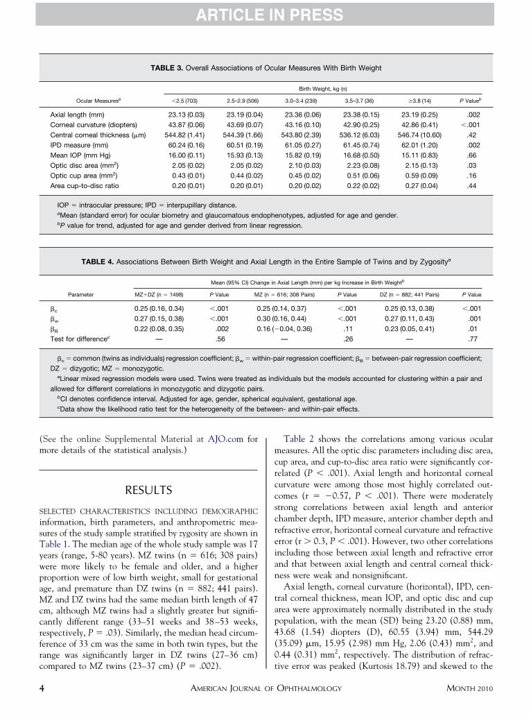
Table 2 shows the correlations among various oculareasures. All the optic disc parameters including disc area,
up area, and cup-to-disc area ratio were significantly cor-elated (P � .001). Axial length and horizontal cornealurvature were among those most highly correlated out-omes (r � �0.57, P � .001). There were moderatelytrong correlations between axial length and anteriorhamber depth, IPD measure, anterior chamber depth andefractive error, horizontal corneal curvature and refractiverror (r � 0.3, P � .001). However, two other correlationsncluding those between axial length and refractive errornd that between axial length and central corneal thick-ess were weak and nonsignificant.Axial length, corneal curvature (horizontal), IPD, cen-
ral corneal thickness, mean IOP, and optic disc and cuprea were approximately normally distributed in the studyopulation, with the mean (SD) being 23.20 (0.88) mm,3.68 (1.54) diopters (D), 60.55 (3.94) mm, 544.2935.09) �m, 15.95 (2.98) mm Hg, 2.06 (0.43) mm2, and.44 (0.31) mm2, respectively. The distribution of refrac-
lar Measures With Birth Weight
Birth Weight, kg (n)
3.0–3.4 (239) 3.5–3.7 (36) �3.8 (14) P Valueb
23.36 (0.06) 23.38 (0.15) 23.19 (0.25) .002
43.16 (0.10) 42.90 (0.25) 42.86 (0.41) �.001
543.80 (2.39) 536.12 (6.03) 546.74 (10.60) .42
61.05 (0.27) 61.45 (0.74) 62.01 (1.20) .002
15.82 (0.19) 16.68 (0.50) 15.11 (0.83) .66
2.10 (0.03) 2.23 (0.08) 2.15 (0.13) .03
0.45 (0.02) 0.51 (0.06) 0.59 (0.09) .16
0.20 (0.02) 0.22 (0.02) 0.27 (0.04) .44
enotypes, adjusted for age and gender.
ression.
ngth in the Entire Sample of Twins and by Zygositya
Axial Length (mm) per kg Increase in Birth Weightb
616; 308 Pairs) P Value DZ (n � 882; 441 Pairs) P Value
.14, 0.37) �.001 0.25 (0.13, 0.38) �.001
.16, 0.44) �.001 0.27 (0.11, 0.43) .001
0.04, 0.36) .11 0.23 (0.05, 0.41) .01
— .26 — .77
air regression coefficient; �B � between-pair regression coefficient;
ividuals but the models accounted for clustering within a pair and
quivalent, gestational age.
en- and within-pair effects.
f Ocu
)
)
)
)
)
)
)
)
doph
ial Le
nge in
Z (n �
.25 (0
.30 (0
.16 (�
thin-p
as ind
s.
ical e
ive error was peaked (Kurtosis 18.79) and skewed to the
OPHTHALMOLOGY MONTH 2010
l0csr
ababartoam(ab(
rg(tpwmas0ciaps
D0
wolb
ttsGccawTbc�wsa
whPf0p1b9
wgw00ts
etwe
V
eft (skewness �2.22) and the median in the right eye wasD (range, �16.50–6.38). The distribution of anterior
hamber depth was peaked (Kurtosis 9.49) and slightlykewed to the left (skewness �1.65) and the median in theight eye was 3.65 mm (range, 1.65–4.46).
Using standard linear regression with each twin treateds an individual, Table 3 shows the associations betweenirth weight and ocular measures after controlling for agend gender. Birth weight was significantly associated withoth axial length and corneal curvature (both horizontalnd vertical) in this model. However, no significantelationship between birth weight and central cornealhickness was observed, nor between birth weight andther ocular measures including IPD, mean IOP, optic cuprea, or optic cup-to-disc area ratio. Birth weight was onlyarginally significantly associated with optic disc area
P � .03), but this association did not persist in furtherdjustment for other confounders. Neither anterior cham-er depth nor refractive error was related to birth weightP � .49 and P � .18, respectively, data not shown).
Table 4 presents the results from the linear mixedegression models for the associations between birth wei-ht and axial length fitted for the whole sample of twinsMZ � DZ) and separately by zygosity (MZ or DZ). Bothhe within-pair (�w 0.27, 95% CI 0.15, 0.38) and between-air (�B 0.22, 95% CI 0.08, 0.35) associations of birtheight with axial length were significant and of a similaragnitude, after adjusting for age, gender, spherical equiv-
lent, and gestational age. These associations remainedignificant even after adjustment for current height (�w
.28, 95% CI 0.15, 0.28; �B 0.20, 95% CI 0.05, 0.34) givenurrent height may be associated with axial length.20 Thismplies that the association between lower birth weightnd shorter axial length was not merely attributable toeople with lower birth weight growing up to be shorter intature.
The within-pair association was of similar magnitude inZ twins (�w 0.30, 95% CI 0.16, 0.44) and MZ twins (�w
TABLE 5. Associations Between Birth Weight and Corne
Parameter
Mean (95% CI) Change in
MZ � DZ (n � 1498) P Value M
�c �0.76 (�0.95, �0.56) �.001 �
�w �0.82 (�1.09, �0.55) �.001 �
�B �0.69 (�0.98, �0.41) �.001 �
Test for differencec — .53
�c � common (twins as individuals) regression coefficient; �w � wi
DZ � dizygotic; MZ � monozygotic.aLinear mixed regression models were used. Twins were treated
allowed for different correlations in monozygotic and dizygotic pairbCI denotes confidence interval. Adjusted for age, gender, currencData show the likelihood ratio test for the heterogeneity of the b
.27, 95% CI 0.11, 0.43). No significant differences of p
BIRTH WEIGHT AND OCULAOL. XX, NO. X
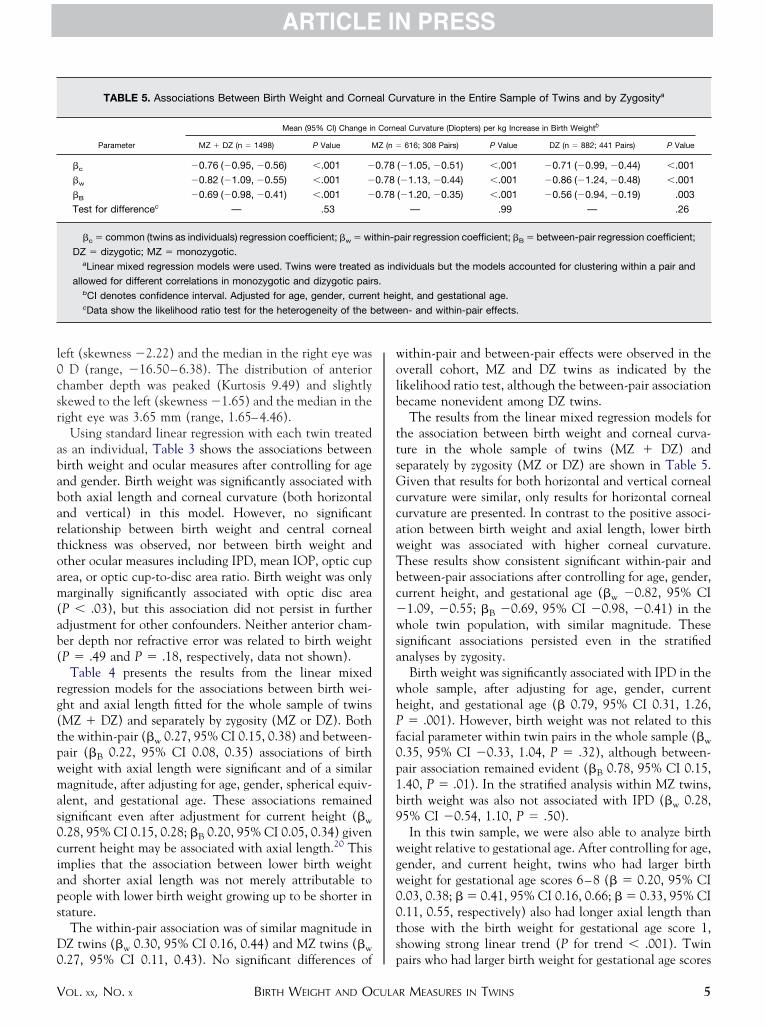
ithin-pair and between-pair effects were observed in theverall cohort, MZ and DZ twins as indicated by theikelihood ratio test, although the between-pair associationecame nonevident among DZ twins.The results from the linear mixed regression models for
he association between birth weight and corneal curva-ure in the whole sample of twins (MZ � DZ) andeparately by zygosity (MZ or DZ) are shown in Table 5.iven that results for both horizontal and vertical corneal
urvature were similar, only results for horizontal cornealurvature are presented. In contrast to the positive associ-tion between birth weight and axial length, lower birtheight was associated with higher corneal curvature.hese results show consistent significant within-pair andetween-pair associations after controlling for age, gender,urrent height, and gestational age (�w �0.82, 95% CI1.09, �0.55; �B �0.69, 95% CI �0.98, �0.41) in thehole twin population, with similar magnitude. These
ignificant associations persisted even in the stratifiednalyses by zygosity.
Birth weight was significantly associated with IPD in thehole sample, after adjusting for age, gender, currenteight, and gestational age (� 0.79, 95% CI 0.31, 1.26,� .001). However, birth weight was not related to this
acial parameter within twin pairs in the whole sample (�w
.35, 95% CI �0.33, 1.04, P � .32), although between-air association remained evident (�B 0.78, 95% CI 0.15,.40, P � .01). In the stratified analysis within MZ twins,irth weight was also not associated with IPD (�w 0.28,5% CI �0.54, 1.10, P � .50).In this twin sample, we were also able to analyze birth
eight relative to gestational age. After controlling for age,ender, and current height, twins who had larger birtheight for gestational age scores 6–8 (� � 0.20, 95% CI.03, 0.38; � � 0.41, 95% CI 0.16, 0.66; � � 0.33, 95% CI.11, 0.55, respectively) also had longer axial length thanhose with the birth weight for gestational age score 1,howing strong linear trend (P for trend � .001). Twin
rvature in the Entire Sample of Twins and by Zygositya
al Curvature (Diopters) per kg Increase in Birth Weightb
616; 308 Pairs) P Value DZ (n � 882; 441 Pairs) P Value
(�1.05, �0.51) �.001 �0.71 (�0.99, �0.44) �.001
(�1.13, �0.44) �.001 �0.86 (�1.24, �0.48) �.001
(�1.20, �0.35) �.001 �0.56 (�0.94, �0.19) .003
— .99 — .26
air regression coefficient; �B � between-pair regression coefficient;
ividuals but the models accounted for clustering within a pair and
ght, and gestational age.
en- and within-pair effects.
al Cu
Corne
Z (n �
0.78
0.78
0.78
thin-p
as ind
s.
t hei
airs who had larger birth weight for gestational age scores
R MEASURES IN TWINS 5

agp
bTsaP
I
pwamtoalotmpfdmupcagabwtaceo
srsswswowbafc
fsCclbaadftlac
lriepdtscwasSbbvcfiic
ccbdostc
tatsteboo
6
lso had flatter corneas than those with birth weight forestational age score 1 (P for trend � .001). These trendsersisted in the stratified analyses by zygosity.There was no gender difference in the relationships
etween birth weight and any ocular biometry measures.he significant associations of lower birth weight with
horter axial length (� 0.23, 95% CI 0.07, 0.39, P � .006)nd more curved corneas (� �0.62, 95% CI �0.94, �0.31,� .001) remained in preterm twins (n � 555).
DISCUSSION
N THIS COHORT COMPRISING 308 MZ AND 441 DZ TWIN
airs who participated in the ATES, we showed that twinsith lower birth weight tended to have shorter axial lengthnd more steeply curved corneas. These associations re-ained evident even in within-pair assessment of MZ
wins, after adjusting for age, gender, gestational age, andther relevant confounders. The between-pair effect of thessociations between lower birth weight and shorter axialength and more curved corneas were also significant andf similar magnitude to the within-pair effect. Our studyhus supports the hypothesis that impaired fetal develop-ent has a long-term effect on ocular biometry measures,
ossibly linked to specific individual factors (eg, differentetal nutrient supply lines),19 independent of possibleeterminants that would be constant across twin pairs (eg,aternal nutrition, general maternal health, and any other
nmeasured shared factors across twin pairs). The within-air associations of birth weight with axial length andorneal curvature were evident even among MZ twinslone, indicating that genetic differences in the inheritedenome are unlikely to explain these associations.21 Thesessociations persisted in supplementary analyses of twinsorn prematurely, and remained significant when birtheight for gestational age was used as a parameter to assess
he birth size effect that was independent of gestationalge. However, birth weight was not related to anteriorhamber depth, refraction status, IPD, and glaucomatousndophenotypes (eg, central corneal thickness, IOP, andptic disc measures) in this twin study.The current study, to our knowledge, is the first twin
tudy to investigate the association of birth size with aange of ocular measures. Our study demonstrates thatmaller birth size (eg, birth weight) is associated withhorter axial length and more curved corneas in twins,hich is in line with the findings reported in 2 recent
tudies from singleton schoolchildren populations.3,4 Ourithin-pair analysis in the present cohort extends thesebservations to twins and adds further insights intohether the associations of birth weight with oculariometric structure found in previous studies in childrenre attributable to shared environment (eg, maternalactors), common genetic factors, or other pathways asso-
iated with twin individuals. Thus, our findings provide cAMERICAN JOURNAL OF
urther insights into the recent studies performed iningleton schoolchildren populations, one in Singaporehinese individuals (7–9 years)3 and another in an ethni-
ally diverse (predominately Caucasian) Australian popu-ation (6-year-olds).4 Given that twins are more likely toe born prematurely than singletons and maternal factorsssociated with gestation duration may differ between twinnd singleton pregnancies,22 within-pair analysis (stan-ardized with gestation duration) and using birth weightor gestational age for analysis thus add important evidencehat these associations were independent of gestationength, suggesting that the findings from this twin study arepplicable beyond twins with the gestation profile of thisohort.
Our twin study shows that birth weight appears to haveittle effect on refraction status, which is consistent witheports from the singleton pediatric populations,3,4 provid-ng strong evidence for the hypothesis that compensatorymmetropization occurs to maintain the optimal refractiveower although impaired fetal growth may alter the ocularimensions.3,23 The finding that birth weight is not relatedo anterior chamber depth is well supported by the 2 recenttudies from singleton schoolchildren populations.3,4 Theurrent twin study, however, adds no evidence that birtheight is associated with any optic disc dimensions such asrea, or cup area, or cup-to-disc area ratio, and thus does notupport the findings from a cross-sectional analysis from theydney Childhood Eye Study, which identified an associationetween smaller birth size (eg, lower birth weight, shorterirth length, and smaller head circumference) and decreasedertical disc diameter, increased cup diameter, and largerup-to-disc ratio.5 This discrepancy clearly implicates thatuture work is required in this area. Nevertheless, our findings consistent with an animal study suggesting no long-termmpact of fetal growth restriction on the diameter of ganglionell axons in the optic nerve.24
In a 18-year follow-up study, which consisted of 302hildren with birth weight less than 2000 g and 237 full-termontrols (only 137 of 537 were followed for 18 years), lowirth weight was found to permanently compromise theevelopment of facial parameters such as IPD.25 In contrast,ur within-pair association of smaller birth weight withhorter IPD became nonsignificant, particularly within MZwin pairs, indicating that this association may likely beonfounded by some shared genetic factors.
Strengths of our study include its large sample of twins,he collection of extensive ocular measures, and the use ofstandardized protocol to measure the optic disc parame-
ers from digitized photographs. Findings from our studyhould be interpreted within the context of several poten-ial limitations. First, residual confounding may partlyxplain some of the associations, particularly for theetween-pair assessments. Our analyses also could not ruleut the possibility that the within-pair birth weight andcular biometry (eg, axial length, corneal curvature) asso-
iations observed may also reflect postnatal environmentalOPHTHALMOLOGY MONTH 2010
iSgnabspwtaamnTnti
(aeb
sacffdIwcglt
TELLA(i(ADsoHPREi
V
nfluences or later individual-specific epigenetic change.econd, birth parameters other than birth weight andestational age (birth length and head circumference) wereot available in the ATES. Although a subset of the twinslso participated in the TIHS, which collected data onirth length and head circumference, the number is toomall to allow meaningful analysis with sufficient statisticalower. Third, the collection of variables such as birtheight and gestational age from self-administered ques-
ionnaires increases the likelihood of measurement error,nd thus may have weakened, at least to some extent, anyssociations we observed. Nevertheless, reliability assess-ent of the data collected from self-administered question-aires in the ATES and from medical birth records in theIHS was fairly high. Fourth, this regression analysis doesot provide full genetic analysis.19 Fifth, proper informa-ion on chorion type was not available to assist our
nterpretation.26,27 Finally, 54.5% of the twin participants r2205–2206.
1
1
1
1
1
1
BIRTH WEIGHT AND OCULAOL. XX, NO. X
816/1498) were under 17 years in the current study, andproportion of these children and adolescents who will
ventually develop refractive error in adulthood may note evident by this early stage.Consistent with previous observations in 2 singleton
chool-aged children populations, lower birth weight isssociated with shorter axial length and more curvedorneas in this twin population. These findings reflect thatetus-specific factors rather than shared maternal or geneticactors may influence the long-term development of ocularimensions. Refraction, anterior chamber depth, IPD,OP, and optic disc parameters are unrelated to birtheight. Although they may not be directly applicable tolinical settings, findings from this twin study add to therowing literature that impaired fetal growth may have aong-lasting effect on ocular biometry, and that despitehese major neonatal influences, emmetropization remains
elatively stable during later years of life.HE AUSTRALIAN TWIN REGISTRY IS SUPPORTED BY A NATIONAL HEALTH AND MEDICAL RESEARCH COUNCIL(NHMRC)nabling Grant (2004–2009), Australia. We also thank the following organizations for their financial support: Clifford Craig Medical Research Trust,aunceston, Australia; Ophthalmic Research Institute of Australia (ORIA), Sydney, Australia; Foundation for Children, Sydney, Australia; Peggy andeslie Cranbourne Foundation, Melbourne, Australia; NHMRC (Project Grant 2005 -2007) Australia; Jack Brockhoff Foundation, Melbourne,ustralia; National Eye Institute (NEI) Project Grant (2007-2010), Bethesda, Maryland, USA; and the American Health Assistance Foundation
AHAF), Maryland, USA. Centre for Eye Research Australia receives operational infrastructure support from the Victorian government. David Mackeys the recipient of the Pfizer Australia Senior Research Fellowship. Involved in conception and design (C.S., A.L.P., A.W.H., D.A.M.); data collectionC.S., S.A.B., L.S.K., J.R.M., J.M.B., J.B.R., A.W.H., D.A.M.); analysis and interpretation of the data and drafting of the manuscript (C.S., A.L.P.,.W.H., T.D., D.A.M.); and critical revision of the manuscript for important intellectual content (C.S., A.L.P., J.R.M., A.W.H., N.G.M., M.J.W., T.D.,.A.M.). Written informed consent was obtained from all participants or their legal guardians, with the participants’ assent prior to examination. This
tudy was approved by the Human Research ethics committees of the Royal Victorian Eye and Ear Hospital, the Royal Hobart Hospital and the Universityf Tasmania, and the Queensland Institute of Medical Research, as well as the Australian Twin Registry, and adhered to the tenets of the Declaration ofelsinki. The authors thank the participants in the ATES for their important contributions. We would also like to thank Fleur O’Hare, Sandra Staffieri, Johanoulsen, Justin Sherwin, Robert Macmillan, Byoung Sung Chu, Katherine Smallcombe, Olivia Bigault, Colleen Wilkinson, Robin Wilkinson, Rachael Adams,obyn Troutbeck, Jonathan Yeoh, Ya Ling Ma, Trent Roydhouse, Lindsey Scotter, Katarina Creese, Vishal Jhanji, Sonya Bennett, Christine Chen, Annldridge, Marlene Grace, Yingfeng Zheng, Jian Zhang, Mingguang He, and Amy Cohn for helping examine twins. In addition, we appreciate the assistance
n recruiting twins from Thanuja Gunasekera, Jenny Boadle, Kim Dorrell, Shyamali Dharmage, and John Hopper.
REFERENCES
1. Barker DJ. Fetal origins of coronary heart disease. BMJ1995;311(6998):171–174.
2. Barker DJ, Bagby SP. Developmental antecedents of cardio-vascular disease: a historical perspective. J Am Soc Nephrol2005;16(9):2537–2544.
3. Saw SM, Tong L, Chia KS, et al. The relation between birthsize and the results of refractive error and biometry measure-ments in children. Br J Ophthalmol 2004;88(4):538–542.
4. Ojaimi E, Robaei D, Rochtchina E, Rose KA, Morgan IG,Mitchell P. Impact of birth parameters on eye size in apopulation-based study of 6-year-old Australian children.Am J Ophthalmol 2005;140(3):535–537.
5. Samarawickrama C, Huynh SC, Liew G, Burlutsky G,Mitchell P. Birth weight and optic nerve head parameters.Ophthalmology 2009;116(6):1112–1118.
6. Sun C, Ponsonby AL, Wong TY, et al. Effect of birthparameters on retinal vascular caliber: the Twins Eye Studyin Tasmania. Hypertension 2009;53(3):487–493.
7. Dwyer T, Morley R, Blizzard L. Twins and fetal originshypothesis: within-pair analyses. Lancet 2002;359(9324):
8. Mackey DA, MacKinnon JR, Brown SA, et al. Twins EyeStudy in Tasmania (TEST): Rationale and methodology torecruit and examine twins. Twin Res Hum Genet 2009;12(5):441–454.
9. Dwyer T, Ponsonby AL, Couper D. Tobacco smoke exposureat one month of age and subsequent risk of SIDS—aprospective study. Am J Epidemiol 1999;149(7):593–602.
0. Wright MJ, Martin NG. Brisbane adolescent twin study:Outline of study methods and research projects. Aust J Psy-chol 2004;56(2):65–78.
1. Hopper JL. The Australian Twin Registry. Twin Res 2002;5(5):329–336.
2. Morley R, Carlin JB, Dwyer T. Maternal calcium supplemen-tation and cardiovascular risk factors in twin offspring. Int JEpidemiol 2004;33(6):1304–1309.
3. Organization UNCsFaWH. Low Birthweight: Country, Re-gional and Global Estimates. New York: UNICEF; 2004.
4. Morgan JE, Sheen NJ, North RV, Choong Y, Ansari E.Digital imaging of the optic nerve head: monoscopic andstereoscopic analysis. Br J Ophthalmol 2005;89(7):879–884.
5. Spitz E, Moutier R, Reed T, et al. Comparative diagnoses oftwin zygosity by SSLP variant analysis, questionnaire, and
dermatoglyphic analysis. Behav Genet 1996;26(1):55–63.R MEASURES IN TWINS 7

1
1
1
1
2
2
2
2
2
2
2
2
8
6. Toh T, Kearns LS, Scotter LW, Mackey DA. Post-cyclople-gia myopic shift in an older population. Ophthalmic Epide-miol 2005;12(3):215–219.
7. Dwyer T, Blizzard L, Morley R, Ponsonby AL. Within pairassociation between birth weight and blood pressure at age 8 intwins from a cohort study. BMJ 1999;319(7221):1325–1329.
8. Armitage P, Berry G, Matthews JNS. Stataistical Methods inMedical Research. 3rd ed. Oxford, UK: Blackwell ScientificPublications; 1994:422–436.
9. Carlin JB, Gurrin LC, Sterne JA, Morley R, Dwyer T.Regression models for twin studies: a critical review. Int JEpidemiol 2005;34(5):1089–1099.
0. Saw SM, Chua WH, Hong CY, et al. Height and itsrelationship to refraction and biometry parameters in Singa-pore Chinese children. Invest Ophthalmol Vis Sci 2002;43(5):1408–1413.
1. Morley R, Dwyer T. Studies of twins: what can they tell usabout the fetal origins of adult disease? Paediatr Perinat
Epidemiol 2005;19(Suppl 1):2–7.AMERICAN JOURNAL OF
2. Rolett A, Kiely JL. Maternal sociodemographic characteris-tics as risk factors for preterm birth in twins versus singletons.Paediatr Perinat Epidemiol 2000;14(3):211–218.
3. Grosvenor T, Goss DA. Role of the cornea in emmetropiaand myopia. Optom Vis Sci 1998;75(2):132–145.
4. Loeliger M, Duncan J, Louey S, Cock M, Harding R, Rees S.Fetal growth restriction induced by chronic placental insuf-ficiency has long-term effects on the retina but not the opticnerve. Invest Ophthalmol Vis Sci 2005;46(9):3300–3308.
5. Fledelius HC. Inhibited growth and development as perma-nent features of low birth weight. A longitudinal study of eyesize, height, head circumference, interpupillary distance andexophthalmometry, as measured at age of 10 and 18 years.Acta Paediatr Scand 1982;71(4):645–650.
6. Leon DA. The foetal origins of adult disease: interpreting theevidence from twin studies. Twin Res 2001;4(5):321–326.
7. Phillips DI, Davies MJ, Robinson JS. Fetal growth and thefetal origins hypothesis in twins—problems and perspectives.
Twin Res 2001;4(5):327–331.OPHTHALMOLOGY MONTH 2010
●
raasfama
pe
aoBp�tfdg
R
V
SUPPLEMENTAL MATERIAL: EXPANDEDMETHODS
STATISTICAL ANALYSIS: The first multivariable linearegression model was constructed for axial length, adjusted forge, gender, spherical equivalent, and gestational age. Welso performed additional adjustment for current height. Theecond multivariable linear regression model was constructedor corneal curvature, adjusted for age, gender, current height,nd gestational age. The third multivariable linear regressionodel was constructed for interpupillary distance, adjusted for
ge, gender, current height, and gestational age.We also used the Australian national birth weight
ercentiles by gestational age based on twin dataS1 to
stimate birth weight for gestational age, which may allowBIRTH WEIGHT AND OCULAOL. XX, NO. X
n assessment of the relationship between birth weight andcular measures that is independent of gestational age.irth weight for gestational age was divided into 8 differentercentile categories (�5, �5 to 10, �10 to 25, �25 to 50,50 to 75, �75 to 90, �90 to 95, and �95, corresponding
o birth weight for gestational scores 1 to 8). Birth weightor gestational score under the 10th percentile was used toefine small for gestational age and as a proxy for fetalrowth restriction.
REFERENCESoberts CL, Lancaster PA. National birthweight percentiles by
gestational age for twins born in Australia. J Paediatr Child
Health 1999;35:278–282.R MEASURES IN TWINS 8.e1

CShde
8
Biosketch
ong Sun, MD, PhD, is currently a research fellow based at Murdoch Children’s Research Institute, Melbourne, Australia.he received her medical degree from Nanjing University, following by a residency in ophthalmology. Dr Sun completeder MPH and then PhD at the University of Melbourne in 2010. Her PhD examined the genetic and environmentaleterminants of a novel marker for microvascular changes associated with systemic vascular diseases. Her research interestsxtend to cardiovascular epidemiology.
AMERICAN JOURNAL OF OPHTHALMOLOGY.e2 MONTH 2010
PwcgS
V
Biosketch
rofessor Mackey has extensively studied large pedigrees with Leber Hereditary Optic Neuropathy. Creating one of theorld’s largest glaucoma biobanks, his Glaucoma Inheritance Study in Tasmania has helped define phenotype-genotypeorrelations in myocilin and other glaucoma genes. The Twins Eye Study in Tasmania and Brisbane is investigating theenetic environmental basis on ocular biometry related to glaucoma and myopia. He also leads the Norfolk Island Eyetudy and the Western Australian Raine Eye Health Study.
BIRTH WEIGHT AND OCULAR MEASURES IN TWINSOL. XX, NO. X 8.e3