Embed Size (px)
Citation preview
PLANT HEALTH PROGRESS Vol. 16, No. 3, 2015 Page 115
Plant Health Research
Association of Diaporthe longicolla with Black Zone Lines on Mature Soybean Plants
Taylor R. Olson, Ahmed Gebreil, and Ana Micijevic, Department of Plant Sciences, South Dakota State University, Brookings 57007; Carl A. Bradley, Department of Crop Sciences, University of Illinois, Urbana 61801 (current address: Department of Plant Pathology, University of Kentucky Research and Education Center, Princeton 42445); Kiersten A. Wise, Department of Botany and Plant Pathology, Purdue University, West Lafayette, IN 47907; Daren S. Mueller, Department of Plant Pathology and Microbiology, Iowa State University, Ames 50011; Martin I. Chilvers, Department of Plant, Soil and Microbial Sciences, Michigan State University, East Lansing 48824; and Febina M. Mathew, Department of Plant Sciences, South Dakota State University, Brookings 57007
Accepted for publication 23 August 2015. Published 28 August 2015.
ABSTRACT
Olson, T. R., Gebreil, A., Micijevic, A., Bradley, C. A., Wise, K. A., Mueller, D. S., Chilvers, M. I., and Mathew, F. M. 2015. Association of Diaporthe longicolla with black zone lines on mature soybean plants. Plant Health Progress doi:10.1094/PHP-RS-15-0020.
In 2014, during a survey for soybean (Glycine max L.) diseases in Illinois, Indiana, Iowa, Michigan, and South Dakota, zone lines were observed on the lower stems of soybean plants. The survey was per-formed by sampling two to three fields per state. In each field, at least five plants exhibiting zone lines were collected. Isolations were made from the zone lines by plating 1-cm pieces on potato dextrose agar. A total of 90 isolates producing black stromata in concentric patterns and alpha conidia were tentatively identified as Diaporthe longicolla (Hobbs) Santos, Vrandecic and Phillips. DNA was extracted from the mycelia of 10 representative isolates and the identity was confirmed by sequencing
the internal transcribed spacer (ITS) region. Additionally, phylogenetic analysis combining translation elongation factor-1α and actin sequences was performed and the ten isolates grouped with the D. longicolla ex-type cultures in a well-supported clade (94% bootstrap support). A patho-genicity test was performed in the greenhouse by inserting D. longicolla-infested toothpicks into the lower stems of the soybean plants. To complete Koch’s postulates, D. longicolla was re-isolated from the zone lines of the inoculated plants and the pathogen identity was confirmed by sequencing the ITS gene.
INTRODUCTION Species of Diaporthe Nitschke cause Phomopsis seed decay,
pod and stem blight, and stem canker of soybean (Glycine max (L.) Merrill). While Diaporthe longicolla (Hobbs) Santos, Vrandecic and Phillips (syn. Phomopsis longicolla Hobbs) causes Phomopsis seed decay, D. sojae Lehman causes pod and stem blight. Stem canker of soybean is divided into northern stem canker caused by D. caulivora (Athow and Caldwell) Santos, Vrandecic and Phillips, and southern stem canker caused by D. aspalathi Janse van Rensburg, Castlebury and Crous. These diseases are generally observed on soybean after flowering in the growing season and can cause yield loss under favorable conditions (Hartman et al. 1999; Wrather et al. 1997).
Diaporthe spp. primarily have been described based on morphology and the characteristic disease symptoms they cause on their hosts. However, given the inter- and intra-species variability, morphological characteristics are inadequate or unreliable for differentiation of Diaporthe spp. (Udayanga et al. 2014). Recently, molecular phylogenies, especially those derived from DNA sequence analyses of the internal transcribed spacer (ITS) region, translation elongation factor-1α (EF-1α), and actin (ACT) gene regions, have been used to identify Diaporthe species (Mathew et al. 2015a; Udayanga et al. 2014; Santos et al. 2011).
In 2014, during a late-season survey of soybean diseases in
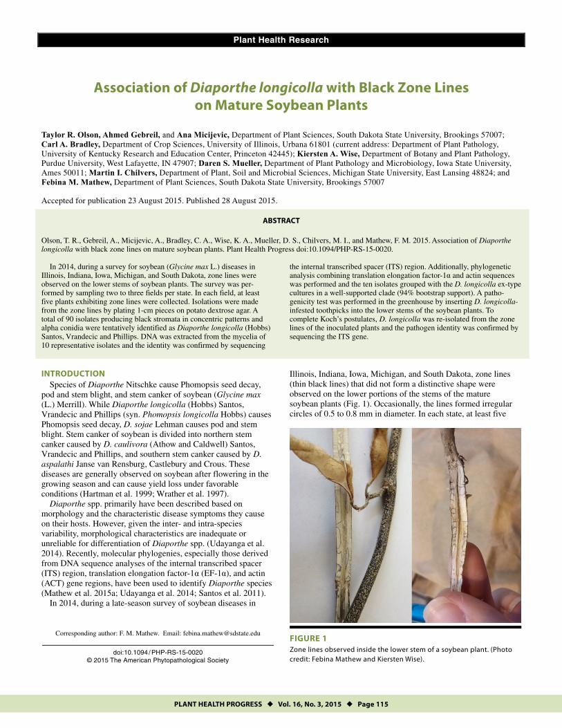
Illinois, Indiana, Iowa, Michigan, and South Dakota, zone lines (thin black lines) that did not form a distinctive shape were observed on the lower portions of the stems of the mature soybean plants (Fig. 1). Occasionally, the lines formed irregular circles of 0.5 to 0.8 mm in diameter. In each state, at least five
FIGURE 1 Zone lines observed inside the lower stem of a soybean plant. (Photo credit: Febina Mathew and Kiersten Wise).
Corresponding author: F. M. Mathew. Email: [email protected]
doi:10.1094 / PHP-RS-15-0020 © 2015 The American Phytopathological Society
PLANT HEALTH PROGRESS Vol. 16, No. 3, 2015 Page 116
plants were collected near or after maturation from two to three arbitrarily selected soybean fields and examined for stems containing zone lines (Table 1).
A study by Barr (1978) showed that Diaporthe spp. have “stromatic tissues composed of prosenchymatous hyphae mixed with substrate tissues in entostromatic regions, that often form dark marginal zones above in epidermal tissues and frequently deep in wood beneath perithecia or groups of perithecia.” More recently, zone lines were characterized as a symptom produced by either Macrophomina phaseolina (Tassi) Goid, Fusarium spp., or Diaporthe spp. on soybean (Ghissi et al. 2014; Zaccaron et al. 2014). The objective of this study was to establish the identity of the Diaporthe spp. associated with black zone lines produced on the lower stems of mature soybean plants in Illinois, Indiana, Iowa, Michigan, and South Dakota.
ISOLATION AND IDENTIFICATION OF THE CAUSAL AGENT Stem samples (approximately 90) collected from the five states
were washed in tap water for 2 min; and approximately 1 cm-long pieces were cut from the zone lines of the infected stems. The pieces from each stem sample were surface-disinfested in sodium hypochlorite (0.05%) and then ethanol (70%) for 1 min each, rinsed in sterile distilled water four times, and blotted between sterile filter paper. Four pieces were placed on potato dextrose agar (PDA; Becton, Dickinson and Company, Franklin Lakes, NJ) acidified to pH 4.5 with 85% lactic acid. Plates were incubated at 25oC for 7 to 10 days under fluorescent light with a photoperiod of 12 h daily. Mycelial colonies appeared white, dense, and floccose, and large black stromata were formed in concentric patterns or scattered. Alpha conidia exuded from the pycnidial ostiole in creamy-to-yellowish drops and were ellipsoid and biguttulate with an average length of 6.6 μm and width of 2.1 μm. Beta conidia and perithecia did not form on PDA. Ninety isolates (one isolate per symptomatic plant) from the five states were tentatively identified D. longicolla based on these morphological characteristics (Santos et al. 2011). From the 90 symptomatic stem samples collected from the five states, only D. longicolla was isolated from the zone lines on the soybean stems. The 90 D. longicolla isolates included 3 from Illinois, 20 from Indiana, 30 from Iowa, 30 from Michigan, and 7 from South Dakota.
To perform molecular identification of the causal agent, a total of 10 representative isolates were selected and DNA was extracted from the lyophilized mycelium scraped from the surface of 10 day old cultures growing on PDA using the FastDNA Spin
Kit (MP Biomedicals, Solon, OH) (Table 1). The Diaporthe isolates were identified to species by amplifying and sequencing of the internal transcribed spacer (ITS) regions using primers ITS1 and ITS2 (White et al. 1990). Approximately 600 bp of the ITS region was amplified from the 10 isolates. Forward and reverse sequences were assembled into contigs using BioEdit software (v7.2.5) (Hall 1999). Analysis of the edited sequences was performed using the Basic Local Alignment Search Tool Nucleotide (BLASTn) searches at the GenBank database (National Center for Biotechnology Information, http://www.ncbi.nlm.nih.gov). A BLASTn search of GenBank performed for the ITS sequences of the 10 isolates showed that the best match was P. longicolla (syn. D. longicolla) strain STAM-35 (GenBank Accession No. AY745021) from soybean in the United States with identities ranging from 98% to 100%. Phylogenetic analyses of the ITS sequences using maximum parsimony method placed the 10 isolates in the group containing isolates of D. longicolla and D. sojae from soybean in the United States (bootstrap value of 85%; data not presented).
In order to establish a well-resolved phylogeny and clarify the phylogenetic position, the 10 isolates were characterized by phylogenetic analyses of two gene fragments, which included EF1-α and ACT regions. The intron region of the EF1-α gene was amplified using primers EF1-728F and EF1-986R (Carbone and Kohn 1999). For the sequencing of the partial actin gene, frag-ments of ACT gene were amplified using the primers ACT-512F and ACT-783R (Carbone and Kohn 1999). All DNA samples were sequenced (GenScript USA Inc., Piscataway, NJ) using the respective primers. DNA sequences generated in this study have been deposited under GenBank Accession Nos. KR815475 to KR815494 and KT021543 to KT021552 (Table 1).
The EF1-α and ACT sequences of the 10 D. longicolla isolates were aligned using the default parameters of ClustalX (Thompson et al. 1997) and adjusted manually by visual examination using the Molecular Evolutionary Genetics Analysis (MEGA) software v6 (Tamura et al. 2013) prior to being exported as NEXUS files for subsequent analyses. The outgroup Chrysoporthella hodges-iana Gryzenhout and Wingfield (Isolate CMW9995) was obtained from GenBank (GQ290152 for EF1-α sequence and GQ290170 for ACT sequence). The 45 sequences in the combined data set (including the outgroup C. hodgesiana) comprised 688 bp of aligned sequence. Phylogenies based on maximum parsimony were inferred using MEGA, and heuristic searches for the most parsimonious trees were conducted using the tree bisection-
TABLE 1 Diaporthe longicolla isolates used in this study, the genes sequenced, and the corresponding GenBank Accession Nos.
Isolatesa,b Locationb Species Identityc
GenBank Accession Numbers
ITS EF1-α ACT IL1 Champaign County, IL D. longicolla KR815475 KR815485 KT021543 IL2 Champaign County, IL D. longicolla KR815476 KR815486 KT021544 IN1 Knox County, IN D. longicolla KR815477 KR815487 KT021545 IN2 Porter County, IN D. longicolla KR815478 KR815488 KT021546 IA1 Montgomery County, IA D. longicolla KR815479 KR815489 KT021547 IA2 Lee County, IA D. longicolla KR815480 KR815490 KT021548 MI1 Ingham County, MI D. longicolla KR815481 KR815491 KT021549 MI2 Ingham County, MI D. longicolla KR815482 KR815492 KT021550 SD1 Brookings County, SD D. longicolla KR815483 KR815493 KT021551 SD2 Miner County, SD D. longicolla KR815484 KR815494 KT021552
a A total of 10 isolates (two representative isolates from each state) were used for identification.
b IL = Illinois, IN = Indiana, IA = Iowa, MI = Michigan, and SD = South Dakota. c Species identity was established based on sequence analysis of the internal transcribed spacer region (ITS), elongation factor subunit 1-α (EF1-α) and
actin (ACT) gene regions.

PLANT HEALTH PROGRESS Vol. 16, No. 3, 2015 Page 117
regrafting (TBR) algorithm (Nei and Kumar 2000) with search level 1. The initial trees were obtained by random addition (10 replicates). Gaps were treated as missing data, and relative support for branches was estimated with 1,000 bootstrap replica-tions (Felsenstein 1985). Prior to the combined analyses, the concordance of the two gene datasets was evaluated with the partition-homogeneity test (Farris et al. 1994) implemented with PAUP* v4.0b10 (Sinauer Associates, Inc., Sunderland, MA; Swofford 2002) using 1,000 random repartitions (Felsenstein 1985), and with MAXTREES set to 5,000. The null hypothesis of congruence was rejected if P < 0.001 (Dettman et al. 2003). For the combined dataset, sequences were concatenated using Mesquite v2.75 (Maddison and Maddison 2011).
The EF1-α and ACT genes were combined for characterization of the 10 isolates based on the results of the partition-homo-geneity test (P = 0.002), indicating that the EF1-α and ACT trees reflect the same underlying phylogeny. The maximum parsimony analysis for the combined dataset resulted in five most-parsi-monious trees and a consensus tree was inferred from the five trees (length = 262; Fig. 2). The consistency index, retention index, and rescaled consistency index were 0.75, 0.92, and 0.74, respectively. The D. longicolla isolates from the five states formed a monophyletic group with the ex-type cultures CBS 179 and CBS 180 that was supported by a bootstrap value of 94% (Fig. 2).
The cultural morphology and DNA sequence analysis of the 10 isolates from the five states corresponded to the description of D. longicolla (syn. P. longicolla) (Santos et al. 2011).
PATHOGENICITY OF DIAPORTHE LONGICOLLA ISOLATES The D. longicolla isolates were assayed for pathogenicity using
a modified toothpick inoculation method (Ghissi et al. 2014; Keeling 1982). Seeds of a commercial soybean cultivar of relative maturity 1.8 (Monsanto Company, St. Louis, MO) were sown into a potting mix (Sunshine Mix #1, Sun Grow Horticulture Products, Belleview, WA) in 7.5-liter, circular, plastic pots, one seed per pot and grown for 4 weeks at 22°C in the greenhouse under a 14-h photoperiod with a light intensity of 450 μE/m2/s and watered daily. The trial was conducted in a completely randomized design with six replicates (pots) per treatment (D. longicolla isolate), and the experiment was repeated once.
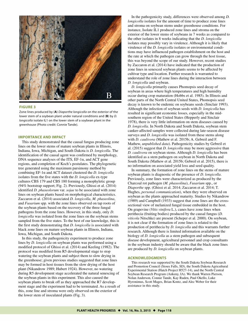
To obtain inoculum, the isolates were grown on PDA with sterilized wood toothpicks placed on top of the media. After a week of incubation, the toothpicks were removed from the PDA and inserted 2 to 3 mm deep into the lower stems (above the cotyledon) of soybean plants that were at approximate develop-mental stages V3 to V4 (Fehr et al. 1971). The toothpicks and stems were wrapped with Parafilm to avoid rapid dehydration. Six plants were inoculated per isolate and one toothpick was inserted per plant. Six control plants were treated by inserting one sterile, non-infested toothpick into each stem. After inoculation, plants were kept in the greenhouse for 7 weeks at 22°C with a 14-h photoperiod. From the beginning of seed development (R5) plant growth stage until maturation, the soybean plants were not watered. Instead, the soybean plants were allowed to dry slowly in the greenhouse at R5 developmental stage so that the fungal inoculum on the inoculated plants would be dried in situ. At developmental stage R7 (beginning maturity), soybean plants were removed from the pots and washed, and examined for zone lines on the soybean stems (Fig. 3). Upon examination, it was observed that the occurrence of zone lines on the soybean stems varied among the ten D. longicolla isolates. For example, zone lines appeared on the exterior of the lower stems of the soybean plants 7 weeks after inoculation with isolate IL1 compared to 8
weeks with the other isolates (Fig. 3). Zone lines were not observed on any of the control plants.
To complete Koch’s postulates, D. longicolla was re-isolated from the inoculated soybean plants, and the identity was confirmed via sequencing of the ITS region using primers ITS1 and ITS2 (White et al. 1990). The pathogen was not recovered from any of the control plants.
FIGURE 2 Consensus of five most-parsimonious trees (length = 262) resulting from the alignment of 688 characters of the combined EF1-α and ACT gene region of the Diaporthe longicolla isolates using the maximum-parsimony analyses. The consistency index, retention index, and rescaled consistency index were 0.75, 0.92, and 0.74, respectively. Bootstrap values greater than 50 are shown. D. longicolla isolates recovered from soybean fields in the five states have state labels (IL = Illinois, IN = Indiana, IA = Iowa, MI = Michigan, and SD = South Dakota). The ex-type cultures CBS 179 and CBS 180 are labelled ‘dl’ for D. longicolla. Chrysoporthella hodgesiana (isolate CMW9995) was used as the outgroup (National Center for Biotechnology Information Accession No. GQ290152 for EF1-α sequence and GQ290170 for ACT sequence). Evolutionary analyses were conducted in MEGA6 (Tamura et al. 2013).
PLANT HEALTH PROGRESS Vol. 16, No. 3, 2015 Page 118
IMPORTANCE AND IMPACT This study demonstrated that the causal fungus producing zone
lines on the lower stems of mature soybean plants in Illinois, Indiana, Iowa, Michigan, and South Dakota is D. longicolla. The identification of the causal agent was confirmed by morphology, DNA sequence analyses of the ITS, EF-1α, and ACT gene regions, and completion of Koch’s postulates. The phylogenetic tree generated using the maximum parsimony method by combining EF-1α and ACT dataset clustered the D. longicolla isolates from the five states with the D. longicolla ex-type cultures CBS 179 and CBS 180 forming a monophyletic group (94% bootstrap support; Fig. 2). Previously, Ghissi et al. (2014) identified D. phaseolorum var. sojae to be associated with zone lines on soybean plants based on morphological characteristics. Zaccaron et al. (2014) associated D. longicolla, M. phaseolina, and Fusarium spp. with the zone lines observed on tap roots of the soybean plants based on the recovery of the three fungal pathogens from the zone lines. However, in this study, only D. longicolla was isolated from the zone lines on the soybean stems sampled from the five states. To the best of our knowledge, this is the first study demonstrating that D. longicolla is associated with black zone lines on mature soybean plants in Illinois, Indiana, Iowa, Michigan, and South Dakota.
In this study, the pathogenicity experiment to produce zone lines by D. longicolla on soybean plants was performed using a modified protocol of Ghissi et al. (2014) and Keeling (1982). The protocol was modified from R5 developmental stage to stop watering the soybean plants and subject them to slow drying in the greenhouse; given previous studies suggested that zone lines may be formed in host tissues from the slow drying of the host plant (Nikandrow 1989; Hubert 1924). However, no watering during R5 development stage accelerated the natural senescing of the soybean plants in this experiment. This also caused the soybean plants to break off as they approached the R7 develop-ment stage and the experiment had to be terminated. As a result of this, zone line and stroma were only observed on the exterior of the lower stem of inoculated plants (Fig. 3).
In the pathogenicity study, differences were observed among D. longicolla isolates for the amount of time to produce zone lines and stroma on soybean stems under greenhouse conditions. For instance, Isolate IL1 produced zone lines and stroma on the exterior of the lower stems of soybeans in 7 weeks as compared to the other isolates in 8 weeks indicating that the D. longicolla isolates may possibly vary in virulence. Although it is likely that virulence of the D. longicolla isolates or environmental condi-tions may have influenced pathogen establishment on the host and the rate at which the pathogen can grow through the host tissue, this was beyond the scope of our study. However, recent studies by Zaccaron et al. (2014) have indicated that the production of zone lines in senesced soybean plants seems to be affected by cultivar type and location. Further research is warranted to understand the role of zone lines during the interaction between D. longicolla and soybean.
D. longicolla primarily causes Phomopsis seed decay of soybean in areas where high temperatures and high humidity occur during crop maturation (Hobbs et al. 1985). In Illinois and other parts of the North Central United States, Phomopsis seed decay is known to be endemic on soybeans seeds (Sinclair 1993). Although the infection of soybean seeds with D. longicolla has resulted in significant economic losses, especially in the mid-southern region of the United States (Hepperly and Sinclair 1978), there is very little information on stem diseases caused by D. longicolla. In North Dakota and South Dakota, soybean stem canker-affected samples were collected during late-season disease surveys and D. longicolla was isolated from these stems along with D. caulivora (Mathew et al. 2015b; A. Gebreil and F. Mathew, unpublished data). Pathogenicity studies by Gebreil et al. (2015) suggest that D. longicolla may be more aggressive than D. caulivora on soybean stems. Although D. longicolla has been identified as a stem pathogen on soybean in North Dakota and South Dakota (Mathew et al. 2015b; Gebreil et al. 2015), there is no information on associated yield loss and seed quality.
In summary, the formation of zone lines on the stems of mature soybean plants is diagnostic of the presence of D. longicolla. Previously, zone lines were characterized as produced by the Charcoal rot pathogen (M. phaseolina), Fusarium spp., or Diaporthe spp. (Ghissi et al. 2014; Zaccaron et al. 2014; T. Hughes, personal communication), when they were observed on soybean as the plants approached maturity. Studies by Nikandrow (1989) and Campbell (1933) suggest that zone lines are the cross-sectional view of melanized fungal tissue embedded in the host. On grapevine (Vitis vinifera L.), canes have zone lines when perithecia (fruiting bodies) produced by the causal fungus (D. viticola Nitschke) are present (Scheper et al. 2000). On soybean, it is not clear if the formation of zone lines is related to the production of perithecia by D. longicolla and this warrants further research. Although there is limited information available on the biology of D. longicolla as a stem pathogen and subsequent disease development, agricultural personnel and crop consultants in the soybean industry should be aware that the black zone lines are produced by D. longicolla on soybean plants.
ACKNOWLEDGMENTS This research was supported by the South Dakota Soybean Research
and Promotion Council (Sioux Falls, SD), the South Dakota Agricultural Experimental Station (Hatch Project H527-14), and the North Central Soybean Research Program (Ankeny, IA). We thank Warren Pierson, Nolan Anderson, Connie Tande, Kay Ruden, Paul Okello, Luke Hyronimus, Scott Mages, Brian Kontz, and Alec Weber for their assistance in this study.
FIGURE 3 Zone lines produced by (A) Diaporthe longicolla on the exterior of the lower stem of a soybean plant under natural conditions and (B) by D. longicolla isolate IL1 on the lower stem of a soybean plant in the greenhouse (Photo credit: Connie Tande).
PLANT HEALTH PROGRESS Vol. 16, No. 3, 2015 Page 119
LITERATURE CITED
Barr, M. E. 1978. The Diaporthales in North America with emphasis on Gnomonia and its segregates. Mycol. Mem. 7:1-232.
Campbell, A. H. 1933. Zone lines in plant tissues. I. The black lines formed by Xylaria polymorpha (Pers.) Grev. in hardwoods. An. Appl. Biol. 20:123-145.
Carbone, I., and Kohn, L. M. 1999. A method for designing primer sets for speciation studies in filamentous ascomycetes. Mycologia 91:553-556.
Farris, J. S., Kallersjo, M., Kluge, A. G., and Bult, C. 1994. Testing significance of congruence. Cladistics 10:315-320.
Fehr, W. R., Caviness, C. E., Burmood, D. T., and Pennington, J. S. 1971. Stage of development descriptions for soybeans, Glycine max (L.) Merrill. Crop Sci. 11:929-931.
Felsenstein, J. 1985. Confidence limits on phylogenies: An approach using the bootstrap. Evolution 39:783-791.
Gebreil, A., Micijevic, A., Weber, A., Hyronimus, L., and Mathew, F. 2015. Characterization of Diaporthe species infecting soybeans (Glycine max L.) in South Dakota. Proceedings of the 100th Anniversary Meeting of South Dakota Academy of Science. (In press.) http://www.sdaos.org/proceedings/.
Ghissi, V. V., Reis, E. M., and Deuner, C. C. 2014. Etiology of Phomopsis root rot in soybean. Summa Phytopathol. 40:270-272.
Hall, T. A. 1999. BioEdit: A user-friendly biological sequence alignment editor and analysis program for Windows 95/98/NT. Nucl. Acids. Symp. Ser. 41:95-98.
Hartman, G. L., Sinclair, J. B., and Rupe, J. C. 1999. Compendium of Soybean Diseases, 4th ed. American Phytopathology Society, St. Paul, MN.
Hepperly, P. R., and Sinclair, J. B. 1978. Quality losses in Phomopsis-infected soybean seeds. Phytopathology 68:1684-1687.
Hobbs, T. W., Schmitthenner, A. F., and Kuter, G. A. 1985. A new Phomopsis species from soybean. Mycologia. 77:535-544.
Hubert, E. E. 1924. The diagnosis of decay in wood. J. Agric. Res. 29:523-567.
Keeling, B. L. 1982. A seedling test for resistance to soybean stem canker caused by Diaporthe phaseolorum var. caulivora. Phytopathology 72:807-809.
Maddison, W. P., and Maddison, D. R. 2011. Mesquite: A modular system for evolutionary analysis. Version 2.75 http://mesquiteproject.org.
Mathew, F. M., Alananbeh, K., Jordahl, J. G., Meyer, S. M., Castlebury, L. A., Gulya, T. J., and Markell, S. G. 2015a. Phomopsis stem canker: A reemerging threat to sunflower (Helianthus annuus) in the United States. Phytopathology 105:990-997.
Mathew, F. M., Castlebury, L. A., Alananbeh, K., Jordahl, J. G., Taylor, C. A., Meyer, S. M., Lamppa, R. S., Pasche, J. A., and Markell, S. G. 2015b. Identification of Diaporthe longicolla on dry edible pea, dry edible bean, and soybean in North Dakota. Plant Health Progress doi:10.1094/PHP-RV-14-0045.
Nei, M., and Kumar, S. 2000. Molecular evolution and phylogenetics. Oxford University Press, New York, NY.
Nikandrow, A. 1989. Zone lines diagnostic of Phomopsis sp. in Lucerne crown rot. Australas. Plant Pathol. 18:86-89.
Santos, J. M., Vrandečić, K., Ćosić, J., Duvnjak, T., and Phillips, A. J. L. 2011. Resolving the complex of Diaporthe/Phomopsis species on soybean in Croatia. Persoonia 27:9-19.
Scheper, R. W. A., Crane, D. C., Whisson, D. L., and Scott, E. S. 2000. The Diaporthe teleomorph of Phomopsis Taxon 1 on grapevine. Mycol. Res. 104:226-231.
Sinclair, J. B. 1993. Phomopsis seed decay of soybeans—a prototype for studying seed disease. Plant Dis. 77:329-334.
Swofford, D. L. 2002. Phylogenetic Analysis Using Parsimony (*and other methods), Version 4.0b10. Sinauer Associates, Sunderland, MA.
Tamura, K., Stecher, G., Peterson D., Filipski A., and Kumar, S. 2013. MEGA6: Molecular Evolutionary Genetics Analysis version 6.0. Mol. Biol. Evol. 30:2725-2729.
Udayanga, D., Castlebury, L. A., Rossman, A. Y., Chukeatirote, E., and Hyde, K. D. 2014. The Diaporthe sojae species complex: Phylogenetic re-assessment of pathogens associated with soybean, cucurbits and other field crops. Fungal Biol. 119:383-407.
White, T. J., Bruns, T., Lee, S., and Taylor, J. 1990. Amplification and direct sequencing of fungal and ribosomal RNA genes for phylogenetics. Pages 315-322 in: PCR Protocols: A Guide to Methods and Applications. Academic Press, Inc., San Diego, CA.
Wrather, J. A., Anderson, T. R., Arsyad, D. M., Gai, J., Ploper, L. D., Porta-Puglia, A., Ram, H. H., and Yorinori, J. T. 1997. Soybean disease loss estimates for the top 10 soybean producing countries in 1994. Plant Dis. 81:107-110.
Zaccaron, M. L., Rupe, J. C., and Holland, R. T. 2014. Association of Phomopsis longicolla and Macrophomina phaseolina with zone lines in soybean roots at maturity. Proc. 41st Annu. Meet. South. Soybean Dis. Workers 41:8.