Embed Size (px)
Citation preview

Turk Neurosurg 26(1):153-165, 2016 | 153
Corresponding author: Su-Min GENG E-mail: [email protected]
Case Report
DOI: 10.5137/1019-5149.JTN.12145-14.2
Received: 15.07.2014 / Accepted: 27.10.2014
Yu-kun ZHANG, Su-Min GENG, Pi-Nan LIU, Gang LV
Beijing Tiantan Hospital, Capital Medical University, Department of Neurosurgery, Beijing, China
Association of Craniovertebral Junction Anomalies, Klippel-Feil Syndrome, Ruptured Dermoid Cyst and Mirror Movement in One Patient: A Unique Case and Literature Review
ABSTRACT
█ CASE REPORTHistory. A 28-year-old unmarried woman presented with a five-month history of unsteady gait, which had worsened one month before admission. The onset of this symptom occurred without any obvious cause. She did not complain of headache, nausea or vomiting episodes. In addition, since birth, the patient had experienced involuntary movement of the contralateral hand while performing voluntary movements with the other hand, although this symptom had never previously caused distress. There was no family history of medical disorders.
Physical Examination. The patient, whose neck was short with a high neck/height ratio (1:20.5, normal 1:9) (32), had a low posterior hairline, extending to the level of the C7 spinous process (Figure 7A). Upon physical examination, the examiner was able to palpate one apparent pit in the
█ InTRODuCTIOnDermoid cysts (DCs) are rare neoplasms, accounting for 0.1–0.7% of all intracranial tumors (20, 29). In 1936, Love and Kernohan first reported the association of DCs of the posterior cranial fossa with cervical fusion anomalies, particularly Klippel-Feil syndrome (KFS) (20). Subsequently, only 23 other cases of KFS-associated posterior cranial fossa DC (1, 3, 4, 7-10, 13, 15, 19, 20, 23, 25, 27, 28, 34, 35, 40, 41, 44) have been reported in the English literature, as listed in Table I. Because these diseases are very rare, the association between DC and KFS may be pathophysiologically significant. We present the first report of a patient with a craniocervical ruptured DC accompanied by craniovertebral junction (CVJ) anomalies, KFS and mirror movement.
The Klippel-Feil syndrome (KFS) has been reported to be associated with intracranial neoplasms, most frequently epidermoid or dermoid cysts. To our knowledge, however, patients who present with a posterior fossa dermoid cyst (DC) and KFS are extremely rare with only 24 previously reported cases in the English literature worldwide. Therefore, we present the first report of a patient with a craniocervical ruptured DC accompanied by craniovertebral junction (CVJ) anomalies, KFS and mirror movement. Meanwhile, a literature review of KFS accompanying with posterior fossa DC discusses these conditions from the embryological, anatomical, clinical and therapeutic perspectives. Additionally, the combination of CVJ anomalies, KFS and DC may represent a new syndrome that has previously gone unnoticed.
KEywORDS: Craniovertebral junction, Dermoid cyst, Klippel-Feil syndrome, Mirror movement, Rupture

154 | Turk Neurosurg 26(1):153-165, 2016
Zhang yK. et al: CVJ, KFS, DC, Mirror Movement
nape of the patient’s neck (Figure 7A), and flexion-extension motions were maintained to a limited extent (Figures 1B, C). Mirror movement was easily elicited and primarily confined to the hand. The patient had gait ataxia, as well as apparent clumsiness in performing the heel-knee-tibia test and finger-to-nose test; Romberg’s sign was positive. The other neurological examinations were negative.
Radiographic Examination. Plain radiography and computed tomography (CT) of the head and neck revealed congenital fusions (classified as Type I of KFS) of C2-C7 and T1-T3 associated with platybasia or basilar invagination (Figures 1A-C, 2A, B) (11). A three-dimensionally (3D) reconstructed CT of CVJ demonstrated spina bifida occulta from C2 to C7 (Figure 3A-C). Most notably, CT showed a low-density, calcified,
Figure 1: Plain cervical spine radiographs. A) Plain radiograph shows congenital fusions (Type I of KFS) (double arrowheads) with 153° NTB angle of Welcker joining the nasion (N), tuberculum (T), and basion (B). The normal angle should be less than 130° (29), platybasia with an increased NTB angle. B, C) Flexion and extension of cervical spine.
Figure 2: Midsagittal reconstructed images from cervical spine CT. A) Double arrowheads reveal congenital fusions (Type I of KFS) (11). Radiographic criteria for basilar invagination with dens protrudes >2.5mm (a=8.0mm) above Chamberlain’s line between hard palate and opisthion (20). B) Another criteria with dens protrudes >5.0mm (b=9.8mm) above McGregor’s line between hard palate and the lowest point of occiput (20). Besides, there are two lesions (asterisk and black arrow).
A B C
A B

Turk Neurosurg 26(1):153-165, 2016 | 155
Zhang yK. et al: CVJ, KFS, DC, Mirror Movement
circumscribed mass lesion (measured as -70~170 Hu) around the foramen magnum. In addition, another lesion (measured as -120 Hu) was found in the upper thoracic spinal canal. CT indicated that the second lesion was likely to be a lipoma (Figure 2A, B).
Magnetic resonance imaging (MRI) revealed one oval intradural mass located in the craniocervical area, measuring 28.6 mm anteroposteriorly and 50.3 mm rostrocaudally. The lesion was heterogeneous and had areas of hyper-, iso, and hypo-intensity on T1-weighted images and areas of hyperintensity on T2-weighted images (Figure 4A-C). In addition, multiple small, short T1WI and long T2WI signal lesions were scattered in the brain fissures, subarachnoid spaces and ventricles (Figure 4A, Figure 5A-D). These lesions were assumed to be fatty droplets derived from spontaneous rupture of the DC (Figure 5A).
3D CTA of the vertebral artery (VA) (Figure 6A-E) demonstrated that the third segment of the right vertebral artery (RVA) had an
abnormal course, leaving the transverse foramen of the axis and passing into the foramen magnum below the occipitalized lateral mass of C1; this was classified as a Type I VA at the CVJ with occipitalization of the atlas (42). The second segment of the left vertebral artery (LVA) was obviously distorted from the osseous fusion of C2-C7. In addition, there was a fenestration of the vertebrobasilar artery.
Surgery. With the use of microsurgical instruments, total resection of the lesion was performed (Figure 7A-C). The anterior portion of the lesion was adherent to the dorsal surface of the brainstem, where an inconspicuous cleft was observed, as shown in the preoperative MRI (Figure 5A). Next, hydrocortisone was used to extensively irrigate the resection bed, in order to prevent postoperative chemical meningitis. No occipitocervical (OC) fixation or fusion was performed during the surgery. After the successful operation, a postoperative neck collar was prescribed for one month.
Figure 3: 3D CT reconstruction of craniovertebral region. A) Posterior view rendering of spina bifida occulta of C1-C7. B) Lateral view showing of defects of posterior arch of atlas. C. Anterior view indicating of scoliosis of congenital fusion (double arrowheads), bifid anterior arch of atlas, and atlantooccipital joint asymmetry, cervical and upper thoracic spine concave to right (white arrow) and left (black arrow) respectively.
Figure 4: Preoperative MRI of head and neck. A) Sagittal T1WI reveals one heterogeneous mass (asterisk) and scattered droplets (arrows). B) Axial T2WI shows a hyper-intense mass (asterisk). C) Coronal contrast-enhanced T1WI of the lesion (asterisk).
A B C
A B C

156 | Turk Neurosurg 26(1):153-165, 2016
Zhang yK. et al: CVJ, KFS, DC, Mirror Movement
Follow-up. After six months of follow-up, the patient’s pre-operative symptoms had resolved, with the exception of mir-ror movement. Compared with the preoperative MRI findings (Figures 4A-C, 5A-D), postoperative MRI of the head showed no recurrence of the lesion; the fat droplets were stable with-out enlargement, reduction or apparent displacement (Figure 9).
Pathological Findings. Microscopic examination of the excised mass revealed the presence of a classic DC with calcification (Figure 8).
Postoperative Course. The course was uneventful, and the patient was discharged with no further neurological deficits on the 10th day after the surgery.
Figure 5: A series of MR images showing the scattered fat droplets. A) There is an inconspicuous cleft (arrow). B, C) They show the hyperintense fat droplets (arrows) on T2WI. D) It describes the hyperintense fat droplets ( arrows) on T1WI.
A B C
D

Turk Neurosurg 26(1):153-165, 2016 | 157
Zhang yK. et al: CVJ, KFS, DC, Mirror Movement
Figure 6: 3D CTA of vertebral artery.A) It shows abnormal course of the third segment of RVA, occipitalization of the right lateral mass of C1 (black arrow), and distortion of the second segment of LVA (white arrow). B, C) They reveal normal course of the first segment of bilateral VA (double arrowheads). D) It shows the fenestration (white arrow) of vertebrobasilar artery and distortion of LVA. E) It shows the asymmetry of sternoclavicular joint.
Figure 7: Intraoperative images of the patient. A) It shows the low occipital headline (black arrow), short neck (double arrowheads) and one apparent pit (white arrow). B) It reveals bulgy cerebellar tissue (R, L) and tumor (asterisk). C) the contents in the cyst (asterisk). (R, L shorting for right, left cerebellar hemisphere).
A B C
D E
A B C

158 | Turk Neurosurg 26(1):153-165, 2016
Zhang yK. et al: CVJ, KFS, DC, Mirror Movement
years (ranging from 1 to 61 years). Pediatric cases represented 56% of patients, indicating that approximately half of such patients are not diagnosed until adulthood. The follow-up period ranged from 3 months to 6 years among the 17 cases with sufficient information. Tumor recurrence was not reported in these patients during the follow-up period.
Klippel-Feil Syndrome
KFS is associated with the classic triad of a short neck, a low posterior hairline and a limited range of motion of the neck; it is caused by congenital fusion of two or more cervical vertebrae (11, 26). Analysis of the 25 cases shows that the classic KFS triad is present in 37.5% of patients. The lack of population screening studies has made it impossible to determine the exact incidence and prevalence of KFS (39). KFS can be classified into three subtypes (11)—type I, II or III:
Type I is defined as having multiple cervical or upper thoracic vertebral fusions;
Type II is defined as having isolated fusions at 1 or 2 cervical interspaces;
Type III is defined as having fusions in the cervical spine combined with lower thoracic or lumbar fusion.
Therefore, the current case was classified as Type I of KFS. As shown in Tables I and II, vertebral fusions were described in 21
█ DISCuSSIOnAnalysis of the 25 cases in Table II present more details concerning the association of KFS with DCs. These cases included 14 women and 11 men, with an average age of 19.7
Figure 8: Photomicrograph demonstrating a classic DC with a lining of smooth muscle cells, sebaceous gland and keratinous material with calcification (H& E, × 200).
Figure 9: T1 MRI follow-up scans show the fat droplets (arrows) in the brain.

Turk Neurosurg 26(1):153-165, 2016 | 159
Zhang yK. et al: CVJ, KFS, DC, Mirror Movement
of DCs. Complete resection of DCs was achieved in 86% of cases. Additionally, 87.5% (21/24) of patients did not require OC fusion and did not demonstrate instability; instead, there was one patient with mild restriction of neck movement after OC fusion.
Craniovertebral Junction Anomalies
The bony CVJ can be divided into two components: a central pillar, which consists of the clivus and a central pivot (including the dens and C2 vertebral body), and a bistratal ring around the central pivot. They are the foramen magnum ring superiorly and the atlantal ring inferiorly (29). Therefore, developmental anomalies at the CVJ can also be classified into those related to the two components. As shown in Tables I and II, atlas assimilation is the most common CVJ anomaly, followed by platybasia, basilar invagination, atlantoaxial dislocation (AAD) and spina bifida occulta. A defect in the posterior arch of C1 also occurred in the present patient.
Mirror Movement
Mirror movement, also known as synkinesia or Kallmann’s syndrome (22), occurs when voluntary movements of one side of the body are copied involuntarily by the other side of the body. This synkinesia manifests as a rare disorder that is usually associated with KFS (31, 35). It may represent neurological impairment or agenesis of the pyramidal decussation (44), cerebral cortex, corpus callosum, medulla oblongata or spinal cord (31).
To date, however, according to Table II, only three cases of KFS and posterior fossa DC have been reported with mirror movement. In our case, we believe that neurological impairment in the pyramidal decussation and the medulla oblongata, due to long-term compression by a congenital tumor, may be the cause of mirror movement.
█ COnCLuSIOnThis is the first reported case of associated CVJ anomalies, KFS, ruptured DC and mirror movement in one patient. A literature review discusses these conditions from multiple aspects. Several hypotheses (4, 15) have been proposed to explain the association among posterior fossa DC, KFS, and CVJ anomalies. Additionally, the combination of CVJ anomalies, KFS and DC may represent a new syndrome that has previously gone unnoticed.
cases, with 62% classified as type I, 33.3% classified as type II, and 4.7% classified as type III. Furthermore, it is unclear whether the diagnosis of KFS is a discrete entity or if it is one condition in a spectrum of congenital spinal deformities (12). As described in Table I, congenital deformities associated with KFS and DCs include Sprengel’s deformity, asymmetry of the sternoclavicular joint, fusion of the ribs or rudimentary cervical ribs, pigeon chest, palatal fissure, and basilar artery fenestration et al.
In recent years, numerous studies of KFS have been reported, particularly regarding the genetic etiology, such as a paracentric inversion on 8q (6), mutations of MEOX1 gene (2, 24), Notch signaling pathway (33, 36), Pax 1 and Pax 9 (5), or HOX gene (21). However, most of these conditions have only been confirmed in animal studies, and further studies are warranted to investigate the exact genetic origins of KFS.
Dermoid Cysts
The common view is that there is a focal failure of cleavage between the neuroectoderm and cutaneous ectoderm between the third and fifth weeks of embryogenesis (15, 41). This results in the incorporation of cutaneous ectoderm, which forms the skin epithelium and all skin appendages, into the neural tube. Additionally, dermoid or epidermoid cysts of the CNS may be produced (4, 10, 34, 37). The clinical manifestations of DCs are non-specific and insidious, unless rupture occurs (14). Symptoms in patients with unruptured DCs result from compression of adjacent neurovascular structures. As shown in tables 1 and 2, DC patients were often admitted with signs (41.7%) of elevated intracranial pressure (rICP), in addition to cerebellar signs (33.3%). Rarely, ruptured intracranial DCs could result in dissemination of fatty material into cisterns, producing variable neurological symptoms, such as chemical meningitis (43), seizures (18), visual deficits (38), hydrocephalus (14), transient cerebral ischemia (45), or no symptoms, just as in our case. Additionally, rupture is usually spontaneous and is due to an increase in the size of the tumor’s contents, together with head movements and/or brain pulsations; however, rupture has been reported following head trauma (16, 30). According to Table II, DCs located in the midline of the posterior fossa were found in 21 cases (84%), DCs extending to the craniocervical junction (including the present case) were found in 3 cases (12%), and a DC in the cerebellar hemisphere was found in one case (4%), showing that the midline of the posterior fossa is the most common site

160 | Turk Neurosurg 26(1):153-165, 2016
Zhang yK. et al: CVJ, KFS, DC, Mirror MovementTa
ble
I: Li
tera
ture
Rev
iew
of 2
5 C
ases
of K
lippe
l-Fei
l Syn
drom
e w
ith D
erm
oid
Cys
t of t
he P
oste
rior F
ossa
(Inc
ludi
ng th
e Pr
esen
t Cas
e)
Auth
ors
& ye
ar
Age
(yrs
)Se
x
Clin
ical
man
ifest
atio
n
Der
mal
si
nus/
Infe
ctio
n
Rad
iolo
gica
lm
odal
ityLo
catio
nR
uptu
reFu
sion
CVJ
M
alfo
rmat
ions
Scol
iosi
sO
ther
M
alfo
rmat
ions
Trea
t-m
ent
Com
plic
a-tio
nsFo
llow
-up
Love
et a
l.,
1936
(20)
30, F
rICP
rigid
ity o
f nec
kN
o/N
oPl
ain
x-ra
yve
ntric
ulog
raph
y
Mid
line
of
post
erio
r fo
ssa
No
C2-
C3
--N
oN
oTo
tal r
esN
oN
o re
c of
7
mon
s
Logu
e et
al.,
19
52 (1
9)61
, FrIC
P2m
m d
impl
e ab
ove
occi
pita
l pro
tube
ranc
eYe
s/N
oPl
ain
x-ra
yM
idlin
e of
po
ster
ior
foss
aN
oBo
dies
and
la
min
ae o
f C
2-C
5--
----
Tota
l res
----
Tytu
s et
al.,
19
56 (4
1)5,
FrIC
PYe
s/N
oPl
ain
x-ra
yM
idlin
e of
po
ster
ior
foss
aN
o
Mul
tiple
fu
sion
s th
roug
h th
e w
hole
spi
ne
----
--To
tal r
esN
oN
o re
c of
4
yrs
Robe
rts e
t al
., 19
57 (3
5)
2yrs
10m
ons,
M
rICP
men
ingi
tisa
swel
ling
belo
w th
e oc
cipi
tal p
rotu
bera
nce
Yes/
Yes
Plai
n x-
ray
Mid
line
of
post
erio
r fo
ssa
No
Mul
tiple
fu
sion
s th
roug
h
who
le c
ervi
cal
verte
brae
----
--To
tal r
es--
--
Whi
ttle
et a
l.,
1983
(44)
2, F
head
tilt
to ri
ght
mirr
or m
ovem
ent
gait
atax
iatri
ad o
f KFS
No/
No
Plai
n x-
ray
CT
scan
Met
rizam
ide
mye
logr
aphy
Mid
line
of
post
erio
r fo
ssa
No
Bodi
es o
f C
2-C
5
Spin
a bi
fida
occu
lta a
t lev
el o
f C
6-C
7--
--To
tal r
esN
oN
o re
c of
1
yrs
Diek
man
n-G
uiro
y et
al.,
19
89 (8
)42
, Fas
eptic
men
ingi
tistri
ad o
f KFS
No/
Yes
Plai
n x-
ray
MRI
CVJ
--C
2-C
5Sp
ina
bifid
a--
--To
tal r
esN
o--
Dick
ey e
t al.,
19
91 (7
)14
, F
rICP
verti
cal n
ysta
gmus
dysd
iado
chok
ines
iam
irror
mov
emen
t
Yes/
No
Plai
n x-
ray
CT
scan
Mid
line
of
post
erio
r fo
ssa
No
Bodi
es a
nd
lam
inae
of
C4-
C6
bodi
es o
f C
1-C
2la
min
ae o
f C
2-C
3
Occ
ipita
lizat
ion
of p
oste
rior a
rch
of C
1
Scol
iosi
s of
che
st,
conc
ave
to th
e rig
ht
Spre
ngel
’s de
form
ityan
terio
r fus
ion
of fi
rst a
nd
seco
nd ri
bshe
mi-v
erte
bra
at T
3
Parti
alRe
sN
o--
Kenn
edy
et
al.,
1998
(15)
40, F
head
ache
gait
atax
ia
abse
nce
of th
e rig
ht
corn
eal r
eflex
and
gap
re
flex
--/-
-Pl
ain
x-ra
yM
RI
Mid
line
of
post
erio
r fo
ssa
No
----
----
----
--
Muz
umda
r et
al.,
2001
(25)
12, F
torti
collis
shor
t and
web
bed
ne
ck h
igh
arch
ed
pala
te
No/
No
Plai
n x-
ray
MRI
Mid
line
of
post
erio
r fo
ssa
No
bodi
es o
f C
2-C
5la
min
ae o
f C
2-C
7
AAD
Occ
ipita
lizat
ion
of
the
atla
s--
bifid
uvu
lapi
geon
-Che
st
Tota
l res
OC
fu
sion
No
rec
of
1 yr
s
8, M
head
ache
shor
t nec
kto
rtico
llistru
ncal
ata
xia
Yes/
No
MRI
Mid
line
of
post
erio
r fo
ssa
No
----
Scol
iosi
s of
cer
vica
l sp
ine
--Pa
rtial
res
No
No
rec
of
9 m
ons

Turk Neurosurg 26(1):153-165, 2016 | 161
Zhang yK. et al: CVJ, KFS, DC, Mirror Movement
Auth
ors
& ye
ar
Age
(yrs
)Se
x
Clin
ical
man
ifest
atio
n
Der
mal
si
nus/
Infe
ctio
n
Rad
iolo
gica
lm
odal
ityLo
catio
nR
uptu
reFu
sion
CVJ
M
alfo
rmat
ions
Scol
iosi
sO
ther
M
alfo
rmat
ions
Trea
t-m
ent
Com
plic
a-tio
nsFo
llow
-up
Akso
y et
al.,
20
01 (1
)16
, Msu
bocc
ipita
l lum
ptri
ad o
f KFS
Yes/
No
Plai
n x-
ray
CT
scan
MRI
Mid
line
of
post
erio
r fo
ssa
No
C3-
C7
Occ
ipita
lizat
ion
of p
oste
rior a
rch
of C
1--
Rudi
men
tary
ce
rvic
al ri
bs o
n th
e le
ft--
----
Hin
ojos
a et
al
., 20
01 (1
3)23
, F
rICP
2mm
dim
ple
abov
e oc
cipi
tal
prot
uber
ance
triad
of K
FS
Yes/
No
Plai
n x-
ray
MRI
Mid
line
of
post
erio
r fo
ssa
No
Bodi
es a
nd
lam
inae
of
C4-
C5
----
--To
tal r
es--
No
rec
of
1 yr
s
Shar
ma
et
al.,
2001
(37)
4, M
rICP
head
tilt
to le
ftpe
a-si
aed
occi
pita
l lu
mp
rest
ricte
d ne
ck
mob
ility
and
stiff
ness
trunc
al a
taxi
a
--/-
-Pl
ain
x-ra
yC
T sc
anM
RI
Mid
line
of
post
erio
r fo
ssa
No
C2-
C3
Occ
ipita
lizat
ion
of p
oste
rior a
rch
of C
1
Scol
iosi
s of
cer
vica
l sp
ine,
co
ncav
e to
the
left
hem
i-ve
rtebr
a at
the
right
sid
e of
C
5-7
Parti
alRe
sN
o--
Gon
zále
z-Da
rder
et a
l.,
2002
(10)
36, M
rICP
juve
nile
dia
bete
sat
axia
triad
of K
FS
Yes/
No
Plai
n x-
ray
CT
scan
MRI
Mid
line
of
post
erio
r fo
ssa
No
C3-
C6
abse
nce
of
post
erio
r arc
h of
C1
--C
ervi
cal a
nd
dors
al ri
bsTo
tal r
esN
oN
o re
c of
1.
5 yr
s
Oer
tel e
t al.,
20
02 (2
7)54
, F--
No/
--M
RI
Mid
line
of
post
erio
r fo
ssa
an
d ce
r he
mis
pher
e
No
C3-
C6
No
----
Mic
ro-
surg
ical
re
sN
oN
o re
c of
3
mon
s
Cal
dare
lli et
al
., 20
04 (3
)5,
MRe
curre
nt
hype
rpyr
exia
Yes/
Yes
MRA
Mid
line
of
post
erio
r fo
ssa
No
----
----
Tota
l res
CSF
leak
-ag
eN
o re
c of
41
mon
s
Cha
ndra
et
al.,
2005
(4)
25, M
torti
collis
bila
tera
l pap
illede
ma
No/
No
Plai
n x-
ray
MRI
Mid
line
of
post
erio
r fo
ssa
No
C2-
C3
AAD
Occ
ipita
lizat
ion
of th
e at
las
plat
ybas
ia
----
Tota
l res
OC
fu
sion
No
No
rec
of
8 m
ons
and
mild
re
stric
-tio
n of
ne
ck
mov
e-m
ents
23, M
head
ache
neck
pai
nre
stric
ted
nec
k m
ovem
ents
No/
No
Plai
n x-
ray
MRI
Mid
line
of
post
erio
r fo
ssa
No
C2-
C3
basi
lar
inva
gina
tion
Occ
ipita
lizat
ion
of C
1 ar
chAA
D
----
res
OC
fu
sion
Asep
tic
men
ingi
tisN
o re
c of
15
mon
s
Tabl
e I:
Con
t.

162 | Turk Neurosurg 26(1):153-165, 2016
Zhang yK. et al: CVJ, KFS, DC, Mirror MovementAu
thor
s &
year
Age
(yrs
)Se
x
Clin
ical
man
ifest
atio
n
Der
mal
si
nus/
Infe
ctio
n
Rad
iolo
gica
lm
odal
ityLo
catio
nR
uptu
reFu
sion
CVJ
M
alfo
rmat
ions
Scol
iosi
sO
ther
M
alfo
rmat
ions
Trea
t-m
ent
Com
plic
a-tio
nsFo
llow
-up
Pai e
t al.,
20
07 (2
8)
3, M
rICP
inte
rmitt
ent i
rrita
bilit
yN
o/N
oPl
ain
x-ra
yC
T sc
anM
RI
Mid
line
of
post
erio
r fo
ssa
No
C2-
C4
----
Cer
ebel
lar
tons
illar
hern
iatio
nre
s--
No
rec
of
2 y
rs
1, M
feve
rvo
miti
ngoc
cipi
tal l
ump
Yes/
--Pl
ain
x-ra
yC
T sc
anM
RI
Righ
t cer
he
mis
pher
eN
o
Fusi
on o
f se
vera
l ce
rvic
al
verte
bral
----
Seve
ral h
emi-
verte
brae
of
thor
acic
spi
nere
sPs
eudo
-m
enin
go-
cele
--
2, M
hesd
ache
epis
taxi
soc
cipi
tal l
ump
Yes/
--C
T sc
anM
RI
Mid
line
of
post
erio
r fo
ssa
No
----
----
Tota
l res
No
No
rec
Turg
ut ,
2009
(4
0)4,
F
head
ache
head
tilt
to le
ftpe
a-si
zed
occi
pita
l lu
mp
triad
of K
FS
Yes/
No
Plai
n x-
ray
CT
scan
MRI
/MRS
Ultr
asou
ndEc
hoca
rdio
-gr
aphy
Mid
line
of
post
erio
r fo
ssa
No
C4-
C7
No
Scol
iosi
s of
cer
vica
l sp
ine,
co
ncav
e to
the
left
No
Tota
l res
CSF
leak
-ag
eN
o re
c of
10
mon
s
Ram
zan
et
al.,
2011
(34)
5, F
Torti
collis
to le
ftoc
cipi
tal l
ump
triad
of K
FSce
r sig
ns o
n th
e rig
ht
Yes/
No
Plai
n x-
ray
CT
scan
MRI
Mid
line
of
post
erio
r fo
ssa
No
C3-
C5
No
--N
oTo
tal r
esN
oN
o re
c of
3
mon
s
Mcl
augh
lin e
t al
., 20
13 (2
3)47
, F
rICP
occi
pita
l lum
ptri
ad o
f KFS
cer s
igns
para
pare
sis
of lo
wer
ex
trem
ity w
ith m
arke
d pa
tella
r hyp
erre
flexi
ase
nsor
y de
ficit
Yes/
No
Plai
n x-
ray
CT
scan
MRI
Cra
nioc
er-
vico
Thor
acic
No
Bodi
es a
nd
lam
inae
of
C2-
T6N
o40
° th
orac
ic
kyph
osis
¨ pala
tal fi
ssur
e¨ ve
rtebr
al a
rtery
an
terio
r to
the
lesi
on
Tota
l res
No
No
rec
of
6 y
rs
Cur
rent
cas
e28
, F
head
tilt
to le
ftm
irror
mov
emen
ttri
ad o
f KFS
cer s
igns
No/
No
Plai
n x-
ray
CT
scan
CTA
MRI
Ultr
asou
ndEc
hoca
rdio
-gr
aphy
Cra
nioc
ervi
-ca
l jun
ctio
nYe
sC
1-C
7T1
-T3
Plat
ybas
iaBa
sila
r in
vagi
natio
nbi
fid a
nter
ior C
1 ar
chde
fect
s of
po
ster
ior C
1 ar
chSp
ina
bifid
a oc
culta
at l
evel
of
C1-
C7
Scol
iosi
s
of c
ervi
cal
spin
e,
conc
ave
to th
e le
ft,Sc
olio
sis
of
firs
t to
third
sp
ine,
co
ncav
e to
the
right
Basi
lar
arte
ry fe
nest
ra-
tion
asym
met
ry o
f st
erno
clav
icul
ar
join
tin
trasp
inal
lip
oma
Tota
l res
No
No
rec
of
6 m
ons
Abbr
evia
tions
: AAD
atla
ntoa
xial d
isloc
atio
n; c
er c
ereb
ellu
m; C
cer
vical
ver
tebr
a; C
SF c
ereb
rosp
inal
flui
d; C
T co
mpu
ted
tom
ogra
phy;
CTA
com
pute
d to
mog
raph
y an
giog
raph
y; C
VJ c
rani
over
-te
bral
junc
tion;
KFS
Klip
pel-F
eil s
yndr
ome;
mon
s m
onth
s; M
RI m
agne
tic re
sona
nce
imag
ing;
MR
S m
agne
tic re
sona
nce
spec
trosc
opy;
No.
num
ber;
OC
occ
ipito
cerv
ical
; rIC
P ra
ised
intra
cran
ial
pres
sure
; rec
recu
rrenc
e; re
s re
sect
ion;
T th
orac
ic v
erte
bra;
yrs
yea
rs; -
- unm
entio
ned.
Tabl
e I:
Con
t.
Turk Neurosurg 26(1):153-165, 2016 | 163
Zhang yK. et al: CVJ, KFS, DC, Mirror Movement
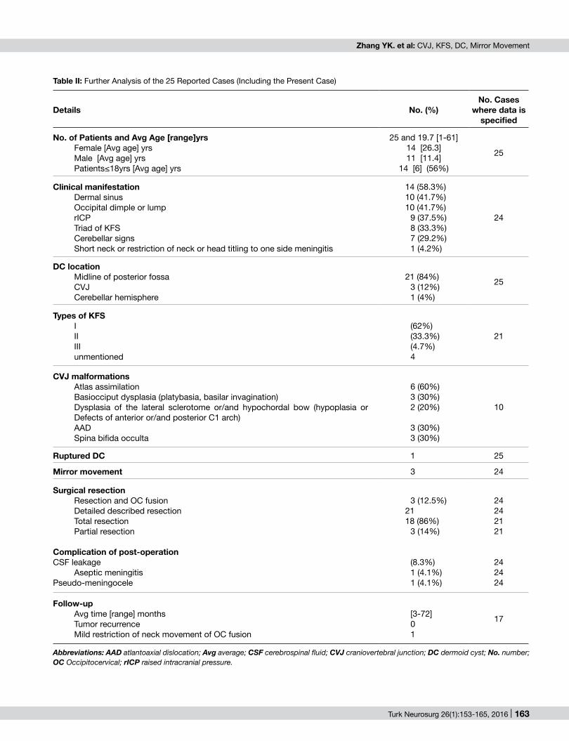
Table II: Further Analysis of the 25 Reported Cases (Including the Present Case)
Details no. (%)no. Cases
where data is specified
no. of Patients and Avg Age [range]yrs Female [Avg age] yrsMale [Avg age] yrsPatients≤18yrs [Avg age] yrs
25 and 19.7 [1-61]14 [26.3]11 [11.4]
14 [6] (56%)
25
Clinical manifestationDermal sinusOccipital dimple or lumprICPTriad of KFSCerebellar signsShort neck or restriction of neck or head titling to one side meningitis
14 (58.3%)10 (41.7%)10 (41.7%)
9 (37.5%)8 (33.3%)7 (29.2%)1 (4.2%)
24
DC locationMidline of posterior fossaCVJCerebellar hemisphere
21 (84%)3 (12%)1 (4%)
25
Types of KFSIIIIIIunmentioned
(62%)(33.3%)(4.7%)4
21
CVJ malformationsAtlas assimilationBasiocciput dysplasia (platybasia, basilar invagination)Dysplasia of the lateral sclerotome or/and hypochordal bow (hypoplasia or Defects of anterior or/and posterior C1 arch) AADSpina bifida occulta
6 (60%)3 (30%)2 (20%)
3 (30%)3 (30%)
10
Ruptured DC 1 25
Mirror movement 3 24
Surgical resection Resection and OC fusionDetailed described resectionTotal resection Partial resection
Complication of post-operationCSF leakage
Aseptic meningitisPseudo-meningocele
3 (12.5%)21 18 (86%)
3 (14%)
(8.3%)1 (4.1%)1 (4.1%)
24242121
242424
Follow-up Avg time [range] monthsTumor recurrence Mild restriction of neck movement of OC fusion
[3-72]01
17
Abbreviations: AAD atlantoaxial dislocation; Avg average; CSF cerebrospinal fluid; CVJ craniovertebral junction; DC dermoid cyst; No. number; OC Occipitocervical; rICP raised intracranial pressure.

164 | Turk Neurosurg 26(1):153-165, 2016
Zhang yK. et al: CVJ, KFS, DC, Mirror Movement
19. Logue V, Till K: Posterior fossa dermoid cysts with special reference to intracranial infection. J Neurol Neurosurg Psychiatry 15: 1-12, 1952
20. Love JG, Kernohan JW: Dermoid and epidermoid tumors (cholesteatomas) of central nervous system. JAMA 107: 1876-1883, 1936
21. Manzanares M, Wada H, Itasaki N, Trainor PA, Krumlauf R, Holland PW: Conservation and elaboration of Hox gene regulation during evolution of the vertebrate head. Nature 408: 854-857, 2000
22. Mayston MJ, Harrison LM, Quinton R, Stephens JA, Krams M, Bouloux PM: Mirror movement in X-linked Kallmann’s syndrome. I. A neurophysiological study. Brain 12: 1199-1216, 1997
23. Mclaughlin N, Weil AG, Demers J, Shedid D: Klippel-Feil syndrome associated with a craniocervico-thoracic dermoid cyst. Surg Neurol Int 4:S61-66, 2013
24. Mohamed JY, Faqeih E, Alsiddiky A, Alshammari MJ, Ibrahim NA, Alkuraya FS: Mutations in MEOX1, Encoding Mesenchyme Homebox 1, cause Klippel-Feil anomaly. Am J Hum Genet 92: 157-161, 2013
25. Muzumdar D, Goel A: Posterior cranial fossa dermoid in association with Craniovertebral and cervical spinal anomaly: Report of two cases. Pediatr Neurosurg 35:158-161, 2001
26. Nagib MG, Maxwell RE, Chou SN: Klippel-Feil syndrome in children: Clinical features and management. Childs Nerv Syst 1: 255-263, 1985
27. Oertel J, Piek J, Müller JU, Vogelgesang S, Warzok R, Gaab RM: Posterior fossa squamous cell carcinoma due to dedifferentiation of a dermoid cyst in Klippel-Feil syndrome case illustration. J Neurosurg 97:1244, 2002
28. Pai VV, Lowe LH, Castillo M, Huang BY, Shao L: Posterior fossa dermoid cysts in association with Klippel-Feil syndrome: Report of three cases. AJNR Am J Neuroradiol 28:1926-1928, 2008
29. Pang D, Thompson D: Embryology and bony malformations of the craniovertebral junction. Childs Nerv Syst 27:523-564, 2011
30. Park SK, Cho KG: Recurrent intracranial dermoid cyst after subtotal removal of traumatic rupture. Clin Neurol Neurosurg 114: 421-424, 2012
31. Parmar PN, Seta AA, Solanki AB: Congenital mirror movements in nodular heterotopia and partial block vertebrae. J Pediatr Neurol 1: 43-49, 2013
32. Pinninti R, Thirulogachandar E, Noorul Ameen KH: An interesting clinical association of short neck with an unusual laryngeal anomaly. J Craniovert Jun Spine 2: 86-88, 2011
33. Pourquié O, Kusumi K: When body segmentation goes wrong. Clin Genet 60: 409-416, 2001
34. Ramzan A, Khursheed N, Rumana M, Abrar W, Ashish J: Posterior fossa dermoid with Klippel-Feil syndrome in a child. Pediatr Neurol 45:197-199, 2011
35. Roberts AP: A case of intracranial dermoid cyst associated with the Klippel-Feil deformity and recurrent meningitis. Arch Dis Child 33:222-225, 1958
36. Saga Y, Takeda H: The making of the somite: Molecular events in vertebrate segmentation. Nat Rev Genet 2: 835-845, 2001
37. Sharma MS, Sharma BS, Yadav A, Khosla VK: Posterior fossa dermoid in association with Klippel-Feil syndrome--a short report. Neurol India 49:210-212, 2001
38. Skovrlj B, Mascitelli JR, Steinberger JM, Weiss N: Progressive visual loss following rupture of an intracranial dermoid cyst. J Clin Neurosci 21: 159-161, 2014
█ REFEREnCES1. Aksoy FG, Aksoy OG, Gomori JM: Klippel-Feil syndrome in
association with posterior fossa suboccipital dermoid cyst. Eur Radiol 11:142-144, 2001
2. Bayrakli F, Guclu B, Yakicier C, Balaban H, Kartal U, Erguner B, Sagiroglu MS, Yuksel S, Ozturk AR, Kazanci B, Ozum U, Kars HZ: Mutation in MEOX1 gene causes a recessive Klippel-Feil syndrome subtype. BMC Genetics 14: 95, 2013
3. Caldarelli M, Massimi L, Kondageski C, Rocco CD: Intracranial midline dermoid and epidermoid cysts in children. J Neurosurg Pediatr 100: 473-480, 2004
4. Chandra PS, Gupta A, Mishra NK, Mehta VS: Association of craniovertebral and upper cervical anomalies with dermoid and epidermoid cysts: Report of four cases. Neurosurgery 56:1155, 2005
5. Chi N, Epstein JA: Getting your Pax straight: Pax proteins in development and disease. Trends Genet 18: 41-47, 2002
6. Clarke RA, Singh S, Mckenzie H, Kearsley JH, Yip MY: Familial Klippel-Feil syndrome and Paracentric inversion inv(8)(q22.2q23.3). Am J Hum Genet 57: 1364-1370, 1995
7. Dickey W, Hawkins SA, Kirkpatrick DH, Gray WJ: Posterior fossa dermoid cysts and the Klippel-Feil syndrome. J Neurol Neurosurg Psychiatry 54: 1016-1017, 1991
8. Diekmann-Guiroy B, Huang PS: Klippel-Feil syndrome in association with a Craniocervical dermoid cyst presenting as aseptic meningitis in an adult: Case report. Neurosurgery 25:652-655, 1989
9. Gallea G, Popa T, Hubsch C, Valabregue R, Brochard V, Kundu P, Schmitt B, Bardinet E, Bertasi E, Flamand-Roze C, Alexandre N, Delmaire C, Méneret A, Depienne C, Poupon C, Hertz-Pannier L, Cincotta M, Vidailhet M, Lehericy S, Meunier S, Roze E: RAD51 deficiency disrupts the corticospinal lateralization of motor control. Brain 136: 3333-3346, 2013
10. González-Darder JM, Feliu-Tatay R, Pesudo-Martínez JV, Vera-Román JM: Klippel-Feil syndrome associated with posterior fossa dermoid cyst. Case report. Neurol Res 24:501-504, 2002
11. Gunderson CH, Greenspan RH, Glaser GH, Lubs HA: The Klippel-Feil syndrome: Genetic and clinical reevaluation of cervical fusion. Medicine 46: 491-512, 1967
12. Hensinger RN, Lang JE, Macewen GD: Klippel-Feil syndrome: A constellation of associated anomalies. J Bone Joint Surg 56-A: 1246-1253, 1974
13. Hinojosa M, Tatagiba M, Harada K, Samii M: Dermoid cyst in the posterior fossa accompanied by Klippel-Feil syndrome. Childs Nerv Syst 17:97-100, 2001
14. Karabulut N, Oguzkurt L: Tetraventricular hydrocephalus due to ruptured intracranial dermoid cyst. Eur Radiol 10: 1810-1811, 2000
15. Kennedy PT, McAuley DJ: Association of posterior fossa dermoid cyst and Klippel-Feil syndrome. AJNR Am J Neuroradiol 19:195-196, 1998
16. Kim IN, Jung S, Jung TY, Kang SS, Kim TS: Traumatic rupture of an intracranial dermoid cyst. J Clin Neurosci 15: 469-471, 2008
17. Kim KH, Cho JH: Ruptured intracranial dermoid cyst associated with rupture of cerebral aneurysm. J Korean Neurosurg Soc 50: 453-456, 2011
18. Kucera JN, Roy P, Murtagh R: Ruptured intracranial dermoid cyst manifesting as new onset seizure: A case report. Neuroradiology 5: 10-18, 2011

Turk Neurosurg 26(1):153-165, 2016 | 165
Zhang yK. et al: CVJ, KFS, DC, Mirror Movement
43. Wang YM, Chang TP, Lo CP, Tu MC: Spontaneous rupture of intracranial dermoid cyst with chemical meningitis. J Emerg Med 44: e275-e276, 2013
44. Whittle IR, Besser M: Congenital neural abnormalities presenting with mirror movements in a patient with Klippel-Feil syndrome. J Neurosurg 59: 891-894, 1983
45. Zheng K, Mao BY, Ma L, Jiang S: Ruptured intracranial dermoid cyst with infarction in the basal ganglia: Case report. Neurol Med Chir (Tokyo) 50: 254-256, 2010
39. Tracy MR, Dormans JP, Kusumi K: Klippel-Feil syndrome: Clinical features and current understanding of etiology. Clin Orthop Relat Res 424:183-190, 2004
40. Turgut M: Klippel-Feil syndrome in association with posterior fossa dermoid tumor. Acta Neurochir 151: 269-279, 2009
41. Tytus JS, Pennybacker J: Pearly tumours in relation to the central nervous system. J Neurol Neurosurg Psychiatry 19: 241-259, 1956
42. Wang SL, Wang C, Liu Y, Yan M, Zhou HT: Anomalous vertebral artery in craniovertebral junction with occipitalization of the atlas. Spine 34:2838-2842, 2009