Embed Size (px)
Citation preview
1 3
World J UrolDOI 10.1007/s00345-014-1265-x
OrIgInal artIcle
Association between the bladder wall thickness and urodynamic findings in patients with spinal cord injury
Jose Ailton Fernandes Silva · Marcia de Castro Diniz Gonsalves · Rogerio Teles de Melo · Fabricio Borges Carrerette · Ronaldo Damião
received: 27 September 2013 / accepted: 15 February 2014 © Springer-Verlag Berlin Heidelberg 2014
in patients with ScI and cannot replace urodynamic evaluation.
Keywords neurogenic lower urinary tract dysfunction · Urodynamics · Ultrasound · Bladder wall thickness · Spinal cord injury
Introduction
Spinal cord injury (ScI) is usually cause of either temporary or permanent neurologic deficits and leads to damage across all body systems. Perhaps one of the most critical changes is in the ability of the body to control bladder function. Depending on the level of injury, the manifestations of lower urinary tract (lUt) dysfunction vary and lead to a variety of urologic problems. Suprasacral spinal cord lesions induce neurogenic detrusor overactivity (nDO) and concurrent det-rusor sphincter dyssynergia (DSD), which impairs the stor-age and emptying functions of the lUt [1]. the subsequent high intravesical pressure leads to a reduced bladder capacity and incontinence, as well as potential structural deteriora-tion of the bladder wall and the upper urinary tract. an ele-vated storage pressure, either due to low bladder compliance (<20 ml/cm H2O) or to nDO (leak point pressure ≥40 cm H2O), is a major risk factor for renal deterioration [2–4].
the management of urinary issues begins at the time of injury and must continue throughout the life of a patient with ScI. Protecting renal function, rehabilitating the lUt, avoiding complications of the lower and upper urinary tract and preserving quality of life are the goals of treatment. the current treatment options rely initially on bladder emp-tying by clean intermittent catheterization (cIc) and thus on oral antimuscarinic agents to reduce the bladder pres-sure and increase the bladder capacity [5].
Abstract Purpose to investigate whether ultrasonographic bladder wall thickness (BWt) correlates with urodynamic param-eters in patients with spinal cord injury (ScI).Methods two hundred and seventy-two patients with ScI were enrolled in the study. all of the patients under-went bladder ultrasonography and urodynamic study. the anterior bladder wall was measured and compared to uro-dynamic data.Results the mean age of the patients was 37.4 years. the mean BWt was 3.9 mm. BWt was significantly higher in the patients with neurogenic detrusor overactivity asso-ciated with detrusor sphincter dyssynergia (nDO/DSD) compared to those without sphincter dyssynergia (4.2 vs. 3.6 mm, respectively, p < 0.001) and in those with com-pliance <20 ml/cm H2O. nevertheless, rOc curve analysis [rOc = 0.624, 95 % cI (0.530, 0.718), p = 0.011] showed that no meaningful BWt measurement cutoff could be made to predict an elevated detrusor pressure in the storage phase.Conclusions Increased BWt was present in patients with low bladder compliance and nDO/DSD. no BWt cutoff value to predict an elevated detrusor pressure was found. therefore, the measurement of BWt has no clinical role
J. a. F. Silva (*) · F. B. carrerette · r. Damião State University of rio de Janeiro, rio de Janeiro, Brazile-mail: [email protected]
M. c. D. gonsalves the SaraH network of rehabilitation Hospitals, rio de Janeiro, Brazil
r. t. de Melo the SaraH network of rehabilitation Hospitals, Belo Horizonte, Minas gerais, Brazil
World J Urol
1 3
there has recently been a growing interest in measuring bladder wall thickness (BWt) due to its association with urodynamic findings and its role in upper urinary tract dam-age. Historically, it was believed that bladder trabeculation was a marker of bladder outlet obstruction (BOO). With respect to BOO, the literature varies. there is evidence that increased BWt can be a useful parameter in the evaluation of men with benign prostatic hyperplasia. normal values for BWt in adults have been established, mean of 3.0 in women, 3.3 in men and 3.8 in men with lUt symptoms [6]. On the other hand, Blatt et al. [7] showed no significant difference among patients with normal urodynamic, BOO and detrusor overactivity (2.0; 2.1 and 1.9, respectively). an increase in BWt has also been observed in children with neurogenic bladder dysfunction, DSD and low-com-pliance bladders [8, 9]. although the impact of ScI on the lUt it is well known, there is only one study published to evaluate the relationship between DWt and urodynamic parameters [10].
the purpose of this study was to investigate whether ultrasonographic measurement of BWt correlates with urodynamic parameters in patients with ScI.
Materials and methods
Between July 2010 and august 2011, 272 patients with neurogenic lUt dysfunction due to ScI were evaluated and included in this study. Only patients who were over 18 years of age and who had suffered from ScI for more than 1 year were included. all of the patients underwent a physical examination and a renal function test (creatinine and cystatin c), as well as bladder ultrasonography and urodynamic study. cases who had bladder stones, who pre-sented acute urinary tract infection (UtI), with history of open bladder surgery and patients who were managed by chronic indwelling catheters (urethral or suprapubic) were excluded.
all of the methods and criteria were based on the stand-ardization provided by the International continence Soci-ety and on the Urodynamic good Practice guidelines [11, 12]. the study was approved by the local ethics committee.
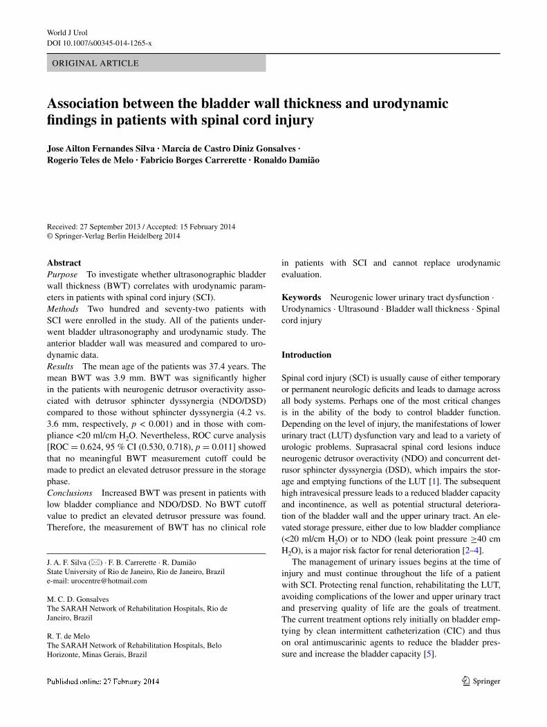
Ultrasound scanning of the bladder was performed from the suprapubic region with a linear multi frequency 7.5-MHz transducer (iU22 Philips, amsterdam, north Holland, the netherlands) with the patient in the supine position. the anterior bladder wall was measured approximately midway between the anterior wall midline and the lateral bladder wall. the BWt was represented by all 3 layers: mucosa/submucosal, detrusor and adventitia (Fig. 1). the bladder volume was calculated as the product of the longi-tudinal, transversal and sagittal diameter, as well as a cor-rection factor of 0.52. the patients were asked to fill their
bladders by drinking, and the ultrasound was performed when they felt the normal desire to void or just before the cIc was performed.
Multichannel urodynamics studies (Medtronic Duet System, version 8.20, Minneapolis, Minnesota, United States of america) consisted of cystometry, pressure flow study and external urethral sphincter electromyography with surface electrodes. the tests were performed with the patient in supine position. an 8-French transurethral double lumen catheter was used to measure the intravesi-cal pressure. Intra-abdominal pressure was simultaneously measured using a rectal balloon catheter. Filling water cys-tometry was performed by infusion of a room temperature saline solution at rate of 15–30 ml/min. the urodynamic parameters comprised maximum cystometric capacity (Mcc), maximum detrusor pressure (pdet max), reflex vol-ume, DSD and compliance. Pdet max was measured during involuntary contraction in the storage phase. low bladder compliance (<20 ml/cm H2O) and nDO (leak point pres-sure ≥40 cm H2O) were considered an elevated storage pressure.
We analyzed the relationship between BWt and urody-namic parameters while also considering other variables, such as gender, distribution of paralysis (paraplegia and tetraplegia), voiding method and continence.
Statistical analysis was carried out using the Statisti-cal Package for Social Sciences, version 20, for Windows (IBM corporation, armonk, new York, United States of america). categorical data were tabulated, and continuous variables were summarized using means and standard devi-ations (SD). Student’s t test was used to compare the quan-titative variables between independent groups; bivariate analysis associated with the Pearson's correlation coef-ficient was used to test the association between quantita-tive variables; and the chi-square test was used to test the
Fig. 1 Ultrasonographic measurement of BWt of the anterior blad-der wall
World J Urol
1 3
categorical variables. Differences were considered to be significant at a value of p < 0.05.
Results
One hundred and seventy-six (64.7 %) of the patients were paraplegic, and 95 (35.3 %) were tetraplegic. eight-een patients were excluded from the study. the reason was acute UtI in four, bladder stones in three, indwelling cathe-ter in two, history of open bladder surgery in two and BWt was not possible to measure in seven patients because of low bladder volume.
the mean ± SD age was 37.4 ± 13.5 years, and the ages ranged from 18 to 84 years. the average ± SD length of time since ScI was 6.6 ± 6.2 years, with a range from 1 to 30 years. two hundred and thirteen of the patients were men (78.3 %), with a ratio of almost 4:1 (male/female). approximately 78.0 % (213 cases) of the patients emp-tied their bladder through cIc, 57.3 % (156 cases) took antimuscarinic agents and 71.3 % (194 cases) presented urinary incontinence. the renal function (creatinine and cystatin c) was normal in all of the patients.
Ultrasonography
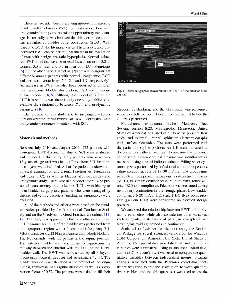
Mean bladder volume during BWt measurement was 310 ml (range 90–770 ml). the mean BWt was 3.9 mm and was significantly higher in the patients with nDO/DSD
compared to those without sphincter dyssynergia (4.2 vs. 3.6 mm, respectively, p < 0.001). BWt was also sig-nificantly higher in male patients, patients with recurrent UtI, patients who emptied their bladder through cIc and patients with low bladder compliance (table 1). By multi-ple linear regression analysis, a significant relation between BWt and bladder compliance was found (R = −0.230, 95 % cI [−0.341, −0.106], p < 0.001). However, no sig-nificant relation between pdet max and BWt was demon-strated (p = 0.107). rOc curve analysis (rOc = 0.624, 95 % cI [0.530, 0.718], p = 0.011) showed that no mean-ingful BWt measurement cutoff could be found to predict an elevated detrusor pressure in the storage phase. Pear-son’s correlation coefficient indicated that there was a weak negative relationship between the length of time since ScI and BWt (r = −0.14, p = 0.018).
Urodynamic evaluation
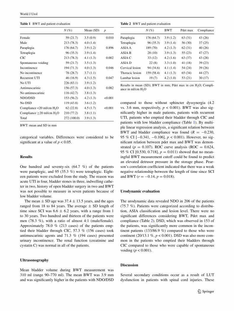
the urodynamic data revealed nDO in 206 of the patients (75.7 %). Patients were categorized according to distribu-tion, aSIa classification and lesion level. there were no significant differences considering BWt, Pdet max and compliance (table 2). DSD, which was observed in 153 of the patients, was significantly more common in the incon-tinent patients (133/86.9 %) compared to those who were continent (20/13.1 %, p < 0.001). DSD was also more com-mon in the patients who emptied their bladders through cIc compared to those who were capable of spontaneous voiding (p < 0.001).
Discussion
Several secondary conditions occur as a result of lUt dysfunction in patients with spinal cord injuries. these
Table 1 BWt and patient evaluation
BWt: mean and SD in mm
N (%) Mean (SD) p
Female 59 (21.7) 3.5 (0.9) 0.010
Male 213 (78.3) 4.0 (1.4)
Paraplegia 176 (64.7) 3.9 (1.2) 0.896
tetraplegia 96 (35.3) 3.9 (1.4)
cIc 213 (78.3) 4.1 (1.3) 0.002
Spontaneous voiding 59 (21.7) 3.5 (1.3)
Incontinence 194 (71.3) 4.0 (1.3) 0.046
no incontinence 78 (28.7) 3.7 (1.1)
recurrent UtI 46 (16.9) 4.3 (1.5) 0.047
no UtI 226 (83.1) 3.9 (1.2)
antimuscarinic 156 (57.3) 4.0 (1.3) 0.082
no antimuscarinic 116 (42.7) 3.8 (1.3)
nDO/DSD 153 (56.2) 4.2 (1.3) <0.001
no DSD 119 (43.8) 3.6 (1.2)
compliance <20 ml/cm H2O 62 (22.8) 4.5 (1.7) <0.001
compliance ≥20 ml/cm H2O 210 (77.2) 3.8 (1.1)
total 272 (100.0) 3.9 (1.3)
Table 2 BWt and patient evaluation
results in mean (SD); BWt in mm; Pdet max in cm H2O; compli-ance in ml/cm H2O
N (%) BWt Pdet max compliance
Paraplegia 176 (64.7) 3.9 (1.2) 63 (31) 43 (26)
tetraplegia 96 (35.3) 3.9 (1.4) 56 (30) 37 (25)
aSIa a 189 (70) 4.2 (1.3) 62 (31) 40 (26)
aSIa B 28 (10) 3.9 (1.3) 55 (23) 47 (27)
aSIa c 33 (12) 4.2 (1.6) 63 (37) 43 (28)
aSIa D 22 (8) 3.3 (1.0) 41 (18) 39 (23)
cervical lesion 94 (34.6) 4.1 (1.4) 54 (24) 39 (26)
thoracic lesion 159 (58.4) 4.1 (1.3) 65 (34) 44 (27)
lumbar lesion 19 (7) 4.2 (1.0) 53 (21) 30 (17)
World J Urol
1 3
conditions are caused by impaired lUt regulation, result-ing in high intravesical pressure. Sustained high intravesi-cal pressure, either due to low bladder compliance or to nDO, if not treated appropriately, further places the upper urinary tract at risk overtime [2]. Unfortunately, the history, level of injury, and signs and symptoms alone are not suf-ficient to determine whether a person is experiencing high intravesical pressure. therefore, urodynamic testing is the “gold standard” diagnostic study of neurologic evaluation to determine bladder and external urethral sphincter func-tion [3, 13].
Measurements of the BWt or the detrusor wall thick-ness (DWt) have received increasing interest as a nonin-vasive test to diagnose BOO. In experimental animals with BOO, the bladder weight and wall thickness increase due to smooth muscle hypertrophy and the deposition of con-nective tissue [14]. these histological changes in the det-rusor during BOO have been confirmed in humans [15, 16]. consequently, it is hypothesized that BWt reflects the workload of the bladder and provides information about the urethral resistance.
Sonographic measurements of the bladder wall have confirmed a low intra- and inter-observer variability, sug-gesting that this technique might be suitable for routine use in patients [17, 18]. the measurement of the anterior blad-der wall is simple, rapid and noninvasive. In healthy adult males and females, bladder thickness decreases rapidly between 50 and 250 ml of bladder filling (or until 50 % of bladder capacity) but reaches a plateau thereafter [19]. In this study, the measurement of BWt was performed with a full bladder or just before cIc on those who were managed through bladder catheterization.
Based on the sample studied, 78.3 % of the patients were under cIc, and 57.3 % took antimuscarinic agents, which is the standard recommended treatment for neu-rogenic bladder due to ScI. the average time since ScI was 6.6 years. therefore, most of the patients had already received the appropriate medical and guidance evaluation with regard to the best management of neurogenic bladder.
BWt was significantly higher in male patients, patients with nDO/DSD, patients with recurrent UtI, patients who emptied their bladder through cIc and patients with low bladder compliance. as was observed in the present study, previous studies have shown significantly higher BWt in males than in females [6, 19]. the higher BWt observed in patients with nDO/DSD is most likely a result of BOO that caused smooth muscle hypertrophy. recurrent UtI is a common finding in patients with neurogenic lUt dys-function. Inflammation may affect the urothelium, result-ing in an increase in BWt. the increased BWt that was observed in patients under cIc is most likely a result of the nDO/DSD that predominated in this group. Unfortu-nately, there was no control group which does not allow
the definition of the normal BWt value in this group of patients.
It is interesting to note that in the current study, the length of time since ScI did not affect significantly BWt. this observation is consistent with our clinical experience. We have seen many patients who developed unfavorable clinical features with severe lUt dysfunction quickly after a ScI. However, there is no doubt that the adequate man-agement of neurogenic bladder plays an important role in protecting the upper and lUt. regarding the use of anti-muscarinic drugs, patients presented no difference in BWt. there was no information given as to when these drugs were started by patients. earlier antimuscarinic blockade can probably prevent structural changes in the bladder wall that would result in increased BWt, but further studies are needed to confirm this assumption.
like in the other studies [7, 10], in our institution, the anterior bladder wall is also routinely used to evaluate lUt dysfunction and we have published a study in chil-dren using this technique. It is not possible to guarantee that the same point of the anterior bladder wall was taken in all patients. Moreover, there is the fact that morphologi-cal variations are common in neurogenic bladder. In most studies, BWt measurement is taken while performing the urodynamic study which in daily medical routine is often not possible. Most studies agree that BWt decreases with bladder filling.
considering the fact that bladder capacity can be extremely variable in patients with neurogenic lUt dys-function, it seems very hard to find any cutoff value for BWt to predict an elevated detrusor pressure. the study showed a wide range of bladder volumes, which associated with a single measurement, may have influenced the find-ings of BWt. However, this range of bladder volumes rep-resents exactly what we have observed in daily routine of this so specific population. Our study reproduced the daily routine of the patients with the BWt being measured when they felt the desire to void or just before the cIc.
Despite the BWt being significantly increased in some groups, it has not been possible to establish a cutoff value considering low bladder compliance and Pdet max. Based on this method, the study showed that the measurement of BWt has no clinical role in patients with ScI and therefore cannot replace urodynamic evaluation. additional studies are necessary to evaluate whether BWt overtime could add insight into which patients may benefit from more aggres-sive treatment at an earlier stage.
a number of confounding variables and the lack of a standardized method have resulted in discrepancies among studies [20]. Issues that may affect the measurement of BWt include the lack of technique standardization, choos-ing between measuring the thickness of the total bladder wall or the detrusor layer alone, exclusion criteria related
World J Urol
1 3
to bladder volume and positioning of the ultrasound probe, principally in neurogenic bladder, in which morphologi-cal variations are common. another limitation of this study was the diagnosis of DSD based on the use of surface elec-tromyography electrodes. a standardized methodology is desirable but has not been established as further validation studies are required in primarily asymptomatic as well as in symptomatic populations.
Acknowledgments We are grateful to the nursing team that sup-ported this work. We are also indebted to the statistician Marcelo anzanello for his valuable statistical analysis.
Conflict of interest the authors declare no conflict of interest.
Ethical standard all patients studies have been approved by the appropriate ethics committee and have therefore been performed in accordance with the ethical standards laid down in the 1964 Decla-ration of Helsinki and its later amendments. all persons gave their informed consent prior to their inclusion in the study.
References
1. Kaplan Sa, chancellor MB, Blaivas Jg (1991) Bladder and sphincter behavior in patients with spinal cord lesions. J Urol 146(1):113–117
2. gerridzen rg, thijssen aM, Dehoux e (1992) risk factors for upper tract deterioration in chronic spinal cord injury patients. J Urol 147(2):416–418
3. Mcguire eJ, Woodside Jr, Borden ta, Weiss rM (1981) Prog-nostic value of urodynamic testing in myelodysplastic patients. J Urol 126(2):205–209
4. Weld KJ, graney MJ, Dmochowski rr (2000) Differences in bladder compliance with time and associations of bladder man-agement with compliance in spinal cord injured patients. J Urol 163(4):1228–1233
5. abrams P, larsson g, chapple c, Wein aJ (1999) Factors involved in the success of antimuscarinic treatment. BJU Int 83(Suppl 2):42–47
6. Hakenberg OW, linne c, Manseck a, Wirth MP (2000) Bladder wall thickness in normal adults and men with mild lower urinary tract symptoms and benign prostatic enlargement. neurourol Urodyn 19(5):585–593
7. Blatt aH, titus J, chan l (2008) Ultrasound measurement of bladder wall thickness in the assessment of voiding dysfunction. J Urol 179(6):2275–2278
8. tanaka H, Matsuda M, Moriya K, Mitsui t, Kitta t, nonomura K (2008) Ultrasonographic measurement of bladder wall thickness as a risk factor for upper urinary tract deterioration in children with myelodysplasia. J Urol 180(1):312–316
9. Ukimura O, Kojima M, Inui e, Ochiai a, naya Y, Kawauchi a et al (1998) noninvasive evaluation of bladder compliance in children using ultrasound estimated bladder weight. J Urol 160(4):1459–1462
10. Pannek J, Bartel P, gocking K, Frotzler a (2013) clinical use-fulness of ultrasound assessment of detrusor wall thickness in patients with neurogenic lower urinary tract dysfunction due to spinal cord injury: urodynamics made easy? World J Urol 31(3):659–664
11. abrams P, cardozo l, Fall M, griffiths D, rosier P, Ulmsten U et al (2002) the standardisation of terminology of lower uri-nary tract function: report from the standardisation sub-commit-tee of the International continence Society. neurourol Urodyn 21(2):167–178
12. Schafer W, abrams P, liao l, Mattiasson a, Pesce F, Spangberg a et al (2002) good urodynamic practices: uroflowmetry, fill-ing cystometry, and pressure-flow studies. neurourol Urodyn 21(3):261–274
13. Watanabe t, rivas Da, chancellor MB (1996) Urodynamics of spinal cord injury. Urol clin n am 23(3):459–473
14. levin rM, Haugaard n, O’connor l, Buttyan r, Das a, Dixon JS et al (2000) Obstructive response of human bladder to BPH vs. rabbit bladder response to partial outlet obstruction: a direct comparison. neurourol Urodyn 19(5):609–629
15. gilpin Sa, gosling Ja, Barnard rJ (1985) Morphological and morphometric studies of the human obstructed, trabeculated uri-nary bladder. Br J Urol 57(5):525–529
16. elbadawi a, Yalla SV, resnick nM (1993) Structural basis of geriatric voiding dysfunction. IV. Bladder outlet obstruction. J Urol 150(5 Pt 2):1681–1695
17. Manieri c, carter SS, romano g, trucchi a, Valenti M, tubaro a (1998) the diagnosis of bladder outlet obstruction in men by ultrasound measurement of bladder wall thickness. J Urol 159(3):761–765
18. naya Y, Kojima M, Honjyo H, Ochiai a, Ukimura O, Watanabe H (1998) Intraobserver and interobserver variance in the meas-urement of ultrasound-estimated bladder weight. Ultrasound Med Biol 24(5):771–773
19. Oelke M, Hofner K, Jonas U, Ubbink D, de la rosette J, Wijkstra H (2006) Ultrasound measurement of detrusor wall thickness in healthy adults. neurourol Urodyn 25(4):308–317
20. Bright e, Oelke M, tubaro a, abrams P (2010) Ultrasound esti-mated bladder weight and measurement of bladder wall thick-ness—useful noninvasive methods for assessing the lower urinary tract? J Urol 184(5):1847–1854