Embed Size (px)
Citation preview
1521-0081/66/2/413–434$25.00 http://dx.doi.org/10.1124/pr.113.008052PHARMACOLOGICAL REVIEWS Pharmacol Rev 66:413–434, April 2014U.S. Government work not protected by U.S. copyright
ASSOCIATE EDITOR: MARK P. MATTSON
G Protein–Coupled Receptor Oligomerization Revisited:Functional and Pharmacological Perspectives
Sergi Ferré, Vicent Casadó, Lakshmi A. Devi, Marta Filizola, Ralf Jockers, Martin J. Lohse, Graeme Milligan, Jean-Philippe Pin,and Xavier Guitart
Integrative Neurobiology Section, National Institute on Drug Addiction, Intramural Research Program, National Institutes of Health,Department of Health and Human Services, Baltimore, Maryland (S.F., X.G.); Department of Biochemistry and Molecular Biology, Faculty
of Biology, University of Barcelona, Barcelona, Spain (V.C.); Centro de Investigación Biomédica en Red sobre EnfermedadesNeurodegenerativas, Barcelona, Spain (V.C.); Department of Pharmacology and Systems Therapeutics (L.A.D.) and Department of
Structural and Chemical Biology (M.F.), Icahn School of Medicine at Mount Sinai, Mount Sinai, New York; Institut National de la Santéet de la Recherche Médicale, U1016, Institut Cochin, Paris, France (R.J.); Centre National de la Recherche Scientifique Unité Mixte de
Recherche 8104, Paris, France (R.J.); Universite Paris Descartes, Sorbone Paris Cite, Paris, France (R.J.); Institute of Pharmacology andToxicology and Rudolf Virchow Center, University of Würzburg, Würzburg, Germany (M.J.L.); Molecular Pharmacology Group, Institute ofMolecular, Cell and Systems Biology, College of Medical, Veterinary and Life Sciences, University of Glasgow, Glasgow, Scotland, United
Kingdom (G.M.); Centre National de la Recherche Scientifique, Unité Mixte de Recherche 5203, Institut de Génomique Fonctionnelle,Université Montpellier, Montpellier, France (J.-P.P.); and Institut National de la Santé et de la Recherche Médicale, U661, Montpellier,
France (J.-P.P.)
Abstract. . . . . . . . . . . . . . . . . . . . . . . . . . . . . . . . . . . . . . . . . . . . . . . . . . . . . . . . . . . . . . . . . . . . . . . . . . . . . . . . . . . . . 413I. Morphologic Aspects of G Protein–Coupled Receptor Oligomerization . . . . . . . . . . . . . . . . . . . . . . . . . 414
A. The Search for the Predominant Oligomeric G Protein–Coupled Receptor Species . . . . . . . . . 414B. The Search for Oligomer Interfaces . . . . . . . . . . . . . . . . . . . . . . . . . . . . . . . . . . . . . . . . . . . . . . . . . . . . . . 416C. Determinants of G Protein–Coupled Receptor Heteromerization . . . . . . . . . . . . . . . . . . . . . . . . . . 417
II. Functional Aspects of G Protein–Coupled Receptor Oligomerization . . . . . . . . . . . . . . . . . . . . . . . . . . 418A. Principles of Allosterism and Properties of Allosteric Modulators . . . . . . . . . . . . . . . . . . . . . . . . . 418B. Allosterism in Receptor Oligomers: Modulation of Ligand Affinity. . . . . . . . . . . . . . . . . . . . . . . . . 419C. Allosterism in Receptor Oligomers: The Two-State Dimer Model . . . . . . . . . . . . . . . . . . . . . . . . . . 421D. Allosterism in Receptor Oligomers: Allosteric Modulation of Intrinsic Efficacy
and Functional Asymmetry . . . . . . . . . . . . . . . . . . . . . . . . . . . . . . . . . . . . . . . . . . . . . . . . . . . . . . . . . . . . . . 424III. G Protein–Coupled Receptor Oligomers as Novel Targets for Drug Development . . . . . . . . . . . . . . 426
A. Localization of Receptor Oligomers in Native Tissues. . . . . . . . . . . . . . . . . . . . . . . . . . . . . . . . . . . . . 426B. Do Receptor Heteromers Constitute Possible Targets for Drug Development? . . . . . . . . . . . . . 428C. Approaches for the Identification of Receptor-Heteromer Selective Compounds . . . . . . . . . . . . 430Acknowledgments . . . . . . . . . . . . . . . . . . . . . . . . . . . . . . . . . . . . . . . . . . . . . . . . . . . . . . . . . . . . . . . . . . . . . . . . . . . 431References . . . . . . . . . . . . . . . . . . . . . . . . . . . . . . . . . . . . . . . . . . . . . . . . . . . . . . . . . . . . . . . . . . . . . . . . . . . . . . . . . . 431
Abstract——Most evidence indicates that, as forfamily C G protein–coupled receptors (GPCRs), familyA GPCRs form homo- and heteromers. Homodimersseem to be a predominant species, with potential
dynamic formation of higher-order oligomers, particularlytetramers. Although monomeric GPCRs can activateG proteins, the pentameric structure constitutedby one GPCR homodimer and one heterotrimeric
This work was supported by the Intramural Research Program of the National Institutes of Health [National Institute on Drug Abuse] (toS.F. and X.G.); the National Institutes of Health National Institute on Drug Abuse [Grants DA026434 and DA034049 (to M.F.) and GrantsDA008863 and DA019521 (to L.A.D.)]; “Ministerio de Ciencia y Tecnología” [Grant SAF2011-23813], “Centro de Investigación Biomédica enRed sobre Enfermedades Neurodegenerativas” [Grant PI2011/02-7], and “Fondation Jerome Lejeune” [FJL-01/01/2013] (to V.C.); “AgenceNationale de la Recherche” [Grants ANR RPIB 2012 “MED-HET-REC-2”, ANR-11-IDEX-0005-01, and ANR-11-LABX-0071 (to R.J.) and ANR-12-BSV2-0015 and ARN-09-BIOT-018 (to J.-P.P.)]; “Fondation Recherche Médicale” [Grant FRM DEQ20130326503], “Institut National de laSanté et de la Recherche Médicale,” and “Centre National de la Recherche Scientifique” (to R.J.); European Research Council Advanced Grant“Topas” (to M.J.L.); and Medical Research Council [Grant G0900050] (to G.M.).
Address correspondence to: Sergi Ferré, Integrative Neurobiology Section, National Institute on Drug Abuse, Intramural ResearchProgram, National Institutes on Drug Abuse, Department of Health and Human Services, 333 Cassell Drive, Baltimore, Maryland 21224.E-mail: [email protected]
dx.doi.org/10.1124/pr.113.008052.
413
by guest on Novem
ber 17, 2018D
ownloaded from
G protein may provide a main functional unit, andoligomeric entities can be viewed as multiplesof dimers. It still needs to be resolved if GPCRheteromers are preferentially heterodimers or if theyare mostly constituted by heteromers of homodimers.Allosteric mechanisms determine a multiplicity ofpossible unique pharmacological properties of GPCRhomomers and heteromers. Some general mechanismsseem to apply, particularly at the level of ligand-bindingproperties. In the frame of the dimer-cooperativitymodel, the two-state dimer model provides the mostpractical method to analyze ligand–GPCR interactions
when considering receptor homomers. In addition toligand-binding properties, unique properties for eachGPCR oligomer emerge in relation to different intrinsicefficacy of ligands for different signaling pathways(functional selectivity). This gives a rationale for theuse of GPCR oligomers, and particularly heteromers,as novel targets for drug development. Herein, wereview the functional and pharmacological propertiesof GPCR oligomers and provide some guidelines forthe application of discrete direct screening and high-throughput screening approaches to the discovery ofreceptor-heteromer selective compounds.
I. Morphologic Aspects of G Protein–CoupledReceptor Oligomerization
A. The Search for the Predominant Oligomeric GProtein–Coupled Receptor Species
Although G protein–coupled receptors (GPCRs) wereinitially thought to be, and function exclusively as,monomeric entities, evidence accumulated over the pasttwo decades indicates that they can form homomers andheteromers in intact cells (Bouvier, 2001; Milligan andBouvier, 2005; Pin et al., 2007; Ferré et al., 2009). It isnow well accepted that family C GPCRs (e.g., metabo-tropic glutamate, calcium-sensing receptors, GABAB,and sweet and umami taste receptors) form constitutivehomo- or heteromers (Kniazeff et al., 2011). Suchobservations raised a long debated question aboutwhether family A (rhodopsin-like) GPCR dimers werealso constitutive and required for G protein activation.A clear demonstration that this is not the case camefrom studies in which monomeric entities were trappedinto nanodiscs. In such experiments, it was demon-strated that b2-adrenergic, rhodopsin, and m-opioidreceptors function effectively as monomers (Bayburtet al., 2007; Whorton et al., 2007, 2008; Kuszak et al.,2009). Also, monomeric rhodopsin in solution activatedits G protein transducin at the diffusion limit (Ernstet al., 2007). Further support for the functionality ofmonomeric GPCR units came when isolated monomeric
seven transmembrane (7TM) domains of family CGPCRs were shown to be capable of fully activating Gprotein when directly activated by synthetic smallmolecules (El Moustaine et al., 2012). Finally, a 1:1stoichiometry has also been found to be sufficient forrhodopsin–b-arrestin binding (Tsukamoto et al., 2010;Bayburt et al., 2011). But those observations did notexclude that class A GPCR oligomers can spontaneouslyform in living cells, as it is well demonstrated withfamily C GPCRs, and raised the question of theirfunctional significance. In fact, the three GPCRs shownto be functional as strict monomers were shown also toexist as dimers or higher-order oligomers in living cells(see below). One of the research groups that demon-strated the functionality of GPCR monomers in nano-discs found evidence for the existence of stable b2
receptor oligomers, mostly tetramers, after reconstitu-tion into phospholipid vesicles (Fung et al., 2009). Also,rhodopsin–b-arrestin binding stoichiometry in isolatedrod outer segment membranes was found to depend onthe percentage of activated receptors, increasing from1:1 to 2:1 (Sommer et al., 2012).
An explosion of data supporting the existence ofhomo- and heteromers of GPCRs in intact cells (storedin the GPCR Oligomerization Knowledge Base; http://www.gpcr-okb.org; Khelashvili et al., 2010) came withthe widespread use of biophysical techniques, suchas resonance energy transfer [bioluminescence and
ABBREVIATIONS: A, concentration of radioligand; B, concentration of the nonradioactive competing compound in radioligand bindingcompetition experiments; BRET, bioluminescence resonance energy transfer; CYM51010, ethyl 1-[4-(acetylamino)benzyl]-4-(2-phenylethyl)piperidine-4-carboxylate; CGS 21680, 4-[2-[[6-amino-9-(N-ethyl-b-D-ribofuranuronamidosyl)-9H-purin-2-yl]amino]ethyl]benzenepropanoicacid; DAB, dimer radioligand/competitor modulation index in radioligand competition experiments; DC, dimer cooperativity index inradioligand binding saturation experiments; DCB, dimer cooperativity index for the competing ligand in radioligand binding competitionexperiments; DDS, discrete direct screening; FCS, fluorescence correlation spectroscopy; FRET, fluorescence resonance energy transfer;GPCR, G protein–coupled receptor; GRK, GPCR kinase; H8, helix 8; HTS, high-throughput screening; KD1 and KD2, macroscopic equilibriumdissociation constants for the binding of A to the first and second receptor in the receptor homodimer in radioligand binding saturationexperiments; KDA1 and KDA2, macroscopic equilibrium dissociation constants for the binding of A to the first and second receptor in thereceptor homodimer in radioligand binding competition experiments; KDAB, equilibrium dissociation constant of B on a receptor homodimersemi-occupied by A in radioligand binding competition experiments; KDB1 and KDB2, macroscopic equilibrium dissociation constants for thebinding of B to the first and second receptor in the receptor homodimer in radioligand binding competition experiments; KDBA, equilibriumdissociation constant of A binding to a receptor homodimer semi-occupied by B in radioligand binding competition experiments; MAPK,mitogen-activated protein kinase; MPTP, 1-methyl-4-phenyl-1,2,3,6-tetrahydropyridine; MSX-3, 3,7-dihydro-8-[(1E)-2-(3-methoxyphenyl)ethenyl]-7-methyl-3-[3-(phosphonooxy)propyl-1-(2-propynyl)-1H-purine-2,6-dione; OTA, d(CH2)
5[Tyr(Me)2-Thr4-Orn8-Tyr(NH2)9]-oxytocin;
PLA, proximity ligation assay; RGS20, protein that regulates the speed of G protein signal transduction; RT, total amount of receptordimers; SCH-442416, 2-(2-furanyl)-7-[3-(4-methoxyphenyl)propyl]-7H-pyrazolo[4,3-e][1,2,4]triazolo[1,5-c]pyrimidin-5-amine; SKF-83959,6-chloro-7,8-dihydroxy-3-methyl-1-(3-methylphenyl)-2,3,4,5-tetrahydro-1H-3-benzazepine; TIR-FM, total internal reflection fluorescencemicroscopy; TM, transmembrane domain; ZM-241385, 4-(2-[7-amino-2-(2-furyl)[1,2,4]triazolo[2,3-a][1,3,5]triazin-5-ylamino]ethyl)phenol.
414 Ferré et al.
fluorescence resonance energy transfer (BRET andFRET, respectively)], fluorescence complementation, orcombination of these techniques (Milligan and Bouvier,2005; Gandía et al., 2008; Pin et al., 2007, 2009; Bacartet al., 2008; Carriba et al., 2008; Guo et al., 2008;Cabello et al., 2009; Ferré et al., 2009; Urizar et al.,2011). These techniques, however, have largely fallenshort in answering questions about the size of theoligomer complexes and their possible dynamic nature.Initial evidence for GPCR oligomerization in a physio-logically relevant system came from atomic forcemicroscopy experiments in native disk membranesfrom mice, which showed rhodopsin to be arranged inparacrystalline arrays of dimers (Fotiadis et al., 2003).These studies raised severe criticism (Chabre and leMaire, 2005), and more recent studies using single-molecule techniques have begun to address the detailsof the spatial and temporal organization of GPCRcomplexes in living cells by directly observing the stateand behavior of individual proteins in the cell. Single-molecule total internal reflectance fluorescence micros-copy (TIR-FM) was first used to track the position ofindividual molecules of muscarinic acetylcholine M1
and N-formyl peptide receptors using fluorescentlylabeled ligands (Hern et al., 2010; Kasai et al., 2011).Those ligands had such slow off-rates that they may beconsidered essentially irreversibly bound. Both studiessuggested a transient (second-scale) formation anddissociation of dimers, with 30–40% proportion ofdimers at any given time. These conclusions requirethat the fluorescent ligands bind with similar affinityto both protomers in the dimer (see below fordiscussion of ligand cooperativity within receptordimers/oligomers; see Section II.B), and it is possiblethat the fluorescent ligand might stabilize the receptorin a nonrepresentative conformational state. Despitethese concerns, similar studies have reached equiva-lent conclusions on the state of the muscarinicacetylcholine M2 receptor in cardiac muscle (Nena-sheva et al., 2013). More recently, TIR-FM was usedtogether with SNAP-tag technology to directly labelcell-surface GPCRs with organic fluorophores to dy-namically monitor individual b1- and b2-adrenoceptorsas well as GABAB receptors on the surface of living,transiently transfected cells (Calebiro et al., 2013).This study showed that all three receptors form dimersand higher-order oligomers (also with estimations ofdynamic second-scale receptor–receptor interactions)in a proportion dependent on the subtype of receptorand on the receptor density. At low densities, mono-meric species were predominant for the b1-adrenocep-tor, whereas b2-adrenoceptors displayed a higherproportion of dimers. A step-wise appearance of firstdimers and successively trimers, tetramers, andupwards to higher-order complexes appeared withprogressive increase in receptor density. At densitiescomparable to receptor expression in native tissue,
dimers and higher-order oligomers were the predom-inant species for both receptors, and agonists did notmodify the oligomerization status (Calebiro et al.,2013). As expected (Maurel et al., 2008; Comps-Agraret al., 2011), GABAB receptors showed mostly dimericand tetrameric species at lower densities, but higher-order species also appeared with increased expressionlevels (Calebiro et al., 2013).
Fluorescence correlation spectroscopy (FCS) is an-other indirect but useful technique to determine theoligomer status of protein clusters. It measures themolecular brightness of a fluorescence-tagged protein,which provides an estimate of the number of fluorescentmolecules by recording fluctuations in fluorescenceintensity arising from individual fluorescent molecules(Chen et al., 2003). FCS has been used to study m-opioidreceptor homo- and heteromerization with d-opioidreceptors, and number and brightness measurementssuggested that m-opioid receptors exist primarily asdimers that oligomerize with d-opioid receptors intotetramers (Golebiewska et al., 2011). The recent FCSwith a particle counting histogram approach by Herrick-Davis et al. (2013) also provides support for homodimersbeing the predominant, and perhaps only, species forseveral GPCRs, including a1B-adrenoceptor, b2-adreno-ceptor, serotonin 5-HT2A and 5-HT2c, muscarinic ace-tylcholine M1 and M2, and dopamine D1 receptors. Aswith the results obtained with TIR-FM, the homodimerconfiguration was unaltered by agonist treatment(Calebiro et al., 2013; Herrick-Davis et al., 2013). Unlikethe fast dynamic behavior of GPCR monomers/dimersshown in TIR-FM experiments (Hern et al., 2010; Kasaiet al., 2011; Calebiro et al., 2013), however, the dimerswere stable over a 10-fold range of receptor expressionlevels (Herrick-Davis et al., 2013). This is also in linewith other findings that suggest much more stableinteractions, such as higher-order dopamine D2 receptoroligomers, over a high range of receptor expression (Guoet al., 2008) and stable b2-adrenoceptor tetramers inphospholipid vesicles (Fung et al., 2009). Stability, atleast over a substantial time period, of GPCR dimers/oligomers is also inferred from experiments that in-dicate these complexes are generated at an early stageof biosynthesis. In fact, it has been suggested that early-stage dimerization may be required for effective foldingand maturation of the receptor (Salahpour et al., 2004;Bulenger et al., 2005; Milligan, 2010, 2013). The samereasoning has been used based on evidence for cointern-alization, with the capacity of a selective ligand of one ofthe receptors in a GPCR heteromer to cointernalize thetwo different receptors constituting the heteromer(Hillion et al., 2002; Milligan, 2010, 2013; Ward et al.,2011; Tadagaki et al., 2012). Negative results shouldalso be acknowledged, such as the recent study byGavalas et al. (2013), which measured the recruitmentof subsets of histidine-tagged GPCR protomers intoartificial microdomains (containing immobilized metal
G Protein–Coupled Receptor Oligomerization 415
affinity chromatography or streptavidin beads) on thesurface of living cells and determines the simultaneouscorecruitment of untagged protomers. The study onlyshowed evidence for corecruitment of metabotropicglutamate receptor protomers, but not b2-adrenergicor m-opioid protomers, into such artificial microdomains.Apart from differences between different receptors orwhen considering receptor homomers or heteromers, itmight be that a number of the approaches used to studyoligomerization are unable to resolve fluctuations inreceptor interactions occurring on a second or subsecondscale. On the other hand, mammalian transfected cellsmight be lacking elements that increase the stability ofthe interfaces that determine the receptor–receptorinteractions, which might also be different dependingon the cellular compartment studied.If different interfaces are involved in receptor oligo-
merization, we could expect dimers to be predominantspecies, determined by the most stable interaction,followed by tetramers (dimers of dimers), determined bya weaker interaction. Recently calculated estimates of therelative stability of different, putative dimeric interfacesof different GPCR subtypes using extended biasedmolecular dynamics simulations in explicit lipid-waterenvironments argue in favor of a variable strength ofassociation depending on the specific residue compositionor shape of the interface, despite an overall transiency inreceptor–receptor interactions (Johnston et al., 2012).Notably, simulations of b1- and b2-adrenoceptors sug-gested a model of oligomerization in which more stablehomodimers involving TM1 diffuse through the mem-brane and transiently interact with other protomers/dimers via other TM helices (e.g., TM4). In agreementwith these predictions are the results obtained in a recentstudy with the muscarinic acetylcholine M3 receptor,using quantitative FRET spectrometry techniques withcontrolled expression of the energy donor-tagged species(Patowary et al., 2013). Mathematical analysis of theFRET efficiencies obtained from spectral unmixing wascompatible with the M3 receptor existing as stabledimeric complexes, a large fraction of which interacteddynamically to form tetramers that were specificallywithin a rhombic organization rather than a square orlinear configuration (Patowary et al., 2013).
B. The Search for Oligomer Interfaces
A very intriguing set of inferences in the field ofGPCR oligomerization has been based on recentlyobtained high-resolution crystallographic structures,including those of the chemokine CXCR4, the m-opioidand k-opioid receptors, the b1-adrenoceptor, and thesmoothened receptor (Wu et al., 2010, 2012; Mangliket al., 2012; Huang et al., 2013; Wang et al., 2013). Eachof these crystallized as parallel dimers and/or tet-ramers. Although some of the observed interfaces mayreflect artifacts of crystal packing and the conditionsemployed for crystallization, a tentative first suggestion
from these studies is the existence of different dimerinterfaces for different GPCR homodimers. Neverthe-less, some TM domains have been observed more oftenthan others. TM5 and TM6 residues constituted themain interfaces for chemokine CXCR4 and m-opioidreceptor crystallized dimers, although there weremarked differences between them, with only a fewcontacts between specific residues for chemokineCXCR4 and extensive contacts throughout the length ofthese TM helices in m-opioid receptor dimers (Wu et al.,2010; Manglik et al., 2012). Involvement of TM6 wasalso suggested early on for b2-adrenoceptor dimers(Hebert et al., 1996) and for the leukotriene receptorBLT1 (Baneres and Parello, 2003) by the use of in-terfering synthetic peptides with the same sequenceas TM6. Also, by using cysteine cross-linking tech-niques, TM5 was suggested to be involved in homodimer-ization of dopamine D2, muscarinic M3, and serotonin5-HT2C receptors (Guo et al., 2005; Mancia et al., 2008;Hu et al., 2012).
Apart from the TM5-TM6 interface, crystallizedchemokine CXCR4 dimers also showed contacts at theintracellular ends of TM3 and TM4 (Wu et al., 2010),and m-opioid dimers also showed a second, lessprominent symmetric interface, involving TM1, TM2,and, also, helix 8 (H8; the helix adjacent to TM7 runningalong the internal membrane surface) (Manglik et al.,2012). A TM1–TM2–H8 interface (with slightly differentcontact residues) was also found in crystals of k-opioidreceptor dimers (Wu et al., 2012), rhodopsin (Ruprechtet al., 2004; Salom et al., 2006), opsin (ligand-freerhodopsin; Park et al., 2008), and b1-adrenoceptor(Huang et al., 2013). An additional interface involvingTM4 and TM5 was also obtained for the b1-adrenocep-tor and smoothened receptor (Wang et al., 2013).Notably, the two crystallographic interfaces of theb1-adrenoceptor (TM1–TM2–H8 and TM4–TM5) weresuggested to be physiologically relevant with cysteine-cross-linking experiments (Huang et al., 2013). A modelof the potential rhodopsin dimer was initially built bydocking the rhodopsin crystal structure into the unit cellconstraints determined by atomic force microscopy. Themodel included a primary interface between protomersinvolving TM4 and TM5, which implied asymmetricbinding of one heterotrimeric G protein to each proto-mer (Liang et al., 2003; Fotiadis et al., 2004). The modelalso suggested a secondary interface holding rows ofdimers together, which was later shown to involve TM1,TM2, and H8 (Salom et al., 2006). The possibleinvolvement of a TM1–TM2–H8 interface in rhodopsinquaternary organization was recently supported bycross-linking experiments of endogenous cysteines ofrhodopsin in disk membranes (Knepp et al., 2012).
It is important to remember that crystal structuresare not only the result of specific crystallization con-ditions, but they correspond to static, ligand-specificconformational states of receptors stripped of their
416 Ferré et al.
natural lipid environment. For GPCRs, the majority ofcrystal structures that are currently available refer toantagonist-bound (inactive) structures. The inferreddimeric interfaces may, therefore, depend on thosespecific conformational states. As suggested by Huanget al. (2013), the comparison of the differences in theinterfaces observed from the crystallized structures ofthe antagonist-bound m-opioid and chemokine CXCR4receptors and the ligand-free b1-adrenoceptor mightsuggest that the TM5 interface can partner in the inter-action with TM4 or TM6, depending on the conforma-tion of the receptor. As mentioned by Manglik et al.(2012), the TM5-TM6 interface inferred by the crystalstructure of m-opioid dimers could preclude eitherprotomer from properly coupling to G protein, becausethe agonist-induced receptor–G protein interactiondepends on rearrangements of TM5 and TM6 withinthe seven-helical domain bundle. Therefore, it is im-portant to assess if, by stabilizing different receptorconformations, different ligands also promote differentdimeric interfaces. By cross-linking substituted cys-teines, Guo et al. (2005) suggested TM4 to be a maininterface in the dopamine D2 receptor oligomer. Cross-linking of a different set of cysteines in TM4 was slowedby inverse agonists and accelerated by agonists. In fact,cross-linking of the latter set of cysteines locked thereceptor in an active state, strongly suggesting thata conformational change at the TM4 dimer interface ispart of dopamine D2 receptor activation (Guo et al.,2005). Mancia et al. (2008), also using the cysteine cross-linking approach, found two interfaces in serotonin5-HT2C receptor dimers involving TM4–TM5 and TM1,with only TM4–TM5 being selectively sensitive toreceptor activation. In summary, although a pattern ofsimilar interfaces of GPCR homomers seems to beemerging, different interfaces can be found in differentoligomers and even in different conformations of thesame oligomer. Although agonists did not modify thedynamics of b2-adrenoceptor oligomerization in TIR-FMexperiments (Calebiro et al., 2013) or the stability ofb2-adrenoceptor oligomers in FCS–photon-counting his-togram experiments (Herrick-Davis et al., 2013) or inFRET experiments in receptors into reconstituted phos-pholipid vesicles, an inverse agonist did modify thestability of b2-adrenoceptor oligomers in this latterpreparation (Fung et al., 2009). Thus, selected ligands(which can stabilize specific conformations of the recep-tor) may still modify GPCR oligomeric interfaces and,therefore, the dynamics of receptor oligomerization.
C. Determinants of G Protein–CoupledReceptor Heteromerization
When attempting to understand the predominantoligomeric species within receptor heteromers, anadditional issue is whether the complexes exist asheterodimers or as hetero-oligomers generated fromindividual homodimers. The lack of detection of trimers
in situations in which interconversion between dimersand tetramers of the muscarinic M3 receptor wasobserved (Patowary et al., 2013) is consistent with adimer + dimer model and with the organization of classC GABAB dimers and tetramers (Comps-Agrar et al.,2012). Some indirect biochemical data also support thismodel for class A receptors. These include cooperativebinding of certain ligands to adenosine A2A and A1
receptor heteromers (see below and Orru et al., 2011a).Furthermore, oligomerization of more than two differ-ent GPCRs has been suggested from studies withsequential BRET-FRET, BRET plus bimolecular fluo-rescence complementation (Carriba et al., 2008; Cabelloet al., 2009; Navarro et al., 2010). Tentatively, theseapparent heterotrimers could represent heteromultim-ers of homomers, as suggested for adenosine A2A–
dopamine D2–cannabinoid CB1 receptor heteromers(Navarro et al., 2010).
Apart from interactions between TM domains, severalstudies have provided evidence for disulfide bridgesbetween extracellular domains of class C GPCR homo-mers (Kniazeff et al., 2011) and for a key role ofelectrostatic interactions between intracellular receptordomains in receptor heteromerization (Woods andFerré, 2005). These electrostatic interactions dependon the very asymmetric and disordered structure ofintracellular domains and have been suggested to beinvolved in several receptor heteromers (Ciruela et al.,2004; Woods and Ferré, 2005; Navarro et al., 2010;O’Dowd 2012, 2013). Several general features of theregions involved in these interactions have been out-lined: one region, in one of the receptors, containsa series of adjacent arginine residues, and the otherregion, in the other receptor, contains acidic residues,with several adjacent residues or at least one phosphor-ylated serine. Once established, the polyarginine–phosphate electrostatic interaction possesses a strongstability. Thus, these bonds can withstand fragmenta-tion by mass spectrometric collision-induced dissocia-tion at energies similar to those that fragment covalentbonds, and they demonstrate an extremely low dissoci-ation constant by surface plasmon resonance (Woodsand Ferré, 2005). If electrostatic interactions betweenintracellular domains are more predominant in receptorheteromers (not reported yet in receptor homomers) andare stronger than those provided by TM interfaces, wecould speculate that some receptor heteromers could bemore stable than receptor homomers. An example ofa functionally relevant electrostatic interaction is theone involved in adenosine A2A–dopamine D2 receptorheteromerization, with the arginine-rich domain local-ized in the N-terminal portion of the long thirdintracellular loop of the dopamine D2 receptor and theacidic domain in the distal part of the long C terminus ofthe adenosine A2A receptor (Ciruela et al., 2004;Navarro et al., 2010). Mutation- or peptide-mediated dis-ruption of the A2A–D2 receptor electrostatic interaction
G Protein–Coupled Receptor Oligomerization 417
produces a profound destabilization of the quaternarystructure of the heteromer (Navarro et al., 2010) withdisappearance of significantly relevant A2A–D2 recep-tor interactions in brain tissue, such as the adenosineA2A receptor–mediated inhibition of dopamine D2 receptor–induced depression of striatal neuronal firing (Azdad et al.,2009).In summary, most evidence indicates that, as for
family C GPCRs, which can be found as strict homo-dimers, heterodimers (Doumazane et al., 2011), as wellas stable heteromers (Comps-Agrar et al., 2012), familyA GPCRs form homo- and heteromers in heterologoussystems. Homodimers seem to be a predominant specieswith potential dynamic formation of higher-order olig-omers, particularly tetramers. It still needs to be resolvedif class A GPCR heteromers are preferentially hetero-dimers or if they are mostly constituted by heteromersof homodimers. In the next sections, we will review theevidence that GPCR homo- and heteromers constitutefunctional and pharmacologic units.
II. Functional Aspects of G Protein–CoupledReceptor Oligomerization
A. Principles of Allosterism and Properties ofAllosteric Modulators
Allosterism can be defined as the process by which theinteraction of a chemical or protein at one location ona protein or macromolecular complex (the allosteric site)influences the binding or function of the same oranother chemical or protein at a topographically distinctsite (Christopoulos and Kenakin, 2002). This definitionprovides a framework to understand the biochemicalproperties of GPCR homomers and heteromers. Assuggested by Kenakin and Miller (2010), it is useful todefine allosterism in terms of three interacting species:the “modulator,” a ligand or protein that binds to the“conduit” (usually a protein; the GPCR protomer oroligomer in this review), which transmits the thermo-dynamic allosteric energy to the “guest,” the target ofthe allosteric modulation. With GPCRs, we can thenconsider three different kinds of allosterism dependingon the location of the target of the allosteric modulation.If the target of the allosteric modulation is anotherligand cobinding with the allosteric modulator, this isreferred to as classic allostery. If the target of theallosteric modulation resides in the cytosol, it may becalled cytosolic allosterism. Finally, if the target of theallosteric modulation interacts with the conduit of theallosteric modulation along the plane of the membrane,this is referred to as lateral allosterism, with a mainexample being allosterism in receptor oligomers (Kena-kin and Miller, 2010).An example of classic allosterism would be the case of
a ligand that modulates allosterically the effect of anorthosteric agonist. An orthosteric agonist has two mainindependent properties: an affinity for the receptor and
an intrinsic efficacy, which determines the power of theagonist to induce a functional response. The allostericmodulator can have different and independent effects onthese properties of distinct orthosteric agonists (Kenakinand Miller, 2010; Smith and Milligan, 2010). Therefore,there may be two general effects of allosteric modu-lators on orthosteric ligands: on their affinity and onintrinsic efficacy. Analysis of functional responses willdetermine the contribution of both allosteric effects.Another property of allosteric modulators is saturabil-ity. A negative allosteric modulator that selectivelymodifies the affinity of an orthosteric agonist will dis-place the functional dose-response curve to higher con-centrations, but only up to a certain extent. By contrast,for a competitive orthosteric antagonist, such a "rightshift" would continue ad infinitum. This ceiling effect ofclassic allosteric modulators can have important ther-apeutic implications by reducing overdose effects, com-pared with orthosteric ligands (Kenakin and Miller,2010; Smith and Milligan, 2010).
In cytosolic allosterism (as defined by Kenakin andMiller, 2010), modulated proteins are cytosolic signalingproteins, such as G proteins, GPCR kinases (GRKs), andb-arrestins. Importantly, this type of allosterism canlead to functional selectivity, i.e., the ability of a ligandto selectively promote a specific cellular signaling event(Reiter et al., 2012). The rationale is that differentligands can stabilize different conformations of thereceptor. If different cytosolic proteins that mediatedifferent signaling interact with different residues orphosphorylated residues of the GPCR, it may be ex-pected that some conformations can favor or impair thebinding of a particular signaling protein or that they caninduce a different conformation of the cytosolic protein,leading to biased agonism or biased antagonism (Reiteret al., 2012). This can also have important therapeuticimplications, i.e., when a particular signaling pathwayor end point is associated with a therapeutic response,whereas another is associated with nonwanted or sideeffects. Agonist binding to GPCRs and G protein activa-tion are rapidly followed by several coordinated eventscommon to most GPCRs. These include recruitment ofGRKs that phosphorylate the receptor at multipleintracellular sites, followed by the recruitment ofb-arrestins, which trigger receptor endocytosis. But, inaddition to canonical G protein–mediated signaling,GPCRs can also bind to other cytosolic adaptors, in-cluding b-arrestins, which elicit G protein–independentsignaling through activation of mitogen-activated proteinkinase (MAPK) and Akt. Most known endogenous andexogenous ligands can signal through both signalingmechanisms. There are already, however, several exam-ples of biased ligands that preferentially signal throughb-arrestins over G proteins. In some cases, this may bea therapeutically beneficial effect, in others an unwantedone. Carvedilol, for instance, has been reported to be ab-arrestin–biased ligand acting at b2-adrenoceptors (but
418 Ferré et al.
also at b1-adrenoceptors), which may contribute to itsclinical value in heart failure beyond its b-blockerproperty (Reiter et al., 2012). The work by Lefkowitzand coworkers indicates that a mechanism behindfunctional selectivity can be a ligand-dependent “phos-phorylation barcoding” (concept developed by Tobinet al., 2008). Thus, the b-arrestin–biased agonism ofcarvedilol appears to depend on its ability to impairGRK2- and promote GRK6-mediated phosphorylation(Nobles et al., 2011). Another mechanism is the dif-ferential structural conformations that lead to a pref-erential binding for specific G proteins. For instance,different agonists of the cannabinoid CB1 receptordifferentially regulate the binding of the three homol-ogous Gi proteins to the receptor. Whereas one ligand(desacetyllevonantradol) behaved as an agonist for Gi1
and Gi2 and an inverse agonist for Gi3, another ligand[(R)-methanandamide] behaved in exactly the oppositemanner, as an inverse agonist for Gi1 and Gi2 and anagonist for Gi3 (Mukhopadhyay and Howlett, 2005).
B. Allosterism in Receptor Oligomers: Modulation ofLigand Affinity
Evidence for the existence of GPCR homomers (andheteromers) could already be deduced some time agofrom analysis of radioligand-binding experiments, withthe evidence of complex binding, such as upwardconcave nonlinear Scatchard plots in saturation experi-ments and as biphasic curves in agonist–antagonistcompetitive-inhibition experiments. Such complex curveswere usually explained by two nonoverlapping models(reviewed in Casadó et al., 2007). First is the mono-mer–G protein model, which considers receptors asmonomers with two independent populations not inequilibrium. One population would be bound and theother would not be bound to G proteins, with thereceptors coupled to G proteins having higher affinityfor the agonist. Receptors with high affinity could thenbe converted into low affinity with the addition of GTP,because it would uncouple the G protein from thereceptor (De Lean et al., 1980). To explain the complexradioligand binding curves, this first mechanism wouldassume a preexisting proportion of both populations ofreceptors and, therefore, a limited pool of G proteins.The second model, the dimer-cooperativity model, con-siders oligomerization, or at least GPCR dimers. In thiscase, allosteric communication through the two proto-mers allows negative cooperativity, meaning that thebinding of a ligand to the first protomer decreases theaffinity of the ligand for the second protomer. Cooper-ativity (positive or negative) is a particular type ofallosteric modulation in receptor oligomers, where theprotomers of a homodimer are the conduit of the allo-steric modulation and the same ligand is the allostericmodulator (binding to the first protomer) and themodulated target (binding to the second protomer).This mechanism does not assume a limited pool of
G proteins, which is always present and acts as anadditional allosteric modulator that increases theaffinity of the agonist, and provides the conformationof the dimer that allows negative cooperativity ofa ligand through the protomers.
In apparent support for the first mechanism, in thestudy by Whorton et al. (2007) with reconstitution ofmonomers of b2-adrenoceptors and G proteins in high-lipoprotein nanoparticles, antagonist/agonist competi-tive inhibition curves showed biphasic curves, and theaddition of a GTP analog converted the low affinitypopulation into high affinity. The study also showed,however, that the proportion of receptors with highaffinity state increases up to 100% by increasing the Gprotein pool, indicating that in situ, in membranepreparations of brain tissue, the detection of twopopulations of receptors would indicate a limited poolof G proteins. In this case, the monomeric model withtwo G protein–dependent populations of receptors couldexplain the upward concave Scatchard plots and thebiphasic antagonist/agonist competition curves (Casadóet al., 2007). The upward concave Scatchard curveswould be the result of the addition of two curves thatdescribe the two independent populations, with highand low affinities for the agonist. The biphasic compe-tition curves would be the result of two different affin-ities of the agonist, which would identify both populations,displacing the antagonist that would have the sameaffinity for both populations. However, some earlystudies indicated that antagonist binding could also bemodulated by guanine nucleotides, which, according tothe monomer–G protein model, would suggest that an-tagonist binding would also be dependent on G proteincoupling (Burgisser et al., 1982; De Lean et al., 1982;Klotz et al., 1990). Dissociation kinetic experimentscan resolve the enigma. Investigation of the dissocia-tion kinetics of a tracer ligand in the absence and pres-ence of a second ligand represents a sensitive methodto detect cooperative interactions between two topo-graphically distinct binding sites. Thus, ligands thatcompete for the same site on a monomeric receptorshould not influence one another’s dissociation kinet-ics. In contrast, allosteric modulation between two si-multaneously bound and interacting sites, either withina receptor monomer or across a receptor dimer oroligomer, should alter ligand dissociation (May et al.,2007). This analysis has been used to demonstratehomomerization of several GPCRs (Urizar et al., 2005;Albizu et al., 2006, 2010; Springael et al., 2006; Mayet al., 2011). Therefore, at present, there is no doubtthat negative cooperativity is an allosteric property ofsome ligands that bind to GPCR homomers.
It could be argued that the dimer-cooperativitymodel is an artifact of membrane preparations, wherethere would not be a limited pool of G proteins for thespecific receptor under study compared with the in situsituation. Negative cooperativity has not only been
G Protein–Coupled Receptor Oligomerization 419
observed in membrane preparations from differenttissues, including brain, and from artificial systems,such as transfected cells, but by measuring dissociationkinetics with fluorescent GPCR ligands, negative co-operativity can also be demonstrated in living cells(May et al., 2011), indicating that it is not an artifact ofmembrane preparation and that it can be of functionaland pharmacological significance. Finally, a significantstudy that strongly supports the dimer-cooperativitymodel not only in living cells, but also in native tissues,involved homogeneous time-resolved FRET with fluo-rescent ligands (Albizu et al., 2010). Homogeneous time-resolved FRET is based on an energy transfer betweena lanthanide (europium or terbium) and a compatiblefluorophore (lanthanide’s long-lasting fluorescenceallows measurement of its fluorescence after a laserpulse at time points when background fluorescence hasdisappeared, greatly increasing signal-to-noise ratio)(Gandia et al., 2008). Having ligands for oxytocin recep-tors separately fused to donor and acceptor moleculesdemonstrated the existence of dimers, both in trans-fected cells and in native tissue (mammary glands;Albizu et al., 2010). Furthermore, the differentialFRET obtained with labeled agonists and antagonists,with significantly less FRET obtained with agonists,indicated a stoichiometry of one agonist molecule perdimer consistent with negative cooperativity, and thiswas only observed with agonists and not antagonists(Albizu et al., 2010)Results from radioligand binding experiments,
therefore, can indicate the existence of GPCR oligo-merization. Furthermore, apart from negative cooper-ativity, the monomeric–G protein model fails to explainother complex radioligand-binding data, such as down-ward concave Scatchard plots, as reported with themixed vasopressin-oxytocin receptor antagonist OTA(d(CH2)
5[Tyr(Me)2-Thr4-Orn8-Tyr(NH2)9]-oxytocin) (Albizu
et al., 2006, 2010). A dimer-cooperativity model couldexplain these findings by the existence of positive co-operativity (Albizu et al., 2006, 2010). Other results ofradioligand-binding experiments that cannot be explainedby the monomer–G protein model are biphasic antagonist/antagonist competition curves—for instance, thoserecently described for the adenosine A2A receptorantagonists ZM-241385 [4-(2-[7-amino-2-(2-furyl)[1,2,4]triazolo[2,3-a][1,3,5]triazin-5-ylamino]ethyl)phenol] andSCH-442416 [2-(2-furanyl)-7-[3-(4-methoxyphenyl)pro-pyl]-7H-pyrazolo[4,3-e][1,2,4]triazolo[1,5-c]pyrimidin-5-amine] as radioligand and competing compounds,respectively (Orru et al., 2011a). These complexcompetitive-inhibition curves could be explained bymeans of the dimer-cooperativity model. In this case,we have two possibilities: first, allosteric interactionswith the same ligand, i.e., negative cooperativity,where the binding of one ligand, SCH-442416, to thefirst protomer modifies the affinity of the binding ofthe same ligand to the second protomer; or second,
allosteric interactions between different ligands in thereceptor homomer, where the binding of one ligand,i.e., ZM-241385, to the first protomer changes theaffinity of the other ligand, SCH-442416, to the secondprotomer. This implies different types of allosterism,and their differentiation could have important thera-peutic implications (see below and Fig. 1). Negativecooperativity has also been reported for some seroto-nin 5-HT2A receptor antagonists, like the atypicalantipsychotics clozapine and risperidone, but not thetypical antipsychotic haloperidol (Brea et al., 2009).Importantly, there was a correlation between thebinding behavior of the different antipsychotics andtheir properties as antagonists of a serotonin 5-HT2A
receptor-mediated signaling. Thus, negative co-operativity could explain a biphasic inhibition ofserotonin-stimulated arachidonic acid release (Breaet al., 2009).
What would be the functional significance of nega-tive (or positive) cooperativity of a receptor homomer?In a framework of symmetric signaling properties ofboth protomers in a receptor homodimer, we wouldexpect two different levels of ligand-mediated signalingthat would depend on the concentration of the ligand.Negative cooperativity could provide a mechanism thatprotects the biologic system against acute elevations ofthe endogenous ligand (Agnati et al., 2005). Positivecooperativity, on the other hand, could provide anamplificatory mechanism, although to our knowledge,no clear examples of positive cooperativity of endoge-nous ligands have been reported (but see below aboutsymmetry).
Regarding allosterism in receptor oligomers, andaccording to Kenakin and Miller (2010), two differentscenarios have to be considered. First, the receptordimer becomes the new conduit of the allosteric mod-ulation, the allosteric modulator binds to one of theprotomers and the modulated target binds to the otherprotomer. When the allosteric modulator and the tar-get of the allosteric modulation are the same ligandand the protomers are identical (GPCR homomers), theresult of this allosterism is positive or negative co-operativity. When the allosteric modulator and thetarget of the allosteric modulation are different and theprotomers identical, the result is allosteric interactionsbetween different ligands in the receptor homomer.The same can apply, with different ligands and dif-ferent protomers, in receptor heteromers. As an ex-ample, adenosine A2A receptor ligands modulate theaffinity of dopamine D2 receptor ligands in the A2A–D2
receptor heteromer. This is a well known receptorheteromer localized in one of the two main neuronalpopulations in the striatum and suggested to bea target for the treatment of Parkinson’s disease (Ferréet al., 2008; Azdad et al., 2009). In the case of thed-m-opioid receptor heteromer, it was shown that bind-ing and signaling by morphine or m receptor agonists
420 Ferré et al.
were potentiated by d-opioid receptor antagonists, andreciprocally, binding and signaling by d-opioid receptoragonists were potentiated by m receptor selectiveantagonists (Gomes et al., 2004, 2011). Studies carriedout with the d-opioid–cannabinoid CB1 receptor het-eromer have also revealed allosteric modulations ofcannabinoid CB1 receptor ligands on d-opioid receptorligand binding properties (Bushlin et al., 2012;Rozenfeld et al., 2012). Both in recombinant systemsexpressing both receptors and endogenous tissues,binding and consequently signaling by d opioid re-ceptor could be potentiated by a low, nonsignaling doseof cannabinoid CB1 receptor agonist or a selectiveantagonist (Bushlin et al., 2012). These unique prop-erties, taken with the fact that the d-opioid–CB1
receptor heteromers are upregulated during neuro-pathic pain (Bushlin et al., 2012), make this receptorheteromer an attractive target for the treatment of thisdisease.In the second scenario of allosterism in receptor
oligomers (Kenakin and Miller, 2010), one of theprotomers becomes the allosteric modulator, which issometimes referred in the literature as ligand-independent allosteric modulation in the receptor het-eromer. As we will see later, dopamine D2 receptorselectively modifies the binding of SCH-442416 to theadenosine A2A receptor in the A2A–D2 receptor heteromer(Orru et al., 2011a). The melatonin MT1-GPR50 receptorheteromer constitutes a particular example of thisallosteric modulation, where the presence of the orphanGPR50 receptor has a negative allosteric effect on
melatonin binding to the MT1 receptor (Levoye et al.,2006a).
C. Allosterism in Receptor Oligomers: The Two-StateDimer Model
In view of the existence of clear experimentalevidence supporting the dimer-cooperativity model,there is a need to develop new models of analysis ofradioligand-binding experiments that consider GPCRsas oligomers or, at least, GPCR dimers. From all themodels that consider receptors as monomers, the mostcommonly used is the two-independent-site model,which can explain the upward concave Scatchard plotsand the biphasic competition curves. This is assumingthe existence of two independent interconvertiblepopulations of receptors with two different affinitiesfor the agonists. This model, however, has seriousdrawbacks. When trying to resolve complex radioligand-binding data, when using the two-independent-sitemodel, the values of the equilibrium dissociation con-stants and number of receptors obtained vary signifi-cantly depending on the concentration of the radioligand(Casadó et al., 2009a), indicating a lack of robustness.
For the analysis of radioligand-binding experiments,several models that consider receptors as dimers/oligomers have been developed (Durroux, 2005; Casadóet al., 2007; Rovira et al., 2009; Giraldo, 2013). Thesemodels, however, involve quite complex initial mecha-nistic equations, with a high number of constants, whichmake them quite unpractical for the analysis of radio-ligand binding experiments. Nevertheless, the further
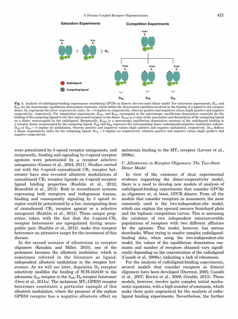
Fig. 1. Analysis of radioligand-binding experiments considering GPCRs as dimers: the two-state dimer model. For saturation experiments, KD1 andKD2 are the macroscopic equilibrium dissociation constants, which define the dissociation equilibria involved in the binding of a ligand to the receptordimer. DC represents the dimer cooperativity index. DC = 0 implies no cooperativity, whereas positive and negatives values imply positive and negativecooperativity, respectively. For competition experiments, KDB1 and KDB2 correspond to the macroscopic equilibrium dissociation constants for thebinding of the competing ligand to the first and second receptor in the dimer. KDAB is a value of the association and dissociation of the competing ligandon a dimer semioccupied by the radioligand. Reciprocally, KDBA is a macroscopic equilibrium dissociation constant of the radioligand binding toa receptor dimer semioccupied by the competing ligand. DAB and DBA represent the corresponding dimer radioligand/competitor modulation indexes.DAB or DBA = 0 implies no modulation, whereas positive and negatives values imply positive and negative modulation, respectively. DCB definesa dimer cooperativity index for the competing ligand. DCB = 0 implies no cooperativity, whereas positive and negative values imply positive andnegative cooperativity.
G Protein–Coupled Receptor Oligomerization 421
development of empirical equations from one of thesemodels, the two-state dimer model, makes it particularlyinsightful and easy to use (Franco et al., 2005, 2006;Casadó et al., 2007, 2009a,b). This is not entirely obviouswhen looking at the initial equations that include all themicroscopic equilibrium isomerization and dissociationconstants: seven for the binding of one ligand for satu-ration experiments, and 11 for two ligands for competitionexperiments (described in detail in Casadó et al., 2007).Hence empirical equations were derived that disclosea lower number of constants, macroscopic equilibriumdissociation constants, and this allows for a practicalanalysis of radioligand-binding data when consideringthe receptors as dimers (analyzed in Casadó et al., 2007)Equation for saturation experiments:
Abound ¼ �KD2Aþ 2 A2� RT=
�KD1KD2 þ KD2Aþ A2�
A is the concentration of radioligand and RT is the totalamount of receptor dimers (the traditional Bmax fromthe two-independent-site model would be twice thisvalue). KD1 and KD2 are the macroscopic equilibriumdissociation constants, which define the dissociationequilibria involved in the binding of a ligand to thereceptor dimer as a whole (not to be confused with theintrinsic equilibrium dissociation constants) (Fig. 1).KD1 represents the equilibrium between free ligand,empty dimer, and semi-occupied dimer (the first ligandoccupying a dimer). KD2 represents the equilibriumbetween free ligand and the occupied and semioccupieddimer (the second ligand occupying a dimer in whichone binding site is already occupied by the first ligand).Assuming symmetry, the rate at which the ligand bindsto the dimer is proportional to the number of unoccupiedreceptors in the dimer, therefore twice for the emptycompared with the semi-occupied dimer. The rate atwhich the ligand dissociates from the dimer is pro-portional to the number of occupied receptors in thedimer, therefore twice in the occupied compared withthe semi-occupied dimer. This implies, and it is easy todemonstrate, that when the intrinsic affinity of bothreceptors in the dimer is the same, i.e., when there is nocooperativity, KD2 = 4KD1. The two-state dimer modelalso introduces a dimer cooperativity index, DC, which isdefined as log (4KD1/KD2). DC = 0 implies no coopera-tivity, whereas positive and negatives values implypositive and negative cooperativity, respectively (Fig. 1).Equation for competitive-inhibition experiments:
Abound ¼ �KDA2Aþ 2 A2
þKDA2A B=KDAB�RT=
�KDA1KDA2 þ KDA2A
þ A2 þ KDA2A B=KDAB þ KDA1KDA2B=KDB1
þKDA1KDA2B2=ðKDB1KDB2Þ�
Although the equation might look complex, there arejust a few constants to be determined. A is again the
concentration of radioligand (fixed in competitionexperiments). B is the concentration of the competingcompound. RT is again the total amount of receptordimers. KDA1 and KDA2 are the macroscopic equilibriumdissociation constants for the binding of A, the radio-ligand, to the first and second receptor in the dimer,equivalent to KD1 and KD2 described above for satura-tion experiments. These constant values would havebeen calculated from previous saturation experimentswith the radioligand and, therefore, they are alreadyknown. KDB1 and KDB2 correspond to the macroscopicequilibrium dissociation constants for the binding of B,the competing compound, to the first and secondreceptor in the dimer (Fig. 1). And there is one moreconstant, KDAB, which can be considered as a hybridequilibrium dissociation constant, a value of the associ-ation and dissociation of B on a dimer semi-occupied by A(Fig. 1). KDAB is instrumental to explain a biphasic curvein competitive-inhibition experiments by means of a non-cooperative binding. It measures an allosteric interactionbetween the two different ligands in the dimer, by whichthe binding of the radioligand to the first protomerdecreases or increases the affinity of the competitor to thesecond protomer. It can easily be demonstrated thatwhen the binding of the radioligand to one protomer inthe dimer does not modify the binding of the competingligand to the other empty protomer, KDAB = 2KDB1 (Figs.1 and 2). Reciprocally, a KDBA value, which is theequilibrium dissociation constant of A binding to a re-ceptor dimer semi-occupied by B, can be deduced (Figs. 1and 2). The relation between these parameters is:
KDBA ¼ KDABKDA1=KDB1
The two-state dimer model also introduces two dimerradioligand/competitor modulation indexes, DAB andDBA, which are defined as log (2KDB1/KDAB) and log(2KDA1/KDBA), respectively. DAB or DBA = 0 implies nomodulation, whereas positive and negatives valuesimply positive and negative modulation, respectively(Figs. 1 and 2). Furthermore, we can also define a dimercooperativity index for the competing ligand, DCB, as log(4KDB1/KDB2). DCB = 0 implies no cooperativity, whereaspositive and negative values imply positive and nega-tive cooperativity, respectively (Figs. 1 and 2). Finally,the two-state dimer model allows the calculation of theconcentration of the competitor providing half satura-tion = (KDB1KDB2)
1/2, independent of the biphasic ormonophasic nature of the competition curve or of theradioligand concentration (Casadó et al., 2009a).
Considering the evidence of receptor oligomerizationand the validity of the dimer-cooperativity model, thetwo-state dimer model provides a better approach thanthe monomer–G protein models for fitting data thateventually give more accurate and physiologic relevantparameters. In terms of accuracy, the two-statedimer model is significantly more robust than the
422 Ferré et al.
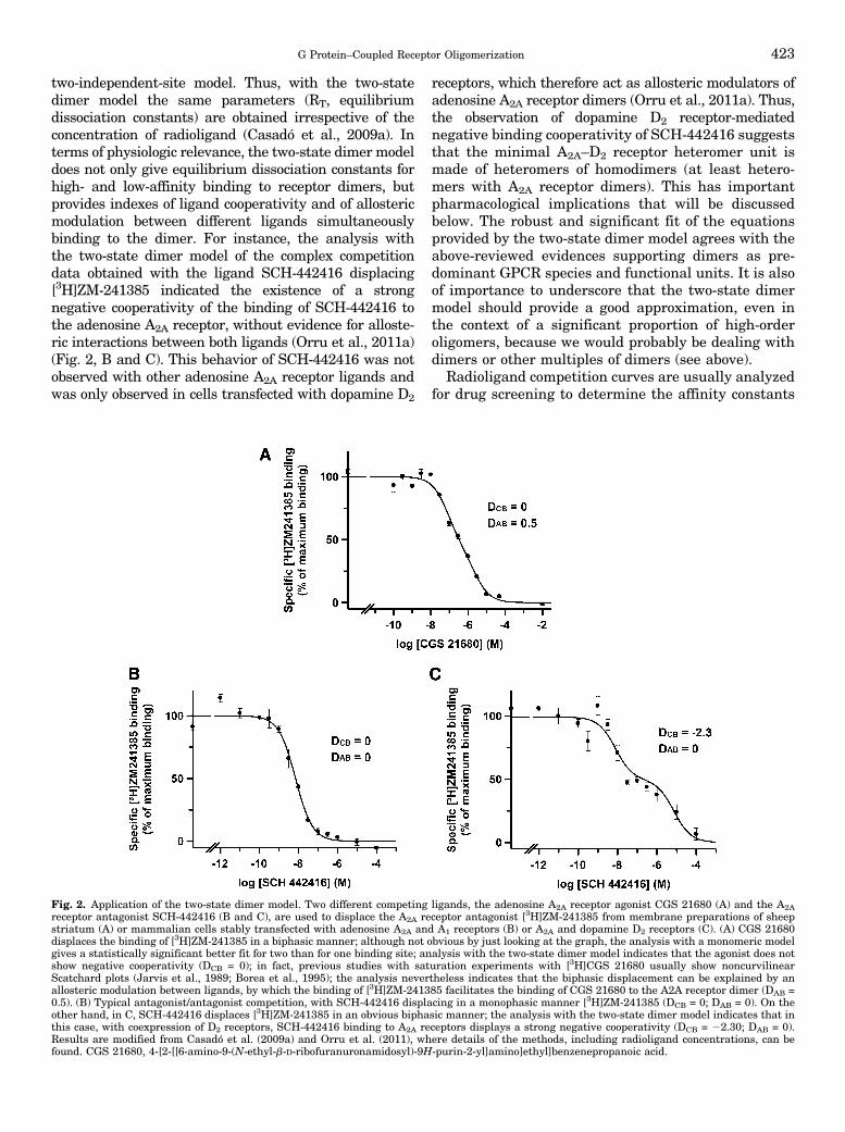
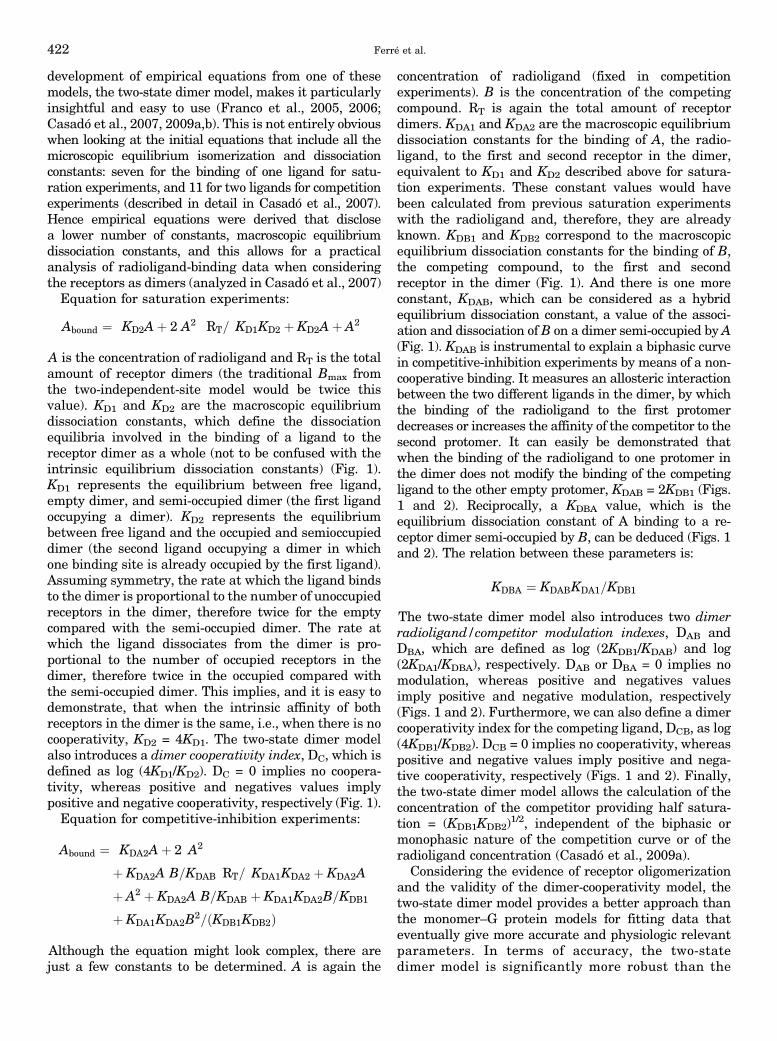
two-independent-site model. Thus, with the two-statedimer model the same parameters (RT, equilibriumdissociation constants) are obtained irrespective of theconcentration of radioligand (Casadó et al., 2009a). Interms of physiologic relevance, the two-state dimer modeldoes not only give equilibrium dissociation constants forhigh- and low-affinity binding to receptor dimers, butprovides indexes of ligand cooperativity and of allostericmodulation between different ligands simultaneouslybinding to the dimer. For instance, the analysis withthe two-state dimer model of the complex competitiondata obtained with the ligand SCH-442416 displacing[3H]ZM-241385 indicated the existence of a strongnegative cooperativity of the binding of SCH-442416 tothe adenosine A2A receptor, without evidence for alloste-ric interactions between both ligands (Orru et al., 2011a)(Fig. 2, B and C). This behavior of SCH-442416 was notobserved with other adenosine A2A receptor ligands andwas only observed in cells transfected with dopamine D2
receptors, which therefore act as allosteric modulators ofadenosine A2A receptor dimers (Orru et al., 2011a). Thus,the observation of dopamine D2 receptor-mediatednegative binding cooperativity of SCH-442416 suggeststhat the minimal A2A–D2 receptor heteromer unit ismade of heteromers of homodimers (at least hetero-mers with A2A receptor dimers). This has importantpharmacological implications that will be discussedbelow. The robust and significant fit of the equationsprovided by the two-state dimer model agrees with theabove-reviewed evidences supporting dimers as pre-dominant GPCR species and functional units. It is alsoof importance to underscore that the two-state dimermodel should provide a good approximation, even inthe context of a significant proportion of high-orderoligomers, because we would probably be dealing withdimers or other multiples of dimers (see above).
Radioligand competition curves are usually analyzedfor drug screening to determine the affinity constants
Fig. 2. Application of the two-state dimer model. Two different competing ligands, the adenosine A2A receptor agonist CGS 21680 (A) and the A2Areceptor antagonist SCH-442416 (B and C), are used to displace the A2A receptor antagonist [3H]ZM-241385 from membrane preparations of sheepstriatum (A) or mammalian cells stably transfected with adenosine A2A and A1 receptors (B) or A2A and dopamine D2 receptors (C). (A) CGS 21680displaces the binding of [3H]ZM-241385 in a biphasic manner; although not obvious by just looking at the graph, the analysis with a monomeric modelgives a statistically significant better fit for two than for one binding site; analysis with the two-state dimer model indicates that the agonist does notshow negative cooperativity (DCB = 0); in fact, previous studies with saturation experiments with [3H]CGS 21680 usually show noncurvilinearScatchard plots (Jarvis et al., 1989; Borea et al., 1995); the analysis nevertheless indicates that the biphasic displacement can be explained by anallosteric modulation between ligands, by which the binding of [3H]ZM-241385 facilitates the binding of CGS 21680 to the A2A receptor dimer (DAB =0.5). (B) Typical antagonist/antagonist competition, with SCH-442416 displacing in a monophasic manner [3H]ZM-241385 (DCB = 0; DAB = 0). On theother hand, in C, SCH-442416 displaces [3H]ZM-241385 in an obvious biphasic manner; the analysis with the two-state dimer model indicates that inthis case, with coexpression of D2 receptors, SCH-442416 binding to A2A receptors displays a strong negative cooperativity (DCB = 22.30; DAB = 0).Results are modified from Casadó et al. (2009a) and Orru et al. (2011), where details of the methods, including radioligand concentrations, can befound. CGS 21680, 4-[2-[[6-amino-9-(N-ethyl-b-D-ribofuranuronamidosyl)-9H-purin-2-yl]amino]ethyl]benzenepropanoic acid.
G Protein–Coupled Receptor Oligomerization 423
for putative agonists and antagonist. If the targetreceptor is assumed to be primarily in its monomericform, monomeric models should be used. However, ifhomomers have already been described for the targetreceptor, the two-state dimer model should be the mostappropriate choice. In the absence of previous assump-tions and in the absence of obvious biphasic competi-tion curves, monomeric models could be used to provideinitial estimates of drug affinities, because they implya lesser number of constants to handle. When notobvious as biphasic, monomeric models can also beused to establish if the competition curves fit signifi-cantly better for one or two binding sites. In the lattercase we should proceed with the analysis with the two-state model (see Fig. 2A). Nevertheless, when lookingfor the most appropriate model, the best approachwould be implementing both a monomer-based modeland a two-state dimer model and comparing results(preferably using a statistical test; see Casadó et al.,2009a). It is important to realize that choosing eithermodel should not imply discarding the possibility ofthe existence of mixtures of monomers and dimers/oligomers.
D. Allosterism in Receptor Oligomers: AllostericModulation of Intrinsic Efficacy andFunctional Asymmetry
The two-state dimer model assumes that both proto-mers have the same initial ligand binding propertiesand, therefore, the same probability to bind the firstligand molecule and the same ability of the first ligand–protomer complex to allosterically modulate the bindingproperties of the second protomer. That is, there shouldbe an initial symmetry of both protomers in a receptordimer at the binding level. Are there symmetricsignaling properties of both protomers in a receptordimer? This requires examining functional responsesand to address questions about the minimal functionalGPCR-G protein unit. Furthermore, above we onlyconsidered allosteric modulation of the affinity ofligands, whereas allosteric modulation of the intrinsicefficacy of the ligands should also be considered. Whendealing with dimers, the most accepted model, atpresent, is that two protomers can only accommodateone heterotrimeric G protein complex (reviewed inMaurice et al., 2011). Extensive biochemical evidenceindicates the involvement of at least two distinct Gprotein regions in interactions with the receptor. Theprimary docking site is in the C terminus of the Gasubunit, which penetrates into the crevice created in theintracellular surface of the receptor by the movement ofcytoplasmic regions of TM5 and TM6 upon binding ofthe agonist (Oldham and Hamm, 2008; Rasmussenet al., 2011). Another putative site is in the C terminusof the Gg subunit. These regions are 55 Å apart in theGabg heterotrimer, a distance greater than the width ofthe monomeric GPCR (approximately 45 Å), indicating
that for both contacts to take place simultaneously, oneheterotrimeric G protein must contact two receptorprotomers (Oldham and Hamm, 2008). Rasmussen et al.(2011) recently resolved the crystal structure of theactive state ternary complex composed of agonist-occupied monomeric b2-adrenoceptor and nucleotide-free Gs heterotrimer. One of the most interestingfindings was the lack of direct interactions betweenthe b2-adrenoceptor and Gbg. Given that the hetero-trimer is required for efficient coupling to a GPCR, theseresults are consistent with the existence of b2-adreno-ceptor dimers in cell membranes with one protomerinteracting with Ga and the second promoter interact-ing with the Gbg subunit (Rasmussen et al., 2011).Although a sequential interaction of simultaneouslyincompatible contacts could still be possible (Herrmannet al., 2004), this sequential fit mechanism would still becompatible with the existence of GPCR homodimersand with the pentameric structure consisting of oneGPCR homodimer and one heterotrimeric G protein asa minimal functional unit (Baneres and Parello, 2003;Han et al., 2009; Pellissier et al., 2011). Even if weconsider tetramers as the predominant oligomericspecies, the most common minimal receptor–G proteinstoichiometry would still probably be 2:1, and oligomericentities can be viewed as multiples of dimers, as, forinstance, suggested by the crystallographic structure ofm-opioid receptor and b1-adrenoceptor (Manglik et al.,2012; Huang et al., 2013).
The apparent asymmetric pentameric structure ofGPCRs (homodimer plus heterotrimeric G protein)might seem incompatible with the two-state dimermodel and the dimer-cooperativity model, because theyassume there is an initial symmetry between bothprotomers regarding ligand binding properties (thesame probability to bind the first ligand molecule) andthat the G protein is always interacting with thereceptor (Casadó et al., 2007). In this respect, twoopposing models have been presented to explain theencounter between G proteins and the activated re-ceptor. In the "collision coupling"model, the interactionsoccur as a result of the free lateral diffusion, and Gproteins only interact with the agonist-bound receptor.In the "precoupling" model, G proteins are alreadyinteracting with receptors before agonist binding andthe ligand modifies this interaction by creating theconformational change in the receptor (crevice) thatallows the a subunit to “tightly” bind the receptor andinduce G protein activation (reviewed in Oldham andHamm, 2008). About the symmetry, the two-state dimermodel only assumes an initial symmetry (at least interms of ligand-binding probability) and fits very wellwith the precoupling model of receptor–G proteininteraction. The two-state dimer model accepts thatthe ligand binding to the first protomer determines anasymmetric function of the pentameric functional unit.According to the model, this ligand-induced asymmetry
424 Ferré et al.
determines the allosteric modulations at the bindinglevel, such as the reduced affinity of the ligand for thesecond protomer (negative cooperativity).What is the functional response that a ligand can
produce when binding to one or both protomers ina GPCR homodimer? Is the binding of an agonist to oneof the protomers enough to elicit a full functionalresponse? Does the asymmetric constraint of G proteincoupling to the two protomers determine an asymmetricfunctional response? Most experimental data agree withthe model that proposes that ligand occupancy to thefirst protomer is enough to produce a significant Gprotein activation and functional response. Occupancyof the second protomer will then usually potentiate(Kniazeff et al., 2004; Pellissier et al., 2011), but couldalso reduce (Han et al., 2009) or not alter significantly(Hlavackova et al., 2005), the functional response(irrespective of the allosteric modulations at the bindinglevel).Therefore, when the ligand binds to the second
protomer in a homodimer, it will often act as an al-losteric modulator of the intrinsic efficacy of the ligandwhen binding to the first protomer.We should also consider G protein–independent
signaling, such as b-arrestin–dependent signaling.Similar to G proteins, GPCR oligomerization raisesthe question of whether GRK and b-arrestin binding toGPCRs occur in an asymmetric manner. The possibleasymmetric nature of the receptor–b-arrestin interac-tion is still a matter of debate. As with G protein bindingand activation, studies with artificially reconstitutedmonomeric GPCRs show that receptor dimers are notrequired for their functional interaction with GRK andb-arrestins (Tsukamoto et al., 2010; Bayburt et al.,2011). These results, however, do not exclude thepossibility that GRK or b-arrestins bind to GPCRdimers (Modzelewska et al., 2006; Sommer et al.,2012). GPCR oligomerization potentially providesa larger platform to accommodate the different GPCRinteracting proteins necessary for receptor function(reviewed in Maurice et al., 2011). This is demonstratedin the complex of melatonin MT1 receptor dimers, Gi
protein and RGS20 (a protein that regulates the speedof G protein signal transduction), which both binddirectly to helix 8 of the receptor (Maurice et al., 2010).In this complex, protomer 1 binds the ligand and the Gprotein and protomer 2 binds RGS20. In this case,RGS20, by slowing down the decay time of G proteininactivation, thus participates in prolonged signaltransduction (Maurice et al., 2010).Several examples of allosteric modulation of intrinsic
efficacy have been reported for GPCR heteromers. As forligand affinity, there are cases of allosteric modulationof the intrinsic efficacy of a ligand of one of theprotomers by the other protomer or by a ligand thatbinds to the other protomer. As an example of the latter,in the a2A-adrenoceptor–m-opioid receptor heteromer,
morphine binding to the m-opioid receptor inhibits a2A-adrenoceptor signaling (Jordan et al., 2003). This hasbeen related to a morphine-induced conformationalchange of the a2A-adrenoceptor, as detected by dynamicintramolecular FRET techniques (Vilardaga et al.,2008). These studies indicated that activation of them-opioid receptor component in the a2A-adrenoceptor–m-opioid receptor heteromer modulates a2A-adrenoceptorreceptor signaling by a direct conformational changethat propagates from the m to the a2A-adrenoceptorwithin 0.4 seconds, slightly faster than the rate of Gprotein activation, implicating a direct allosteric mod-ulation through the receptor heteromer (Vilardagaet al., 2008).
As an example of an allosteric modulation of intrinsicefficacy of a ligand of one of the protomers by the otherprotomer in the GPCR heteromer, in the metabotropicglutamate mGlu2-serotonin 5-HT2A receptor heteromer,glutamate produces a stronger mGlu2-mediated signal-ing and serotonin produces a weaker serotonin 5-HT2A–
mediated signaling compared with when each receptoris expressed alone (Fribourg et al., 2011). In the samereceptor heteromer, the serotonin 5-HT2A receptorantagonist clozapine also induced a positive allostericmodulation of the intrinsic efficacy of glutamate(Fribourg et al., 2011). These results could have impli-cations for the understanding of the clinical efficacyof this compound in schizophrenia and establish themGlu2–5-HT2A receptor heteromer as a promising tar-get for the treatment of this disease.
Considering, therefore, receptor heteromers in theframe of allosteric modulation of GPCRs, there areexamples of allosteric modulation by specific ligands ofsome receptors of the affinity and intrinsic efficacy ofligands for other receptors (see above). There are alsosignificant examples of functional selectivity, where oneof the protomers of the heteromer acts as an allostericmodulator that “forces” the other receptor protomerto signal predominantly through a distinct signalingpathway. An example is that of the d-m-opioid receptorheteromer (Rozenfeld and Devi, 2010). Under normalphysiologic conditions, the m-opioid receptor is foundmostly in the homomeric state, and morphine stimula-tion induces G protein–mediated signaling, which isinvolved in its analgesic effects, and low b-arrestin–mediated signaling, which promotes unwanted effects,such as tolerance to the analgesic effects (Raehal et al.,2011). In contrast, upon chronic treatment with mor-phine, the abundance of d-m-opioid receptor heteromersincreases (Gupta et al., 2010), and morphine stimula-tion induces b-arrestin–mediated signaling (Rozenfeldand Devi, 2007), which is thought to contribute to thetolerance to the analgesic effect of morphine upon itschronic administration (Raehal et al., 2011). Notably,occupancy of the d-opioid receptor protomer witha selective d-opioid receptor antagonist (Rozenfeld andDevi, 2007) or with a bivalent ligand in the d-m-opioid
G Protein–Coupled Receptor Oligomerization 425
receptor heteromer (see below and Daniels et al., 2005)allows for the restoration of G protein–biased agonismof morphine and, therefore, its analgesic effects (Rozen-feld and Devi, 2010).Other dramatic examples of functional selectivity in
receptor heteromers are changes in G protein coupling,such as with the angiotensin AT1–cannabinoid CB1
receptor heteromer (Rozenfeld et al., 2011). Although, inthe normal liver angiotensin AT1 receptor signals viaGq, under conditions of alcohol-induced liver fibrosiscannabinoid, CB1 receptor is upregulated and hetero-merizes with angiotensin AT1 receptor and this, in turn,leads to Gi-mediated signaling by angiotensin AT1
receptor that can be blocked by cannabinoid CB1
receptor antagonist, suggesting that AT1–CB1 receptorheteromer represents a disease-specific and potentiallytissue-specific therapeutic target. Another example ofchange in G protein coupling is the dopamine D1–
histamine H3 receptor heteromer, where dopamine D1
receptor agonists activates Gi instead of Gs proteins(inhibition of cAMP formation) (Ferrada et al., 2009).Another important allosteric property of the D1–H3
heteromer is the ability of H3 receptor agonists toinhibit dopamine D1 receptor–mediated G protein andb-arrestin signaling, which allows histamine to providea brake on dopamine D1 receptor–mediated effects,including cell death (Moreno et al., 2014). This hetero-mer also binds the two-transmembrane sigma s1
receptors, which are well known targets for cocaine.Binding of cocaine to the D1–H3–s1 complex disruptsthe allosteric properties of the D1–H3 heteromer allowsdopamine D1 receptor to couple to Gs and to eliminatethe histamine H3 receptor–mediated signaling brake,which promotes cell death (Moreno et al., 2014).Therefore D1-H3 heteromers may provide a new thera-peutic target for the D1 receptor–mediated neurotoxiceffects of cocaine.Another recent example of functional selectivity in
receptor heteromers is the dopamine D2–ghrelin GHS1a
receptor heteromer (Kern et al., 2012). These receptorsare colocalized in the brain, in the hypothalamus andbrain stem (Guan et al., 1997). Interestingly, ghrelinreceptors seem to be orphan receptors in certainlocalizations, because the peptide ghrelin, which isproduced in the stomach, can only reach the arcuatenucleus but not other hypothalamic nuclei and otherbrain regions. In the hypothalamus, heteromerizationwith ghrelin GHS1a receptor modifies dopamine D2
receptor signaling, resulting in Gbg-dependent mobili-zation of Ca2+ (Kern et al., 2012). The anorexigeniceffects of dopamine D2 receptor agonists seem to bemediated by the hypothalamic D2–GHS1a receptorheteromer, which therefore might become an importanttherapeutic target for eating disorders.In summary, the allosteric analysis of GPCR homo-
mers and heteromers provides overwhelming addi-tional evidence for the results of in vitro experiments
and experiments in heterologous systems, supportingthe existence of functionally and pharmacologicallyrelevant GPCR oligomers. The evidence points to thepentameric structure constituted by one GPCR homo-dimer and one heterotrimeric G protein as a mainminimal functional unit and oligomeric entities can beviewed as multiples of dimers. Allosteric mechanismsdetermine a multiplicity of possible unique pharmaco-logical properties of receptor homomers and hetero-mers. Some general mechanisms seem to apply,particularly at the level of ligand-binding properties.When considering receptor homomers, the two-statedimer model provides the most practical method toanalyze ligand–GPCR interactions. If possible, similarpractical models would need to be developed to studyligand–GPCR interactions for receptor heteromers andfor dynamically changing oligomers. In addition toligand-binding properties, unique properties for eachGPCR oligomer seem to emerge in relation to differentintrinsic efficacy of different ligands (intrinsic efficacy)for different signaling pathways (functional selectiv-ity). This provides a rationale for the use of GPCRoligomers, and particularly heteromers, as novel tar-gets for drug development.
III. G Protein–Coupled Receptor Oligomers asNovel Targets for Drug Development
A. Localization of Receptor Oligomers inNative Tissues
Before considering the possible role of GPCRoligomers in vivo, and particularly receptor hetero-mers, as targets for drug development, we must firstreaddress questions about the demonstration of re-ceptor homo- and heteromers in native tissues, becausemost of the studies discussed above have been per-formed in heterologous expression systems, particu-larly in transfected mammalian cells. Next, we need toaddress the questions about localization in situ andsignificance of receptor homomers and heteromers invivo (Levoye et al., 2006b; Pin et al., 2007; Ferré et al.,2007, 2009). Efforts toward addressing these importantquestions have been hampered by the lack of appro-priate tools and techniques that would allow their cleardemonstration in native tissues. Biochemical techni-ques such as coimmunoprecipitation cannot provideunambiguous evidence for direct protein–protein inter-actions, and biophysical techniques, such as BRET andFRET, are difficult to implement in tissues. In contrastto heterologous systems, where wild-type or mutatedreceptors fused with tags or fluorescent proteins can beeasily expressed, the expression level of receptors innative tissues is difficult to modify and their sequencescannot be modified unless using knock-in strategies.Nevertheless, quantitative FRET in situ has been usedto explore the presence of dopamine D1–D2 receptorheteromers in brain tissue (Hasbi et al., 2011).
426 Ferré et al.
An alternative approach to label and detect nativeGPCR oligomers has been the use of antibodies orfluorescent ligands. However, they need to be highlyspecific and to provide a reliable signal, and the levelsof GPCR oligomers should be abundant or highlyregulated. Recent efforts to identify tools that fit someof these criteria have begun to yield promising results.Using labeled ligands that could demonstrate proxim-ity of endogenous receptors, the study by Albizu et al.(2010) provided quite a clear demonstration of thepresence of oxytocin receptor homomers in the mam-mary gland. They showed differential FRET withlabeled agonists and antagonists in tissue as theyhad seen with transfected cells, implying the existenceof oxytocin receptor homomers. This is therefore anindirect but clear demonstration of the existence ofreceptor homomers in native tissues.Efforts to generate heteromer-selective antibodies
using a subtractive immunization strategy have begunto yield useful monoclonal antibodies with heteromerselectivity, because they recognize the epitope only inwild-type animals but not in animals lacking eitherprotomers of the heteromer (Gomes et al., 2013a,c).These heteromer-selective antibodies have helped notonly studies examining heteromer levels and regulationin endogenous tissue but also studies investigating theextent of heteromer-specific signaling (Gomes et al.,2013a,c). In the case of d-m-opioid receptor heteromers,the antibodies were helpful in demonstrating morphine-induced increases in heteromer abundance, as well ascharacterizing heteromer-mediated signaling (Guptaet al., 2010). The AT1–CB1 receptor heteromer selectiveantibody was useful in demonstrating upregulation ofthe heteromer in hepatic stellate cells after alcohol-induced fibrosis (Rozenfeld et al., 2011). The d-opioid–CB1 receptor heteromer selective antibody was useful indemonstrating increases in d-opioid–CB1 receptor het-eromers in the brain after peripherally elicited neuro-pathic pain (Bushlin et al., 2012). Finally, the d-k-opioidreceptor heteromer selective antibody was critical indemonstrating a role for this heteromer in sensory paintransmission, because the antibody was able to aug-ment the effect of d receptor agonist-mediated anti-nociception (Berg et al., 2012). Together these studiesdemonstrate that GPCR heteromer antibodies, if foundto be highly selective, are critical and important tools instudies of GPCR heteromers in native tissue.The proximity ligation assay (PLA) is an antibody-
based method that also provides an approximation tothe demonstration of receptor heteromers in tissues. Ithas been used for instance for the demonstration of closeproximity of adenosine A2A and dopamine D2 receptorsin brain slices (Trifilieff et al., 2011). In PLA, primaryantibodies labeling the two putative protomers ofthe receptor heteromer are labeled with two differentspecies-specific secondary antibodies conjugated tocomplementary oligonucleotides. When the antibodies
are in close proximity, the complementary DNA strandscan be ligated, amplified, and visualized with a fluores-cent probe. The maximal distance between the second-ary antibodies in this assay is ;16 nm, only slightlylarger than that for resonance energy transfer betweenfluorophores (;10 nm) (Soderberg et al., 2006). How-ever, PLA does not demonstrate GPCR heteromeriza-tion, but allows a validation in vivo of the molecularproximity of two GPCRs, giving the frame for hetero-merization to take place in vivo.
Another indirect but easier and straightforwarddemonstration depends on the identification of a bio-chemical property (“biochemical fingerprint”) of thereceptor oligomer (Ferré et al., 2009). To ascertaina “biochemical fingerprint” of the receptor heteromer,the putative biochemical property should be disruptedwith molecular or chemical tools that significantlydestabilize the quaternary structure of the heteromer(such as introducing mutations of key determinantresidues at the oligomerization interfaces or usingcompeting peptides that harbor the same sequence asthat of the receptor heteromer interface). The recentlyreported dopamine D2–D4 receptor heteromer servesas a good example. A human polymorphism of thedopamine D4 receptor, D4.7 (with seven repeats ofa proline-reach 16-amino acid sequence in the thirdintracellular loop) differs from the most commonvariant, D4.4 (with four repeats) in that it does notheteromerize with dopamine D2 receptor and it does notshow a pharmacological D2–D4 receptor interaction(Gonzalez et al., 2012). This interaction consists ofa dopamine D2 receptor agonist-mediated potentiationof MAPK activation induced by a dopamine D4 receptoragonist (Gonzalez et al., 2012). Thus, a synergisticMAPK activation could be used as a biochemicalfingerprint of the heteromer, which could only beobserved in mice expressing wild-type dopamine D4
receptor (which also heteromerized with mouse wild-type dopamine D2 receptor), but not in knock-in trans-genic mice expressing the dopamine D4.7 human variant(Gonzalez et al., 2012). The use of transgenic animals istherefore a useful indirect method to study receptorheteromer function. An additional example is the recentstudy about the role of MT1–MT2 receptor heteromerson the effect of melatonin on rod photoreceptor lightsensitivity, which was abolished in melatonin receptorMT1
2/2 and MT22/2 mice (Baba et al., 2013). The
involvement of MT1–MT2 receptor heteromers in thiseffect was further underscored by the lack of effect ofmelatonin in transgenic mice overexpressing a nonfunc-tional melatonin MT2 receptor mutant that competeswith the formation of functional MT1–MT2 heteromersin photoreceptor cells (Baba et al., 2013). These experi-ments are of therapeutic interest for the improvement ofphotoreceptor functioning in several clinical conditionsand applications in the pathogenesis of age-relatedmacular degeneration and glaucoma.
G Protein–Coupled Receptor Oligomerization 427
Therefore, use of several complementary approachesis needed to address the identification of GPCRoligomers in native tissues. It is essential that theseapproaches are critically evaluated and validated usingartificial systems, such as mammalian transfectedcells, especially when looking for specific biochemicalproperties of a GPCR oligomer. It is not enough justreproducing some findings in native tissues previouslyfound in the artificial systems. A unique biochemicalfinding in mammalian transfected cells that expressreceptor heteromers (which can be demonstrated withproperly applied biophysical techniques) could well berelated to interactions between the two receptors thatdo not depend on oligomerization. As mentioned above,we need to obtain and implement tools that specificallydisrupt GPCR oligomerization or the quaternarystructure of the GPCR oligomer. When possible, thosetools should also be applied in situ. For instance, byselectively interfering with the oligomer interfaces,particular mutations or the in vitro application or invivo delivery of peptides altering the quaternarystructure of a GPCR oligomer (as first demonstratedwith biophysical techniques in artificial systems)should selectively alter the detection of the putativebiochemical property of the GPCR oligomer.
B. Do Receptor Heteromers Constitute Possible Targetsfor Drug Development?
GPCRs are targeted by many therapeutic drugsaiming at restoring or ameliorating a variety of healthdisorders and remain a primary focus of biomedicalresearch and programs of pharmacological drug dis-covery. However, despite the proven success of GPCRsas drug targets, clinically useful ligands do not exist forthe majority of GPCRs. A main contributing factor isthe lack of selectivity of orthosteric ligands acrossmembers of a GPCR subfamily (or that certain GPCRsdo not have roles in major disease phenotypes), withthe obvious selectivity problem, which leads to un-wanted side effects. An alternative approach alreadyfound successful for ligand-gated ion channels, such asbenzodiazepines, positive allosteric modulators ofGABAA receptors, is the development of GPCR allo-steric modulators (ligands that modulate the affinity orintrinsic efficacy of orthosteric agonists of the sameGPCR unit) (Conn et al., 2009). Allosteric modulatorscan potentially overcome the selectivity problem. Theorthosteric binding sites across members of a GPCRsubfamily for a particular endogenous ligand, such asnoradrenaline or dopamine, are highly conserved, butGPCRs offer numerous possible selective allostericbinding sites across different receptor subtypes. Fur-thermore, allosteric modulators possess pharmacolog-ically appealing properties, such as saturability. Inrecent years, significant progress has been made in thediscovery of various GPCR allosteric modulators,although not without difficulties, such as generating
a structure–activity relationship in an allosteric com-pound series, or likelihood of lack of cross-speciesactivity for validation/efficacy studies (Conn et al.,2009).
Receptor heteromers, with their allosteric properties,give rise to a new kind of pharmacological target. Theability of one of the protomers to act as an allostericmodulator of the second protomer in the receptorheteromer gives the possibility of finding selectiveligands for the protomer acting as conduit of theallosteric modulation. A proof of concept was providedby the dramatically decreased binding affinity (strongnegative cooperativity) of the adenosine A2A receptorantagonist SCH-442416 in cells cotransfected withdopamine D2 receptors compared with cells onlyexpressing adenosine A2A receptors or also expressingadenosine A1 (Orru et al., 2011a) (Fig. 2, B and C).Several other antagonists, such as MSX-3, also showeda less dramatic yet significantly decreased affinity foradenosine A2A receptors when cotransfected withdopamine D2 receptors (Orru et al., 2011a). Theseresults are important from a practical pharmacologicalpoint of view, because different striatal subpopulationsof adenosine A2A receptors modulate different striatalfunctions (Orru et al., 2011a,b), and these subpopula-tions are constituted by different adenosine A2A receptorheteromers. A2A–D2 receptor heteromers are selectivelylocalized postsynaptically in GABAergic striatopallidalneurons, where they exert a key role in the modulationof the activity of these neurons (see above and Azdadet al., 2009). On the other hand, A1–A2A receptorheteromers are selectively localized presynaptically inthe striatal glutamatergic terminals that make synapticcontact with GABAergic striatonigral neurons (Quirozet al., 2009). Through these heteromers, postsynapticblockade of adenosine A2A receptors produces locomotoractivation, and presynaptic blockade of adenosine A2A
receptors produces inhibition of corticostriatal neuro-transmission (Ciruela et al., 2006; Quiroz et al., 2009;Orru et al., 2011a). The postsynaptic effect hasimplications for Parkinson’s disease and, based on theexistence of A2A–D2 receptor interactions, adenosineA2A receptor antagonists are being used for thetreatment of Parkinson’s disease (see below andArmentero et al., 2011). On the other hand, presynapticeffects can have implications for drug addiction, buta concomitant postsynaptic adenosine A2A receptorblockade will determine opposing effects. In this regard,a low dose of the selective adenosine A2A receptorantagonist MSX-3 (shown to be preferentially presyn-aptic in rodents; Orru et al., 2011a) decreased tetrahy-drocannabinol and anandamide self-administration insquirrel monkeys, whereas a slightly higher dose hadpotentiating effects (Justinová et al., 2011). The ap-pearance of potentiating rather than suppressing effectson cannabinoid reinforcement at the slightly higherdose of MSX-3 would likely preclude the use of such
428 Ferré et al.
a compound as a pharmacologic treatment of cannabisabuse. As expected, recent results showed a significantantagonistic effect of SCH-442416 over a much widerrange of doses than with MSX-3 (Justinová et al.,submitted manuscript), strongly supporting the use ofthis or other similar compounds for the treatment ofdrug abuse.When the receptor heteromer is the conduit for the
allosteric modulation, the allosteric modulator acts verymuch the same way as a classic allosteric modulator.The difference is that the binding of the allostericmodulator takes place in the orthosteric site of one ofthe receptors in the heteromer and the modulatedligand binds to the orthosteric site of the other receptor.For instance, adenosine A2A receptor antagonists in-crease the affinity of the dopamine D2 receptor fordopamine, which as mentioned earlier is being used asa strategy for the treatment of Parkinson’s disease. Infact, evidence has already been provided for an increasein the therapeutic index of L-dopa with adenosine A2A
receptor antagonists (Armentero et al., 2011). Further-more, adenosine A2A receptor antagonists can poten-tially be useful as monotherapy in early stages ofParkinson’s disease where some symptoms seem to berelated to the loss of a tonic dopamine D2 receptor–mediated inhibition of adenosine A2A receptor signaling(Ferré et al., 2008; Armentero et al., 2011). Schizophre-nia is another pathologic condition where the A2A–D2
receptor heteromer could be used as a therapeutictarget (Ferré, 1997). Thus, different from dopamine D2
receptor antagonists, adenosine A2A receptor agonistsbehave as atypical antipsychotics (antipsychotics withlow extrapyramidal side effects) in animal models(Rimondini et al., 1997). This profile could be relatedto the allosteric and therefore saturable effect ondopamine D2 receptor–mediated signaling. So far, thecardiovascular effects of adenosine A2A receptor ago-nists preclude their use as antipsychotics, but ligandswith selectivity for the A2A–D2 receptor heteromerwould be devoid of these peripheral side effects becauseof the lack of peripheral A2A–D2 receptor interactions(Schindler et al., 2004). In the A2A–D2 receptor hetero-mer as target for Parkinson’s disease and schizophrenia,we deal with an allosteric modulation of the affinity ofdopamine D2 receptor ligands, although changes inintrinsic efficacy might also take place. As mentionedabove, another candidate target for antipsychotic treat-ment is the mGlu2–5HT2A receptor heteromer, wherethe atypical antipsychotic clozapine, in addition toblocking serotonin 5-HT2A, induces an allosteric modu-lation of the intrinsic efficacy of glutamate (Fribourget al., 2011). Functional selectivity is also being ad-dressed in the search for antipsychotics with lowliability for side effects.A particular value of receptor heteromers as targets
for drug development is that they can be involved inthe pathophysiology of the disorder being targeted. In
this regard, the receptor heteromer would be diseasespecific (Ferré et al., 2010; Gomes et al., 2013a). Thedopamine D1–D3 receptor heteromer seems to beinvolved in the very common and troublesome second-ary effect of L-dopa treatment in Parkinson’s disease,L-dopa–induced dyskinesia (reviewed in Ferré et al.,2010). The dopamine D1 receptor in the D1-D3 receptorheteromer has significantly more affinity for dopamineor other agonists than when not forming heteromerswith dopamine D3 receptors (Fiorentini et al., 2008;Marcellino et al., 2008), which results in a significantincrease in the potency of dopamine D1 receptoragonists to signal through the G protein and potentiatecAMP accumulation (Fiorentini et al., 2008). This isa very significant finding, because it is well knownthat, under normal conditions, striatal dopamine D1
receptors have a much lower affinity for dopamine thandopamine D2 or D3 receptors (Missale et al., 1998).There is a substantial amount of evidence thatindicates that dopamine D1 and D3 receptors arecolocalized in GABAergic striatonigral neurons (whichconstitutes the direct striatal efferent pathway), wherethey play a key role in the development of L-dopa–induced dyskinesia in Parkinson’s disease. The work-ing hypothesis is that dopamine denervation followedby chronic L-dopa treatment leads to an increaseddopamine D1 receptor signaling promoted by anupregulation of dopamine D3 receptors and by anincrease in the expression of D1–D3 receptor hetero-mers, which produces an imbalance in basal gangliaprocessing due to an increased activation of the directpathway (reviewed in Ferré et al., 2010). Significantly,treatment with dopamine D3 receptor partial agonistsor antagonists attenuates L-dopa–induced dyskinesiain 1-methyl-4-phenyl-1,2,3,6-tetrahydropyridine(MPTP)–treated monkeys (Bezard et al., 2003; Visanjiet al., 2009). Finally, an increase in dopamine D3
receptor density has been observed in human cocainefatalities (Staley and Mash, 1996), suggesting thata concomitant increase in D1–D3 receptor heteromersmay also occur. The D1–D3 receptor heteromer there-fore emerges as a target for the treatment of L-dopa–induced dyskinesia and cocaine addiction.
As mentioned earlier, the availability of theheteromer-selective antibodies has helped exploredisease-specific modulation of their abundance andhence define them as novel therapeutic targets (Gomeset al., 2013c). A potential role for d-m-opioid receptorheteromers in morphine tolerance was implied bystudies that demonstrated an increase in the abun-dance of d-m-opioid receptor heteromers in the brainand in dorsal root ganglion cells in response toa paradigm of morphine treatment that leads todevelopment of tolerance (Gupta et al., 2010). AT1–
CB1 receptor heteromers are upregulated in alcohol-induced liver fibrosis, and this leads to a shift insignaling from a Gi to Gq-coupled pathway implicating
G Protein–Coupled Receptor Oligomerization 429
the CB1R–AT1R heteromer as a potential target for thetreatment of liver fibrosis (Rozenfeld et al., 2011).Finally, the d-opioid–CB1 receptor heteromer wasfound to be upregulated during neuropathic pain andthe heteromer exhibited unique allosteric modulation,making this an attractive target for the development oftherapeutic for the treatment of pain (Bushlin et al.,2012).
C. Approaches for the Identification ofReceptor-Heteromer Selective Compounds
Despite their pharmacologic potential, there are veryfew studies reporting the identification of receptor-heteromer selective compounds. A receptor-heteromerselective compound would not only include ligands withsignificantly different affinity or intrinsic efficacy forone or both receptors in a receptor heteromer (comparedwith the same biochemical properties of the ligands forthe same receptors when not forming heteromers), itwould also include compounds that would exert signif-icant allosteric modulations in the receptor heteromer.Two general approaches were recently used for thediscovery of receptor-heteromer selective compounds:discrete direct screening (DDS) and high-throughputscreening (HTS). DDS is used when a series of ligandstargeting either of the receptors in the heteromer isavailable. In this case, relatively smaller number ofligands would allow the use of labor-intensive techni-ques to demonstrate differences in affinity (for agonistsor antagonists) or intrinsic efficacy (agonists). The mostimportant step for this strategy to be successful is theavailability of appropriate cell lines. The study by Orruet al. (2011a) provides a successful example of DDS ofadenosine A2A receptor antagonists in different compa-rable cell lines expressing only adenosine A2A receptorsor A1–A2A or A2A–D2 receptor heteromers. As mentionedabove, radioligand-binding experiments and analysis ofthe data with the two-state dimer model demonstrateda very specific behavior, a negative cooperativity of theadenosine A2A receptor antagonist SCH-442416 in cellsexpressing A2A–D2 receptor heteromers (Fig. 2, B andC). This biochemical result predicted a preferentialstriatal presynaptic profile of SCH-442416, whichtotally correlated with in vivo experiments in rodentsand monkeys. Notably, preclinical data suggest that thiscompound could be useful in the treatment of drugaddiction (see Section III.B). SCH-442416 can now beused as a lead compound for further chemical optimi-zation leading to novel compounds exhibiting even loweraffinity for the A2A–D2 receptor heteromer (striatalpostsynaptic adenosine A2A receptors), whereas keepinga significant affinity for A1–A2A receptor heteromers(striatal presynaptic adenosine A2A receptors). Thesame DDS approach could be used to find adenosineA2A receptor antagonists useful for Parkinson’s diseasethat would have preferential affinity for the A2A–D2
versus the A1–A2A receptor heteromer or that would
have stronger allosteric modulatory effects of theaffinity or intrinsic efficacy of dopamine D2 receptoragonists. Other receptor-heteromer selective compoundsobtained by the DDS approach include the putative D1–
D2 receptor heteromer compound SKF-83959 [6-chloro-7,8-dihydroxy-3-methyl-1-(3-methylphenyl)-2,3,4,5-tetrahy-dro-1H-3-benzazepine] (Rashid et al., 2007; but see Chunet al., 2013) and clozapine, which not only acts as a potentserotonin 5-HT2A receptor antagonist but also increasesthe intrinsic efficacy of glutamate in the mGlu2–5-HT2A
receptor heteromer (Fribourg et al., 2011).DDS using bivalent ligands constitutes another
approach for targeting receptor-heteromers (recentlyreviewed in Hiller et al., 2013). A bivalent ligand consistsof two pharmacophoric entities linked by an appropriatespacer. In principle, it should be possible to targetGPCR heteromers by selection of adequate, potent, andreceptor-selective pharmacophores. The proof of princi-ple came from the work of Daniels et al. (2005), whoreported the effects of bivalent ligands incorporating thepharmacophores of the m-opioid receptor agonist oxy-morphone and the d-opioid receptor antagonist naltrin-dole tethered by spacers of different lengths to selectivelylabel d-m-opioid receptor heteromers. Only ligands withappropriate spacer length produced no tolerance to theiranalgesic effects (Daniels et al., 2005). Absence oftolerance was therefore most likely due to the appropri-ate tethering of the m-opioid receptor agonist andd-opioid receptor antagonist and again supported theinvolvement of d-m-opioid receptor heteromer in thetolerance to the analgesic effects of opioids. The sameresearch group recently published results that suggestthat some bivalent ligands can very selectively interactwith the putative m-opioid–glutamate metabotropicmGlu5 receptor heteromer, with potent antinociceptiveeffects (Akgun et al., 2013). Although clinical applicationof these big molecules might not be feasible for phar-macokinetic reasons, the same as any receptor-heteromerselective compound, bivalent ligands can provide impor-tant tools for the identification and study of the functionalsignificance of receptor heteromers.
HTS is the most current initial strategy in theprocess of drug development. Availability of a simple,quick, and robust assay is critical for enabling HTS(reviewed in Franco et al., 2010). A frequently usedassay for initial screens is the second-messenger cell-based assay using promiscuous G protein that is able toelicit a calcium response. Another common strategy isbased on b-arrestin translocation that occurs as a re-sult of receptor activation-dependent recruitment ofb-arrestins. This latter approach was recently usedfor the identification of d-m-opioid receptor heteromerselective compounds, because a previous study sug-gested that this heteromer preferentially recruitsb-arrestin (Rozenfeld and Devi, 2007). Screening ofa small-molecule library was performed with theb-arrestin recruitment assay in cell lines expressing
430 Ferré et al.
d-m-opioid receptor heteromers; the hits were subject toa secondary screening using cells expressing onlyd-opioid or m-opioid receptors (Gomes et al., 2013b).The compound named CYM51010 [ethyl 1-[4-(acetyl-amino)benzyl]-4-(2-phenylethyl)piperidine-4-carboxylate]was identified as a ligand that preferentially activatesd-m-opioid receptor heteromer (Gomes et al., 2013b).The compound showed strong intrinsic efficacy not onlyfor b-arrestin but also for G protein–mediated signal-ing [guanosine 59-3-O-(thio)triphosphate binding]. Thed-m-opioid receptor-heteromer selective antibody wasable to block not only the CYM51010-mediatedsignaling in heterologous cells, but also intrathecalanalgesia in a rodent model of thermal pain (Gomeset al., 2013b). The significant intrinsic efficacy of thecompound for d-m-opioid receptor–induced b-arrestinand G protein–mediated signaling (compared withmorphine) makes CYM51010 an attractive candidatefor the development of a new analgesic with lowertolerance than morphine. In fact, using an in vivo assayof analgesia (tail-flick assay), CYM51010 was found toexert similar antinociceptive effects than morphine butwith less tolerance (Gomes et al., 2013b). Compoundssuch as these that selectively target the heteromers arelikely to serve as valuable tools to explore the phys-iologic role of heteromers in biologic systems.In summary, GPCR heteromers offer very promising
new targets for drug development. First, they tend toexhibit unique localization (for instance a type-specificneuron in the brain). Second, their distinct allostericproperties provide a framework for saturability, ligandselectivity, and functional selectivity. These are allproperties that are now eagerly pursued in possiblynew marketable compounds. Very few studies haveaddressed the search for receptor-heteromer selectivecompounds, which is quite surprising given the con-tinuous discovery of functionally significant receptorheteromers (for recent review, see Gomes et al., 2013a).Compounds or reagents (monovalent antibodies ormembrane permeant peptides) specifically targetingthe heteromer interface are likely not only to serve asmuch needed tools to explore the physiologic signifi-cance of GPCR heteromers, but also serve as "leads" fordevelopment of highly selective drugs for the treatmentof a variety of disorders. We provide some guidelinesfor the application of DDS and HTS approaches to thediscovery of receptor-heteromer selective compounds.This should definitively serve as a key element inGPCR drug discovery programs.
Acknowledgments
This article was written as a result of a round-table meeting of thecoauthors organized by Esteve Foundation in S’Agaro (Girona, Spain)in May 2013.
Authorship Contributions
Wrote or contributed to the writing of the manuscript: Ferré,Casadó, Devi, Filizola, Jockers, Lohse, Milligan, Pin, Guitart.
ReferencesAgnati LF, Fuxe K, and Ferré S (2005) How receptor mosaics decode transmittersignals. Possible relevance of cooperativity. Trends Biochem Sci 30:188–193.
Akgün E, Javed MI, Lunzer MM, Smeester BA, Beitz AJ, and Portoghese PS (2013)Ligands that interact with putative MOR-mGluR5 heteromer in mice with in-flammatory pain produce potent antinociception. Proc Natl Acad Sci USA 110:11595–11599.
Albizu L, Balestre MN, Breton C, Pin JP, Manning M, Mouillac B, Barberis C,and Durroux T (2006) Probing the existence of G protein-coupled receptor dimersby positive and negative ligand-dependent cooperative binding. Mol Pharmacol70:1783–1791.
Albizu L, Cottet M, Kralikova M, Stoev S, Seyer R, Brabet I, Roux T, Bazin H,Bourrier E, and Lamarque L, et al. (2010) Time-resolved FRET between GPCRligands reveals oligomers in native tissues. Nat Chem Biol 6:587–594.
Armentero MT, Pinna A, Ferré S, Lanciego JL, Müller CE, and Franco R (2011) Past,present and future of A(2A) adenosine receptor antagonists in the therapy ofParkinson’s disease. Pharmacol Ther 132:280–299.
Azdad K, Gall D, Woods AS, Ledent C, Ferré S, and Schiffmann SN (2009) DopamineD2 and adenosine A2A receptors regulate NMDA-mediated excitation in accumbensneurons through A2A-D2 receptor heteromerization. Neuropsychopharmacology34:972–986.
Baba K, Benleulmi-Chaachoua A, Journé AS, Kamal M, Guillaume JL, Dussaud S,Gbahou F, Yettou K, Liu C, and Contreras-Alcantara S, et al.. (2013) HeteromericMT1/MT2 melatonin receptors modulate photoreceptor function. Sci Signal 6:ra89.
Bacart J, Corbel C, Jockers R, Bach S, and Couturier C (2008) The BRET technologyand its application to screening assays. Biotechnol J 3:311–324.
Banères JL and Parello J (2003) Structure-based analysis of GPCR function: evidencefor a novel pentameric assembly between the dimeric leukotriene B4 receptorBLT1 and the G-protein. J Mol Biol 329:815–829.
Bayburt TH, Leitz AJ, Xie G, Oprian DD, and Sligar SG (2007) Transducin activationby nanoscale lipid bilayers containing one and two rhodopsins. J Biol Chem 282:14875–14881.
Bayburt TH, Vishnivetskiy SA, McLean MA, Morizumi T, Huang CC, Tesmer JJ,Ernst OP, Sligar SG, and Gurevich VV (2011) Monomeric rhodopsin is sufficient fornormal rhodopsin kinase (GRK1) phosphorylation and arrestin-1 binding. J BiolChem 286:1420–1428.
Berg KA, Rowan MP, Gupta A, Sanchez TA, Silva M, Gomes I, McGuire BA,Portoghese PS, Hargreaves KM, and Devi LA, et al. (2012) Allosteric interactionsbetween d and k opioid receptors in peripheral sensory neurons. Mol Pharmacol81:264–272.
Bézard E, Ferry S, Mach U, Stark H, Leriche L, Boraud T, Gross C, and Sokoloff P(2003) Attenuation of levodopa-induced dyskinesia by normalizing dopamine D3receptor function. Nat Med 9:762–767.
Borea PA, Dalpiaz A, Varani K, Guerra L, and Gilli G (1995) Binding thermody-namics of adenosine A2a receptor ligands. Biochem Pharmacol 49:461–469.
Bouvier M (2001) Oligomerization of G-protein-coupled transmitter receptors. NatRev Neurosci 2:274–286.
Brea J, Castro M, Giraldo J, López-Giménez JF, Padín JF, Quintián F, Cadavid MI,Vilaró MT, Mengod G, and Berg KA, et al. (2009) Evidence for distinct antagonist-revealed functional states of 5-hydroxytryptamine(2A) receptor homodimers. MolPharmacol 75:1380–1391.
Bulenger S, Marullo S, and Bouvier M (2005) Emerging role of homo- and hetero-dimerization in G-protein-coupled receptor biosynthesis and maturation. TrendsPharmacol Sci 26:131–137.
Bürgisser E, De Lean A, and Lefkowitz RJ (1982) Reciprocal modulation of agonistand antagonist binding to muscarinic cholinergic receptor by guanine nucleotide.Proc Natl Acad Sci USA 79:1732–1736.
Bushlin I, Gupta A, Stockton SD Jr, Miller LK, and Devi LA (2012) Dimerization withcannabinoid receptors allosterically modulates delta opioid receptor activity duringneuropathic pain. PLoS ONE 7:e49789.
Cabello N, Gandía J, Bertarelli DC, Watanabe M, Lluís C, Franco R, Ferré S, Luján R,and Ciruela F (2009) Metabotropic glutamate type 5, dopamine D2 and adenosine A2areceptors form higher-order oligomers in living cells. J Neurochem 109:1497–1507.
Calebiro D, Rieken F, Wagner J, Sungkaworn T, Zabel U, Borzi A, Cocucci E, Zürn A,and Lohse MJ (2013) Single-molecule analysis of fluorescently labeled G-protein-coupled receptors reveals complexes with distinct dynamics and organization. ProcNatl Acad Sci USA 110:743–748.
Carriba P, Navarro G, Ciruela F, Ferré S, Casadó V, Agnati L, Cortés A, Mallol J,Fuxe K, and Canela EI, et al. (2008) Detection of heteromerization of more thantwo proteins by sequential BRET-FRET. Nat Methods 5:727–733.
Casadó V, Cortés A, Ciruela F, Mallol J, Ferré S, Lluis C, Canela EI, and Franco R(2007) Old and new ways to calculate the affinity of agonists and antagonistsinteracting with G-protein-coupled monomeric and dimeric receptors: the receptor-dimer cooperativity index. Pharmacol Ther 116:343–354.
Casadó V, Cortés A, Mallol J, Pérez-Capote K, Ferré S, Lluis C, Franco R, and CanelaEI (2009b) GPCR homomers and heteromers: a better choice as targets for drugdevelopment than GPCR monomers? Pharmacol Ther 124:248–257.
Casadó V, Ferrada C, Bonaventura J, Gracia E, Mallol J, Canela EI, Lluís C, CortésA, and Franco R (2009a) Useful pharmacological parameters for G-protein-coupledreceptor homodimers obtained from competition experiments. Agonist-antagonistbinding modulation. Biochem Pharmacol 78:1456–1463.
Chabre M and le Maire M (2005) Monomeric G-protein-coupled receptor as a func-tional unit. Biochemistry 44:9395–9403.
Christopoulos A and Kenakin T (2002) G protein-coupled receptor allosterism andcomplexing. Pharmacol Rev 54:323–374.
Chun LS, Free RB, Doyle TB, Huang XP, Rankin ML, and Sibley DR (2013) D1-D2dopamine receptor synergy promotes calcium signaling via multiple mechanisms.Mol Pharmacol 84:190–200.
Ciruela F, Burgueño J, Casadó V, Canals M, Marcellino D, Goldberg SR, Bader M,Fuxe K, Agnati LF, and Lluis C, et al. (2004) Combining mass spectrometry and
G Protein–Coupled Receptor Oligomerization 431
pull-down techniques for the study of receptor heteromerization. Direct epitope-epitope electrostatic interactions between adenosine A2A and dopamine D2receptors. Anal Chem 76:5354–5363.
Ciruela F, Casadó V, Rodrigues RJ, Luján R, Burgueño J, Canals M, Borycz J, RebolaN, Goldberg SR, and Mallol J, et al. (2006) Presynaptic control of striatal gluta-matergic neurotransmission by adenosine A1-A2A receptor heteromers. J Neurosci26:2080–2087.
Conn PJ, Christopoulos A, and Lindsley CW (2009) Allosteric modulators of GPCRs:a novel approach for the treatment of CNS disorders.Nat Rev Drug Discov 8:41–54.
Chen Y, Wei LN, and Müller JD (2003) Probing protein oligomerization in living cellswith fluorescence fluctuation spectroscopy. Proc Natl Acad Sci USA 100:15492–15497.
Comps-Agrar L, Kniazeff J, Brock C, Trinquet E, and Pin JP (2012) Stability ofGABAB receptor oligomers revealed by dual TR-FRET and drug-induced cellsurface targeting. FASEB J 26:3430–3439.
Comps-Agrar L, Kniazeff J, Nørskov-Lauritsen L, Maurel D, Gassmann M, Gregor N,Prézeau L, Bettler B, Durroux T, and Trinquet E, et al. (2011) The oligomeric statesets GABA(B) receptor signalling efficacy. EMBO J 30:2336–2349.
Daniels DJ, Lenard NR, Etienne CL, Law PY, Roerig SC, and Portoghese PS (2005)Opioid-induced tolerance and dependence in mice is modulated by the distancebetween pharmacophores in a bivalent ligand series. Proc Natl Acad Sci USA 102:19208–19213.
De Lean A, Kilpatrick BF, and Caron MG (1982) Dopamine receptor of the porcineanterior pituitary gland. Evidence for two affinity states discriminated by bothagonists and antagonists. Mol Pharmacol 22:290–297.
De Lean A, Stadel JM, and Lefkowitz RJ (1980) A ternary complex model explainsthe agonist-specific binding properties of the adenylate cyclase-coupled beta-adrenergic receptor. J Biol Chem 255:7108–7117.
Doumazane E, Scholler P, Zwier JM, Trinquet E, Rondard P, and Pin JP (2011) Anew approach to analyze cell surface protein complexes reveals specific hetero-dimeric metabotropic glutamate receptors. FASEB J 25:66–77.
Durroux T (2005) Principles: a model for the allosteric interactions between ligandbinding sites within a dimeric GPCR. Trends Pharmacol Sci 26:376–384.
El Moustaine D, Granier S, Doumazane E, Scholler P, Rahmeh R, Bron P, Mouillac B,Banères JL, Rondard P, and Pin JP (2012) Distinct roles of metabotropic glutamatereceptor dimerization in agonist activation and G-protein coupling. Proc Natl AcadSci USA 109:16342–16347.
Ernst OP, Gramse V, Kolbe M, Hofmann KP, and Heck M (2007) Monomeric Gprotein-coupled receptor rhodopsin in solution activates its G protein transducin atthe diffusion limit. Proc Natl Acad Sci USA 104:10859–10864.
Ferrada C, Moreno E, Casadó V, Bongers G, Cortés A, Mallol J, Canela EI, Leurs R,Ferré S, and Lluís C, et al. (2009) Marked changes in signal transduction uponheteromerization of dopamine D1 and histamine H3 receptors. Br J Pharmacol157:64–75.
Ferré S (1997) Adenosine-dopamine interactions in the ventral striatum. Implica-tions for the treatment of schizophrenia. Psychopharmacology (Berl) 133:107–120.
Ferré S, Baler R, Bouvier M, Caron MG, Devi LA, Durroux T, Fuxe K, George SR,Javitch JA, and Lohse MJ, et al. (2009) Building a new conceptual framework forreceptor heteromers. Nat Chem Biol 5:131–134.
Ferré S, Ciruela F, Woods AS, Lluis C, and Franco R (2007) Functional relevance ofneurotransmitter receptor heteromers in the central nervous system. TrendsNeurosci 30:440–446.
Ferré S, Lluis C, Lanciego JL, and Franco R (2010) Prime time for G-protein-coupledreceptor heteromers as therapeutic targets for CNS disorders: the dopamine D₁-D₃receptor heteromer. CNS Neurol Disord Drug Targets 9:596–600.
Ferré S, Quiroz C, Woods AS, Cunha R, Popoli P, Ciruela F, Lluis C, Franco R, AzdadK, and Schiffmann SN (2008) An update on adenosine A2A-dopamine D2 receptorinteractions: implications for the function of G protein-coupled receptors. CurrPharm Des 14:1468–1474.
Fiorentini C, Busi C, Gorruso E, Gotti C, Spano P, and Missale C (2008) Reciprocalregulation of dopamine D1 and D3 receptor function and trafficking by hetero-dimerization. Mol Pharmacol 74:59–69.
Fotiadis D, Liang Y, Filipek S, Saperstein DA, Engel A, and Palczewski K (2003)Atomic-force microscopy: Rhodopsin dimers in native disc membranes.Nature 421:127–128.
Fotiadis D, Liang Y, Filipek S, Saperstein DA, Engel A, and Palczewski K (2004) TheG protein-coupled receptor rhodopsin in the native membrane. FEBS Lett 564:281–288.
Franco R, Canela EI, Casadó V, and Ferré S (2010) Platforms for the identification ofGPCR targets, and of orthosteric and allosteric modulators. Expert Opin DrugDiscov 5:391–403.
Franco R, Casadó V, Mallol J, Ferrada C, Ferré S, Fuxe K, Cortés A, Ciruela F, LluisC, and Canela EI (2006) The two-state dimer receptor model: a general model forreceptor dimers. Mol Pharmacol 69:1905–1912.
Franco R, Casadó V, Mallol J, Ferré S, Fuxe K, Cortés A, Ciruela F, Lluis C,and Canela EI (2005) Dimer-based model for heptaspanning membrane receptors.Trends Biochem Sci 30:360–366.
Fribourg M, Moreno JL, Holloway T, Provasi D, Baki L, Mahajan R, Park G, AdneySK, Hatcher C, and Eltit JM, et al. (2011) Decoding the signaling of a GPCRheteromeric complex reveals a unifying mechanism of action of antipsychoticdrugs. Cell 147:1011–1023.
Fung JJ, Deupi X, Pardo L, Yao XJ, Velez-Ruiz GA, Devree BT, Sunahara RK,and Kobilka BK (2009) Ligand-regulated oligomerization of beta(2)-adrenoceptorsin a model lipid bilayer. EMBO J 28:3315–3328.
Gavalas A, Lan TH, Liu Q, Corrêa IR Jr, Javitch JA, and Lambert NA (2013) Seg-regation of family A G protein-coupled receptor protomers in the plasma mem-brane. Mol Pharmacol 84:346–352.
Gandía J, Lluís C, Ferré S, Franco R, and Ciruela F (2008) Light resonance energytransfer-based methods in the study of G protein-coupled receptor oligomerization.Bioessays 30:82–89.
Giraldo J (2013) Modeling cooperativity effects in dimeric G protein-coupled recep-tors. Prog Mol Biol Transl Sci 115:349–373.
Golebiewska U, Johnston JM, Devi L, Filizola M, and Scarlata S (2011) Differentialresponse to morphine of the oligomeric state of m-opioid in the presence of d-opioidreceptors. Biochemistry 50:2829–2837.
Gomes I, Fujita W, Chandrakala MV, and Devi LA (2013a) Disease-specific hetero-merization of G-protein-coupled receptors that target drugs of abuse. Prog Mol BiolTransl Sci 117:207–265.
Gomes I, Fujita W, Gupta A, Saldanha SA, Negri A, Pinello CE, Roberts E, FilizolaM, Hodder P, and Devi LA (2013b) Identification of a m-d opioid receptorheteromer-biased agonist with antinociceptive activity. Proc Natl Acad Sci USA110:12072–12077.
Gomes I, Gupta A, and Devi LA (2013c) G-protein-coupled heteromers: regulation indisease. Methods Enzymol 521:219–238.
Gomes I, Gupta A, Filipovska J, Szeto HH, Pintar JE, and Devi LA (2004) A role forheterodimerization of mu and delta opiate receptors in enhancing morphine an-algesia. Proc Natl Acad Sci USA 101:5135–5139.
Gomes I, Ijzerman AP, Ye K, Maillet EL, and Devi LA (2011) G protein-coupledreceptor heteromerization: a role in allosteric modulation of ligand binding. MolPharmacol 79:1044–1052.
González S, Rangel-Barajas C, Peper M, Lorenzo R, Moreno E, Ciruela F, Borycz J,Ortiz J, Lluís C, and Franco R, et al. (2012) Dopamine D4 receptor, but not theADHD-associated D4.7 variant, forms functional heteromers with the dopamineD2S receptor in the brain. Mol Psychiatry 17:650–662.
Guan XM, Yu H, Palyha OC, McKee KK, Feighner SD, Sirinathsinghji DJ, Smith RG,Van der Ploeg LH, and Howard AD (1997) Distribution of mRNA encoding thegrowth hormone secretagogue receptor in brain and peripheral tissues. Brain ResMol Brain Res 48:23–29.
Guo W, Shi L, Filizola M, Weinstein H, and Javitch JA (2005) Crosstalk in G protein-coupled receptors: changes at the transmembrane homodimer interface determineactivation. Proc Natl Acad Sci USA 102:17495–17500.
Guo W, Urizar E, Kralikova M, Mobarec JC, Shi L, Filizola M, and Javitch JA (2008)Dopamine D2 receptors form higher order oligomers at physiological expressionlevels. EMBO J 27:2293–2304.
Gupta A, Mulder J, Gomes I, Rozenfeld R, Bushlin I, Ong E, Lim M, Maillet E, JunekM, and Cahill CM, et al. (2010) Increased abundance of opioid receptor heteromersafter chronic morphine administration. Sci Signal 3:ra54.
Han Y, Moreira IS, Urizar E, Weinstein H, and Javitch JA (2009) Allosteric com-munication between protomers of dopamine class A GPCR dimers modulates ac-tivation. Nat Chem Biol 5:688–695.
Hasbi A, O’Dowd BF, and George SR (2011) Dopamine D1-D2 receptor heteromersignaling pathway in the brain: emerging physiological relevance. Mol Brain 4:26.
Hebert TE, Moffett S, Morello JP, Loisel TP, Bichet DG, Barret C, and Bouvier MA(1996) A peptide derived from a beta2-adrenergic receptor transmembrane domaininhibits both receptor dimerization and activation. J Biol Chem 271:16384–16392.
Hern JA, Baig AH, Mashanov GI, Birdsall B, Corrie JE, Lazareno S, Molloy JE,and Birdsall NJ (2010) Formation and dissociation of M1 muscarinic receptordimers seen by total internal reflection fluorescence imaging of single molecules.Proc Natl Acad Sci USA 107:2693–2698.
Herrmann R, Heck M, Henklein P, Henklein P, Kleuss C, Hofmann KP, and ErnstOP (2004) Sequence of interactions in receptor-G protein coupling. J Biol Chem279:24283–24290.
Herrick-Davis K, Grinde E, Cowan A, and Mazurkiewicz JE (2013) Fluorescencecorrelation spectroscopy analysis of serotonin, adrenergic, muscarinic, and dopa-mine receptor dimerization: the oligomer number puzzle. Mol Pharmacol 84:630–642 10.1124/mol.113.087072.
Hiller C, Kühhorn J, and Gmeiner P(2013) Class A G-protein-coupled receptor(GPCR) dimers and bivalent ligands. J Med Chem 56:6542–6559.
Hillion J, Canals M, Torvinen M, Casadó V, Scott R, Terasmaa A, Hansson A, WatsonS, Olah ME, and Mallol J, et al. (2002) Coaggregation, cointernalization, andcodesensitization of adenosine A2A receptors and dopamine D2 receptors. J BiolChem 277:18091–18097.
Hlavackova V, Goudet C, Kniazeff J, Zikova A, Maurel D, Vol C, Trojanova J, PrézeauL, Pin JP, and Blahos J (2005) Evidence for a single heptahelical domain beingturned on upon activation of a dimeric GPCR. EMBO J 24:499–509.
Hu J, Thor D, Zhou Y, Liu T, Wang Y, McMillin SM, Mistry R, Challiss RA, CostanziS, and Wess J (2012) Structural aspects of M₃ muscarinic acetylcholine receptordimer formation and activation. FASEB J 26:604–616.
Huang J, Chen S, Zhang JJ, and Huang XY (2013) Crystal structure of oligomeric b1-adrenergic G protein-coupled receptors in ligand-free basal state. Nat Struct MolBiol 20:419–425.
Jarvis MF, Schulz R, Hutchison AJ, Do UH, Sills MA, and Williams M (1989) [3H]CGS 21680, a selective A2 adenosine receptor agonist directly labels A2 receptorsin rat brain. J Pharmacol Exp Ther 251:888–893.
Johnston JM, Wang H, Provasi D, and Filizola M (2012) Assessing the relative sta-bility of dimer interfaces in g protein-coupled receptors. PLOS Comput Biol8:e1002649.
Jordan BA, Gomes I, Rios C, Filipovska J, and Devi LA (2003) Functional inter-actions between mu opioid and alpha 2A-adrenergic receptors. Mol Pharmacol64:1317–1324.
Justinová Z, Ferré S, Redhi GH, Mascia P, Stroik J, Quarta D, Yasar S, Müller CE,Franco R, and Goldberg SR (2011) Reinforcing and neurochemical effects of can-nabinoid CB1 receptor agonists, but not cocaine, are altered by an adenosine A2Areceptor antagonist. Addict Biol 16:405–415.
Kasai RS, Suzuki KG, Prossnitz ER, Koyama-Honda I, Nakada C, Fujiwara TK,and Kusumi A (2011) Full characterization of GPCR monomer-dimer dynamicequilibrium by single molecule imaging. J Cell Biol 192:463–480.
Kenakin T and Miller LJ (2010) Seven transmembrane receptors as shapeshiftingproteins: the impact of allosteric modulation and functional selectivity on new drugdiscovery. Pharmacol Rev 62:265–304.
432 Ferré et al.
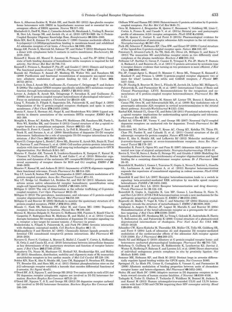
Kern A, Albarran-Zeckler R, Walsh HE, and Smith RG (2012) Apo-ghrelin receptorforms heteromers with DRD2 in hypothalamic neurons and is essential for an-orexigenic effects of DRD2 agonism. Neuron 73:317–332.
Khelashvili G, Dorff K, Shan J, Camacho-Artacho M, Skrabanek L, Vroling B, BouvierM, Devi LA, George SR, and Javitch JA, et al. (2010) GPCR-OKB: the G Protein-Coupled Receptor Oligomer Knowledge Base. Bioinformatics 26:1804–1805.
Klotz KN, Keil R, Zimmer FJ, and Schwabe U (1990) Guanine nucleotide effects on8-cyclopentyl-1,3-[3H]dipropylxanthine binding to membrane-bound and solubilizedA1 adenosine receptors of rat brain. J Neurochem 54:1988–1994.
Knepp AM, Periole X, Marrink SJ, Sakmar TP, and Huber T (2012) Rhodopsin formsa dimer with cytoplasmic helix 8 contacts in native membranes. Biochemistry 51:1819–1821.
Kniazeff J, Bessis AS, Maurel D, Ansanay H, Prézeau L, and Pin JP (2004) Closedstate of both binding domains of homodimeric mGlu receptors is required for fullactivity. Nat Struct Mol Biol 11:706–713.
Kniazeff J, Prézeau L, Rondard P, Pin JP, and Goudet C (2011) Dimers and beyond:The functional puzzles of class C GPCRs. Pharmacol Ther 130:9–25.
Kuszak AJ, Pitchiaya S, Anand JP, Mosberg HI, Walter NG, and Sunahara RK(2009) Purification and functional reconstitution of monomeric mu-opioid recep-tors: allosteric modulation of agonist binding by Gi2. J Biol Chem 284:26732–26741.
Levoye A, Dam J, Ayoub MA, Guillaume JL, Couturier C, Delagrange P, and JockersR (2006a) The orphan GPR50 receptor specifically inhibits MT1 melatonin receptorfunction through heterodimerization. EMBO J 25:3012–3023.
Levoye A, Jockers R, Ayoub MA, Delagrange P, Savaskan E, and Guillaume JL(2006b) Are G protein-coupled receptor heterodimers of physiological relevance?—Focus on melatonin receptors. Chronobiol Int 23:419–426.
Liang Y, Fotiadis D, Filipek S, Saperstein DA, Palczewski K, and Engel A (2003)Organization of the G protein-coupled receptors rhodopsin and opsin in nativemembranes. J Biol Chem 278:21655–21662.
Mancia F, Assur Z, Herman AG, Siegel R, and Hendrickson WA (2008) Ligand sen-sitivity in dimeric associations of the serotonin 5HT2c receptor. EMBO Rep 9:363–369.
Manglik A, Kruse AC, Kobilka TS, Thian FS, Mathiesen JM, Sunahara RK, Pardo L,Weis WI, Kobilka BK, and Granier S (2012) Crystal structure of the m-opioid re-ceptor bound to a morphinan antagonist. Nature 485:321–326.
Marcellino D, Ferré S, Casadó V, Cortés A, Le Foll B, Mazzola C, Drago F, Saur O,Stark H, and Soriano A, et al. (2008) Identification of dopamine D1-D3 receptorheteromers. Indications for a role of synergistic D1-D3 receptor interactions in thestriatum. J Biol Chem 283:26016–26025.
Maurel D, Comps-Agrar L, Brock C, Rives ML, Bourrier E, Ayoub MA, Bazin H, TinelN, Durroux T, and Prézeau L, et al. (2008) Cell-surface protein-protein interactionanalysis with time-resolved FRET and snap-tag technologies: application to GPCRoligomerization. Nat Methods 5:561–567.
Maurice P, Daulat AM, Turecek R, Ivankova-Susankova K, Zamponi F, Kamal M,Clement N, Guillaume JL, Bettler B, and Galès C, et al. (2010) Molecular orga-nization and dynamics of the melatonin MT₁ receptor/RGS20/G(i) protein complexreveal asymmetry of receptor dimers for RGS and G(i) coupling. EMBO J 29:3646–3659.
Maurice P, Kamal M, and Jockers R (2011) Asymmetry of GPCR oligomers supportstheir functional relevance. Trends Pharmacol Sci 32:514–520.
May LT, Leach K, Sexton PM, and Christopoulos A (2007) Allosteric modulation of Gprotein-coupled receptors. Annu Rev Pharmacol Toxicol 47:1–51.
May LT, Bridge LJ, Stoddart LA, Briddon SJ, and Hill SJ (2011) Allosteric inter-actions across native adenosine-A3 receptor homodimers: quantification usingsingle-cell ligand-binding kinetics. FASEB J 25:3465–3476.
Milligan G (2010) The role of dimerisation in the cellular trafficking of G-protein-coupled receptors. Curr Opin Pharmacol 10:23–29.
Milligan G (2013) The prevalence, maintenance, and relevance of G protein-coupledreceptor oligomerization. Mol Pharmacol 84:158-169.
Milligan G and Bouvier M (2005) Methods to monitor the quaternary structure of Gprotein-coupled receptors. FEBS J 272:2914–2925.
Missale C, Nash SR, Robinson SW, Jaber M, and Caron MG (1998) Dopaminereceptors: from structure to function. Physiol Rev 78:189–225.
Moreno E, Moreno-Delgado D, Navarro G, Hoffmann HM, Fuentes S, Rosell-Vilar S,Gasperini P, Rodriguez-Ruiz M, Medrano M, and Mallol J, et al. (2014) Cocainedisrupts histamine H3 receptor modulation of dopamine D1 receptor signaling vias1 receptor: a key target for cocaine toxicity. J Neurosci (in press).
Modzelewska A, Filipek S, Palczewski K, and Park PS (2006) Arrestin interactionwith rhodopsin: conceptual models. Cell Biochem Biophys 46:1–15.
Mukhopadhyay S and Howlett AC (2005). Chemically distinct ligands promote dif-ferential CB1 cannabinoid receptor-Gi protein interactions. Mol Pharmacol 67:2016–2024.
Navarro G, Ferré S, Cordomi A, Moreno E, Mallol J, Casadó V, Cortés A, HoffmannH, Ortiz J, and Canela EI, et al. (2010) Interactions between intracellular domainsas key determinants of the quaternary structure and function of receptor hetero-mers. J Biol Chem 285:27346–27359.
Nenasheva TA, Neary M, Mashanov GI, Birdsall NJ, Breckenridge RA, and MolloyJE (2013) Abundance, distribution, mobility and oligomeric state of M₂ muscarinicacetylcholine receptors in live cardiac muscle. J Mol Cell Cardiol 57:129–136.
Nobles KN, Xiao K, Ahn S, Shukla AK, Lam CM, Rajagopal S, Strachan RT, HuangTY, Bressler EA, and Hara MR, et al. (2011) Distinct phosphorylation sites on theb(2)-adrenergic receptor establish a barcode that encodes differential functions ofb-arrestin. Sci Signal 4:ra51.
O’Dowd BF, Ji X, Nguyen T, and George SR (2012) Two amino acids in each of D1 andD2 dopamine receptor cytoplasmic regions are involved in D1-D2 heteromer for-mation. Biochem Biophys Res Commun 417:23–28.
O’Dowd BF, Nguyen T, Ji X, and George SR (2013) D5 dopamine receptor carboxyltail involved in D5-D2 heteromer formation. Biochem Biophys Res Commun 431:586–589.
OldhamWM and HammHE (2008) Heterotrimeric G protein activation by G-protein-coupled receptors. Nat Rev Mol Cell Biol 9:60–71.
Orru M, Bake�sová J, Brugarolas M, Quiroz C, Beaumont V, Goldberg SR, Lluís C,Cortés A, Franco R, and Casadó V, et al. (2011a) Striatal pre- and postsynapticprofile of adenosine A(2A) receptor antagonists. PLoS ONE 6:e16088.
Orrú M, Quiroz C, Guitart X, and Ferré S (2011b) Pharmacological evidence fordifferent populations of postsynaptic adenosine A2A receptors in the rat striatum.Neuropharmacology 61:967–974.
Park JH, Scheerer P, Hofmann KP, Choe HW, and Ernst OP (2008) Crystal structureof the ligand-free G-protein-coupled receptor opsin. Nature 454:183–187.
Patowary S, Alvarez-Curto E, Xu TR, Holz JD, Oliver JA, Milligan G, and Raicu V(2013) The muscarinic M3 acetylcholine receptor exists as two differently sizedcomplexes at the plasma membrane. Biochem J 452:303–312.
Pellissier LP, Barthet G, Gaven F, Cassier E, Trinquet E, Pin JP, Marin P, DumuisA, Bockaert J, and Banères JL, et al. (2011) G protein activation by serotonin type4 receptor dimers: evidence that turning on two protomers is more efficient. J BiolChem 286:9985–9997.
Pin JP, Comps-Agrar L, Maurel D, Monnier C, Rives ML, Trinquet E, Kniazeff J,Rondard P, and Prézeau L (2009) G-protein-coupled receptor oligomers: two ormore for what? Lessons from mGlu and GABAB receptors. J Physiol 587:5337–5344.
Pin JP, Neubig R, Bouvier M, Devi L, Filizola M, Javitch JA, Lohse MJ, Milligan G,Palczewski K, and Parmentier M, et al. (2007) International Union of Basic andClinical Pharmacology. LXVII. Recommendations for the recognition and no-menclature of G protein-coupled receptor heteromultimers. Pharmacol Rev 59:5–13.
Quiroz C, Luján R, Uchigashima M, Simoes AP, Lerner TN, Borycz J, Kachroo A,Canas PM, Orru M, and Schwarzschild MA, et al. (2009) Key modulatory role ofpresynaptic adenosine A2A receptors in cortical neurotransmission to the striataldirect pathway. ScientificWorldJournal 9:1321–1344.
Raehal KM, Schmid CL, Groer CE, and Bohn LM (2011) Functional selectivity at them-opioid receptor: implications for understanding opioid analgesia and tolerance.Pharmacol Rev 63:1001–1019.
Rashid AJ, O’Dowd BF, Verma V, and George SR (2007) Neuronal Gq/11-coupleddopamine receptors: an uncharted role for dopamine. Trends Pharmacol Sci 28:551–555.
Rasmussen SG, DeVree BT, Zou Y, Kruse AC, Chung KY, Kobilka TS, Thian FS,Chae PS, Pardon E, and Calinski D, et al. (2011) Crystal structure of the b2adrenergic receptor-Gs protein complex. Nature 477:549–555.
Reiter E, Ahn S, Shukla AK, and Lefkowitz RJ (2012) Molecular mechanism ofb-arrestin-biased agonism at seven-transmembrane receptors. Annu Rev Phar-macol Toxicol 52:179–197.
Rimondini R, Ferré S, Ogren SO, and Fuxe K (1997) Adenosine A2A agonists: a po-tential new type of atypical antipsychotic. Neuropsychopharmacology 17:82–91.
Rovira X, Vivó M, Serra J, Roche D, Strange PG, and Giraldo J (2009) Modelling theinterdependence between the stoichiometry of receptor oligomerization and ligandbinding for a coexisting dimer/tetramer receptor system. Br J Pharmacol 156:28–35.
Rozenfeld R, Bushlin I, Gomes I, Tzavaras N, Gupta A, Neves S, Battini L, GusellaGL, Lachmann A, and Ma’ayan A, et al. (2012) Receptor heteromerizationexpands the repertoire of cannabinoid signaling in rodent neurons. PLoS ONE7:e29239.
Rozenfeld R and Devi LA (2007) Receptor heterodimerization leads to a switch insignaling: beta-arrestin2-mediated ERK activation by mu-delta opioid receptorheterodimers. FASEB J 21:2455–2465.
Rozenfeld R and Devi LA (2010) Receptor heteromerization and drug discovery.Trends Pharmacol Sci 31:124–130.
Rozenfeld R, Gupta A, Gagnidze K, Lim MP, Gomes I, Lee-Ramos D, Nieto N,and Devi LA (2011) AT1R-CB₁R heteromerization reveals a new mechanism for thepathogenic properties of angiotensin II. EMBO J 30:2350–2363.
Ruprecht JJ, Mielke T, Vogel R, Villa C, and Schertler GF (2004) Electron crystal-lography reveals the structure of metarhodopsin I. EMBO J 23:3609–3620.
Salahpour A, Angers S, Mercier JF, Lagacé M, Marullo S, and Bouvier M (2004)Homodimerization of the beta2-adrenergic receptor as a prerequisite for cell sur-face targeting. J Biol Chem 279:33390–33397.
Salom D, Lodowski DT, Stenkamp RE, Le Trong I, Golczak M, Jastrzebska B, HarrisT, Ballesteros JA, and Palczewski K (2006) Crystal structure of a photoactivateddeprotonated intermediate of rhodopsin. Proc Natl Acad Sci USA 103:16123–16128.
Schindler CW, Karcz-Kubicha M, Thorndike EB, Müller CE, Tella SR, Goldberg SR,and Ferré S (2004) Lack of adenosine A1 and dopamine D2 receptor-mediatedmodulation of the cardiovascular effects of the adenosine A2A receptor agonistCGS 21680. Eur J Pharmacol 484:269–275.
Smith NJ and Milligan G (2010) Allostery at G protein-coupled receptor homo- andheteromers: uncharted pharmacological landscapes. Pharmacol Rev 62:701–725.
Söderberg O, Gullberg M, Jarvius M, Ridderstråle K, Leuchowius KJ, Jarvius J,Wester K, Hydbring P, Bahram F, and Larsson LG, et al. (2006) Direct observationof individual endogenous protein complexes in situ by proximity ligation. NatMethods 3:995–1000.
Sommer ME, Hofmann KP, and Heck M (2012) Distinct loops in arrestin differen-tially regulate ligand binding within the GPCR opsin. Nat Commun 3:995.
Springael JY, Le Minh PN, Urizar E, Costagliola S, Vassart G, and Parmentier M(2006) Allosteric modulation of binding properties between units of chemokinereceptor homo- and hetero-oligomers. Mol Pharmacol 69:1652–1661.
Staley JK and Mash DC (1996) Adaptive increase in D3 dopamine receptors in thebrain reward circuits of human cocaine fatalities. J Neurosci 16:6100–6106.
Tadagaki K, Tudor D, Gbahou F, Tschische P, Waldhoer M, Bomsel M, Jockers R,and Kamal M (2012) Human cytomegalovirus-encoded UL33 and UL78 hetero-merize with host CCR5 and CXCR4 impairing their HIV coreceptor activity. Blood119:4908–4918.
G Protein–Coupled Receptor Oligomerization 433
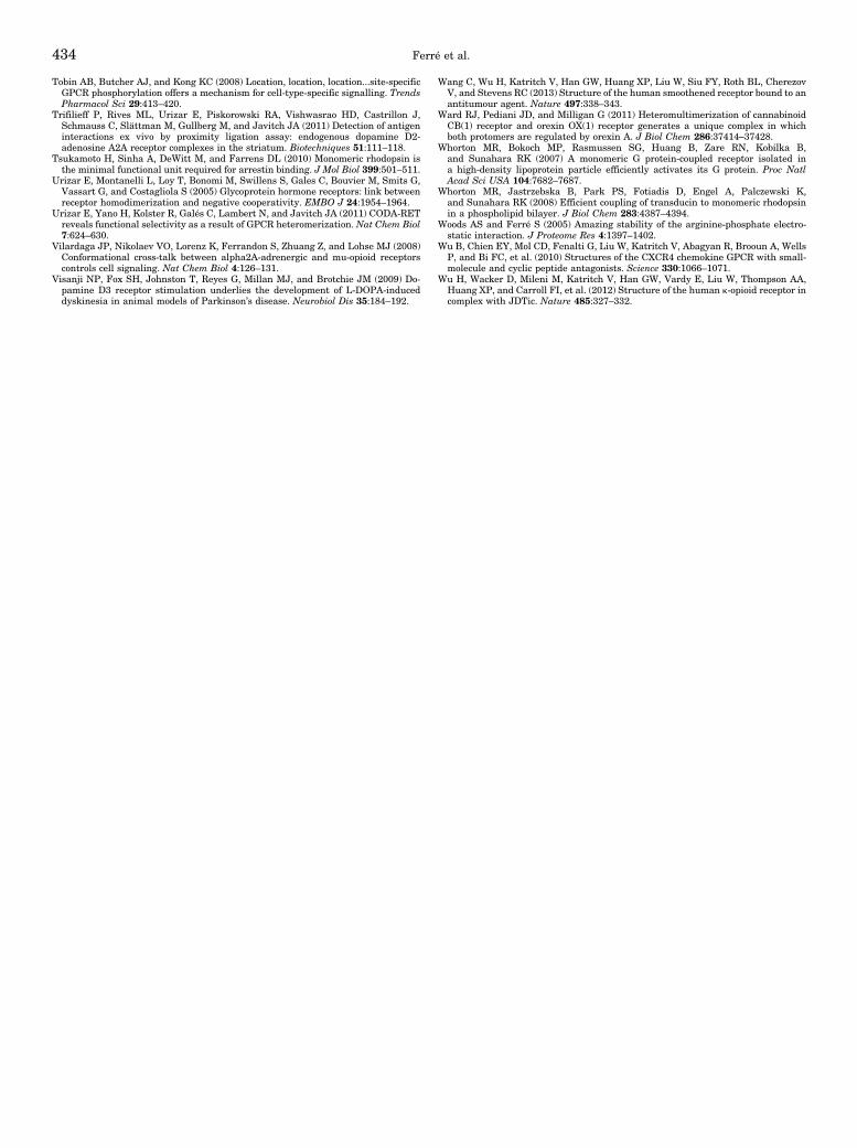
Tobin AB, Butcher AJ, and Kong KC (2008) Location, location, location...site-specificGPCR phosphorylation offers a mechanism for cell-type-specific signalling. TrendsPharmacol Sci 29:413–420.
Trifilieff P, Rives ML, Urizar E, Piskorowski RA, Vishwasrao HD, Castrillon J,Schmauss C, Slättman M, Gullberg M, and Javitch JA (2011) Detection of antigeninteractions ex vivo by proximity ligation assay: endogenous dopamine D2-adenosine A2A receptor complexes in the striatum. Biotechniques 51:111–118.
Tsukamoto H, Sinha A, DeWitt M, and Farrens DL (2010) Monomeric rhodopsin isthe minimal functional unit required for arrestin binding. J Mol Biol 399:501–511.
Urizar E, Montanelli L, Loy T, Bonomi M, Swillens S, Gales C, Bouvier M, Smits G,Vassart G, and Costagliola S (2005) Glycoprotein hormone receptors: link betweenreceptor homodimerization and negative cooperativity. EMBO J 24:1954–1964.
Urizar E, Yano H, Kolster R, Galés C, Lambert N, and Javitch JA (2011) CODA-RETreveals functional selectivity as a result of GPCR heteromerization. Nat Chem Biol7:624–630.
Vilardaga JP, Nikolaev VO, Lorenz K, Ferrandon S, Zhuang Z, and Lohse MJ (2008)Conformational cross-talk between alpha2A-adrenergic and mu-opioid receptorscontrols cell signaling. Nat Chem Biol 4:126–131.
Visanji NP, Fox SH, Johnston T, Reyes G, Millan MJ, and Brotchie JM (2009) Do-pamine D3 receptor stimulation underlies the development of L-DOPA-induceddyskinesia in animal models of Parkinson’s disease. Neurobiol Dis 35:184–192.
Wang C, Wu H, Katritch V, Han GW, Huang XP, Liu W, Siu FY, Roth BL, CherezovV, and Stevens RC (2013) Structure of the human smoothened receptor bound to anantitumour agent. Nature 497:338–343.
Ward RJ, Pediani JD, and Milligan G (2011) Heteromultimerization of cannabinoidCB(1) receptor and orexin OX(1) receptor generates a unique complex in whichboth protomers are regulated by orexin A. J Biol Chem 286:37414–37428.
Whorton MR, Bokoch MP, Rasmussen SG, Huang B, Zare RN, Kobilka B,and Sunahara RK (2007) A monomeric G protein-coupled receptor isolated ina high-density lipoprotein particle efficiently activates its G protein. Proc NatlAcad Sci USA 104:7682–7687.
Whorton MR, Jastrzebska B, Park PS, Fotiadis D, Engel A, Palczewski K,and Sunahara RK (2008) Efficient coupling of transducin to monomeric rhodopsinin a phospholipid bilayer. J Biol Chem 283:4387–4394.
Woods AS and Ferré S (2005) Amazing stability of the arginine-phosphate electro-static interaction. J Proteome Res 4:1397–1402.
Wu B, Chien EY, Mol CD, Fenalti G, Liu W, Katritch V, Abagyan R, Brooun A, WellsP, and Bi FC, et al. (2010) Structures of the CXCR4 chemokine GPCR with small-molecule and cyclic peptide antagonists. Science 330:1066–1071.
Wu H, Wacker D, Mileni M, Katritch V, Han GW, Vardy E, Liu W, Thompson AA,Huang XP, and Carroll FI, et al. (2012) Structure of the human k-opioid receptor incomplex with JDTic. Nature 485:327–332.
434 Ferré et al.


![INDEX [pharmrev.aspetjournals.org]pharmrev.aspetjournals.org/content/pharmrev/6/4/local/back-matter.… · 546 INDEX Drugallergy-Continued dosage ofdrugaffecting, 395 duration of,408](https://img.pdfslide.us/doc/110x75/5e9d1621ae88997f3e7425b1/index-546-index-drugallergy-continued-dosage-ofdrugaffecting-395-duration-of408.jpg)
![INDEX [pharmrev.aspetjournals.org]pharmrev.aspetjournals.org/content/pharmrev/7/4/local/... · 2005-07-23 · INDEX 499 Receptor hypothesis, development, 197 theory fornon-competitive](https://img.pdfslide.us/doc/110x75/5f792a265f98132bd25e45a7/index-2005-07-23-index-499-receptor-hypothesis-development-197-theory-fornon-competitive.jpg)


![Index [pharmrev.aspetjournals.org]pharmrev.aspetjournals.org/content/pharmrev/36/4/local/back-matter… · 338 INDEX Benzamidines, inhibitor ofclassical pathway, 229 Bile acid-dependent](https://img.pdfslide.us/doc/110x75/5e9fe02aa70cf17206521135/index-338-index-benzamidines-inhibitor-ofclassical-pathway-229-bile-acid-dependent.jpg)


![Index [pharmrev.aspetjournals.org]pharmrev.aspetjournals.org/content/pharmrev/43/4/local/back-matter.pdf · primary structure, kidney (fig.), 497 ... pathophysiology, ... schematic](https://img.pdfslide.us/doc/110x75/5b82cde67f8b9a315b8bcf1a/index-primary-structure-kidney-fig-497-pathophysiology-schematic.jpg)