Embed Size (px)
Citation preview
1521-0081/67/1/1–35$25.00 http://dx.doi.org/10.1124/pr.114.009225PHARMACOLOGICAL REVIEWS Pharmacol Rev 67:1–35, January 2015Copyright © 2014 by The American Society for Pharmacology and Experimental Therapeutics
ASSOCIATE EDITOR: ELIOT H. OHLSTEIN
International Union of Basic and ClinicalPharmacology. XCI. Structure, Function, and
Pharmacology of Acid-Sensing Ion Channels and theEpithelial Na+ Channel
Stephan Kellenberger and Laurent Schild
Département de Pharmacologie et de Toxicologie, Université de Lausanne, Lausanne, Switzerland
Abstract . . . . . . . . . . . . . . . . . . . . . . . . . . . . . . . . . . . . . . . . . . . . . . . . . . . . . . . . . . . . . . . . . . . . . . . . . . . . . . . . . . . . . 3I. Introduction . . . . . . . . . . . . . . . . . . . . . . . . . . . . . . . . . . . . . . . . . . . . . . . . . . . . . . . . . . . . . . . . . . . . . . . . . . . . . . . . . 3II. Phylogenetic and Sequence Comparison . . . . . . . . . . . . . . . . . . . . . . . . . . . . . . . . . . . . . . . . . . . . . . . . . . . . . . 3III. Tissue Distribution, Cellular Functions, and Physiologic and Pathologic Roles. . . . . . . . . . . . . . . . . 4
A. Acid-Sensing Ion Channels . . . . . . . . . . . . . . . . . . . . . . . . . . . . . . . . . . . . . . . . . . . . . . . . . . . . . . . . . . . . . . . 41. Tissue Distribution and Cellular Functions. . . . . . . . . . . . . . . . . . . . . . . . . . . . . . . . . . . . . . . . . . . . 42. Physiologic and Pathologic Roles. . . . . . . . . . . . . . . . . . . . . . . . . . . . . . . . . . . . . . . . . . . . . . . . . . . . . . 4
a. Synaptic plasticity and learning. . . . . . . . . . . . . . . . . . . . . . . . . . . . . . . . . . . . . . . . . . . . . . . . . . . . 4b. Fear and anxiety.. . . . . . . . . . . . . . . . . . . . . . . . . . . . . . . . . . . . . . . . . . . . . . . . . . . . . . . . . . . . . . . . . . 5c. Pain sensation. . . . . . . . . . . . . . . . . . . . . . . . . . . . . . . . . . . . . . . . . . . . . . . . . . . . . . . . . . . . . . . . . . . . . 6d. Mechanosensation. . . . . . . . . . . . . . . . . . . . . . . . . . . . . . . . . . . . . . . . . . . . . . . . . . . . . . . . . . . . . . . . . 7e. Neurodegenerative diseases. . . . . . . . . . . . . . . . . . . . . . . . . . . . . . . . . . . . . . . . . . . . . . . . . . . . . . . . 7f. Epileptic seizures. . . . . . . . . . . . . . . . . . . . . . . . . . . . . . . . . . . . . . . . . . . . . . . . . . . . . . . . . . . . . . . . . . 7
B. Epithelial Na+ Channel . . . . . . . . . . . . . . . . . . . . . . . . . . . . . . . . . . . . . . . . . . . . . . . . . . . . . . . . . . . . . . . . . . 81. Function. . . . . . . . . . . . . . . . . . . . . . . . . . . . . . . . . . . . . . . . . . . . . . . . . . . . . . . . . . . . . . . . . . . . . . . . . . . . . . 82. Kidney. . . . . . . . . . . . . . . . . . . . . . . . . . . . . . . . . . . . . . . . . . . . . . . . . . . . . . . . . . . . . . . . . . . . . . . . . . . . . . . . 83. Lungs.. . . . . . . . . . . . . . . . . . . . . . . . . . . . . . . . . . . . . . . . . . . . . . . . . . . . . . . . . . . . . . . . . . . . . . . . . . . . . . . . 94. Gastrointestinal Tract. . . . . . . . . . . . . . . . . . . . . . . . . . . . . . . . . . . . . . . . . . . . . . . . . . . . . . . . . . . . . . . . . 95. Sodium Sensor. . . . . . . . . . . . . . . . . . . . . . . . . . . . . . . . . . . . . . . . . . . . . . . . . . . . . . . . . . . . . . . . . . . . . . . . 9
IV. Structural Aspects . . . . . . . . . . . . . . . . . . . . . . . . . . . . . . . . . . . . . . . . . . . . . . . . . . . . . . . . . . . . . . . . . . . . . . . . . . . 10A. Primary Structure and Subunit Topology . . . . . . . . . . . . . . . . . . . . . . . . . . . . . . . . . . . . . . . . . . . . . . . . . 10
1. Subunit Organization of Epithelial Na+ Channel and Acid-Sensing Ion Channels. . . . . . . 102. Origin and Experimental Conditions of Different Crystal Structures.. . . . . . . . . . . . . . . . . . . 103. Differences between the Open and the Desensitized Structure and between
Different Published Structures. . . . . . . . . . . . . . . . . . . . . . . . . . . . . . . . . . . . . . . . . . . . . . . . . . . . . . . . 114. Difference and Similarities between the Acid-Sensing Ion Channels and
Epithelial Na+ Channel Subunit Organization. . . . . . . . . . . . . . . . . . . . . . . . . . . . . . . . . . . . . . . . . 11B. Subunit Stoichiometry . . . . . . . . . . . . . . . . . . . . . . . . . . . . . . . . . . . . . . . . . . . . . . . . . . . . . . . . . . . . . . . . . . . 12
V. Channel Function and Regulation. . . . . . . . . . . . . . . . . . . . . . . . . . . . . . . . . . . . . . . . . . . . . . . . . . . . . . . . . . . . 12A. Acid-Sensing Ion Channels . . . . . . . . . . . . . . . . . . . . . . . . . . . . . . . . . . . . . . . . . . . . . . . . . . . . . . . . . . . . . . . 12
1. Channel Gating and Ion Selectivity. . . . . . . . . . . . . . . . . . . . . . . . . . . . . . . . . . . . . . . . . . . . . . . . . . . . 12a. Current kinetics. . . . . . . . . . . . . . . . . . . . . . . . . . . . . . . . . . . . . . . . . . . . . . . . . . . . . . . . . . . . . . . . . . . 12b. Ion selectivity.. . . . . . . . . . . . . . . . . . . . . . . . . . . . . . . . . . . . . . . . . . . . . . . . . . . . . . . . . . . . . . . . . . . . . 12c. pH dependence. . . . . . . . . . . . . . . . . . . . . . . . . . . . . . . . . . . . . . . . . . . . . . . . . . . . . . . . . . . . . . . . . . . . 13d. Coupling of activation and steady-state desensitization. . . . . . . . . . . . . . . . . . . . . . . . . . . . . 13
Research in the authors’ laboratories was supported by Swiss National Science Foundation [Grants 310030_135378 (to L.S.) and310030_135542 and 31003A_153419 (to S.K.)].
Address correspondence to: Stephan Kellenberger, Département de Pharmacologie et de Toxicologie, Rue du Bugnon 27, Université deLausanne, CH-1005 Lausanne, Switzerland. E-mail: [email protected]
dx.doi.org/10.1124/pr.114.009225.
1
by guest on Decem
ber 16, 2021D
ownloaded from
2. Regulation. . . . . . . . . . . . . . . . . . . . . . . . . . . . . . . . . . . . . . . . . . . . . . . . . . . . . . . . . . . . . . . . . . . . . . . . . . . . 13a. Regulation by protein-protein interactions. . . . . . . . . . . . . . . . . . . . . . . . . . . . . . . . . . . . . . . . . . 14b. Regulation by ions, small molecules, and proteins. . . . . . . . . . . . . . . . . . . . . . . . . . . . . . . . . . 14
i. Calcium and Other Divalent or Polyvalent Cations. . . . . . . . . . . . . . . . . . . . . . . . . . . . . . . 14ii. Zinc. . . . . . . . . . . . . . . . . . . . . . . . . . . . . . . . . . . . . . . . . . . . . . . . . . . . . . . . . . . . . . . . . . . . . . . . . . . . . 14iii. Neuropeptides. . . . . . . . . . . . . . . . . . . . . . . . . . . . . . . . . . . . . . . . . . . . . . . . . . . . . . . . . . . . . . . . . . . 15iv. Redox Reagents and Free Radicals. . . . . . . . . . . . . . . . . . . . . . . . . . . . . . . . . . . . . . . . . . . . . . . 15v. Arachidonic Acid. . . . . . . . . . . . . . . . . . . . . . . . . . . . . . . . . . . . . . . . . . . . . . . . . . . . . . . . . . . . . . . . 17vi. G Protein–Coupled Receptors. . . . . . . . . . . . . . . . . . . . . . . . . . . . . . . . . . . . . . . . . . . . . . . . . . . . 17vii. Proteases.. . . . . . . . . . . . . . . . . . . . . . . . . . . . . . . . . . . . . . . . . . . . . . . . . . . . . . . . . . . . . . . . . . . . . . . 17viii. Kinases. . . . . . . . . . . . . . . . . . . . . . . . . . . . . . . . . . . . . . . . . . . . . . . . . . . . . . . . . . . . . . . . . . . . . . . . . 17
B. Epithelial Na+ Channel . . . . . . . . . . . . . . . . . . . . . . . . . . . . . . . . . . . . . . . . . . . . . . . . . . . . . . . . . . . . . . . . . . 171. Intrinsic Regulation. . . . . . . . . . . . . . . . . . . . . . . . . . . . . . . . . . . . . . . . . . . . . . . . . . . . . . . . . . . . . . . . . . . 172. Serine Proteases. . . . . . . . . . . . . . . . . . . . . . . . . . . . . . . . . . . . . . . . . . . . . . . . . . . . . . . . . . . . . . . . . . . . . . 183. Hormonal Regulation.. . . . . . . . . . . . . . . . . . . . . . . . . . . . . . . . . . . . . . . . . . . . . . . . . . . . . . . . . . . . . . . . . 18
C. Structure-Function Relationship of Acid-Sensing Ion Channels and Epithelial Na+
Channel . . . . . . . . . . . . . . . . . . . . . . . . . . . . . . . . . . . . . . . . . . . . . . . . . . . . . . . . . . . . . . . . . . . . . . . . . . . . . . . . . 191. The Acidic Pocket and Other Proton-Sensing Sites of Acid-Sensing Ion Channels. . . . . . . 192. The Finger—A Determinant of Subfamily-Specific Activation Mechanisms? . . . . . . . . . . . . 193. The Palm. . . . . . . . . . . . . . . . . . . . . . . . . . . . . . . . . . . . . . . . . . . . . . . . . . . . . . . . . . . . . . . . . . . . . . . . . . . . . 204. The b-Turn and the Extracellular Vestibule—at the Interface between the
Extracellular and Transmembrane Domains. . . . . . . . . . . . . . . . . . . . . . . . . . . . . . . . . . . . . . . . . . . 215. The Transmembrane Domain.. . . . . . . . . . . . . . . . . . . . . . . . . . . . . . . . . . . . . . . . . . . . . . . . . . . . . . . . . 22
a. Components of the channel pore.. . . . . . . . . . . . . . . . . . . . . . . . . . . . . . . . . . . . . . . . . . . . . . . . . . . 22b. The desensitization and closing gate.. . . . . . . . . . . . . . . . . . . . . . . . . . . . . . . . . . . . . . . . . . . . . . . 22c. The degenerin residue and the amiloride binding site. . . . . . . . . . . . . . . . . . . . . . . . . . . . . . . 22d. The selectivity filter.. . . . . . . . . . . . . . . . . . . . . . . . . . . . . . . . . . . . . . . . . . . . . . . . . . . . . . . . . . . . . . . 22
6. Cytoplasmic N and C Termini. . . . . . . . . . . . . . . . . . . . . . . . . . . . . . . . . . . . . . . . . . . . . . . . . . . . . . . . . 237. A Model of Acid-Sensing Ion Channel Gating. . . . . . . . . . . . . . . . . . . . . . . . . . . . . . . . . . . . . . . . . . 23
VI. Pharmacology . . . . . . . . . . . . . . . . . . . . . . . . . . . . . . . . . . . . . . . . . . . . . . . . . . . . . . . . . . . . . . . . . . . . . . . . . . . . . . . 24A. Acid-Sensing Ion Channels . . . . . . . . . . . . . . . . . . . . . . . . . . . . . . . . . . . . . . . . . . . . . . . . . . . . . . . . . . . . . . . 24
1. Small molecules. . . . . . . . . . . . . . . . . . . . . . . . . . . . . . . . . . . . . . . . . . . . . . . . . . . . . . . . . . . . . . . . . . . . . . . 24a. Acid-sensing ion channel inhibitors. . . . . . . . . . . . . . . . . . . . . . . . . . . . . . . . . . . . . . . . . . . . . . . . . 24
i. Amiloride. . . . . . . . . . . . . . . . . . . . . . . . . . . . . . . . . . . . . . . . . . . . . . . . . . . . . . . . . . . . . . . . . . . . . . . 24ii. Nonsteroidal Anti-Inflammatory Drugs. . . . . . . . . . . . . . . . . . . . . . . . . . . . . . . . . . . . . . . . . . 24iii. Other Small-Molecule Inhibitors. . . . . . . . . . . . . . . . . . . . . . . . . . . . . . . . . . . . . . . . . . . . . . . . . 25
b. The acid-sensing ion channel modulator 2-guanidine-4-methylquinazoline. . . . . . . . . . . 252. Toxins. . . . . . . . . . . . . . . . . . . . . . . . . . . . . . . . . . . . . . . . . . . . . . . . . . . . . . . . . . . . . . . . . . . . . . . . . . . . . . . . 25
a. Psalmotoxin1. . . . . . . . . . . . . . . . . . . . . . . . . . . . . . . . . . . . . . . . . . . . . . . . . . . . . . . . . . . . . . . . . . . . . . 25b. APETx2. . . . . . . . . . . . . . . . . . . . . . . . . . . . . . . . . . . . . . . . . . . . . . . . . . . . . . . . . . . . . . . . . . . . . . . . . . . 27c. Mambalgins. . . . . . . . . . . . . . . . . . . . . . . . . . . . . . . . . . . . . . . . . . . . . . . . . . . . . . . . . . . . . . . . . . . . . . . 27d. Mit-toxin. . . . . . . . . . . . . . . . . . . . . . . . . . . . . . . . . . . . . . . . . . . . . . . . . . . . . . . . . . . . . . . . . . . . . . . . . . 27
B. Epithelial Na+ Channel . . . . . . . . . . . . . . . . . . . . . . . . . . . . . . . . . . . . . . . . . . . . . . . . . . . . . . . . . . . . . . . . . . 271. Direct Epithelial Na+ Channel Antagonists. . . . . . . . . . . . . . . . . . . . . . . . . . . . . . . . . . . . . . . . . . . . 27
a. Properties. . . . . . . . . . . . . . . . . . . . . . . . . . . . . . . . . . . . . . . . . . . . . . . . . . . . . . . . . . . . . . . . . . . . . . . . . 27b. Renal effects. . . . . . . . . . . . . . . . . . . . . . . . . . . . . . . . . . . . . . . . . . . . . . . . . . . . . . . . . . . . . . . . . . . . . . . 28c. Pulmonary effects. . . . . . . . . . . . . . . . . . . . . . . . . . . . . . . . . . . . . . . . . . . . . . . . . . . . . . . . . . . . . . . . . . 28
2. Indirect Antagonists.. . . . . . . . . . . . . . . . . . . . . . . . . . . . . . . . . . . . . . . . . . . . . . . . . . . . . . . . . . . . . . . . . . 29
ABBREVIATIONS: AA, arachidonic acid; ASDN, aldosterone-sensitive distal nephron; ASIC, acid-sensing ion channel; ASL, airway surfaceliquid; BASIC, bile acid–sensitive ion channel; BDNF, brain-derived neurotrophic factor; CAP, channel activating protease; cASIC1, chickenASIC1; CCD, cortical collecting duct; CF, cystic fibrosis; CFTR, cystic fibrosis transmembrane conductance regulator; CNS, central nervoussystem; CNT, connecting tubule; DEG, degenerin; DRG, dorsal root ganglion; ENaC, epithelial Na+ channel; ET-1, endothelin-1; GAS, Gly-Ala-Ser; GMQ, 2-guanidine-4-methylquinazoline; KO, knockout; LTP, long-term potentiation; MitTx, Mit-toxin; MS, multiple sclerosis; NAc,nucleus accumbens; NSAIDs, nonsteroidal anti-inflammatory drugs; PcTx1, Psalmotoxin1; PDB, protein data bank; pH50, pH of half-maximalactivation; PHA-1, pseudohypoaldosteronism type 1; PICK1, protein interacting with C kinase; PKA, protein kinase A; PKC, protein kinase C,PNS, peripheral nervous system; S3969, N-(2-hydroxyethyl)-4-methyl-2-(4-methyl-1H-indol-3-ylthio)pentanamide; SSD, steady-state de-sensitization; TM, transmembrane segment; V2R, vasopressin receptor type 2; WIN55,212-2, (R)-(+)-[2,3-dihydro-5-methyl-3-(4-morpholi-nylmethyl)pyrrolo[1,2,3-de]-1,4-benzoxazin-6-yl]-1-naphthalenylmethanone.
2 Kellenberger and Schild
3. Agonists. . . . . . . . . . . . . . . . . . . . . . . . . . . . . . . . . . . . . . . . . . . . . . . . . . . . . . . . . . . . . . . . . . . . . . . . . . . . . . 29C. Bile Acid–Sensitive Ion Channel. . . . . . . . . . . . . . . . . . . . . . . . . . . . . . . . . . . . . . . . . . . . . . . . . . . . . . . . . . 29
VII. Conclusions and Perspectives . . . . . . . . . . . . . . . . . . . . . . . . . . . . . . . . . . . . . . . . . . . . . . . . . . . . . . . . . . . . . . . . 29Acknowledgments . . . . . . . . . . . . . . . . . . . . . . . . . . . . . . . . . . . . . . . . . . . . . . . . . . . . . . . . . . . . . . . . . . . . . . . . . . . 29References. . . . . . . . . . . . . . . . . . . . . . . . . . . . . . . . . . . . . . . . . . . . . . . . . . . . . . . . . . . . . . . . . . . . . . . . . . . . . . . . . . . 29
Abstract——The epithelial Na+ channel (ENaC) andthe acid-sensing ion channels (ASICs) form subfamilieswithin the ENaC/degenerin family of Na+ channels.ENaC mediates transepithelial Na+ transport, therebycontributing to Na+ homeostasis and the maintenance ofblood pressure and the airway surface liquid level. ASICsare H+-activated channels found in central and peripheralneurons, where their activation induces neuronaldepolarization. ASICs are involved in pain sensation, theexpression of fear, and neurodegeneration after ischemia,making them potentially interesting drug targets. Thisreview summarizes the biophysical properties, cellular
functions, and physiologic and pathologic roles of theASIC and ENaC subfamilies. The analysis of thehomologies between ENaC and ASICs and the relationbetween functional and structural information showsmany parallels between these channels, suggesting thatsome mechanisms that control channel activity areshared between ASICs and ENaC. The available crystalstructures and the discovery of animal toxins acting onASICs provide a unique opportunity to address themolecular mechanisms of ENaC and ASIC function toidentify novel strategies for the modulation of thesechannels by pharmacologic ligands.
I. Introduction
The epithelial Na+ channel (ENaC) and acid-sensingion channels (ASICs) represent the mammalian sub-families of the ENaC/degenerin (DEG) family of ionchannels that was discovered at the beginning of thenineties. ENaC has a well established role in Na+
reabsorption in the distal nephron, in the distal colon,and in the control of the liquid film on airway epi-thelia. ENaC is inhibited by the drugs amiloride andtriamterene that are clinically used as potassium-sparing diuretics. ASICs are neuronal channels thatwere discovered based on their primary structurehomology to ENaC. Many of their physiologic andpathologic roles, which include pain sensation, synap-tic plasticity, expression of fear, and neurodegenera-tion after ischemia, have been elucidated in geneticallymodified animal models during the last years andmake them potentially interesting drug targets. Sev-eral animal toxins act on ASICs. Currently, there arehowever no ASIC drugs in clinical use. Crystalstructures of ASICs, but not other ENaC/DEG familymembers, have been published. We review here thephysiologic and pathologic roles of ENaC and ASICs,their structural organization, functional properties, andregulation. Functional and structural parallels and dif-ferences are discussed in the perspective of ENaC andASICs as targets for pharmacological ligands. Otherrecent reviews cover relevant aspects of ASIC and ENaCfunction in more detail than this review: physiologic andpathologic roles of ASICs (Wemmie et al., 2013), reg-ulation of ASIC function (Chu et al., 2011), physiologyand regulation of ENaC (Palmer et al., 2012; Rossier,2014), and ENaC’s role in cystic fibrosis (Hobbs et al.,2013). Information on ASIC and ENaC properties andpharmacology can be found in the IUPHAR/BPS Guideto PHARMACOLOGY (www.guidetopharmacology.org/GRAC/FamilyDisplayForward?familyId=118; and
www.guidetopharmacology.org/GRAC/FamilyDisplay-Forward?familyId=122, respectively).
II. Phylogenetic and Sequence Comparison
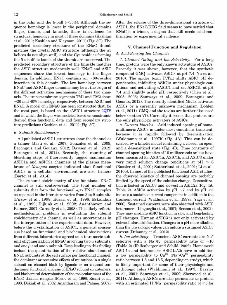
The phylogenetic tree in Fig. 1 is based mainly onsequences of ENaC and ASICs. Because this reviewfocuses on the mammalian ENaC and ASICs, the largeDrosophila and Caenorhabditis elegans subfamilieseach are represented by only one member, pickpocketand MEC-4, respectively. In addition, the peptide-gated FaNaC of Helix aspersa is included. The fourENaC subunits are encoded by the genes SCNN1A-D,whereas the ASIC subunits are encoded by ACCN1-4.The mammalian hiNaC (human intestine Na+ channel)and its rat/mouse ortholog BLINaC (brain-liver-intestineamiloride–sensitive Na+ channel), whose names refer totheir main sites of expression, have recently been re-named to BASIC for “bile acid–sensitive ion channel”after the discovery of their activation by bile acids(Wiemuth et al., 2012; Lefevre et al., 2014). BASIChas a slightly higher sequence homology to ASICsthan to ENaC, is encoded by ACCN5, and also has beencalled ASIC5 despite the fact that it is not activated byprotons and shares only ;30% sequence homology withASICs. ENaC and ASIC genes have in the meantimebeen cloned from many different species. For clarity weinclude in this phylogenetic tree mostly the human andrat sequences and, in addition, some fish ASIC se-quences, which were used for functional studies, chickenASIC1, from which the published crystal structures arederived, as well as Xenopus laevis ENaC sequences.ASIC genes have different splice variants, leading tocurrently at least 8 mammalian subunits, ASIC1a and -1b;ASIC2a and -2b; ASIC3a, -3b, and -3c (only in humans,in other organisms there is only one ASIC3), and ASIC4.Because of the cloning history, most ASICs originally haddifferent names and were later renamed (Kellenberger
ASIC and ENaC Function and Pharmacology 3
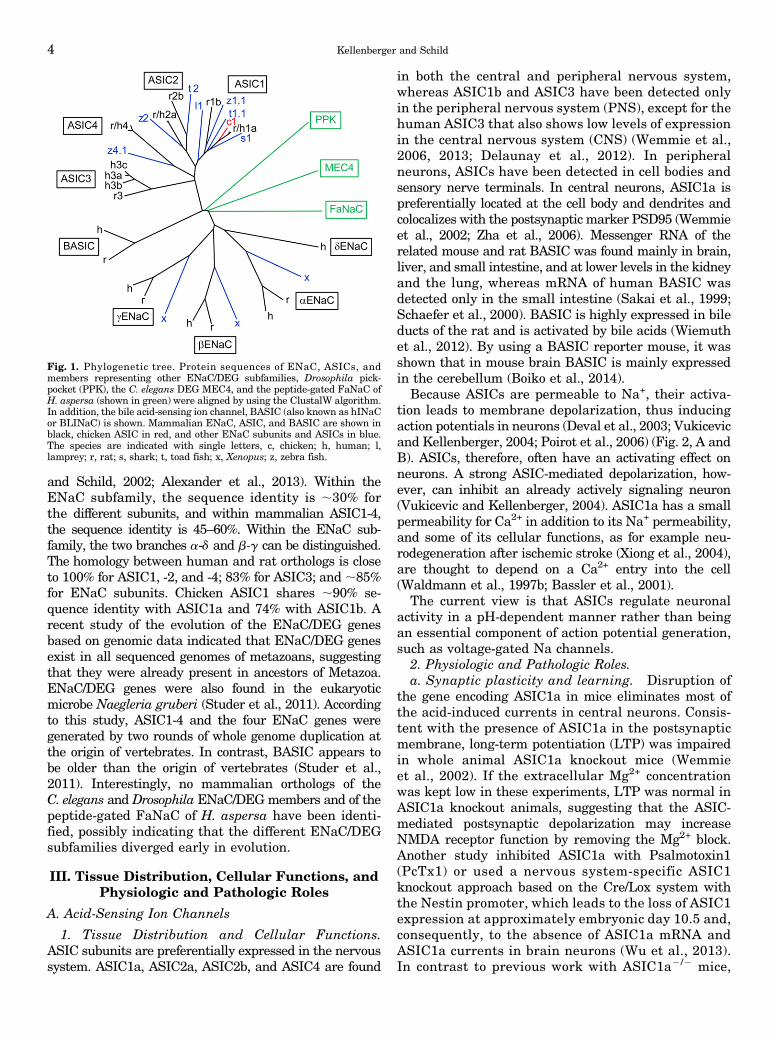
and Schild, 2002; Alexander et al., 2013). Within theENaC subfamily, the sequence identity is ;30% forthe different subunits, and within mammalian ASIC1-4,the sequence identity is 45–60%. Within the ENaC sub-family, the two branches a-d and b-g can be distinguished.The homology between human and rat orthologs is closeto 100% for ASIC1, -2, and -4; 83% for ASIC3; and ;85%for ENaC subunits. Chicken ASIC1 shares ;90% se-quence identity with ASIC1a and 74% with ASIC1b. Arecent study of the evolution of the ENaC/DEG genesbased on genomic data indicated that ENaC/DEG genesexist in all sequenced genomes of metazoans, suggestingthat they were already present in ancestors of Metazoa.ENaC/DEG genes were also found in the eukaryoticmicrobe Naegleria gruberi (Studer et al., 2011). Accordingto this study, ASIC1-4 and the four ENaC genes weregenerated by two rounds of whole genome duplication atthe origin of vertebrates. In contrast, BASIC appears tobe older than the origin of vertebrates (Studer et al.,2011). Interestingly, no mammalian orthologs of theC. elegans and Drosophila ENaC/DEGmembers and of thepeptide-gated FaNaC of H. aspersa have been identi-fied, possibly indicating that the different ENaC/DEGsubfamilies diverged early in evolution.
III. Tissue Distribution, Cellular Functions, andPhysiologic and Pathologic Roles
A. Acid-Sensing Ion Channels
1. Tissue Distribution and Cellular Functions.ASIC subunits are preferentially expressed in the nervoussystem. ASIC1a, ASIC2a, ASIC2b, and ASIC4 are found
in both the central and peripheral nervous system,whereas ASIC1b and ASIC3 have been detected onlyin the peripheral nervous system (PNS), except for thehuman ASIC3 that also shows low levels of expressionin the central nervous system (CNS) (Wemmie et al.,2006, 2013; Delaunay et al., 2012). In peripheralneurons, ASICs have been detected in cell bodies andsensory nerve terminals. In central neurons, ASIC1a ispreferentially located at the cell body and dendrites andcolocalizes with the postsynaptic marker PSD95 (Wemmieet al., 2002; Zha et al., 2006). Messenger RNA of therelated mouse and rat BASIC was found mainly in brain,liver, and small intestine, and at lower levels in the kidneyand the lung, whereas mRNA of human BASIC wasdetected only in the small intestine (Sakai et al., 1999;Schaefer et al., 2000). BASIC is highly expressed in bileducts of the rat and is activated by bile acids (Wiemuthet al., 2012). By using a BASIC reporter mouse, it wasshown that in mouse brain BASIC is mainly expressedin the cerebellum (Boiko et al., 2014).
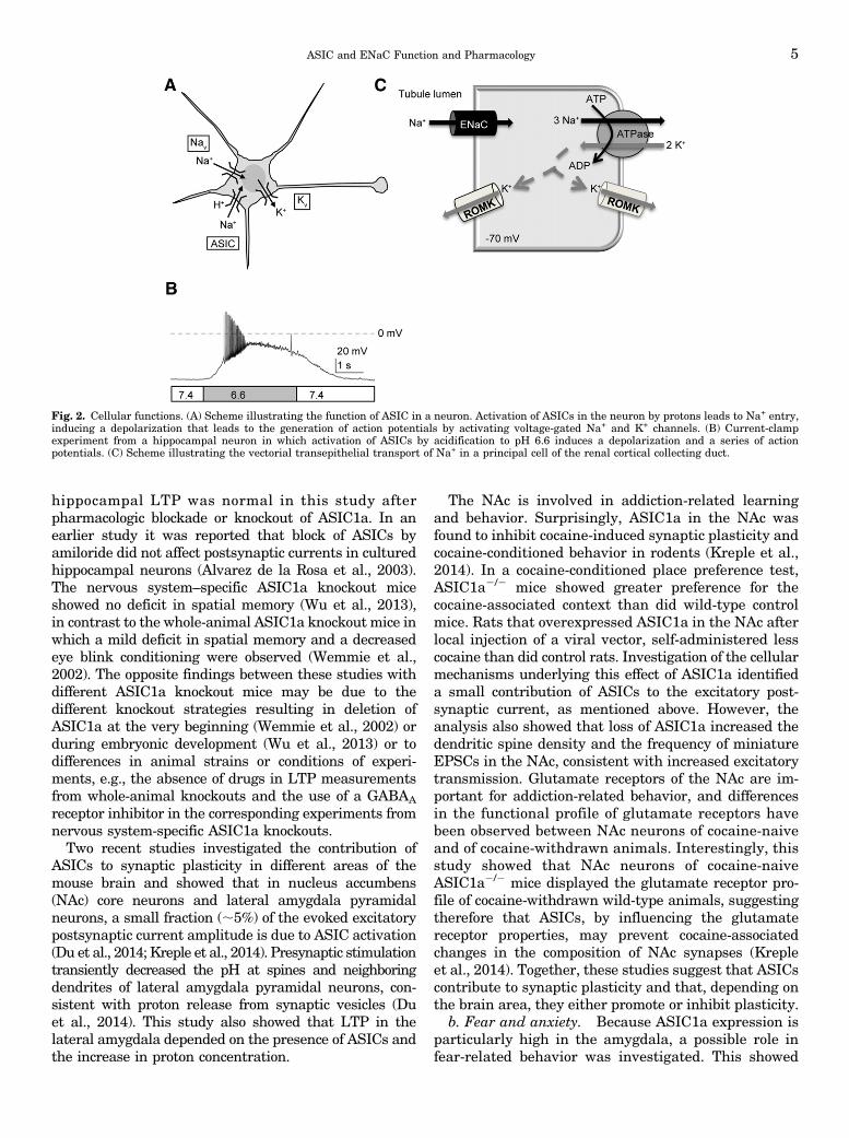
Because ASICs are permeable to Na+, their activa-tion leads to membrane depolarization, thus inducingaction potentials in neurons (Deval et al., 2003; Vukicevicand Kellenberger, 2004; Poirot et al., 2006) (Fig. 2, A andB). ASICs, therefore, often have an activating effect onneurons. A strong ASIC-mediated depolarization, how-ever, can inhibit an already actively signaling neuron(Vukicevic and Kellenberger, 2004). ASIC1a has a smallpermeability for Ca2+ in addition to its Na+ permeability,and some of its cellular functions, as for example neu-rodegeneration after ischemic stroke (Xiong et al., 2004),are thought to depend on a Ca2+ entry into the cell(Waldmann et al., 1997b; Bassler et al., 2001).
The current view is that ASICs regulate neuronalactivity in a pH-dependent manner rather than beingan essential component of action potential generation,such as voltage-gated Na channels.
2. Physiologic and Pathologic Roles.a. Synaptic plasticity and learning. Disruption of
the gene encoding ASIC1a in mice eliminates most ofthe acid-induced currents in central neurons. Consis-tent with the presence of ASIC1a in the postsynapticmembrane, long-term potentiation (LTP) was impairedin whole animal ASIC1a knockout mice (Wemmieet al., 2002). If the extracellular Mg2+ concentrationwas kept low in these experiments, LTP was normal inASIC1a knockout animals, suggesting that the ASIC-mediated postsynaptic depolarization may increaseNMDA receptor function by removing the Mg2+ block.Another study inhibited ASIC1a with Psalmotoxin1(PcTx1) or used a nervous system-specific ASIC1knockout approach based on the Cre/Lox system withthe Nestin promoter, which leads to the loss of ASIC1expression at approximately embryonic day 10.5 and,consequently, to the absence of ASIC1a mRNA andASIC1a currents in brain neurons (Wu et al., 2013).In contrast to previous work with ASIC1a2/2 mice,
Fig. 1. Phylogenetic tree. Protein sequences of ENaC, ASICs, andmembers representing other ENaC/DEG subfamilies, Drosophila pick-pocket (PPK), the C. elegans DEG MEC4, and the peptide-gated FaNaC ofH. aspersa (shown in green) were aligned by using the ClustalW algorithm.In addition, the bile acid-sensing ion channel, BASIC (also known as hINaCor BLINaC) is shown. Mammalian ENaC, ASIC, and BASIC are shown inblack, chicken ASIC in red, and other ENaC subunits and ASICs in blue.The species are indicated with single letters, c, chicken; h, human; l,lamprey; r, rat; s, shark; t, toad fish; x, Xenopus; z, zebra fish.
4 Kellenberger and Schild
hippocampal LTP was normal in this study afterpharmacologic blockade or knockout of ASIC1a. In anearlier study it was reported that block of ASICs byamiloride did not affect postsynaptic currents in culturedhippocampal neurons (Alvarez de la Rosa et al., 2003).The nervous system–specific ASIC1a knockout miceshowed no deficit in spatial memory (Wu et al., 2013),in contrast to the whole-animal ASIC1a knockout mice inwhich a mild deficit in spatial memory and a decreasedeye blink conditioning were observed (Wemmie et al.,2002). The opposite findings between these studies withdifferent ASIC1a knockout mice may be due to thedifferent knockout strategies resulting in deletion ofASIC1a at the very beginning (Wemmie et al., 2002) orduring embryonic development (Wu et al., 2013) or todifferences in animal strains or conditions of experi-ments, e.g., the absence of drugs in LTP measurementsfrom whole-animal knockouts and the use of a GABAA
receptor inhibitor in the corresponding experiments fromnervous system-specific ASIC1a knockouts.Two recent studies investigated the contribution of
ASICs to synaptic plasticity in different areas of themouse brain and showed that in nucleus accumbens(NAc) core neurons and lateral amygdala pyramidalneurons, a small fraction (;5%) of the evoked excitatorypostsynaptic current amplitude is due to ASIC activation(Du et al., 2014; Kreple et al., 2014). Presynaptic stimulationtransiently decreased the pH at spines and neighboringdendrites of lateral amygdala pyramidal neurons, con-sistent with proton release from synaptic vesicles (Duet al., 2014). This study also showed that LTP in thelateral amygdala depended on the presence of ASICs andthe increase in proton concentration.
The NAc is involved in addiction-related learningand behavior. Surprisingly, ASIC1a in the NAc wasfound to inhibit cocaine-induced synaptic plasticity andcocaine-conditioned behavior in rodents (Kreple et al.,2014). In a cocaine-conditioned place preference test,ASIC1a2/2 mice showed greater preference for thecocaine-associated context than did wild-type controlmice. Rats that overexpressed ASIC1a in the NAc afterlocal injection of a viral vector, self-administered lesscocaine than did control rats. Investigation of the cellularmechanisms underlying this effect of ASIC1a identifieda small contribution of ASICs to the excitatory post-synaptic current, as mentioned above. However, theanalysis also showed that loss of ASIC1a increased thedendritic spine density and the frequency of miniatureEPSCs in the NAc, consistent with increased excitatorytransmission. Glutamate receptors of the NAc are im-portant for addiction-related behavior, and differencesin the functional profile of glutamate receptors havebeen observed between NAc neurons of cocaine-naiveand of cocaine-withdrawn animals. Interestingly, thisstudy showed that NAc neurons of cocaine-naiveASIC1a2/2 mice displayed the glutamate receptor pro-file of cocaine-withdrawn wild-type animals, suggestingtherefore that ASICs, by influencing the glutamatereceptor properties, may prevent cocaine-associatedchanges in the composition of NAc synapses (Krepleet al., 2014). Together, these studies suggest that ASICscontribute to synaptic plasticity and that, depending onthe brain area, they either promote or inhibit plasticity.
b. Fear and anxiety. Because ASIC1a expression isparticularly high in the amygdala, a possible role infear-related behavior was investigated. This showed
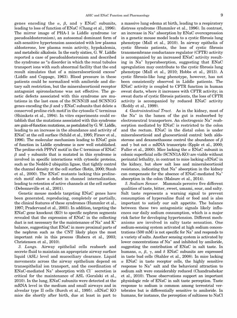
Fig. 2. Cellular functions. (A) Scheme illustrating the function of ASIC in a neuron. Activation of ASICs in the neuron by protons leads to Na+ entry,inducing a depolarization that leads to the generation of action potentials by activating voltage-gated Na+ and K+ channels. (B) Current-clampexperiment from a hippocampal neuron in which activation of ASICs by acidification to pH 6.6 induces a depolarization and a series of actionpotentials. (C) Scheme illustrating the vectorial transepithelial transport of Na+ in a principal cell of the renal cortical collecting duct.
ASIC and ENaC Function and Pharmacology 5

that whole-animal ASIC1a knockout mice displayeddeficits in cue and context fear conditioning as well asin unconditioned fear behaviors (Wemmie et al., 2003,2013; Coryell et al., 2007). The disruption of ASIC1aalso produced antidepressant-like effects in severalbehavioral tests carried out with mice (Coryell et al.,2009). Overexpression of ASIC1a increased fear-related behavior (Wemmie et al., 2004). It was shownthat the amygdala senses acidic pH and thereby evokesfear behavior (Ziemann et al., 2009). It is known thatan increase in CO2 levels induces fear behavior inhumans and has the same effect in mice. Inhibition ordisruption of ASIC1a function impaired CO2-inducedfear behavior (Ziemann et al., 2009). Localized expres-sion of ASIC1a in the amygdala of ASIC1a knockoutmice rescued the fear phenotype, demonstrating thatASIC1a of the amygdala contributes importantly tofear behavior. As mentioned above, it was also shownthat presynaptic stimulation lowers the amygdala pHand activates ASICs in lateral amygdala pyramidalneurons (Du et al., 2014). Intriguingly, a recent studywith rats showed that activation of ASIC1a in thebasolateral amygdala suppressed anxiety-like behavior(Pidoplichko et al., 2014). Activation of ASICs byinjection of ammonium in the basolateral amygdaladecreased the time spent in the center of the open fieldin the open field test, whereas inhibition of ASIC1a byPcTx1 had the opposite effect. Ammonium injectionalso prolonged the latency before the rats entered thedark compartment in the light/dark box test. ASICcurrents were present in both principal cells andinterneurons of the basolateral amygdala in the rat,and ASIC activation led to a higher increase in spon-taneous inhibitory postsynaptic currents than EPSCs inprincipal cells, indicating that the inhibitory effects ofASICs on principal cell signaling prevailed (Pidoplichkoet al., 2014). Because most results obtained with knockoutmice were confirmed with pharmacologic approaches, theopposite results obtained in these studies cannot be dueto a role of ASICs in development. It is therefore mostlikely that species differences are the cause of these con-tradictory results, thus that involvement of ASICs in thebasolateral amygdala signaling may be different betweenmice and rats. Currently, behavioral data from rats onthe role of ASICs in fear behavior is very limited and itwill be necessary to extend such studies to cue and con-text fear conditioning and to CO2-induced fear to de-termine how different the role of ASICs in fear behavior isbetween mice and rats. In humans, single nucleotidepolymorphisms in the noncoding region of the ASIC1agene were found to be associated with panic disorder,amygdala volume, and fear-related reactions (Smolleret al., 2014). It would be interesting to identify the ASICfunction that is changed in these variants.c. Pain sensation. In a number of conditions in-
volving pain, such as inflammation and ischemia, thetissue pH is lowered. The extracellular pH drops for
example in cardiac ischemia to 6.7 (Cobbe and Poole-Wilson, 1980). ASICs in sensory neurons are thereforecandidate receptors for acidification inducing pain.ASIC1a and ASIC2 are also expressed in pain-processing areas of the CNS where they may addition-ally contribute to pain sensation (Wemmie et al., 2013).Studies on human volunteers showed that local in-jection in the skin or iontophoresis of acidic solutionsinduced pain that was prevented by amiloride anddisplayed a pH of half-maximal effect of;6.5, thus closeto the pH of half-maximal activation (pH50) of ASIC1aand ASIC3 (Ugawa et al., 2002; Jones et al., 2004).Several animal studies provided strong evidence for therole of peripheral ASIC3 and ASIC1 in pain sensation.Specific knockdown of ASIC3 with intrathecally admin-istered siRNA in rats prevented inflammation-inducedheat hyperalgesia or flinching after local injection ofirritating substances (Deval et al., 2008). The syntheticcompound 2-guanidine-4-methylquinazoline (GMQ)activates ASIC3 at physiologic pH and inhibits otherASICs (Yu et al., 2010; Alijevic and Kellenberger, 2012).Local injection of GMQ in the mouse paw induced painbehavior that depended on the presence of ASIC3,further confirming the importance of ASIC3 (Yu et al.,2010). A number of studies has demonstrated a contribu-tion of ASIC3 to inflammation- and chronic acidification-related forms of pain (Sluka et al., 2009). Injection of theASIC activator Mit-toxin (MitTx) of the Texas coral snakevenom in the mouse paw induced pain behavior that wasdecreased by ASIC1a disruption (Bohlen et al., 2011).Recently, the ASIC-inhibitory toxin mambalgin-1 fromblack mamba venom was shown to reduce pain behaviorafter peripheral injection due to inhibition of ASIC1b(Diochot et al., 2012).
Intrathecal injection of mambalgin-1 diminishedpain behaviors due to inhibition of ASICs containingthe subunits ASIC1a and/or ASIC2a. Previous workhad shown that the main functional ASICs in thespinal cord were ASIC1a homomers and ASIC1a/2aheteromers (Baron et al., 2008), and that ASIC RNAsin the spinal cord were upregulated by peripheralinflammation (Duan et al., 2007). Administration tothe CNS of the spider toxin PcTx1, a specific inhibitorof ASIC1a, diminished pain behavior, further demon-strating the important role of central ASIC1a in painsensation (Duan et al., 2007; Mazzuca et al., 2007). Arecent study showed that ASIC1a in the spinal cord isthe target of brain-derived neurotrophic factor (BDNF)(Duan et al., 2012). BDNF increased ASIC1a cellsurface expression by inducing phosphorylation of theN-terminal residue Ser25 via the phosphoinositide3-kinase–protein kinase B cascade. Intrathecal injectionof BDNF induced mechanical hyperalgesia, which wasprevented by ASIC1a disruption (Duan et al., 2012).
Migraine is associated with tissue acidification in thedura. Functional changes of several types of ionchannels may contribute to an altered state of neuronal
6 Kellenberger and Schild

excitability of trigeminal afferent neurons innervatingthe dura (Yan and Dussor, 2014). The majority of ratdural afferents display ASIC currents, which are likelymediated by ASIC3-containing channels (Yan et al.,2011). Application of pH 5 synthetic interstitial fluid tothe dura of rats induced facial and hind paw allodynia.Facial allodynia is often associated with migraine (Yanet al., 2011). Amiloride has been reported to inhibitcortical spreading depression in rodents, and improvedaura and headache severity in 4 of 7 patients of a smallclinical trial (Holland et al., 2012).Together, these studies provide clear indications for
nociceptive roles of central and peripheral ASICs.Surprisingly, two studies showed that mice in which allASIC currents were suppressed displayed increasedpain behavior (Mogil et al., 2005; Kang et al., 2012).Mice overexpressing a dominant-negative mutantASIC3 subunit were more sensitive than wild-typecontrols to mechanical pain and chemical/inflammatorypain and developed stronger mechanical hypersensitivityafter inflammation (Mogil et al., 2005). Triple knockoutmice, in which the ASIC1, ASIC2, and ASIC3 genes weresimultaneously disrupted, showed an increased behav-ioral sensitivity to mechanical stimuli and an increasedmechanosensitivity of A-mechanonociceptors (Kang et al.,2012). This indicates that the role of ASICs in nociceptionis still not fully understood despite many importantfindings.d. Mechanosensation. Because related channels in
C. elegans are involved in mechanosensation, a similarpossible role of ASICs was investigated in mammals.Currently there is evidence of ASIC expression inprimary sensory neurons and the afferents of mecha-noreceptors, obtained either by retrograde tracingcombined with functional analysis of isolated sensoryneurons or by immunohistochemistry (reviewed in Chenand Wong, 2013). Disruption of the expression of dif-ferent ASICs produced defects in mechanotransductionin tissues such as the skin, the stomach, the colon, thecochlea, and arterial baroreceptors (reviewed in Chenand Wong, 2013). These effects were most clearly dem-onstrated in the gastrointestinal tract and in arterialbaroreceptors. Mechanotransduction in the gastroin-testinal system is important for the control of gastriccoordination and emptying, colonic motility, and thesensation of pain. Single-fiber recordings on an in vitrovagus-gastroesophageal or colon preparation showedin ASIC12/2 mice enhanced, in ASIC22/2 mice mixed,and in ASIC32/2 mice decreased mechanosensitivity invisceral mechanoreceptors (Jones et al., 2005; Pageet al., 2005). Analysis of the digestive functionshowed that that ASIC12/2 mice had an alterationin upper gastrointestinal functions manifested as aslower gastric emptying, whereas ASIC22/2 miceshowed a decrease in the number of fecal pellets perday, indicating a changed lower bowel function (Pageet al., 2005).
Aortic baroreceptor neurons in the nodose gangliaand their terminals express ASIC2. ASIC22/2 miceshowed hypertension, increased sympathetic and de-creased parasympathetic control of the circulation, anda decreased gain of the baroreflex (Lu et al., 2009). Theimpairment of the baroreflex in ASIC22/2 mice sug-gests that ASIC2 is a determinant of the sensitivity ofarterial baroreceptor and contributes to the autonomiccontrol of the circulation.
So far it has not been possible to demonstratemechanosensitivity of recombinantly expressed ASICsor of ASICs expressed in dorsal root ganglion (DRG)neurons (Drew et al., 2004). Therefore it is possiblethat ASICs need to form complexes with other proteinsto be able to act as mechanosensors.
e. Neurodegenerative diseases. Calcium entry intoneurons is thought to be the most important cause ofneuronal injury after ischemic stroke. Because ische-mic stroke induces a tissue acidification and ASIC1a ispartially permeable to Ca2+, a possible contribution ofASIC1a to this toxicity was investigated. Disruption ofthe ASIC1a gene or inhibition of ASIC1a by PcTx1-containing spider venom reduced the infarct volume inan experimental stroke model by .60%, thus demon-strating an important contribution of ASIC1a to ischemicstroke-induced neurodegeneration (Xiong et al., 2004).Intracerebroventricular administration of PcTx1 up to5 hours after transient middle cerebral artery occlusionreduced the infarct volume by .50% (Pignataro et al.,2007). Subsequent studies showed a protective effect ofASIC gene disruption or inhibition of ASIC function ina number of neurodegenerative diseases, includingmultiple sclerosis (MS), Huntington’s, and Parkinson’sdisease (reviewed in Wemmie et al., 2013). The disrup-tion of the ASIC1a gene reduced the clinical deficit andaxonal degeneration in experimental autoimmune en-cephalomyelitis (Friese et al., 2007). It was furthershown that ASIC1a is upregulated in lesions from ex-perimental autoimmune encephalomyelitis in mice andfrom MS patients (Vergo et al., 2011). Treatment withamiloride was neuroprotective in animal models and inhuman MS patients (Vergo et al., 2011; Arun et al.,2013). Interestingly, an association between the ASIC2gene and MS has been shown in a remote humanpopulation in Sardinia (Bernardinelli et al., 2007).
f. Epileptic seizures. During epileptic seizures thebrain pH is lowered, and it is known that acidosis canend seizures. Disruption of the ASIC1a gene increasedthe severity of seizures, and the opposite effect wasobserved with overexpression of ASIC1a (Ziemann et al.,2008). This study showed that ASIC1a expression isrequired for seizure termination by CO2 inhalation.Because inhibitory interneurons in hippocampus displayhigher ASIC current amplitudes than pyramidal neu-rons it is possible that the interneuron-mediated in-hibition ended the seizures (Ziemann et al., 2008).Observations of seizure inhibition by high concentrations
ASIC and ENaC Function and Pharmacology 7
of amiloride (Ali et al., 2006; N’Gouemo, 2008; Luszczkiet al., 2009) appear to contradict the role of ASIC1a inseizure termination. Because of its hydrophilic nature,amiloride is expected not to cross the blood-brain barrier,and it is currently not clear whether it reaches the brainor whether it exerts anti-seizure effects by othermechanisms. Amiloride is not specific for ASICs, and ifit reaches the brain its effect on seizures may well be dueto inhibition of the Na+/H+ exchanger, which, by loweringthe intracellular pH, may decrease epileptic activity (Aliet al., 2006; Luszczki et al., 2009). Recently, a geneticstudy indicated an association between single nucleo-tide polymorphisms of ASIC1 and temporal lobeepilepsy (Lv et al., 2011). Whether these polymorphismsaffected the expression and function of ASIC1a, how-ever, were not investigated.
B. Epithelial Na+ Channel
1. Function. According to the Koefoed-Johnsen-Ussing model of Na+ absorbing epithelia, Na+ entersthe epithelial cell by facilitated diffusion across theapical membrane through a Na+-selective, amiloride-sensitive channel (Koefoed-Johnsen and Ussing, 1958;Palmer and Andersen, 2008) (Fig. 2C). This Na+ entryacross the apical membrane occurs along a favorableelectrochemical gradient. The epithelial Na+ channelENaC was first identified by patch-clamp in the apicalmembrane of the collecting ducts from rat kidney(Palmer and Frindt, 1986). This epithelial Na+ channelis spontaneously open at the cell surface and ischaracterized by a small conductance (5 pS at physio-logic Na+ concentrations), a half-saturation of ion con-ductance obtained at 70 mM Na+, and a selectivity forNa+ over K+ ions of .100.The primary structure of ENaC was solved by cloning
of three homologous ENaC subunits, a, b and g, thatform the functional channel (Canessa et al., 1993,1994b). Coexpression of the three abg ENaC subunitsis required for maximal expression of ENaC-mediatedNa+ current (Firsov et al., 1996) and to reproduce thebiophysical properties observed in the epithelial Na+
channel of the cortical collecting duct (CCD). The threeENaC subunits a, b and g are expressed and aredetected at the protein level in the kidney, the lung,the colon, and in salivary glands. In skin, blood vessels,the eye, and heart, the presence of ENaC mRNAs wasalso reported (Mauro et al., 2002; Drummond et al.,2004; Krueger et al., 2012). The functional role ofENaC in these tissues is still a matter of debate, and noclear phenotype is associated with the loss of functionof ENaC in these tissues in humans (Riepe, 2009).In addition to the well characterized abg ENaC
subunits, a d subunit has been identified by cloning froma human kidney cDNA library (Waldmann et al., 1995).The d subunit shows highest transcriptional expressionlevels in the ovaries, testis, pancreas, and brain. In het-erologous cell expression systems, the d subunit can
functionally substitute for the a subunit (Waldmannet al., 1995). The d subunit has only been identified inhumans, and nothing is known about its physiologic/pathologic role.
2. Kidney. Most of the filtered Na+ (90%) is reab-sorbed in the proximal tubule and the thick ascendinglimb of the nephron by antiporters or cotransporters, andonly 3–5% of the filtered load of Na+ is reabsorbed in thedistal part. The ENaC subunit proteins are located atthe apical membrane of principal cells lining the distalend of the distal convoluted tubule DCT2, the connect-ing tubule (CNT), and the collecting duct (Loffing et al.,2000a). This correlates with the presence of activeENaC channel detected by electrophysiologic techni-ques ex vivo in the CNT and CCD (Frindt and Palmer,2004). The distal nephron segment is generally calledthe aldosterone-sensitive distal nephron (ASDN) be-cause of the coexpression of ENaC channels with themineralocorticoid receptor in this tubule segment (Malsureet al., 2014).
In the distal nephron and the renal collecting duct,ENaC is the only Na+-selective channel present at theapical membrane of the principal cells and constitutesthe rate-limiting step for Na+ reabsorption. The ENaC-mediated Na+ absorption is under tight hormonalcontrol to precisely match the amount of Na+ excretedin the urine to the daily Na+ intake to maintain a Na+
balance. In principal cells, ENaC is coexpressed at theapical membrane with K+-selective channels namedrat outer medulla K+ (ROMK) channel (Palmer andFrindt, 2007) (Fig. 2C). ENaC and ROMK are electri-cally coupled, and when ENaC-mediated Na+ absorp-tion increases, the membrane depolarization providesan increase in electrical driving force for K+ secretionacross the apical membrane and an increase in renalexcretion of K+.
The role of ENaC in the maintenance of Na+ and K+
homeostasis is best illustrated by two genetic syn-dromes associated with mutations in the ENaC genesleading to either a loss or a gain of function of thechannel. Pseudohypoaldosteronism type-1 (PHA-1) isa rare disease of mineralocorticoid resistance associat-ing hyponatremia, severe hyperkalemia, and metabolicacidosis with high levels of plasma aldosterone. Twoforms of PHA-1 have been identified: an autosomaldominant form with usually mild symptoms restrictedto the kidney that is associated with heterozygous mu-tations in the NR3C2 gene encoding for the mineralo-corticoid receptor (Chang et al., 1996; Geller et al.,1998), and the generalized PHA-1 form that is alsocalled autosomal recessive PHA-1. This second form ofPHA-1 is a multisystem disorder characterized by saltwasting from the kidney, the colon, the sweat glands,and a reduced capacity to reabsorb Na+ in the airways,leading to rhinorrhea, pulmonary congestion, and re-current pulmonary infections. Mutations have beenidentified in the SCNN1A, SCNN1B, and SCNN1G
8 Kellenberger and Schild
genes encoding the a, b, and g ENaC subunits,leading to loss of function of ENaC (Chang et al., 1996).The mirror image of PHA-1 is Liddle syndrome (orpseudoaldosteronism), an autosomal dominant form ofsalt-sensitive hypertension associated with low plasmaaldosterone, low plasma renin activity, hypokalemia,and metabolic alkalosis. In the early sixties, G. W. Liddlereported a case of pseudoaldosteronism and describedthe syndrome as “a disorder in which the renal tubulestransport ions with such abnormal facility that the endresult simulates that of a mineralocorticoid excess"(Liddle and Coppage, 1963). Blood pressure in thesepatients could be normalized with amiloride and die-tary salt restriction, but the mineralocorticoid receptorantagonist spironolactone was not effective. The ge-netic defects associated with this syndrome are mu-tations in the last exon of the SCNN1B and SCNN1Ggenes encoding the b and g ENaC subunits that delete aconserved proline-rich motif in the cytosolic C terminus(Shimkets et al., 1994). In vitro experiments could es-tablish that the mutations associated with this syndromeare gain-of-functionmutations, as postulated byG.W. Liddle,leading to an increase in the abundance and activity ofENaC at the cell surface (Schild et al., 1995; Firsov et al.,1996). The molecular mechanism leading to ENaC gainof function in Liddle syndrome is now well established.The proline-rich PPPxY motif in the C terminus of ENaCb and g subunits that is mutated in this syndrome isinvolved in specific interactions with cytosolic proteins,such as the Nedd4-2 ubiquitin ligase, that tightly controlthe channel density at the cell surface (Rotin, 2000; Staubet al., 2000). The ENaC mutants lacking this proline-rich motif show a defect in channel internalization,leading to retention of active channels at the cell surface(Debonneville et al., 2001).Genetic mouse models targeting ENaC genes have
been generated, reproducing, completely or partially,the clinical features of these syndromes (Hummler et al.,1997; Pradervand et al., 2003). In addition, targeting theENaC gene knockout (KO) to specific nephron segmentsrevealed that the expression of ENaC in the collectingduct is not necessary for the maintenance of Na+ and K+
balance, suggesting that ENaC in more proximal parts ofthe nephron such as the CNT likely plays the mostimportant role in this process (Rubera et al., 2003;Christensen et al., 2010).3. Lungs. Airway epithelial cells reabsorb and
secrete fluid to maintain an appropriate airway surfaceliquid (ASL) level and mucociliary clearance. Liquidmovements across the airway epithelium depend ontransepithelial ion transport, and the coordination ofENaC-mediated Na+ absorption with Cl2 secretion iscritical for the maintenance of ASL (Goralski et al.,2010). In the lung, ENaC subunits were detected at themRNA level in the medium and small airways and inalveolar type II cells (Burch et al., 1995). aENaC KOmice die shortly after birth, due at least in part to
a massive lung edema at birth, leading to a respiratorydistress syndrome (Hummler et al., 1996). In contrast,an increase in Na+ absorption by ENaC overexpressionin a genetic mouse model leads to a cystic fibrosis lungphenotype (Mall et al., 2010). In airway epithelia ofcystic fibrosis patients, the loss of cystic fibrosistransmembrane conductance regulator (CFTR) activityis accompanied by an increased ENaC activity result-ing in Na+ hyperabsorption, suggesting that ENaCupregulation may contribute to the cystic fibrosis lungphenotype (Mall et al., 2010; Hobbs et al., 2013). Acystic fibrosis-like lung phenotype, however, has notbeen consistently observed in Liddle patients. TheENaC activity is coupled to CFTR function in humansweat ducts, where it increases with CFTR activity; insweat ducts of cystic fibrosis patients, the loss of CFTRactivity is accompanied by reduced ENaC activity(Reddy et al., 1999).
4. Gastrointestinal Tract. As in the kidney, most ofthe Na+ in the lumen of the gut is reabsorbed byelectroneutral transporters. An electrogenic Na+ reab-sorption mediated by ENaC is restricted to the colonand the rectum. ENaC in the distal colon is undermineralocorticoid and glucocorticoid control: both aldo-sterone and dexamethasone control the abundance of band g but not a mRNA transcripts (Epple et al., 2000;Fuller et al., 2000). Mice lacking the a ENaC subunit incolonic superficial cells (SCNN1A-KO) are viable withoutperinatal lethality, in contrast to mice lacking aENaC inthe kidney, but show salt loss and mineralocorticoidresistance, indicating that Na+ absorption in the kidneycan compensate for the absence of ENaC-mediated Na+
absorption in the colon (Malsure et al., 2014).5. Sodium Sensor. Mammals perceive five different
qualities of taste, bitter, sweet, umami, sour, and salty.Salt taste represents a warning signal to preventconsumption of hypersaline fluid or food and is alsoimportant to satisfy our salt appetite. The balancebetween these two antagonistic signals likely influ-ences our daily sodium consumption, which is a majorrisk factor for developing hypertension. Different mech-anisms are involved in salty taste sensation. Onesodium-sensing system activated at high sodium concen-trations (500 mM) is not specific for Na+ and responds toa variety of salts. Another sensing system is activated bylower concentrations of Na+ and inhibited by amiloride,suggesting the contribution of ENaC in salt taste. Inhumans, a, b, g, and d ENaC subunits are expressedin taste bud cells (Stahler et al., 2008). In mice lackinga ENaC in taste receptor cells, the highly sensitiveresponse to Na+ salt and the behavioral attraction tosodium salt were considerably reduced (Chandrashekaret al., 2010). These observations support an importantphysiologic role of ENaC in salt taste perception. Tasteresponse to sodium is common among terrestrial ver-tebrates but is differentially sensitive to amiloride. Inhumans, for instance, the perception of saltiness to NaCl
ASIC and ENaC Function and Pharmacology 9
or LiCl remained essentially unchanged by amiloride atconcentrations up to 100 mM (see for review, Halpern,1998). These observations are consistent with the contri-bution of different salt-sensing systems that may varyamong vertebrates and possibly be dependent on factorssuch as the environment or the genetic background.
IV. Structural Aspects
A. Primary Structure and Subunit Topology
1. Subunit Organization of Epithelial Na+ Channeland Acid-Sensing Ion Channels. All DEG/ENaCfamily members share a common membrane topology.ASIC and ENaC subunits are composed of two trans-membrane segments, intracellular N and C termini,and a large extracellular loop that represents morethan half of the mass of the channel protein (Canessaet al., 1994a) (Fig. 3A). Protease digestion and glycosyl-ation site analysis of ENaC showed the presence ofa large extracellular loop, comprising;50 kDa, betweenthe two transmembrane domains. It also indicated thatthe N and C termini are intracellular (Canessa et al.,1994a; Renard et al., 1994; Snyder et al., 1994). Thissubunit topology was confirmed in ASIC2a by analyz-ing engineered glycosylation sites and epitope accessi-bility to antibodies (Saugstad et al., 2004). The largeextracellular domain has essential roles in the regula-tion and control of activity of ENaC and ASICs. Itcontains H+-sensing residues and is involved in theactivation of ASICs. Its specific role in supporting Na+
ion flow through the ENaC pore, however, remainsunclear. The membrane topology was confirmed by thethree-dimensional structures of chicken ASIC1 (cASIC1),the only ENaC/DEG family member that has successfullybeen crystallized so far.The different crystal structures of cASIC1 show
a channel composed of three subunits arranged arounda central ion pore (Fig. 3A). In these structures, theintracellular channel domains are either truncatedor, when present, not resolved (Table 1). The domain
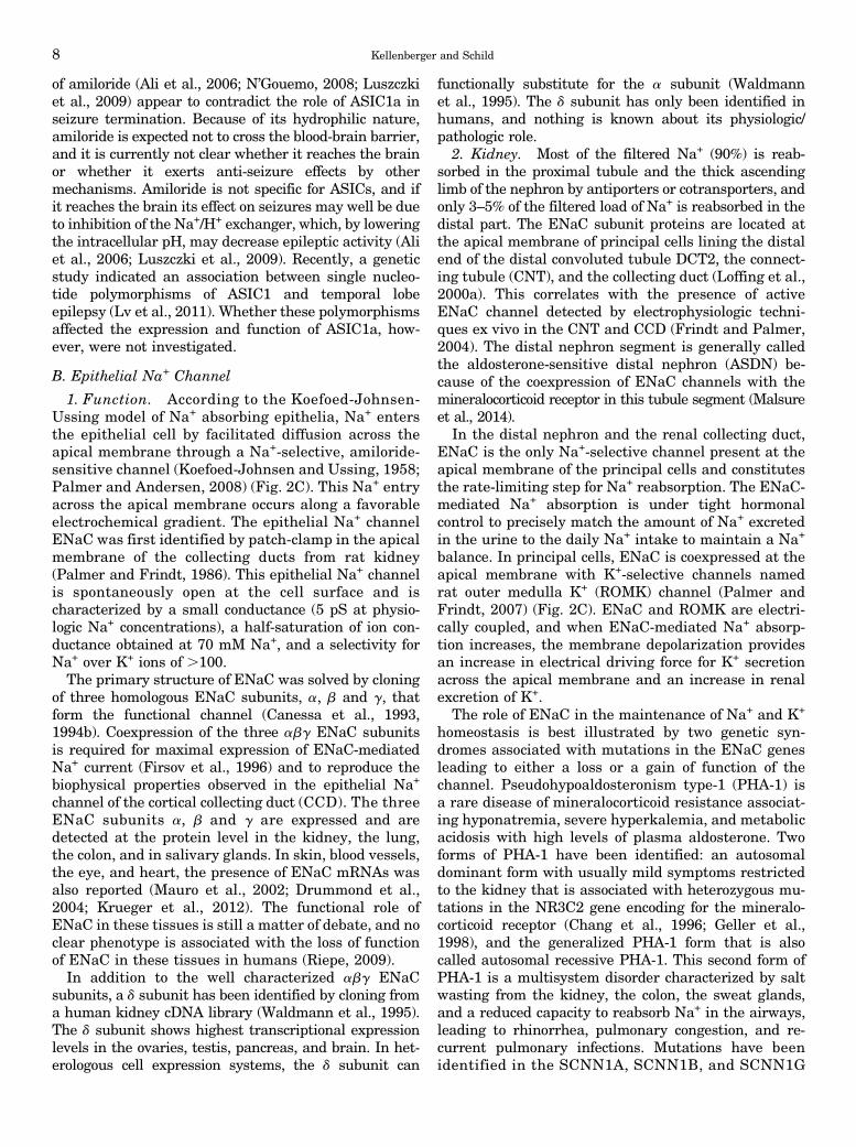
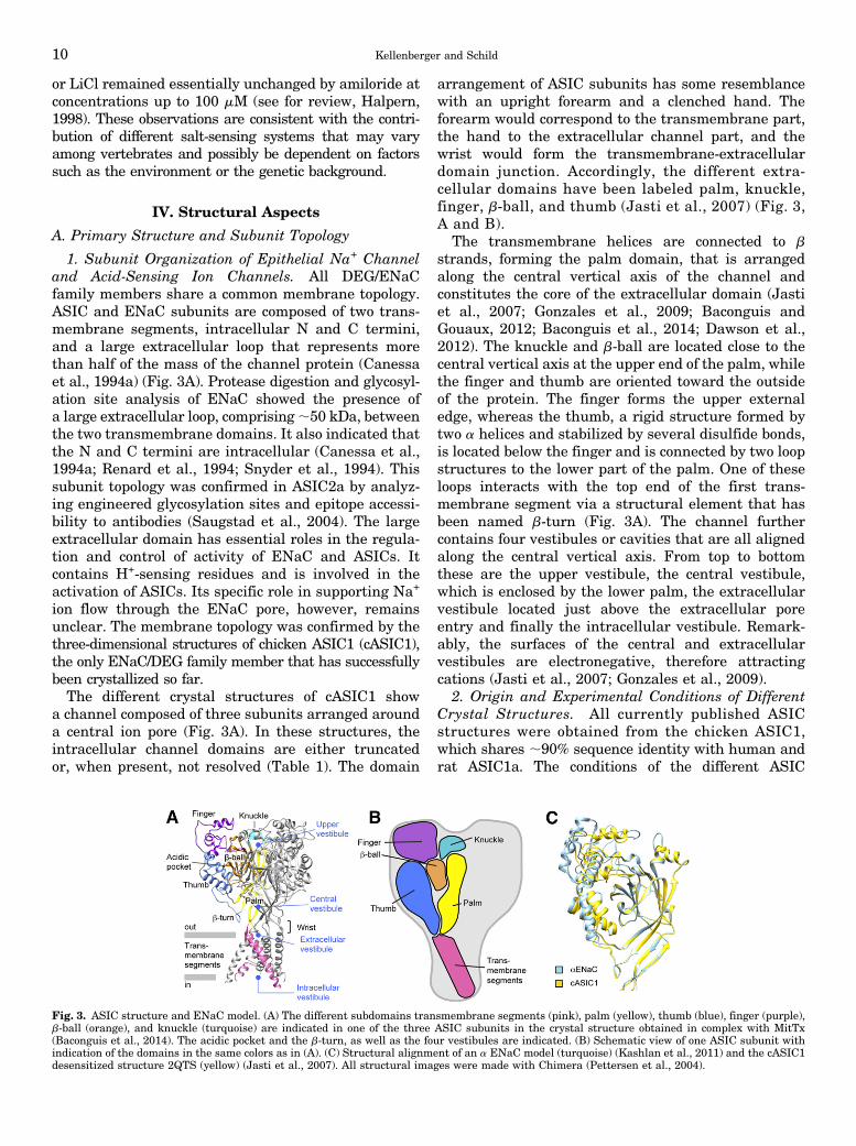
arrangement of ASIC subunits has some resemblancewith an upright forearm and a clenched hand. Theforearm would correspond to the transmembrane part,the hand to the extracellular channel part, and thewrist would form the transmembrane-extracellulardomain junction. Accordingly, the different extra-cellular domains have been labeled palm, knuckle,finger, b-ball, and thumb (Jasti et al., 2007) (Fig. 3,A and B).
The transmembrane helices are connected to bstrands, forming the palm domain, that is arrangedalong the central vertical axis of the channel andconstitutes the core of the extracellular domain (Jastiet al., 2007; Gonzales et al., 2009; Baconguis andGouaux, 2012; Baconguis et al., 2014; Dawson et al.,2012). The knuckle and b-ball are located close to thecentral vertical axis at the upper end of the palm, whilethe finger and thumb are oriented toward the outsideof the protein. The finger forms the upper externaledge, whereas the thumb, a rigid structure formed bytwo a helices and stabilized by several disulfide bonds,is located below the finger and is connected by two loopstructures to the lower part of the palm. One of theseloops interacts with the top end of the first trans-membrane segment via a structural element that hasbeen named b-turn (Fig. 3A). The channel furthercontains four vestibules or cavities that are all alignedalong the central vertical axis. From top to bottomthese are the upper vestibule, the central vestibule,which is enclosed by the lower palm, the extracellularvestibule located just above the extracellular poreentry and finally the intracellular vestibule. Remark-ably, the surfaces of the central and extracellularvestibules are electronegative, therefore attractingcations (Jasti et al., 2007; Gonzales et al., 2009).
2. Origin and Experimental Conditions of DifferentCrystal Structures. All currently published ASICstructures were obtained from the chicken ASIC1,which shares ;90% sequence identity with human andrat ASIC1a. The conditions of the different ASIC
Fig. 3. ASIC structure and ENaC model. (A) The different subdomains transmembrane segments (pink), palm (yellow), thumb (blue), finger (purple),b-ball (orange), and knuckle (turquoise) are indicated in one of the three ASIC subunits in the crystal structure obtained in complex with MitTx(Baconguis et al., 2014). The acidic pocket and the b-turn, as well as the four vestibules are indicated. (B) Schematic view of one ASIC subunit withindication of the domains in the same colors as in (A). (C) Structural alignment of an a ENaC model (turquoise) (Kashlan et al., 2011) and the cASIC1desensitized structure 2QTS (yellow) (Jasti et al., 2007). All structural images were made with Chimera (Pettersen et al., 2004).
10 Kellenberger and Schild

structures presently available are summarized inTable 1. The first published crystal ASIC structure andone of the PcTx1-cASIC1 structures were obtained froma truncated, nonfunctional construct (Jasti et al., 2007;Dawson et al., 2012). The other crystal structures wereobtained from functional but still truncated constructs.ASICs exist in three functional states: closed, open, anddesensitized. Two structures were obtained at acidic pHin the absence of gating modifiers (Protein Data Bank[PDB] ID 2QTS and 3HGC/4NYK) and their trans-membrane region did not contain a continuous porethat would allow ion transport. Therefore, it wasconcluded that these structures likely represent thedesensitized conformation of ASIC1 (Jasti et al., 2007;Gonzales et al., 2009). Recently, crystal structures ofcASIC1 in complex with the gating modifiers PcTx1 orMitTx were published (Baconguis and Gouaux, 2012;Dawson et al., 2012; Baconguis et al., 2014). MitTx is anASIC activator and PcTx1 had been known to be agating modifier that shifts the pH dependence ofactivation and desensitization to more alkaline values,thereby inhibiting human and rat ASIC1a. However,PcTx1 opens cASIC1 at pH 7.4 and rat ASIC1b atslightly acidic pH (see section VI) (Chen et al., 2005,2006; Samways et al., 2009). Both functional cASIC1-toxin complexes showed an incomplete desensitizationat the pH used for crystallization. In contrast to theprevious crystals, all toxin-bound cASIC1 structures con-tained a continuous open pore and therefore likely rep-resent the channel in an open conformation. The cASIC1crystal structures with the highest resolution were thefirst, putatively desensitized structure obtained at 1.9 Åand the open cASIC1-MitTx structure, resolved at 2.1 Å(Jasti et al., 2007; Baconguis et al., 2014). All structuresshowed generally a 3-fold symmetry except for thetransmembrane domain, which was asymmetric in thefirst desensitized structure (2QTS) and the two cASIC1-PcTx1 structures obtained at pH 5.5 (3X3X and 4FZ0).
3. Differences between the Open and the DesensitizedStructure and between Different Published Structures.Comparison of the open PcTx1- and MitTx-complexstructures with that of the desensitized state (PDB ID4NYK) showed no difference in conformation of theknuckle and upper palm domains, suggesting thatthese domains may act as a scaffold for the movementof other channel domains. Compared with the desensi-tized conformation, the lower palm and wrist of theopen conformations undergo a rotation that increasesthe distance between the thumb and the palm domainof adjacent subunits, thus widening the central vestibule.In open ASIC structures the extracellular vestibulebecomes wider than it is in the desensitized structures,and the “desensitization gate” forming an ;8 Å-thickocclusion in the upper half of the transmembrane partopens up (Baconguis and Gouaux, 2012; Grunder andAugustinowski, 2012; Baconguis et al., 2014).
The conformation of the ectodomain is the same inthe two desensitized structures (2QTS, 3HGC) and issimilar between the open structures. In the recentcASIC1-MitTx structure obtained at high resolution,the transmembrane (TM)2 segment is separated intotwo parts at the level of the selectivity filter sequenceGly-Ala-Ser, which adapts a horizontal, extendedconformation. Reinterpretation of the desensitized struc-ture 3HGC showed a similar break in helical structure.In contrast, the first desensitized structure (2QTS) andthe cASIC1-PcTx1 complexes have continuous TM2helices. It is currently not clear which of these porestructures is more realistic, although the cASIC1-MitTxstructure may be favored because it has a higher res-olution, shows a 3-fold symmetry, and seems to be com-patible with known ASIC functions.
4. Difference and Similarities between the Acid-Sensing Ion Channels and Epithelial Na+ ChannelSubunit Organization. In the extracellular domain,ENaC and ASIC1 show the highest sequence homology
TABLE 1ASIC structures
All crystal structures were obtained from chicken ASIC1. The structures 2QTS and 3HGC (4NYK) likely represent the desensitized state, whereas the other structuresrepresent the open state. 2QTS, 3S3W, 4FZ0, and 4FZ1 display a continuous TM2 segment, whereas in 4NYK (reinterpretation of 3HGC) and 4NTW the TM2 is split in twoparts by the selectivity filter GAS motif.
PDB ID Construct Complexwith pH Resolution Symmetry Function Reference
Å
2QTS N and C terminustruncated
— 5.6 1.9 ;3-fold, exceptin TM part
Nonfunctional Jasti et al., 2007
3HGCa (4NYK) C terminus truncated — 6.5 3.0 3-fold Functional Gonzales et al., 20093S3X N and C terminus
truncatedPcTx1 5.5 3.0 ;3-fold, except
in TM partNonfunctional Dawson et al., 2012
4FZ0 Truncation of 13N-terminal and 63C-terminal residues
PcTx1 5.5 2.8 ;3-fold, exceptin TM part
Functional, Na+-selective andamiloride-inhibited at pH 5.5
Baconguis andGouaux, 2012
4FZ1 “ PcTx1 7.25 3.3 3-fold Functional, nonselective, andamiloride-resistant at pH 7.25
Baconguis andGouaux, 2012
4NTWb “ MitTx 5.5 2.1 3-fold Functional, Na+-selective,amiloride-inhibited
Baconguis et al., 2014
aElectron density of this crystal was reinterpreted in the transmembrane segment, available under PDB ID 4NYK; a structure was also obtained from these crystals soakedin Cs+.
bStructures from this crystal were also obtained soaked in Cs+ (2.6 Å, 4NTY) or in amiloride (2.3 Å, 4NTX).
ASIC and ENaC Function and Pharmacology 11
in the palm and the b-ball (;35%). Although the se-quence homology is lower in the peripheral domainsfinger, thumb, and knuckle, there is evidence forstructural homology in most of these domains (Kashlanet al., 2011; Kashlan and Kleyman, 2011) (Fig. 3C). Thepredicted secondary structure of the ENaC thumbmatches the crystal ASIC structure (although the a5helices do not align well), and the Cys residues formingthe 5 disulfide bonds of the thumb are conserved. Thepredicted secondary structure of the knuckle matchesthe ASIC structure moderately well. ENaC and ASICsequences share the lowest homology in the fingerdomain. In addition, ENaC contains an ;80-residueinsertion in this domain. The low homology betweenENaC and ASIC finger domains may be at the origin ofthe different activation mechanisms of these two chan-nels. The transmembrane segments TM1 and TM2 share;20 and 40% homology, respectively, between ASIC andENaC. A model of a ENaC has been constructed that, forthe most part, is based on the cASIC1 structure 2QTSand in which the finger was modeled based on constraintsderived from functional data and from secondary struc-ture predictions (Kashlan et al., 2011) (Fig. 3C).
B. Subunit Stoichiometry
All published cASIC1 structures show the channel asa trimer (Jasti et al., 2007; Gonzales et al., 2009;Baconguis and Gouaux, 2012; Dawson et al., 2012;Baconguis et al., 2014). Recently, the counting ofbleaching steps of fluorescently tagged mammalianASIC1a and ASIC2a channels at the plasma mem-brane of Xenopus oocytes indicated that functionalASICs in a cellular environment are also trimers(Bartoi et al., 2014).The subunit stoichiometry of the functional ENaC
channel is still controversial. The total number ofsubunits that form the functional abg ENaC complexas reported in the literature varies from 3 to 9 subunits(Firsov et al., 1998; Kosari et al., 1998; Eskandariet al., 1999; Dijkink et al., 2002; Anantharam andPalmer, 2007; Carnally et al., 2008). This likely reflectsmethodological problems in evaluating the subunitstoichiometry of a channel as well as uncertainties inthe interpretation of the experimental data. However,before the crystallization of ASIC1, a general consen-sus based on functional and biochemical observationsfrom different laboratories agreed on a tetrameric sub-unit oligomerization of ENaC involving two a subunits,and one b and one g subunit. Data leading to this findinginclude the quantification of the relative abundance ofENaC subunits at the cell surface per functional channel,the dominant or recessive effects of mutations in a singlesubunit on channel block by ligands or on channel con-ductance, functional analysis of ENaC subunit concatemers,and biochemical determination of the molecular mass of theENaC channel complex (Firsov et al., 1998; Kosari et al.,1998; Dijkink et al., 2002; Anantharam and Palmer, 2007).
After the release of the three-dimensional structure ofASIC1, the ENaC/DEG field seems to have settled thatENaC is a trimer, a dogma that still needs solid con-firmation by experimental evidence.
V. Channel Function and Regulation
A. Acid-Sensing Ion Channels
1. Channel Gating and Ion Selectivity. For a longtime, protons were the only known activators of ASICs.Recently it was shown, however, that the syntheticcompound GMQ activates ASIC3 at pH 7.4 (Yu et al.,2010). The spider toxin PcTx1 shifts ASIC pH de-pendencies, inhibiting ASIC1a under physiologic con-ditions and activating cASIC1 and rat ASIC1b at pH7.4 and slightly acidic pH, respectively (Chen et al.,2005, 2006; Samways et al., 2009; Baconguis andGouaux, 2012). The recently identified MitTx activatesASICs by a currently unknown mechanism (Bohlenet al., 2011). GMQ and the toxins are further discussedbelow (section VI). Currently it seems that protons arethe only physiologic activators of ASICs.
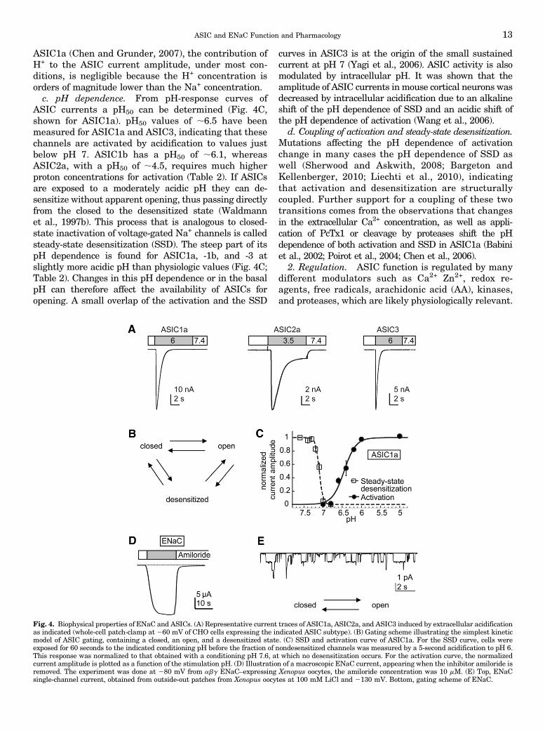
a. Current kinetics. Acid-induced opening of homo-multimeric ASICs is under most conditions transient,because it is rapidly followed by desensitization(Waldmann et al., 1997b) (Fig. 4A). This can be de-scribed by a kinetic model containing a closed, an open,and a desensitized state (Fig. 4B). Time constants ofchannel opening kinetics of 10 milliseconds or less havebeen measured for ASIC1a, ASIC1b, and ASIC3 undervery rapid solution change conditions at pH # 6(Bassler et al., 2001; Sutherland et al., 2001; Li et al.,2010b). In most of the published functional ASIC studies,the observed kinetics of channel opening are probablylimited by the speed of the solution change. Desensitiza-tion is fastest in ASIC3 and slowest in ASIC2a (Fig. 4A;Table 2). ASIC3 activation by pH ;7 and by pH ,5induces a sustained current component in addition to thetransient current (Waldmann et al., 1997a; Yagi et al.,2006). Sustained currents were also observed with ASICheteromers (Lingueglia et al., 1997; Benson et al., 2002).They may mediate ASIC function in slow and long-lastingpH changes. Human ASIC3 is not only activated byextracellular acidification. Changes to a pHmore alkalinethan the physiologic values can induce a sustained ASIC3current (Delaunay et al., 2012).
b. Ion selectivity. Transient ASIC currents are Na+
selective with a Na+/K+ permeability ratio of ;10(Table 2) (Kellenberger and Schild, 2002). HomomericASIC1a and heteromeric ASIC1a/2b have in additiona low permeability to Ca2+ (Na+/Ca2+ permeabilityratio between 1.8 and 18.5, depending on study), whichis likely important for some of their physiologic andpathologic roles (Waldmann et al., 1997b; Bassleret al., 2001; Samways et al., 2009; Sherwood et al.,2011). Although ASICs are also permeable to protonswith an estimated H+/Na+ permeability ratio of ,5 for
12 Kellenberger and Schild
ASIC1a (Chen and Grunder, 2007), the contribution ofH+ to the ASIC current amplitude, under most con-ditions, is negligible because the H+ concentration isorders of magnitude lower than the Na+ concentration.c. pH dependence. From pH-response curves of
ASIC currents a pH50 can be determined (Fig. 4C,shown for ASIC1a). pH50 values of ;6.5 have beenmeasured for ASIC1a and ASIC3, indicating that thesechannels are activated by acidification to values justbelow pH 7. ASIC1b has a pH50 of ;6.1, whereasASIC2a, with a pH50 of ;4.5, requires much higherproton concentrations for activation (Table 2). If ASICsare exposed to a moderately acidic pH they can de-sensitize without apparent opening, thus passing directlyfrom the closed to the desensitized state (Waldmannet al., 1997b). This process that is analogous to closed-state inactivation of voltage-gated Na+ channels is calledsteady-state desensitization (SSD). The steep part of itspH dependence is found for ASIC1a, -1b, and -3 atslightly more acidic pH than physiologic values (Fig. 4C;Table 2). Changes in this pH dependence or in the basalpH can therefore affect the availability of ASICs foropening. A small overlap of the activation and the SSD
curves in ASIC3 is at the origin of the small sustainedcurrent at pH 7 (Yagi et al., 2006). ASIC activity is alsomodulated by intracellular pH. It was shown that theamplitude of ASIC currents inmouse cortical neurons wasdecreased by intracellular acidification due to an alkalineshift of the pH dependence of SSD and an acidic shift ofthe pH dependence of activation (Wang et al., 2006).
d. Coupling of activation and steady-state desensitization.Mutations affecting the pH dependence of activationchange in many cases the pH dependence of SSD aswell (Sherwood and Askwith, 2008; Bargeton andKellenberger, 2010; Liechti et al., 2010), indicatingthat activation and desensitization are structurallycoupled. Further support for a coupling of these twotransitions comes from the observations that changesin the extracellular Ca2+ concentration, as well as appli-cation of PcTx1 or cleavage by proteases shift the pHdependence of both activation and SSD in ASIC1a (Babiniet al., 2002; Poirot et al., 2004; Chen et al., 2006).
2. Regulation. ASIC function is regulated by manydifferent modulators such as Ca2+ Zn2+, redox re-agents, free radicals, arachidonic acid (AA), kinases,and proteases, which are likely physiologically relevant.
Fig. 4. Biophysical properties of ENaC and ASICs. (A) Representative current traces of ASIC1a, ASIC2a, and ASIC3 induced by extracellular acidificationas indicated (whole-cell patch-clamp at 260 mV of CHO cells expressing the indicated ASIC subtype). (B) Gating scheme illustrating the simplest kineticmodel of ASIC gating, containing a closed, an open, and a desensitized state. (C) SSD and activation curve of ASIC1a. For the SSD curve, cells wereexposed for 60 seconds to the indicated conditioning pH before the fraction of nondesensitized channels was measured by a 5-second acidification to pH 6.This response was normalized to that obtained with a conditioning pH 7.6, at which no desensitization occurs. For the activation curve, the normalizedcurrent amplitude is plotted as a function of the stimulation pH. (D) Illustration of a macroscopic ENaC current, appearing when the inhibitor amiloride isremoved. The experiment was done at 280 mV from abg ENaC–expressing Xenopus oocytes, the amiloride concentration was 10 mM. (E) Top, ENaCsingle-channel current, obtained from outside-out patches from Xenopus oocytes at 100 mM LiCl and 2130 mV. Bottom, gating scheme of ENaC.
ASIC and ENaC Function and Pharmacology 13
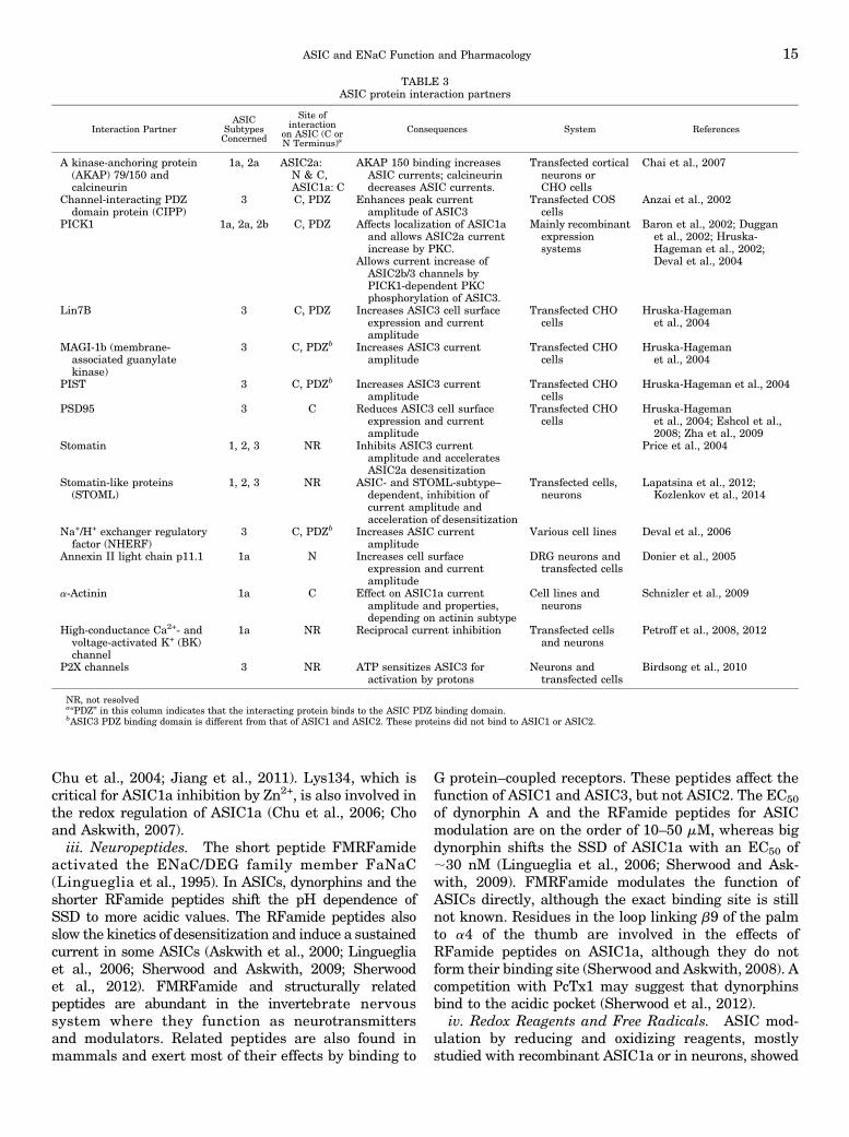
We discuss here the most important modulators ofASIC function and provide an overview in Tables 3and 4. For a more exhaustive discussion of ASICmodulation see also Chu et al. (2011) and Wemmieet al. (2013).a. Regulation by protein-protein interactions. As
many other membrane proteins, ASICs interact withother proteins, some of which have been identified in thelast years, as shown in Table 3 (Wemmie et al., 2006).ASICs were shown to interact mostly with scaffoldingproteins and with other ion channels. PDZ bindingdomains at the cytoplasmic C terminus were involvedin many of these interactions. In many cases, these in-teractions affect the cell surface expression or localizationof ASICs, their regulation, or their gating. Currently,most of these interactions have been shown in cellularsystems and their in vivo relevance is not known.b. Regulation by ions, small molecules, and proteins.i. Calcium and Other Divalent or Polyvalent Cations.
The extracellular Ca2+ concentration affects ASIC pHdependence in a way that suggests a competitionbetween Ca2+ and H+ for a common binding site. Ithas been shown that increasing the extracellular Ca2+
concentration in the range of low micromolars to 10 mMshifts the pH dependence of activation to more acidicvalues in ASIC1a and ASIC3 (Babini et al., 2002; Immkeand McCleskey, 2003). A similar Ca2+-dependent shifthas also been shown for the pH dependence of SSD inASIC1a and ASIC1b, and other divalent or polyvalentcations (Mg2+, Ba2+, spermine) had similar effects as Ca2+
(Babini et al., 2002). The spermine-induced increase inneuronal injury in animal stroke models might be due tosuch a mechanism (Duan et al., 2011). In addition to itseffects on gating, extracellular Ca2+ also inhibits maxi-mal currents by a pore block with an IC50 of the orderof millimolars. The decrease of the unitary conductanceby Ca2+ has been demonstrated in ASIC1a, ASIC2a, andASIC3 (de Weille and Bassilana, 2001; Immke andMcCleskey, 2003; Paukert et al., 2004; Zhang et al.,2006). Depending on the pH conditions, an increase inextracellular Ca2+ concentration can therefore either in-crease current amplitudes due to an acidic shift of theSSD pH dependence or decrease them due to the shift of
the activation pH dependence and the pore block(Waldmann et al., 1997b; Babini et al., 2002; Wu et al.,2004). The Ca2+ blocking site in ASIC1a is formed byE426 and D433 of the extracellular pore entry (Paukertet al., 2004) (cASIC1 numbering, Fig. 6B). Its localizationin other ASICs is not known. The Ca2+ binding sitemediating the shift in pH dependence has not beenidentified yet. The Ca2+ and H+ dependence of ASIC3currents is compatible with a mechanism of H+-inducedopening of ASIC3 by relieving a constitutive pore blockby Ca2+ (Immke and McCleskey, 2003). However, twoarticles strongly indicate that ASIC1a is not opened bythis mechanism. After mutation of the residues formingthe Ca2+ binding site in the ASIC1a pore, the mutantchannels were not constitutively open but still activatedby protons (Paukert et al., 2004). In toadfish ASIC1,lowering of the Ca2+ concentration progressively openedmore channels and did not show properties associatedwith a pore block, such as, e.g., an increase in the meanopen time (Zhang et al., 2006).
Anaerobic metabolism during ischemia can lead toextracellular lactate concentrations of 12–20 mM (nor-mally ;1 mM (Schurr, 2002). At these concentrations,lactate potentiated ASIC1a and ASIC3 currents, as wellas ASIC currents of PNS and CNS neurons (Immke andMcCleskey, 2001; Allen and Attwell, 2002). Lactate isa weak chelator of Ca2+ and other divalent cations. Itappears that the induced reduction of free Ca2+ con-centration is sufficient to increase the ASIC currents(Immke and McCleskey, 2001).
ii. Zinc. ASIC1a is inhibited by nanomolar concen-trations of Zn2+, whereas micromolar concentrations areneeded to inhibit ASIC1b and ASIC3 (Chu et al., 2004;Poirot et al., 2006; Jiang et al., 2010, 2011). Theinhibition of ASIC1b and ASIC3 is pH-independent,whereas an acidic shift of the pH dependence ofactivation contributes to the Zn2+ inhibition of ASIC1a.Interestingly, H+-induced currents mediated by ASIC2-containing channels are potentiated by Zn2+ due to analkaline shift in the pH dependence of activation (Baronet al., 2001). Residues important for the effects of Zn2+
were identified in the finger of ASIC1a and ASIC1b andin the finger and thumb of ASIC2a (Baron et al., 2001;
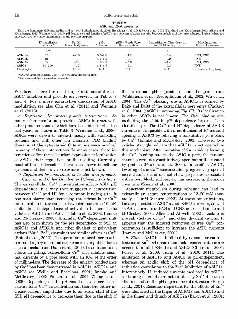
TABLE 2ASIC and ENaC properties
Data are from many different studies and reviews (Sutherland et al., 2001; Hesselager et al., 2004; Poirot et al., 2004; Blanchard and Kellenberger, 2011; Alijevic andKellenberger, 2012; Wemmie et al., 2013). pH dependence and kinetics of ASICs vary between subtypes and also between orthologs of the same subtypes. Typical values areindicated here. For more information, see the relevant chapters.
IC50 AmilorideInhibitiona
Na+/K+
Permeability RatiopH50
ActivationpHD50 Steady-State
DesensitizationDesensitization Time Constant
(at pH Close to pH50)Most Important
Sites of Expression
mM s
ASIC1a 10 6–13 6.2–6.6 ;7.2 ;0.4 CNS, PNSASIC1b 21 ;3 5.9–6.3 ;6.7 ;0.9 PNSASIC2a 28 ;10 4.0–4.9 ;5.6 ;1.4 CNS, PNSASIC3 60–100 ;12 6.4–6.7 ;7.1 ;0.3 PNSENaCabg 0.1 $100 N.A. N.A. N.A. Kidney, colon, lung
N.A., not applicable; pHD50, pH of half-maximal desensitization.aFor transient ASIC current component.
14 Kellenberger and Schild
Chu et al., 2004; Jiang et al., 2011). Lys134, which iscritical for ASIC1a inhibition by Zn2+, is also involved inthe redox regulation of ASIC1a (Chu et al., 2006; Choand Askwith, 2007).iii. Neuropeptides. The short peptide FMRFamide
activated the ENaC/DEG family member FaNaC(Lingueglia et al., 1995). In ASICs, dynorphins and theshorter RFamide peptides shift the pH dependence ofSSD to more acidic values. The RFamide peptides alsoslow the kinetics of desensitization and induce a sustainedcurrent in some ASICs (Askwith et al., 2000; Linguegliaet al., 2006; Sherwood and Askwith, 2009; Sherwoodet al., 2012). FMRFamide and structurally relatedpeptides are abundant in the invertebrate nervoussystem where they function as neurotransmittersand modulators. Related peptides are also found inmammals and exert most of their effects by binding to
G protein–coupled receptors. These peptides affect thefunction of ASIC1 and ASIC3, but not ASIC2. The EC50
of dynorphin A and the RFamide peptides for ASICmodulation are on the order of 10–50 mM, whereas bigdynorphin shifts the SSD of ASIC1a with an EC50 of;30 nM (Lingueglia et al., 2006; Sherwood and Ask-with, 2009). FMRFamide modulates the function ofASICs directly, although the exact binding site is stillnot known. Residues in the loop linking b9 of the palmto a4 of the thumb are involved in the effects ofRFamide peptides on ASIC1a, although they do notform their binding site (Sherwood and Askwith, 2008). Acompetition with PcTx1 may suggest that dynorphinsbind to the acidic pocket (Sherwood et al., 2012).
iv. Redox Reagents and Free Radicals. ASIC mod-ulation by reducing and oxidizing reagents, mostlystudied with recombinant ASIC1a or in neurons, showed
TABLE 3ASIC protein interaction partners
Interaction PartnerASIC
SubtypesConcerned
Site ofinteraction
on ASIC (C orN Terminus)a
Consequences System References
A kinase-anchoring protein(AKAP) 79/150 andcalcineurin
1a, 2a ASIC2a:N & C,ASIC1a: C
AKAP 150 binding increasesASIC currents; calcineurindecreases ASIC currents.
Transfected corticalneurons orCHO cells
Chai et al., 2007
Channel-interacting PDZdomain protein (CIPP)
3 C, PDZ Enhances peak currentamplitude of ASIC3
Transfected COScells
Anzai et al., 2002
PICK1 1a, 2a, 2b C, PDZ Affects localization of ASIC1aand allows ASIC2a currentincrease by PKC.
Mainly recombinantexpressionsystems
Baron et al., 2002; Dugganet al., 2002; Hruska-Hageman et al., 2002;Deval et al., 2004Allows current increase of
ASIC2b/3 channels byPICK1-dependent PKCphosphorylation of ASIC3.
Lin7B 3 C, PDZ Increases ASIC3 cell surfaceexpression and currentamplitude
Transfected CHOcells
Hruska-Hagemanet al., 2004
MAGI-1b (membrane-associated guanylatekinase)
3 C, PDZb Increases ASIC3 currentamplitude
Transfected CHOcells
Hruska-Hagemanet al., 2004
PIST 3 C, PDZb Increases ASIC3 currentamplitude
Transfected CHOcells
Hruska-Hageman et al., 2004
PSD95 3 C Reduces ASIC3 cell surfaceexpression and currentamplitude
Transfected CHOcells
Hruska-Hagemanet al., 2004; Eshcol et al.,2008; Zha et al., 2009
Stomatin 1, 2, 3 NR Inhibits ASIC3 currentamplitude and acceleratesASIC2a desensitization
Price et al., 2004
Stomatin-like proteins(STOML)
1, 2, 3 NR ASIC- and STOML-subtype–dependent, inhibition ofcurrent amplitude andacceleration of desensitization
Transfected cells,neurons
Lapatsina et al., 2012;Kozlenkov et al., 2014
Na+/H+ exchanger regulatoryfactor (NHERF)
3 C, PDZb Increases ASIC currentamplitude
Various cell lines Deval et al., 2006
Annexin II light chain p11.1 1a N Increases cell surfaceexpression and currentamplitude
DRG neurons andtransfected cells
Donier et al., 2005
a-Actinin 1a C Effect on ASIC1a currentamplitude and properties,depending on actinin subtype
Cell lines andneurons
Schnizler et al., 2009
High-conductance Ca2+- andvoltage-activated K+ (BK)channel
1a NR Reciprocal current inhibition Transfected cellsand neurons
Petroff et al., 2008, 2012
P2X channels 3 NR ATP sensitizes ASIC3 foractivation by protons
Neurons andtransfected cells
Birdsong et al., 2010
NR, not resolveda“PDZ” in this column indicates that the interacting protein binds to the ASIC PDZ binding domain.bASIC3 PDZ binding domain is different from that of ASIC1 and ASIC2. These proteins did not bind to ASIC1 or ASIC2.
ASIC and ENaC Function and Pharmacology 15
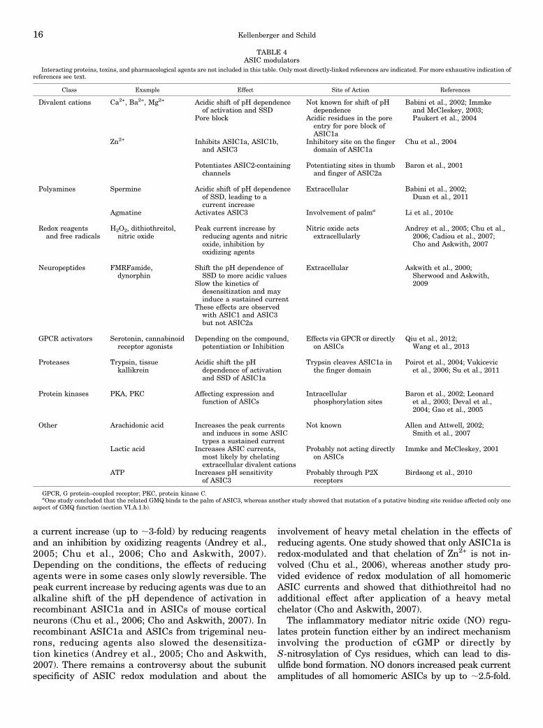
a current increase (up to ;3-fold) by reducing reagentsand an inhibition by oxidizing reagents (Andrey et al.,2005; Chu et al., 2006; Cho and Askwith, 2007).Depending on the conditions, the effects of reducingagents were in some cases only slowly reversible. Thepeak current increase by reducing agents was due to analkaline shift of the pH dependence of activation inrecombinant ASIC1a and in ASICs of mouse corticalneurons (Chu et al., 2006; Cho and Askwith, 2007). Inrecombinant ASIC1a and ASICs from trigeminal neu-rons, reducing agents also slowed the desensitiza-tion kinetics (Andrey et al., 2005; Cho and Askwith,2007). There remains a controversy about the subunitspecificity of ASIC redox modulation and about the
involvement of heavy metal chelation in the effects ofreducing agents. One study showed that only ASIC1a isredox-modulated and that chelation of Zn2+ is not in-volved (Chu et al., 2006), whereas another study pro-vided evidence of redox modulation of all homomericASIC currents and showed that dithiothreitol had noadditional effect after application of a heavy metalchelator (Cho and Askwith, 2007).
The inflammatory mediator nitric oxide (NO) regu-lates protein function either by an indirect mechanisminvolving the production of cGMP or directly byS-nitrosylation of Cys residues, which can lead to dis-ulfide bond formation. NO donors increased peak currentamplitudes of all homomeric ASICs by up to ;2.5-fold.
TABLE 4ASIC modulators
Interacting proteins, toxins, and pharmacological agents are not included in this table. Only most directly-linked references are indicated. For more exhaustive indication ofreferences see text.
Class Example Effect Site of Action References
Divalent cations Ca2+, Ba2+, Mg2+ Acidic shift of pH dependenceof activation and SSD
Not known for shift of pHdependence
Babini et al., 2002; Immkeand McCleskey, 2003;Paukert et al., 2004Pore block Acidic residues in the pore
entry for pore block ofASIC1a
Zn2+ Inhibits ASIC1a, ASIC1b,and ASIC3
Inhibitory site on the fingerdomain of ASIC1a
Chu et al., 2004
Potentiates ASIC2-containingchannels
Potentiating sites in thumband finger of ASIC2a
Baron et al., 2001
Polyamines Spermine Acidic shift of pH dependenceof SSD, leading to acurrent increase
Extracellular Babini et al., 2002;Duan et al., 2011
Agmatine Activates ASIC3 Involvement of palma Li et al., 2010c
Redox reagentsand free radicals
H2O2, dithiothreitol,nitric oxide
Peak current increase byreducing agents and nitricoxide, inhibition byoxidizing agents
Nitric oxide actsextracellularly
Andrey et al., 2005; Chu et al.,2006; Cadiou et al., 2007;Cho and Askwith, 2007
Neuropeptides FMRFamide,dynorphin
Shift the pH dependence ofSSD to more acidic values
Extracellular Askwith et al., 2000;Sherwood and Askwith,2009Slow the kinetics of
desensitization and mayinduce a sustained current
These effects are observedwith ASIC1 and ASIC3but not ASIC2a
GPCR activators Serotonin, cannabinoidreceptor agonists
Depending on the compound,potentiation or Inhibition
Effects via GPCR or directlyon ASICs
Qiu et al., 2012;Wang et al., 2013
Proteases Trypsin, tissuekallikrein
Acidic shift the pHdependence of activationand SSD of ASIC1a
Trypsin cleaves ASIC1a inthe finger domain
Poirot et al., 2004; Vukicevicet al., 2006; Su et al., 2011
Protein kinases PKA, PKC Affecting expression andfunction of ASICs
Intracellularphosphorylation sites
Baron et al., 2002; Leonardet al., 2003; Deval et al.,2004; Gao et al., 2005
Other Arachidonic acid Increases the peak currentsand induces in some ASICtypes a sustained current
Not known Allen and Attwell, 2002;Smith et al., 2007
Lactic acid Increases ASIC currents,most likely by chelatingextracellular divalent cations
Probably not acting directlyon ASICs
Immke and McCleskey, 2001
ATP Increases pH sensitivityof ASIC3
Probably through P2Xreceptors
Birdsong et al., 2010
GPCR, G protein–coupled receptor; PKC, protein kinase C.aOne study concluded that the related GMQ binds to the palm of ASIC3, whereas another study showed that mutation of a putative binding site residue affected only one
aspect of GMQ function (section VI.A.1.b).
16 Kellenberger and Schild
For ASIC1a, it was shown that this was due to analkaline shift in the pH dependence of activation. NOacted directly on the ASIC ectodomain; however, the siteof action is not known. Topical application of the NOdonor glyceryl trinitrate increased acid-induced pain inhuman volunteers (Cadiou et al., 2007).v. Arachidonic Acid. AA is a metabolite of mem-
brane phospholipids produced by phospholipase A2. Itsconcentration is increased in ischemia and inflamma-tion and it has been shown to modulate the function ofseveral channel types (Chu et al., 2011). AA potenti-ated ASIC currents by up to ;2.5-fold in neurons of theCNS and the PNS and of recombinant ASIC1a andASIC3 and in many cases increased the sustainedcurrent fraction (Allen and Attwell, 2002; Smith et al.,2007). It was shown that the AA-induced currentincrease of ASIC3 is due to an alkaline shift of the pHdependence of activation (Deval et al., 2008).vi. G Protein–Coupled Receptors. Serotonin and
other inflammatory mediators, such as nerve growthfactor, increase ASIC mRNA levels (Mamet et al., 2002).Serotonin or subtype-specific 5-HT receptor agonistsalso potentiated ASIC currents in sensory neurons(Deval et al., 2004), either through a direct action onASIC3 (Wang et al., 2013) or via activation of 5-HT2
receptors (Qiu et al., 2012). Both serotonin and the5-HT2 agonist a-methyl-5-HT increased acid-inducednociceptive responses.The cannabinoid receptor agonist WIN55,212-2 [(R)-
(+)-[2,3-dihydro-5-methyl-3-(4-morpholinylmethyl)pyrrolo[1,2,3-de]-1,4-benzoxazin-6-yl]-1-naphthalenylmetha-none] inhibited ASIC currents of DRG neurons by upto ;50% with an IC50 of ;10 nM, without shifting theASIC pH dependence (Liu et al., 2012). This inhibitionwas mediated by cannabinoid receptor type 1.vii. Proteases. Stroke, brain injury, and other path-
ologic conditions can compromise the integrity of theblood-brain barrier and allow blood-derived proteases toaccess the CNS (Gingrich and Traynelis, 2000). Serineproteases modulate the function of various ion channels,including the ASICs (Poirot et al., 2004) and ENaC (seesection V.B.2). Trypsin and other serine proteases shiftthe pH dependence of SSD and activation of ASIC1a tomore acidic values. This decreases ASIC currents underphysiologic pH conditions but increases the current am-plitudes in situations of constitutively lowered pH suchas ischemia. The current modulation is due to cleavage ofthe ASIC1a protein in the finger, at Arg146 (Fig. 5B)(Vukicevic et al., 2006), in the same subdomain as thecleavage of a and g ENaC. Exposure of ASIC1a totrypsin reduced the relative Ca2+ permeability (Neagaet al., 2005), which may reduce the cellular toxicity ofprolonged ASIC activity. Tissue kallikrein that wasshown to cleave ASIC1a, but not ASIC2a, protectedASIC1a-expressing cells from acidosis-induced injury(Su et al., 2011). This study did not test whethercleavage changed the functional properties of ASIC1a;
however, the protective effect is likely due to a changedfunction of ASIC1a.
viii. Kinases. The protein kinase C (PKC) activator1-oleyl-2-acetyl-sn-glycerol potentiates ASIC2a cur-rents only in the presence of protein interacting withC kinase 1 (PICK1) (Baron et al., 2002). PICK1interaction sites were found on ASIC1a, -2a, and 2b(Baron et al., 2002; Duggan et al., 2002; Hruska-Hageman et al., 2002; Deval et al., 2004). The PKCmodulation of ASIC2a currents requires the presenceof the PDZ domain of PICK1 and the PDZ bindingdomain on the C terminus of ASIC2a and involvesphosphorylation of residue T39 in the cytoplasmic Nterminus. A phorbol ester activating PKC enhancesASIC2b/3 currents by inducing an alkaline shift intheir pH dependence of activation (Deval et al., 2004).The presence of PICK1 and its binding site on theASIC2b C terminus are required for this effect, whichinvolves phosphorylation of ASIC3 residues. The basalASIC3 expression in sensory neurons depends on theactivation of neurotrophic tyrosine kinase receptor type 1by nerve growth factor, which in turn leads to activationof the phospholipase C/PKC pathway (Mamet et al.,2003). In contrast to ASIC2, ASIC1a is not phosphory-lated by PKC, but by protein kinase A (Leonard et al.,2003). Protein kinase A phosphorylates residue S479 ofthe C terminus and disrupts the binding of PICK1,thereby affecting the localization of ASIC1a.
In global ischemia, intracellular Ca2+ concentrationsare increased because of, among other mechanisms, theactivity of NMDA receptors, leading to activation of thecalcium/calmodulin-dependent protein kinase II thatphosphorylates the C-terminal residues S478 and S479of ASIC1a (Gao et al., 2005). This sensitizes the channelto protons and thus potentiates its currents.
B. Epithelial Na+ Channel
ENaC is constitutively active at the cell surface andtransits between open and closed conformations withan average open probability of;0.5 (Palmer and Frindt,1996) (Fig. 4, D and E). During its short lifetime at thecell surface, the channel evolves between differentgating modes of high and low open probability beforeits internalization (Garty and Palmer, 1997; Shimketset al., 1997; Kleyman et al., 2001; Palmer et al., 2012).
1. Intrinsic Regulation. Na+ ions constitute one ofthe different factors that directly modulate ENaC atthe cell surface and affect ENaC activity by twomechanisms. The so-called Na+ feedback inhibition isa decrease in ENaC activity upon an increase incytosolic Na+ concentration due to the rapid entry of Na+
ions into the cell. This inhibition develops over a timecourse of minutes and is due to a decrease in both thenumber of ENaC channels at the cell surface and to areduction of their open probability (Frindt et al., 1993,1995; Anantharam et al., 2006). Na+ self-inhibitionrefers to a rapid decrease in ENaC activity after an
ASIC and ENaC Function and Pharmacology 17
increase in extracellular Na+ concentration (Fuchset al., 1977; Horisberger and Chraibi, 2004; Bize andHorisberger, 2007). These regulatory processes tendto limit the Na+ entry into a single cell to optimize Na+
absorption along the aldosterone-sensitive distalnephron.In addition, intracellular perfusion of SH-oxidizing
agents or a decrease in intracellular pH also reducesENaC activity (Chalfant et al., 1999; Kellenbergeret al., 2005), but the physiologic relevance of theseprocesses is less clear. The cytosolic N terminus ofENaC subunits contains a conserved HG motif and Cysresidues that modulate channel gating (Grunder et al.,1997; Kellenberger et al., 2005). The importance of thecytosolic amino terminus as regulatory domain of ENaCwas first evidenced by a mutation in the HG motif ofENaC associated PHA-1 (Chang et al., 1996). The ENaCchannels carrying mutations in the HG motif showaltered gating properties characterized by abnormallylong closures and short channel openings correspondingto a loss of function mutation.2. Serine Proteases. The extracellular domain be-
tween the two transmembrane helices represents morethan half of the mass of ENaC/DEG proteins. A similarfeature is found in the P2X channel family (Gonzaleset al., 2009). Serine proteases regulate ENaC functionby proteolytic cleavage. In vitro studies showed thatsoluble proteases, such as trypsin or chymotrypsin,potentiate ENaC activity, and functional cloning ap-proaches allowed the identification of membrane-bound serine proteases as ENaC partners involved inchannel activation (Vallet et al., 1997; Vuagniauxet al., 2000). Extracellular serine proteases stimulateENaC directly by increasing the open probabilityindependently of a G protein–coupled receptor (Chraibiet al., 1998). Among these proteases, the first identifiedwas the mouse CAP-1 (channel activating protease-1),an ortholog of the human prostasin (Vallet et al., 1997).Subsequently, mouse CAP-2, an ortholog of the humantransmembrane protease serine 4 (TRPMSS4), and mouseCAP-3 (ortholog of the human matriptase or epithin) wereshown to stimulate ENaC to a similar extent (Vuagniauxet al., 2002; Andreasen et al., 2006). Later, the identifi-cation of a cleavage sequence for the furin-like propro-tein convertase in the extracellular domain of the a andg ENaC subunits suggested a possible role of furin inENaC cleavage, ENaC sorting, and the control of ENaCactivity at the cell surface (Hughey et al., 2004; Brunset al., 2007). However, ENaC regulation by serine pro-teases appears quite complex, because cleavage of gENaC by furin is not absolutely required for channelactivation (Harris et al., 2008), suggesting a redundancyof serine proteases in ENaC activation. Proteases cleavea and g ENaC at defined sites in the finger domain(Hughey et al., 2003; Carattino et al., 2008) (Fig. 5C). Ithas been proposed that a double cleavage in the a and gsubunits removes an inhibitory peptide (26 residues in
a and 43 residues in g ENaC), resulting in the activationof ENaC (Sheng et al., 2006; Bruns et al., 2007).
3. Hormonal Regulation. The activity of the chan-nel at the cell surface is tightly regulated by hormones.They control the expression of active ENaC at the cellsurface by interfering at different steps of the bio-synthetic pathway such as transcription, translation,or membrane trafficking. Ultimately, these hormonesare essential for the maintenance of Na+ homeostasisand the control of blood pressure.
Aldosterone is the major regulator of ENaC in theASDN. Aldosterone increases the synthesis of a ENaCbut not of b or g ENaC in the kidney, in contrast to thecolon where aldosterone or dexamethasone increasethe synthesis of b and g only (Masilamani et al., 1999;Epple et al., 2000; Fuller et al., 2000; Loffing et al.,2000b). This increase in the synthesis of a ENaC isaccompanied by the redistribution of the ENaC sub-units from an intracellular pool to the apical membraneof principal cells. Interestingly this also correlates withthe presence of cleaved a and g ENaC subunits at thecell surface (Frindt and Palmer, 2009b). The increase inthe abundance of a ENaC contributes to, but is notsufficient for, the increase in aldosterone-induced netNa+ transport (Frindt et al., 2008), suggesting the con-tribution of other processes essential for the upregulationof ENaC by aldosterone.
Binding of aldosterone to the mineralocorticoidreceptor allows the dimerization of the receptor, itsmigration to the nucleus, and the binding to hormone-responsive elements on the DNA for the induction oftranscriptional responses. Aldosterone-induced pro-teins likely play a role in the redistribution of ENaCto the cell surface by controlling the exocytic pathway.These proteins include SGK1 (serum- and glucocorticoid-regulated kinase 1), the deubiquitylation enzyme USP2-45 and GILZ (glucocorticoid-induced leucine zipper)(Soundararajan et al., 2007). These proteins likely workin concert with the ubiquitin ligase Nedd4-2 that isinvolved in the endocytic pathway. No evidence sup-ports a limiting role of these proteins for the aldosteroneeffect on ENaC, because mouse models with completeknockout of the genes encoding these proteins (Nedd4-2,SGK, USP2-45, GILZ) failed to suppress the response ofENaC-mediated renal Na+ absorption to aldosterone(Fejes-Toth et al., 2008; Suarez et al., 2012; Pouly et al.,2013; Ronzaud et al., 2013). The complete picture of thealdosterone signaling pathway that controls ENaC-mediated Na+ absorption remains therefore largelyunknown.
Dexamethasone upregulates the abundance of dif-ferent Na+ transporters in the kidney and stimulatesENaC-mediated current in the principal cells of the CCD.Dexamethasone has additive effects on the abundance ofa ENaC when coadministered with aldosterone inrats, indicating that dexamethasone regulates ENaCsynthesis by occupying the glucocorticoid receptors
18 Kellenberger and Schild
(Frindt and Palmer, 2012). This glucocorticoid-dependent increase in a ENaC synthesis is not as-sociated with an increase in ENaC activity at the cellsurface, further suggesting that still unidentified regula-tory proteins, and not as previously thought the in-tracellular abundance of ENaC proteins, are rate-limitingfor addressing ENaC channels to the apical membrane ofprincipal cells, (Frindt and Palmer, 2012).A recent report showed that ENaC activity in the late
distal convoluted tubule and the connecting tubule isindependent of aldosterone, suggesting the presence ofother hormones involved in ENaC regulation (Nesterovet al., 2012). Angiotensin II upregulated the activity ofthe Na+/Cl2 cotransporter at the apical membraneof the distal tubule (San-Cristobal et al., 2009) and alsoof ENaC in the distal nephron ex vivo (Mamenko et al.,2012; Sun et al., 2012). Endothelin-1 (ET-1) is a potentvasoconstrictor that regulates systemic blood pressure.In CCD perfused ex vivo, ET-1 significantly inhibitedNa+ absorption; the ENaC-mediated Na+ absorptionwas restored by an ETA receptor inhibitor, suggestingthat ET-1 downregulates ENaC (Lynch et al., 2013).However, deletion of the ET-1 receptor ETA in thekidney of mice did not affect blood pressure and resultedin a modest volume expansion (Stuart et al., 2012), in-dicating that ETA receptors in the nephron have littleeffect on Na+ absorption by the kidney and on bloodpressure. In several cellular models, vasopressin or cAMPagonists stimulated ENaC activity (Schafer and Troutman,1990; Kleyman et al., 1994; Auberson et al., 2003; Snyderet al., 2004). Vasopressin and aldosterone have synergisticeffects on ENaC-mediated Na+ absorption in the collectingduct. The vasopressin-induced upregulation of ENaC ismediated by vasopressin receptor type 2 (V2R). In humans,vasopressin induced an increase in urine osmolality anda reduced Na+ excretion, which is likely due to an increasedENaC-dependent Na+ absorption mediated by stimulationof V2R (Bankir et al., 2005).
C. Structure-Function Relationship of Acid-SensingIon Channels and Epithelial Na+ Channel
Initial information on structure-function aspects ofdegenerin and ENaC was obtained from mutagenesisscreens on C. elegans, and the identification of humandisease mutations on ENaC.ENaC and ASIC activity is temperature-dependent
(Askwith et al., 2001; Chraibi and Horisberger, 2002;Neelands et al., 2010; Blanchard and Kellenberger,2011). This suggests that conformational changes arepart of the mechanisms that control ENaC and ASICactivity and likely transmit binding events in theectodomain to the channel gate. Fluorescence studieshave indeed shown evidence for rapid conformationalchanges in ASIC1a (Passero et al., 2009; Bonifacioet al., 2014). For detailed discussions of ASIC structure-function aspects, see also Grunder and Chen (2010)and Sherwood et al. (2012). An insightful overview of
degenerin structure-function aspects is given byEastwood and Goodman (2012).
1. The Acidic Pocket and Other Proton-Sensing Sitesof Acid-Sensing Ion Channels. The acidic pocket,located between the thumb, the finger, and the b-ballis highly negatively charged and contains several pairsof acidic side chains (Jasti et al., 2007) (Fig. 5A).Because mutation of such acidic residues led to acidicshifts in the pH dependence of activation, it was pro-posed that the acidic pocket forms the pH sensor thatcontrols ASIC activity (Jasti et al., 2007; Li et al., 2009;Sherwood et al., 2009). It was suggested that, in theclosed conformation, the acidic residues are moredistant from each other than in the crystal structuresthat likely represent the open or desensitized confor-mation. Protonation would allow the thumb to ap-proach the finger and b-ball, and this movement wouldinduce activation and possibly desensitization. Muta-tions in the acidic pocket that decreased the interactionenergy between the thumb and the finger or the b-balltended to shift the pH dependence of activation to moreacidic values, suggesting that channel gating is facilitatedby attractive forces between these subdomains (Yanget al., 2009). The mutagenesis and functional analysis ofconserved charged residues of ASIC1a identified acidicresidues and His residues outside the acidic pocket in thefinger and the extracellular vestibule, whose mutationaffected the pH dependence of activation (Paukert et al.,2008). This analysis emphasized the role of residues inthe extracellular vestibule and pore entry for ASICfunction. Another study calculated the pKa values ofAsp, Glu, and His residues in ASIC1a based on thecrystal structure, and functionally analyzed the role ofresidues with pKa values in the gating range ofASIC1a (Liechti et al., 2010). This approach identifiedputative pH-sensing residues for activation or de-sensitization in the acidic pocket and the palm. Therole of palm residues in activation was recentlyconfirmed (Krauson et al., 2013).
2. The Finger—A Determinant of Subfamily-SpecificActivation Mechanisms? Residue Asp108 of the a1finger helix is critical for activation of ASIC1a andASIC3, possibly by building a salt bridge with basicfinger residues (Paukert et al., 2008; Yang et al., 2012),and protease cleavage next to Arg146 of the a2 helixshifts the ASIC1a pH dependence to more acidic values(Vukicevic et al., 2006) (Fig. 5B). Note that we usethroughout this review the amino acid numbering ofcASIC1 for ASIC1a and ASIC1 to facilitate comparisonbetween different orthologs. Residues involved in ASIC1aand -1b inhibition and ASIC2a potentiation by Zn2+ arelocated at different positions within the finger. The fingeris covalently linked to the b-ball and the palm at a sitecontaining residues Asn97, His164, and Gln226. Thisinteraction site is highly conserved, sensitive to mu-tations, and undergoes conformational changes dur-ing ASIC activity (Bargeton and Kellenberger, 2010)
ASIC and ENaC Function and Pharmacology 19

(Fig. 5B). Experiments with fluorophore-quencherpairing and voltage-clamp fluorometry suggest thatthe upper finger helix a2 and the b-ball move awayfrom each other upon channel activation, becauseacidification induced a fluorescence signal increaseof fluorophores placed in the a2 finger helix anddepended on the quenching by the b-ball residueTrp234 (Fig. 5B) (Bonifacio et al., 2014).ENaC is activated by diverse cleaving events in the a
and g subunits (see section V.B.2) (Kleyman et al., 2009;Rossier and Stutts, 2009). Two cleavage sites for furinare present in the finger domain of a ENaC and one inthe finger of g ENaC (Fig. 5C). Elastase, plasmin, andprostasin can cleave g ENaC C terminus to the furincleavage site.3. The Palm. The palm forms a b strand–rich scaffold
at the central part of the channel, reaching from thetransmembrane segments up to the knuckle (Fig. 3,A and B). The lower part of the palm, formed by thetwo short b strands 1 and 12 connected to the TMsegments and by the two long b strands 9 and 10,encloses the central vestibule (Figs. 3A and 6A). Theb1–b2 loop, connecting the lower palm to the b-ball, iscritical for the kinetics of ASIC1 desensitization fromthe open state (Coric et al., 2003). This study showedthat the desensitization kinetics are 25-fold faster intoadfish compared with rat ASIC1a and that this
difference is due to three residues in the b1–b2 loop,Ser-Gln-Leu in rat ASIC1a, located just at the begin-ning of a stretch of residues that are highly conserved inthe ENaC/DEG family. The crystal structure indicatedthat the b1–b2 loop is located close to the b11–b12 loop.Several studies have shown that these loops interactwith each other and codetermine the current kineticsand that mutations in this region can induce sustainedopening (Li et al., 2010a,b; Springauf et al., 2011; Royet al., 2013). Comparison of the open and desensitizedcrystal structures indicated a 180° rotation of thepeptide bond between Ser84 and Gln85 and a swappingof two critical side chains in the b11–b12 loop, Leu414and Asn415 (Baconguis and Gouaux, 2012). Everysecond residue of the lower palm b sheet surroundingthe central vestibule points into the central cavity.Glu417 is a proton-sensing residue for activation (Fig.6A) (Liechti et al., 2010; Krauson et al., 2013). Severalresidues of the lower palm are important for ASIC3activation by GMQ (see section VI.A.1.b) (Yu et al.,2010; Alijevic and Kellenberger, 2012).
The Glu80 side chain is oriented toward the centralcavity (Fig. 6A). When mutated to Cys, this residue ofASIC3 was better accessible to small hydrophilicsulfhydryl reagents in the closed compared with thedesensitized state, consistent with a conformationalchange of the palm upon desensitization (Cushman
Fig. 5. Acidic pocket and finger. The color code for subdomains is the same as in Fig. 3. (A) Acidic pocket, showing acidic side chains of the thumb,finger, loop and b-ball. (B) The three a helices of a finger domain are indicated. The knuckle of a neighboring subunit is highlighted in dark gray. D108,involved in pH-dependent activation of ASIC1a and ASIC3; K134, important for Zn2+ inhibition of ASIC1a; H164, a residue involved in theenhancement of ASIC2a currents by Zn2+, localized with N97 and Q226 in the interaction domain between the finger, the b-ball, and the palm. ASIC1b-C149, important for Zn2+ inhibition of ASIC1b, is located at the position homologous to G163 (not shown); R146, ASIC1a cleavage site by trypsin. (Aand B) Based on the 2QTS structure; (C) a ENaC structural model, indicating the location of furin cleavage sites (F1a and F2a) and homologousposition of cleavage sites in gENaC by furin (F1g), CAP-1, plasmin (Plas.), and elastase (Elas.). The peptide of a ENaC located between the two furinsites and therefore cleaved by furin is highlighted in black.
20 Kellenberger and Schild
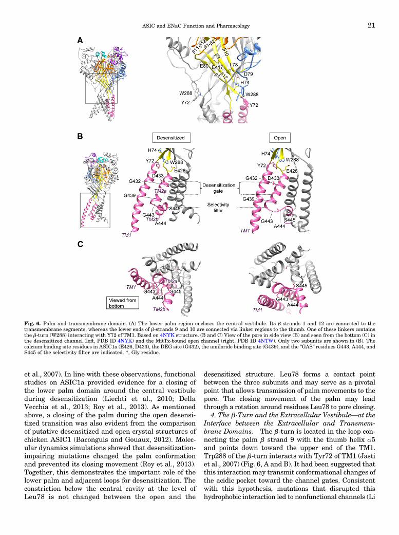
et al., 2007). In line with these observations, functionalstudies on ASIC1a provided evidence for a closing ofthe lower palm domain around the central vestibuleduring desensitization (Liechti et al., 2010; DellaVecchia et al., 2013; Roy et al., 2013). As mentionedabove, a closing of the palm during the open desensi-tized transition was also evident from the comparisonof putative desensitized and open crystal structures ofchicken ASIC1 (Baconguis and Gouaux, 2012). Molec-ular dynamics simulations showed that desensitization-impairing mutations changed the palm conformationand prevented its closing movement (Roy et al., 2013).Together, this demonstrates the important role of thelower palm and adjacent loops for desensitization. Theconstriction below the central cavity at the level ofLeu78 is not changed between the open and the
desensitized structure. Leu78 forms a contact pointbetween the three subunits and may serve as a pivotalpoint that allows transmission of palm movements to thepore. The closing movement of the palm may leadthrough a rotation around residues Leu78 to pore closing.
4. The b-Turn and the Extracellular Vestibule—at theInterface between the Extracellular and Transmem-brane Domains. The b-turn is located in the loop con-necting the palm b strand 9 with the thumb helix a5and points down toward the upper end of the TM1.Trp288 of the b-turn interacts with Tyr72 of TM1 (Jastiet al., 2007) (Fig. 6, A and B). It had been suggested thatthis interaction may transmit conformational changes ofthe acidic pocket toward the channel gates. Consistentwith this hypothesis, mutations that disrupted thishydrophobic interaction led to nonfunctional channels (Li
Fig. 6. Palm and transmembrane domain. (A) The lower palm region encloses the central vestibule. Its b-strands 1 and 12 are connected to thetransmembrane segments, whereas the lower ends of b-strands 9 and 10 are connected via linker regions to the thumb. One of these linkers containsthe b-turn (W288) interacting with Y72 of TM1. Based on 4NYK structure. (B and C) View of the pore in side view (B) and seen from the bottom (C) inthe desensitized channel (left, PDB ID 4NYK) and the MitTx-bound open channel (right, PDB ID 4NTW). Only two subunits are shown in (B). Thecalcium binding site residues in ASIC1a (E426, D433), the DEG site (G432), the amiloride binding site (G439), and the “GAS” residues G443, A444, andS445 of the selectivity filter are indicated. *, Gly residue.
ASIC and ENaC Function and Pharmacology 21
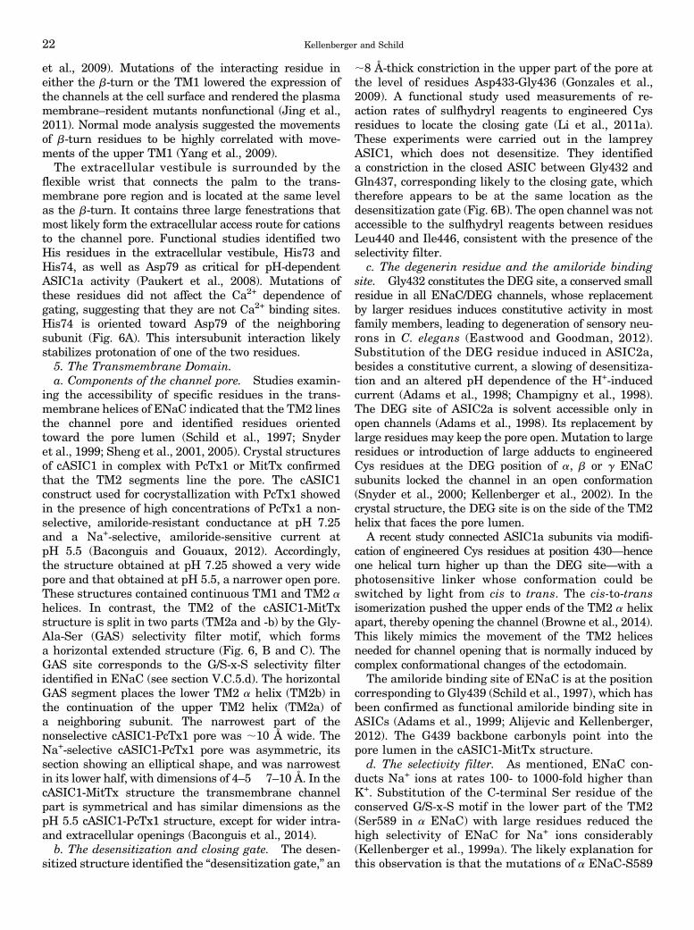
et al., 2009). Mutations of the interacting residue ineither the b-turn or the TM1 lowered the expression ofthe channels at the cell surface and rendered the plasmamembrane–resident mutants nonfunctional (Jing et al.,2011). Normal mode analysis suggested the movementsof b-turn residues to be highly correlated with move-ments of the upper TM1 (Yang et al., 2009).The extracellular vestibule is surrounded by the
flexible wrist that connects the palm to the trans-membrane pore region and is located at the same levelas the b-turn. It contains three large fenestrations thatmost likely form the extracellular access route for cationsto the channel pore. Functional studies identified twoHis residues in the extracellular vestibule, His73 andHis74, as well as Asp79 as critical for pH-dependentASIC1a activity (Paukert et al., 2008). Mutations ofthese residues did not affect the Ca2+ dependence ofgating, suggesting that they are not Ca2+ binding sites.His74 is oriented toward Asp79 of the neighboringsubunit (Fig. 6A). This intersubunit interaction likelystabilizes protonation of one of the two residues.5. The Transmembrane Domain.a. Components of the channel pore. Studies examin-
ing the accessibility of specific residues in the trans-membrane helices of ENaC indicated that the TM2 linesthe channel pore and identified residues orientedtoward the pore lumen (Schild et al., 1997; Snyderet al., 1999; Sheng et al., 2001, 2005). Crystal structuresof cASIC1 in complex with PcTx1 or MitTx confirmedthat the TM2 segments line the pore. The cASIC1construct used for cocrystallization with PcTx1 showedin the presence of high concentrations of PcTx1 a non-selective, amiloride-resistant conductance at pH 7.25and a Na+-selective, amiloride-sensitive current atpH 5.5 (Baconguis and Gouaux, 2012). Accordingly,the structure obtained at pH 7.25 showed a very widepore and that obtained at pH 5.5, a narrower open pore.These structures contained continuous TM1 and TM2 ahelices. In contrast, the TM2 of the cASIC1-MitTxstructure is split in two parts (TM2a and -b) by the Gly-Ala-Ser (GAS) selectivity filter motif, which formsa horizontal extended structure (Fig. 6, B and C). TheGAS site corresponds to the G/S-x-S selectivity filteridentified in ENaC (see section V.C.5.d). The horizontalGAS segment places the lower TM2 a helix (TM2b) inthe continuation of the upper TM2 helix (TM2a) ofa neighboring subunit. The narrowest part of thenonselective cASIC1-PcTx1 pore was ;10 Å wide. TheNa+-selective cASIC1-PcTx1 pore was asymmetric, itssection showing an elliptical shape, and was narrowestin its lower half, with dimensions of 4–5 � 7–10 Å. In thecASIC1-MitTx structure the transmembrane channelpart is symmetrical and has similar dimensions as thepH 5.5 cASIC1-PcTx1 structure, except for wider intra-and extracellular openings (Baconguis et al., 2014).b. The desensitization and closing gate. The desen-
sitized structure identified the “desensitization gate,” an
;8 Å-thick constriction in the upper part of the pore atthe level of residues Asp433-Gly436 (Gonzales et al.,2009). A functional study used measurements of re-action rates of sulfhydryl reagents to engineered Cysresidues to locate the closing gate (Li et al., 2011a).These experiments were carried out in the lampreyASIC1, which does not desensitize. They identifieda constriction in the closed ASIC between Gly432 andGln437, corresponding likely to the closing gate, whichtherefore appears to be at the same location as thedesensitization gate (Fig. 6B). The open channel was notaccessible to the sulfhydryl reagents between residuesLeu440 and Ile446, consistent with the presence of theselectivity filter.
c. The degenerin residue and the amiloride bindingsite. Gly432 constitutes the DEG site, a conserved smallresidue in all ENaC/DEG channels, whose replacementby larger residues induces constitutive activity in mostfamily members, leading to degeneration of sensory neu-rons in C. elegans (Eastwood and Goodman, 2012).Substitution of the DEG residue induced in ASIC2a,besides a constitutive current, a slowing of desensitiza-tion and an altered pH dependence of the H+-inducedcurrent (Adams et al., 1998; Champigny et al., 1998).The DEG site of ASIC2a is solvent accessible only inopen channels (Adams et al., 1998). Its replacement bylarge residues may keep the pore open. Mutation to largeresidues or introduction of large adducts to engineeredCys residues at the DEG position of a, b or g ENaCsubunits locked the channel in an open conformation(Snyder et al., 2000; Kellenberger et al., 2002). In thecrystal structure, the DEG site is on the side of the TM2helix that faces the pore lumen.
A recent study connected ASIC1a subunits via modifi-cation of engineered Cys residues at position 430—henceone helical turn higher up than the DEG site—with aphotosensitive linker whose conformation could beswitched by light from cis to trans. The cis-to-transisomerization pushed the upper ends of the TM2 a helixapart, thereby opening the channel (Browne et al., 2014).This likely mimics the movement of the TM2 helicesneeded for channel opening that is normally induced bycomplex conformational changes of the ectodomain.
The amiloride binding site of ENaC is at the positioncorresponding to Gly439 (Schild et al., 1997), which hasbeen confirmed as functional amiloride binding site inASICs (Adams et al., 1999; Alijevic and Kellenberger,2012). The G439 backbone carbonyls point into thepore lumen in the cASIC1-MitTx structure.
d. The selectivity filter. As mentioned, ENaC con-ducts Na+ ions at rates 100- to 1000-fold higher thanK+. Substitution of the C-terminal Ser residue of theconserved G/S-x-S motif in the lower part of the TM2(Ser589 in a ENaC) with large residues reduced thehigh selectivity of ENaC for Na+ ions considerably(Kellenberger et al., 1999a). The likely explanation forthis observation is that the mutations of a ENaC-S589
22 Kellenberger and Schild
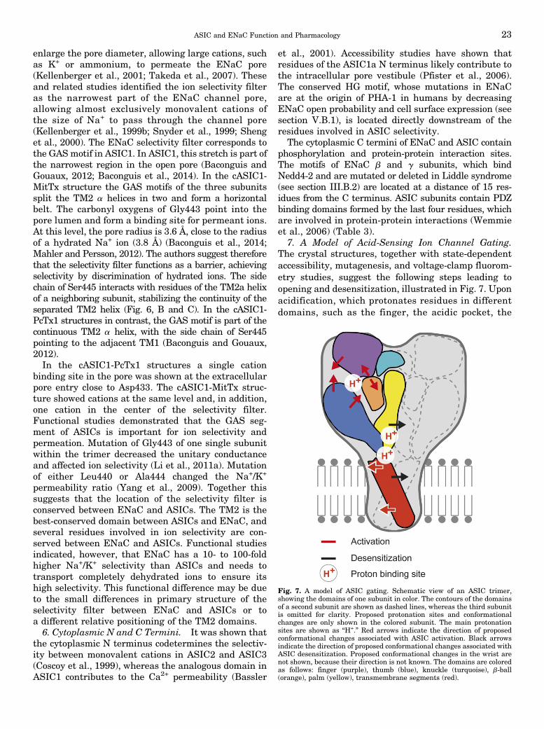
enlarge the pore diameter, allowing large cations, suchas K+ or ammonium, to permeate the ENaC pore(Kellenberger et al., 2001; Takeda et al., 2007). Theseand related studies identified the ion selectivity filteras the narrowest part of the ENaC channel pore,allowing almost exclusively monovalent cations ofthe size of Na+ to pass through the channel pore(Kellenberger et al., 1999b; Snyder et al., 1999; Shenget al., 2000). The ENaC selectivity filter corresponds tothe GASmotif in ASIC1. In ASIC1, this stretch is part ofthe narrowest region in the open pore (Baconguis andGouaux, 2012; Baconguis et al., 2014). In the cASIC1-MitTx structure the GAS motifs of the three subunitssplit the TM2 a helices in two and form a horizontalbelt. The carbonyl oxygens of Gly443 point into thepore lumen and form a binding site for permeant ions.At this level, the pore radius is 3.6 Å, close to the radiusof a hydrated Na+ ion (3.8 Å) (Baconguis et al., 2014;Mahler and Persson, 2012). The authors suggest thereforethat the selectivity filter functions as a barrier, achievingselectivity by discrimination of hydrated ions. The sidechain of Ser445 interacts with residues of the TM2a helixof a neighboring subunit, stabilizing the continuity of theseparated TM2 helix (Fig. 6, B and C). In the cASIC1-PcTx1 structures in contrast, the GAS motif is part of thecontinuous TM2 a helix, with the side chain of Ser445pointing to the adjacent TM1 (Baconguis and Gouaux,2012).In the cASIC1-PcTx1 structures a single cation
binding site in the pore was shown at the extracellularpore entry close to Asp433. The cASIC1-MitTx struc-ture showed cations at the same level and, in addition,one cation in the center of the selectivity filter.Functional studies demonstrated that the GAS seg-ment of ASICs is important for ion selectivity andpermeation. Mutation of Gly443 of one single subunitwithin the trimer decreased the unitary conductanceand affected ion selectivity (Li et al., 2011a). Mutationof either Leu440 or Ala444 changed the Na+/K+
permeability ratio (Yang et al., 2009). Together thissuggests that the location of the selectivity filter isconserved between ENaC and ASICs. The TM2 is thebest-conserved domain between ASICs and ENaC, andseveral residues involved in ion selectivity are con-served between ENaC and ASICs. Functional studiesindicated, however, that ENaC has a 10- to 100-foldhigher Na+/K+ selectivity than ASICs and needs totransport completely dehydrated ions to ensure itshigh selectivity. This functional difference may be dueto the small differences in primary structure of theselectivity filter between ENaC and ASICs or toa different relative positioning of the TM2 domains.6. Cytoplasmic N and C Termini. It was shown that
the cytoplasmic N terminus codetermines the selectiv-ity between monovalent cations in ASIC2 and ASIC3(Coscoy et al., 1999), whereas the analogous domain inASIC1 contributes to the Ca2+ permeability (Bassler
et al., 2001). Accessibility studies have shown thatresidues of the ASIC1a N terminus likely contribute tothe intracellular pore vestibule (Pfister et al., 2006).The conserved HG motif, whose mutations in ENaCare at the origin of PHA-1 in humans by decreasingENaC open probability and cell surface expression (seesection V.B.1), is located directly downstream of theresidues involved in ASIC selectivity.
The cytoplasmic C termini of ENaC and ASIC containphosphorylation and protein-protein interaction sites.The motifs of ENaC b and g subunits, which bindNedd4-2 and are mutated or deleted in Liddle syndrome(see section III.B.2) are located at a distance of 15 res-idues from the C terminus. ASIC subunits contain PDZbinding domains formed by the last four residues, whichare involved in protein-protein interactions (Wemmieet al., 2006) (Table 3).
7. A Model of Acid-Sensing Ion Channel Gating.The crystal structures, together with state-dependentaccessibility, mutagenesis, and voltage-clamp fluorom-etry studies, suggest the following steps leading toopening and desensitization, illustrated in Fig. 7. Uponacidification, which protonates residues in differentdomains, such as the finger, the acidic pocket, the
Fig. 7. A model of ASIC gating. Schematic view of an ASIC trimer,showing the domains of one subunit in color. The contours of the domainsof a second subunit are shown as dashed lines, whereas the third subunitis omitted for clarity. Proposed protonation sites and conformationalchanges are only shown in the colored subunit. The main protonationsites are shown as “H+.” Red arrows indicate the direction of proposedconformational changes associated with ASIC activation. Black arrowsindicate the direction of proposed conformational changes associated withASIC desensitization. Proposed conformational changes in the wrist arenot shown, because their direction is not known. The domains are coloredas follows: finger (purple), thumb (blue), knuckle (turquoise), b-ball(orange), palm (yellow), transmembrane segments (red).
ASIC and ENaC Function and Pharmacology 23

palm, and the wrist (Paukert et al., 2008; Liechtiet al., 2010; Krauson et al., 2013), rapid movements areobserved in the finger and the extracellular vestibule,with the finger a2 helix and the b-ball moving away fromeach other (Passero et al., 2009; Bonifacio et al., 2014)and likely the thumb helices approaching the b-ball andthe finger (Jasti et al., 2007). It is currently not clearwhether the finger movements are transmitted to thepore via the outer structures (thumb and palm-thumbloops) or more centrally via the palm to the extracellularvestibule. These conformational changes lead togetherwith contributions of the palm and the wrist to theopening of the closing gate (Li et al., 2011a), which islocated between Gly432 and Gln437. Slower movementsin the palm (Bonifacio et al., 2014), detected as closingmovement of the lower palm around the central vestibulein accessibility studies in the closed-to-desensitizedtransition (Cushman et al., 2007; Liechti et al., 2010;Della Vecchia et al., 2013; Roy et al., 2013) and doc-umented as a narrower central vestibule in the desensi-tized compared with the open ASIC structure (Baconguisand Gouaux, 2012; Baconguis et al., 2014), are associatedwith desensitization. The closing movement of the palmleads to the closing of the desensitization gate, preventingion conduction (Baconguis and Gouaux, 2012).
VI. Pharmacology
A. Acid-Sensing Ion Channels
1. Small molecules.a. Acid-sensing ion channel inhibitors.i. Amiloride. Amiloride (Fig. 8) inhibits the tran-
sient ASIC currents with IC50 values of the order of10–100 mM, thus with a 100- to 1000-fold lower potencythan its potency for ENaC block (Table 2) (Kellenbergerand Schild, 2002). The sustained component of theASIC3 current is only partially inhibited or evenincreased by amiloride (de Weille et al., 1998; Babinskiet al., 1999; Yagi et al., 2006; Li et al., 2011b).Amiloride induces a conformational change in ASIC2a
that exposes the “DEG site” (located in the pore entry;Fig. 6B) to the extracellular solution, in addition toblocking the pore (Adams et al., 1999). Together theseobservations suggest that amiloride binds to twodifferent sites in ASICs. The homologous residue tothe ENaC blocking site (Schild et al., 1997) is alsonecessary for inhibition of ASIC1a, ASIC2a, and ASIC3(Adams et al., 1999; Alijevic and Kellenberger, 2012)(Fig. 6B). The location of the modulatory amiloridebinding site is not known; however, it was shown thatresidues of the palm are necessary for the amiloride-induced current increase in ASIC3 (Li et al., 2011b). Inthe open MitTx-cASIC1 structure, amiloride was foundin the acidic pocket when the crystallization wascarried out in the presence of the drug, suggesting thatthe modulatory site may be in the acidic pocket(Baconguis et al., 2014). Although amiloride is far frombeing an ideal ASIC inhibitor, because at the micromolarconcentrations needed to inhibit ASICs it also inhibitsENaC, T-type calcium channels, and the sodium-protonexchanger (Kleyman and Cragoe, 1988), it has beensuccessfully used in several studies targeting ASICfunctions. Cortical spreading depression, occurringduring migraine aura, is associated with failure ofbrain ion homeostasis and the release of variousagents, as e.g., protons and AA, which activate orenhance ASIC function. Amiloride blocked corticalspreading depression in rodents and reduced aura andheadache symptoms in human patients (Hollandet al., 2012). Furthermore, a recent study suggestedthat amiloride has a neuroprotective action on MSpatients (Arun et al., 2013). A search for amiloridederivatives with increased potency on ASIC3 resultedin the identification of several substitutions of theNH2 group at position 5 of the pyrazine ring thatincreased the potency by 10-fold (Kuduk et al., 2009b).
ii. Nonsteroidal Anti-Inflammatory Drugs.Inflammation induces a large increase in ASIC mRNAlevels in DRGs, which is prevented by administration ofthe nonsteroidal anti-inflammatory drugs (NSAIDs)
Fig. 8. ENaC/DEG channel modulators and inhibitors. The chemical structures of GMQ, A-317567, amiloride, and benzamil are shown.
24 Kellenberger and Schild
aspirin, diclofenac, and ibuprofen at therapeutic doses(Voilley et al., 2001). In addition, ASIC currents areinhibited by NSAIDs with IC50 values in the order ofhundreds of micromolars to several millimolars (Voilleyet al., 2001; Dorofeeva et al., 2008), thus with 100- to1000-fold lower potency than the inhibition of cyclo-oxygenases by these drugs (Warner et al., 1999). It istherefore likely that the inhibition of ASIC expression,but not the experimentally observed inhibition of ASICcurrents, may contribute to the clinical effects of NSAIDs.iii. Other Small-Molecule Inhibitors. A-317567 is
structurally unrelated to amiloride (Fig. 8) and inhibitsASIC currents in DRG neurons with IC50 values of 2–30 mM (Dube et al., 2005). In contrast to amiloride,A-317567 inhibits transient and sustained ASIC currentswith similar potency. In animal pain models, A-317567was more potent than amiloride. Subsequently, a seriesof indol amidines was tested for inhibition of ASIC3,identifying compounds inhibiting ASIC3 with an IC50
,1 mM that partially reversed mechanical hypersensi-tivity after paw inflammation (Kuduk et al., 2009a).Different anti-protozoal diarylamidines inhibited ASICswith IC50 of 0.3–38 mM (Chen et al., 2010). Nafomastat,a clinically used serine protease inhibitor (Fujii andHitomi, 1981), inhibits ASIC currents, including thesustained component of the ASIC3 current, with an IC50
of ;2–70 mM (Ugawa et al., 2007). More recently, thescreening of a fragment library and subsequent optimi-zation yielded 2-aminopyridine derivatives inhibitingASIC3, with an IC50 of;3 mM (Wolkenberg et al., 2011).ASIC currents are inhibited by the local anestheticstetracaine and lidocaine with IC50 values of ;10 mMand by the K+ channel inhibitor 4-aminopyridine withIC50 values $1 mM (Lin et al., 2011; Boiko et al., 2013;Leng et al., 2013).b. The acid-sensing ion channel modulator 2-guanidine-
4-methylquinazoline. GMQ induces persistent ASIC3currents when applied at pH 7.4 and induces pain-related behavior in wild-type but not ASIC32 /2 mice(Yu et al., 2010). GMQ, which shares some struc-tural similarity with amiloride (Fig. 8), was the firstdiscovered ASIC activator other than protons. It wasshown that mild acidosis and reduction of extra-cellular Ca2+ concentration enhance the GMQ-inducedcurrents. Several residues in the palm domain arecritical for the effects of GMQ (Yu et al., 2011). Covalentmodification of one of these residues, Glu79, is sufficientfor ASIC3 activation, suggesting that GMQ may bind tothe central cavity formed by the lower palm domains(Yu et al., 2010). The potency of GMQ for ASIC3activation is low with an EC50 of ;1 mM at physiologicCa2+ concentrations. It was later shown that, in additionto the synthetic GMQ, the arginine metabolites agmatineand arcaine have qualitatively similar effects on ASIC3(Li et al., 2010c). Agmatine levels are increasedduring trauma, infection, and cancer. In concert withother endogenous ASICmodulators, such as AA, agmatine
may affect ASIC function under physiologic or patho-logic conditions. Agmatine injection induced pain-relatedbehaviors in mice. A recent study addressed themechanisms of action of GMQ and showed that thiscompound induces an acidic shift of the pH dependenceof SSD and an alkaline shift of the pH dependence ofactivation of ASIC3, creating a window current at phys-iologic pH (Alijevic and Kellenberger, 2012). Intrigu-ingly, mutation of Glu79, previously shown to be criticalfor ASIC3 activation by GMQ at physiologic pH, pre-vented the shift of the SSD, but not of the activation pHdependence, suggesting that this residue may rather beinvolved in the effect of GMQ than being part of the GMQbinding site (Alijevic and Kellenberger, 2012). Deletion ofa residue in the lower palm—thus in the same domain—of b ENaC suppressed the effect of the ENaC activatorS3969 [N-(2-hydroxyethyl)-4-methyl-2-(4-methyl-1H-indol-3-ylthio)pentanamide] (Lu et al., 2008). GMQ alsoaffected the pH dependence of ASIC subtypes other thanASIC3—mostly by shifts to more acidic pH—which,however, did not induce currents at physiologic pH(Alijevic and Kellenberger, 2012). At higher concentra-tions GMQ inhibited ASIC currents by a pore block.
2. Toxins. Venom toxins acting on ASICs have im-portantly contributed to the understanding of physio-logic and pathologic functions of ASICs (Baron et al.,2013). The most important ASIC-targeting toxinsare the spider toxin Psalmotoxin1 (PcTx1) (Escoubaset al., 2000), the sea anemone toxin APETx2 (Diochotet al., 2004), and the snake toxins Mambalgin-1–3(Diochot et al., 2012) and MitTx (Bohlen et al., 2011).APETx2 and Mambalgins are inhibitors, MitTx is anactivator of ASICs, and PcTx1 acts depending on theASIC subtype and the pH conditions either as aninhibitor or an activator. PcTx1 and Mambalgin, thetwo toxins for which the mechanism of action has beenelucidated, are gating modifiers. The properties ofthese toxins are summarized in Table 5. Other ASIC-targeting toxins with lower potency or incompleteinhibition are known, such as the sea anemone toxinsUgr 9-1 that inhibit ASIC3 currents with an IC50 of;10 mM (Osmakov et al., 2013) and PhcrTx1, whoseinhibition of DRG neuron ASIC currents is incomplete(;40%) with an IC50 of;100 nM (Rodriguez et al., 2014).These two toxins will not be further discussed here. Allof these ASIC-targeting toxins are minor venom compo-nents, and their use for the organism producing them isnot clear, except for MitTx that induces pain.
a. Psalmotoxin1. PcTx1 of the spider Psalmopoeuscambridgei inhibits homomeric ASIC1a and hetero-meric ASIC1a/2b with nanomolar potency (Escoubaset al., 2000; Sherwood et al., 2011). The structure ofPcTx1 was solved by NMR (Escoubas et al., 2003;Saez et al., 2011) and later by X-ray crystallogra-phy as part of the cASIC1-PcTx1 complex (Baconguisand Gouaux, 2012; Dawson et al., 2012). Composedof a three-stranded antiparallel b sheet, it is folded
ASIC and ENaC Function and Pharmacology 25
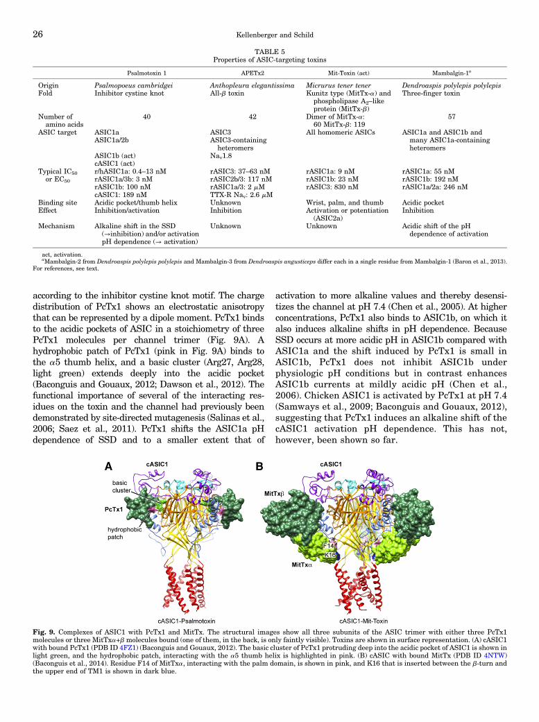
according to the inhibitor cystine knot motif. The chargedistribution of PcTx1 shows an electrostatic anisotropythat can be represented by a dipole moment. PcTx1 bindsto the acidic pockets of ASIC in a stoichiometry of threePcTx1 molecules per channel trimer (Fig. 9A). Ahydrophobic patch of PcTx1 (pink in Fig. 9A) binds tothe a5 thumb helix, and a basic cluster (Arg27, Arg28,light green) extends deeply into the acidic pocket(Baconguis and Gouaux, 2012; Dawson et al., 2012). Thefunctional importance of several of the interacting res-idues on the toxin and the channel had previously beendemonstrated by site-directed mutagenesis (Salinas et al.,2006; Saez et al., 2011). PcTx1 shifts the ASIC1a pHdependence of SSD and to a smaller extent that of
activation to more alkaline values and thereby desensi-tizes the channel at pH 7.4 (Chen et al., 2005). At higherconcentrations, PcTx1 also binds to ASIC1b, on which italso induces alkaline shifts in pH dependence. BecauseSSD occurs at more acidic pH in ASIC1b compared withASIC1a and the shift induced by PcTx1 is small inASIC1b, PcTx1 does not inhibit ASIC1b underphysiologic pH conditions but in contrast enhancesASIC1b currents at mildly acidic pH (Chen et al.,2006). Chicken ASIC1 is activated by PcTx1 at pH 7.4(Samways et al., 2009; Baconguis and Gouaux, 2012),suggesting that PcTx1 induces an alkaline shift of thecASIC1 activation pH dependence. This has not,however, been shown so far.
TABLE 5Properties of ASIC-targeting toxins
Psalmotoxin 1 APETx2 Mit-Toxin (act) Mambalgin-1a
Origin Psalmopoeus cambridgei Anthopleura elegantissima Micrurus tener tener Dendroaspis polylepis polylepisFold Inhibitor cystine knot All-b toxin Kunitz type (MitTx-a) and
phospholipase A2–likeprotein (MitTx-b)
Three-finger toxin
Number ofamino acids
40 42 Dimer of MitTx-a:60 MitTx-b: 119
57
ASIC target ASIC1a ASIC3 All homomeric ASICs ASIC1a and ASIC1b andmany ASIC1a-containingheteromers
ASIC1a/2b ASIC3-containingheteromers
ASIC1b (act) Nav1.8cASIC1 (act)
Typical IC50or EC50
r/hASIC1a: 0.4–13 nM rASIC3: 37–63 nM rASIC1a: 9 nM rASIC1a: 55 nMrASIC1a/3b: 3 nM rASIC2b/3: 117 nM rASIC1b: 23 nM rASIC1b: 192 nMrASIC1b: 100 nM rASIC1a/3: 2 mM rASIC3: 830 nM rASIC1a/2a: 246 nMcASIC1: 189 nM TTX-R Nav: 2.6 mM
Binding site Acidic pocket/thumb helix Unknown Wrist, palm, and thumb Acidic pocketEffect Inhibition/activation Inhibition Activation or potentiation
(ASIC2a)Inhibition
Mechanism Alkaline shift in the SSD(→inhibition) and/or activationpH dependence (→ activation)
Unknown Unknown Acidic shift of the pHdependence of activation
act, activation.aMambalgin-2 from Dendroaspis polylepis polylepis and Mambalgin-3 from Dendroaspis angusticeps differ each in a single residue from Mambalgin-1 (Baron et al., 2013).
For references, see text.
Fig. 9. Complexes of ASIC1 with PcTx1 and MitTx. The structural images show all three subunits of the ASIC trimer with either three PcTx1molecules or three MitTxa+b molecules bound (one of them, in the back, is only faintly visible). Toxins are shown in surface representation. (A) cASIC1with bound PcTx1 (PDB ID 4FZ1) (Baconguis and Gouaux, 2012). The basic cluster of PcTx1 protruding deep into the acidic pocket of ASIC1 is shown inlight green, and the hydrophobic patch, interacting with the a5 thumb helix is highlighted in pink. (B) cASIC with bound MitTx (PDB ID 4NTW)(Baconguis et al., 2014). Residue F14 of MitTxa, interacting with the palm domain, is shown in pink, and K16 that is inserted between the b-turn andthe upper end of TM1 is shown in dark blue.
26 Kellenberger and Schild
b. APETx2. This toxin of the sea anemone Anthopleuraelegantissima inhibits ASIC3 and ASIC3-containingheteromers with IC50 values ranging from 63 nM to2 mM (Diochot et al., 2004). APETx2 shares 64% se-quence identity with the HERG channel inhibitorAPETx1 and 25–35% identity with several Kv- andNav-targeting toxins (Diochot et al., 2004). On the basisof the solution structure of the native APETx2 peptideand on comparison with related toxins, it was suggestedthat the channel-interacting surface may be constitutedby basic and aromatic residues (Chagot et al., 2005). Oneof these aromatic residues, Phe15, was recently shown tobe important for ASIC3 inhibition (Anangi et al., 2012).The site of action of APETx2 is not known. On the basisof the presence of the basic-aromatic toxin surface patchit has been suggested that APETx2 may bind as PcTx1to the acidic pocket (Baron et al., 2013). APETx2 is notselective for ASICs and inhibits voltage-gated Na+
currents in DRG neurons with an IC50 of 2.6 mM,recombinant Nav1.8 (IC50 of 55 nM or 19 mM, dependingon the study), and Nav1.2 (IC50 of 114 nM) (Blanchardet al., 2012; Peigneur et al., 2012).c. Mambalgins. Mambalgin-1 and -2 from black
mamba and Mambalgin-3 from green mamba differonly in one amino acid from each other and inhibitASIC1a, ASIC1b, and ASIC1a-containing heteromerswith IC50 values ranging from 11 to 250 nM (Diochotet al., 2012; Baron et al., 2013). Modeling of the three-dimensional structure of Mambalgins and its determi-nation by NMR (Schroeder et al., 2014) showed thatthey form a new subfamily of three-finger toxins. Thepeptide surface contains a hydrophobic patch and severalclusters of basic or acidic residues. Combination ofmutagenesis and functional analysis showed thatMambalgin-2 binds in the acidic pocket to the thumb a5helix, the b-ball, and to the palm of an adjacent subunit(Salinas et al., 2014; Schroeder et al., 2014). This is veryclose to the PcTx1 binding site, and it is surprising thatPcTx1 and Mambalgin-1 appear not to interact witheach other on ASIC1a (Diochot et al., 2012). Mambalgin-1shifts the activation curve strongly to more acid values,thereby preventing channel opening.d. Mit-toxin. MitTx of the Texas coral snake is for-
med by noncovalent interaction between the twopeptides MitTxa, a Kunitz type peptide, and MitTxb,a phospholipase A2–like protein (Bohlen et al., 2011).This complex resembles the b-bungarotoxins, in whicha Kunitz type peptide and a phospholipase A2 domainare covalently linked by a disulfide bond (Doley andKini, 2009). MitTx induces a slow activation of ASIC1a,ASIC1b, and ASIC3 with EC50 values of 9, 23, and 830nM, respectively. It only poorly activates ASIC2a, butpotentiates ASIC2a currents with an EC50 of 75 nM byinducing a strong alkaline shift of the activation curve.The mechanism of action of MitTx is currently notknown. However, the most recent ASIC structure wasobtained from a cASIC1-MitTx complex, revealing the
structure and binding site of MitTx (Baconguis et al.,2014). Three molecules of MitTx bind to one channeltrimer (Fig. 9B). Seen from the top, the toxin hetero-dimers protrude from the edges of the cASIC1 trimer. Thetoxin heterodimer is held together by extensive inter-actions between the two subunits. It interacts mainlywith the wrist, palm, and thumb domains of cASIC1 butdoes not reach into the acidic pocket as does PcTx1 (Fig.9). The arrangement and possible effect of MitTx iscompared with that of a church key bottle opener, withMitTxb forming the upper part, interacting with residuesof the two thumb a helices, and MitTxa reaching down tothe membrane plane, interacting with residues of thetwo palm-thumb loops. MitTxa inserts its residue Phe14(pink in Fig. 9B) between the bottom of the palmdomains of two adjacent subunits where it interactswith the b1-b2 linker, and Lys16 (dark blue) is placed inthe wrist between the b turn and the upper end of theTM1. These interactions may open the palm and changethe pore conformation to open the channel pore.
B. Epithelial Na+ Channel
1. Direct Epithelial Na+ Channel Antagonists.a. Properties. Amiloride is the prototypical ENaC
blocker. Developed by Cragoe in the early 1960s, it hasbeen widely used as an adjunctive therapy with loopdiuretics or thiazides to minimize hypokalemia (Cragoeet al., 1967). Amiloride is a pyrazinoylguanidine bearingamino groups in positions 3 and 5 and a chloro group atposition 6 of the pyrazine ring (Fig. 8). Amiloride wasfound to be a potent reversible inhibitor of the epithelialNa+ selective conductance in tight epithelia such asthe toad urinary bladder, with an IC50 of 0.1–0.3 mM(Lindemann and Van Driessche, 1977). Pharmacody-namic experiments on tight amphibian epithelial modelsprovided the first clues on the mechanism of block of theepithelial Na+ channels by amiloride. An important ob-servation was that amiloride inhibition depended on thetransmembrane voltage that, if negative, would tend todrive the positively charged amiloride in the membranechannel, and that amiloride interacts competitively withpermeant cations (Palmer and Andersen, 1989; Schildet al., 1997). These observations were interpreted asamiloride binding within the transmembrane part ofthe channel pore, thereby inhibiting the flux of permeantions. Patch-clamp experiments on renal CCD cells pro-vided evidence that amiloride directly interacts witha highly selective Na+ channel at the apical membrane,resulting in a concentration-dependent decrease in theopen-channel probability, by promoting a nonconductingstate of the channel (Palmer and Frindt, 1986).
A large number of amiloride analogs were synthesized(Kleyman and Cragoe, 1988). Among these analogs, themost specific inhibitors of ENaC, such as benzamil, bearhydrophobic substituents on the terminal nitrogen atomof the guanidiniummoiety (Fig. 8) (Kleyman and Cragoe,1988). The positively charged guanidinium is needed for
ASIC and ENaC Function and Pharmacology 27
ENaC block (Palmer, 1984). Amiloride and its analogswere found to be rather poorly specific and inhibit otherNa+ transporters, such as Na+/H+ or Na+/Ca2+ exchangers.A higher specificity of amiloride analogs for the Na+/H+
exchanger is conferred by addition of hydrophobicsubstituents on the 5-amino group of the pyrazine ring(Kleyman and Cragoe, 1988).The identification of the primary structure of ENaC
made experiments possible that provided a more precisepicture of the ENaC block by amiloride. Site-directedmutagenesis coupled to functional expression of ENaCmutants identified specific residues in the TM2 thatwhen mutated considerably decreased the affinity ofthe channel for amiloride. In the center of TM2, a Ser-Gly-Gly ring in the a, b, and g subunits (correspondingto cASIC1-G439; Fig. 6B) was found to contain the mostcritical structural determinants of the high amilorideaffinity of ENaC (Schild et al., 1997). Substitution ofaS583 with residues such as Ala, Gly, or Cys in-creased the IC50 for amiloride by 6- to 25-fold de-pending on the type of mutation. Similar substitutionsin the b or g subunits increased the IC50 by more than1000-fold and 100- to 1000-fold, respectively. Analysisof the kinetics of ENaC block by amiloride revealedthat the decrease of the amiloride affinity in ENaCmutants by 1–3 orders of magnitude was essentiallydue to an increase in the dissociation rate constant ofamiloride from its receptor site (Kellenberger et al.,2003). In other words, mutations of the G/S ring in thepore of ENaC destabilize the binding interaction ofamiloride with its binding site. These experimentsidentified the amiloride binding site in the channelpore of ENaC and confirmed observations from earlyexperiments that amiloride acts on ENaC as a poreblocker (Palmer and Andersen, 1989). The mutationsin the ENaC pore that affect the amiloride potencydecreased to a similar extend the affinity of otherENaC channel blockers such as triamterene or theamiloride analog benzamil, indicating a common poreblocking mechanism (Kellenberger et al., 2003).Currently no allosteric inhibitors other than the pore
blockers such as amiloride and its derivatives areknown to block ENaC activity. However, functionaldomains important for ENaC activity and its modula-tion could represent potential target sites for allostericantagonists. An interesting candidate among them isthe N-terminal intracellular domain with highlyconserved Cys residues that lead to efficient inhibitionof ENaC when modified by sulfhydryl reagents oroxidizing agents (Kellenberger et al., 2005).b. Renal effects. The renal effects of amiloride are
directly related to ENaC inhibition. Amiloride reachesits target in the lumen of the CCD by filtration andsecretion in the proximal tubule by an organic cationtransporter. The ENaC inhibition results in an increasein urinary Na+ excretion and a decrease in K+ secretion.The effect on K+ secretion is due to the decrease in the
apical Na+ conductance, which hyperpolarizes the apicalmembrane and decreases the driving force for K+
secretion from the tubular cell into the lumen of theCCD. Interestingly, K+ secretion in the distal nephrondepends under normal K+ diet entirely on ENaC-mediated Na+ absorption. In contrast, a part of the K+
secretion is independent of ENaC activity under high K+
diet (Frindt and Palmer, 2009a).Because of the limited contribution of ENaC to the
overall reabsorption of the filtered Na+ along thenephron, its effect on Na+ excretion is smaller than thatof loop diuretics or thiazides. Because the K+ secretioncoupled to ENaC-mediated Na+ absorption is the mainsecretory pathway for K+ secretion under standard K+
diet, the effect of amiloride on K+ secretion is larger thanits effect on Na+ absorption.
In a randomized, crossover trial involving younghealthy volunteers under a standardized diet, amiloridewas not different from placebo regarding a short-term 24-hour blood pressure or a Na+ or K+ excretion,consistent with the presence of compensatory mecha-nisms to maintain Na+ and K+ homeostasis in healthysubjects exposed to amiloride (Matthesen et al., 2013).
c. Pulmonary effects. As mentioned previously,ENaC is expressed in airway epithelial cells and iscrucial for the maintenance of an appropriate ASL leveland mucociliary clearance. These processes representthe primary innate defense mechanism against re-spiratory tract infections. Because cystic fibrosis (CF)pathogenesis involves defective mucus clearance, ami-loride was tested as an inhalation therapy for rehydrat-ing ASL, enhancing mucus clearance in the respiratorytract of CF patients. Amiloride failed as an inhalationtherapy (Graham et al., 1993; Grasemann et al., 2007).Among the possible reasons that were evoked to explainthis failure were first a restricted drug delivery associ-ated with conventional inhalation therapy due to thelimited solubility of amiloride, second a limited potencyand a rapid dissociation from the receptor, and thirda rapid absorption by the airway epithelia. The amiloridederivative benzamil is a more potent inhibitor of ENaCbut was not more efficient than amiloride in thetreatment of CF. These observations led to an optimiza-tion strategy of the amiloride structure-activity relation-ship. Interestingly, amiloride shows a high structuraltolerance for substitutions, allowing the generation of avariety of ENaC blockers (Hirsh et al., 2006). Forinstance, modifications of the distal guanidinium groupto introduce a permanent positive charge reproducedfeatures that resemble quaternary amines with a highpotency for ENaC inhibition. This permanent positivecharge introduces an increased polarity of the block-ing molecule that results in an increased solubilityand a restricted permeability across cell membranes.Other amiloride derivatives were obtained by long-chain substitutions at the guanidine nitrogen. One ofthe compounds, 552-02, was shown to be more potent
28 Kellenberger and Schild
and more selective than amiloride, less epithelium-permeant, and able to increase mucus clearance in vivoand ASL volume in CF bronchial epithelial cells (Hirshet al., 2008; Schoenberger and Althaus, 2013).2. Indirect Antagonists. Other ENaC antagonists
indirectly modulate ENaC activity at the cell surface.The best-known examples are the antagonists of themineralocorticoid receptor such as spironolactone oreplerenone. The effects on urinary Na+ or K+ excretionare very similar to those of amiloride, but these drugs donot require access to the tubule lumen. Endothelin andaldosterone have opposing effects on ENaC-mediated Na+
absorption in the distal nephron and the collecting duct.Endothelin antagonists are expected to relieve ENaCfrom inhibition by endothelin, but the physiologic/pharmacological relevance of this phenomenon isunknown.3. Agonists. As already pointed out, the liquid
homeostasis in the adult lung depends on the passiveflow of cations via ENaC and cyclic nucleotide-gatedion channels (Berthiaume and Matthay, 2007). A proofof principle for the development of ENaC agonists isprovided by the covalent modification of Cys residuesintroduced at specific sites in the external vestibule ofthe channel pore, e.g., at the DEG site (section V.C). Thecompound S3969 was recently reported to reversiblystimulate the human ENaC in heterologous cell expres-sion systems (Lu et al., 2008). This compound acts onENaC by increasing the channel open probability withan apparent affinity (EC50) of 1 mM. In addition, a cyclicpeptide was shown to activate ENaC by increasing itsopen probability in heterologous expression systems(Shabbir et al., 2013).The antinatriuretic effect of vasopressin mediated by
the V2R has been attributed to a stimulation of ENaC-dependent Na+ absorption in the ASDN. In normalhealthy volunteers under a high Na+ diet to suppressaldosterone secretion, the potent V2R agonist desmo-pressin decreased Na+ excretion. A 7-day treatmentwith amiloride prevented the vasopressin-induced natri-uresis, consistent with an ENaC-mediated Na+ absorp-tion (Bankir et al., 2005).
C. Bile Acid–Sensitive Ion Channel
Many of the chemical compounds that inhibit ASICshave also been applied to the related BASIC. Amilorideinhibits the constitutive mouse BASIC current and thebile-activated rat BASIC current with IC50 values of 7and 9 mM, respectively (Wiemuth and Grunder, 2010;Wiemuth et al., 2012). The diarylamidines diminazene andnafamostat, previously shown to inhibit ASICs, also blockBASIC currents with IC50 values of 2–6 mM in a voltage-dependent way, suggesting a pore block (Wiemuth andGrunder, 2011; Lefevre et al., 2014). Fenamates such asflufenamic acid are NSAIDs that inhibit the cyclo-oxygenase in their clinical use and have been known tomodulate the function of a large number of ion channels
(Wiemuth and Grunder, 2011). Flufenamic acid and to alesser extent related drugs, such as diclofenac, activateBASIC at millimolar concentrations, thus at concen-trations that are not reached in clinical use of thesedrugs (Wiemuth and Grunder, 2011). It is interesting tonote in this context that diclofenac is known to inhibitASIC3 with an IC50 of ;100 mM (Voilley et al., 2001).
VII. Conclusions and Perspectives
The cloning of ENaC and ASICs boosted the researchon these channels and made possible the investigationof molecular aspects of their function and cellular roles.The physiologic and pathologic roles of both ENaC andASICs have been addressed extensively in geneticmouse models. More recently, tissue-specific knockoutapproaches provided more detailed information on theENaC physiology. Newly discovered ASIC toxinscontributed importantly to the understanding of theinvolvement of ASICs in pain sensation and neuro-degeneration after ischemic stroke. There are still manyopen questions remaining, and further studies are neededto find answers to questions such as the molecular mech-anisms of ENaC regulation by aldosterone or of theinvolvement of ASICs in pain sensation, synaptic plastic-ity, and the expression of fear. Studies addressing thesequestions may identify new drug targets for ENaCregulation and will likely provide more precise informa-tion on ASIC physiology and confirm ASICs as promisingdrug targets.
The recent publication of crystal structures of anASIC channel has stimulated the structure-function in-vestigation of ASICs. Together, newly available struc-tural and functional information allowed elucidatingmolecular mechanisms of ASIC and ENaC function. Bycontinuing these investigations, it will be possible todescribe the molecular mechanisms by which extra- andintracellular factors control ENaC and ASIC activityand to identify specific sites within the channel proteinsthat can be targeted by novel drugs.
Acknowledgments
The authors thank Miguel van Bemmelen, Olivier Staub, EdithHummler, Karolina Gwiazda, Gaetano Bonifacio, and Omar Alijevicfor comments on a previous version of the manuscript.
Authorship Contributions
Wrote or contributed to the writing of the manuscript: Kellenberger,Schild.
ReferencesAdams CM, Snyder PM, Price MP, and Welsh MJ (1998) Protons activate brain Na+
channel 1 by inducing a conformational change that exposes a residue associatedwith neurodegeneration. J Biol Chem 273:30204–30207.
Adams CM, Snyder PM, and Welsh MJ (1999) Paradoxical stimulation of a DEG/ENaCchannel by amiloride. J Biol Chem 274:15500–15504.
Alexander SP, Benson HE, Faccenda E, Pawson AJ, Sharman JL, Catterall WA,Spedding M, Peters JA, and Harmar AJ; CGTP Collaborators (2013) The ConciseGuide to PHARMACOLOGY 2013/14: ion channels. Br J Pharmacol 170:1607–1651.
Ali A, Pillai KP, Ahmad FJ, Dua Y, and Vohora D(2006) Anticonvulsant effect of ami-loride in pentetrazole-induced status epilepticus in mice. Pharmacol Rep 58:242–245.
ASIC and ENaC Function and Pharmacology 29
Alijevic O and Kellenberger S (2012) Subtype-specific modulation of acid-sensing ionchannel (ASIC) function by 2-guanidine-4-methylquinazoline. J Biol Chem 287:36059–36070.
Allen NJ and Attwell D (2002) Modulation of ASIC channels in rat cerebellar Pur-kinje neurons by ischaemia-related signals. J Physiol 543:521–529.
Alvarez de la Rosa D, Krueger SR, Kolar A, Shao D, Fitzsimonds RM, and CanessaCM (2003) Distribution, subcellular localization and ontogeny of ASIC1 in themammalian central nervous system. J Physiol 546:77–87.
Anangi R, Rash LD, Mobli M, and King GF (2012) Functional expression inEscherichia coli of the disulfide-rich sea anemone peptide APETx2, a potentblocker of acid-sensing ion channel 3. Mar Drugs 10:1605–1618.
Anantharam A and Palmer LG (2007) Determination of epithelial Na+ channelsubunit stoichiometry from single-channel conductances. J Gen Physiol 130:55–70.
Anantharam A, Tian Y, and Palmer LG (2006) Open probability of the epithelialsodium channel is regulated by intracellular sodium. J Physiol 574:333–347.
Andreasen D, Vuagniaux G, Fowler-Jaeger N, Hummler E, and Rossier BC (2006)Activation of epithelial sodium channels by mouse channel activating proteases(mCAP) expressed in Xenopus oocytes requires catalytic activity of mCAP3 andmCAP2 but not mCAP1. J Am Soc Nephrol 17:968–976.
Andrey F, Tsintsadze T, Volkova T, Lozovaya N, and Krishtal O (2005) Acid sensingionic channels: modulation by redox reagents. Biochim Biophys Acta 1745:1–6.
Anzai N, Deval E, Schaefer L, Friend V, Lazdunski M, and Lingueglia E (2002) Themultivalent PDZ domain-containing protein CIPP is a partner of acid-sensing ionchannel 3 in sensory neurons. J Biol Chem 277:16655–16661.
Arun T, Tomassini V, Sbardella E, de Ruiter MB, Matthews L, Leite MI, Gelineau-Morel R, Cavey A, Vergo S, and Craner M, et al. (2013) Targeting ASIC1 in primaryprogressive multiple sclerosis: evidence of neuroprotection with amiloride. Brain136:106–115.
Askwith CC, Benson CJ, Welsh MJ, and Snyder PM (2001) DEG/ENaC ion channelsinvolved in sensory transduction are modulated by cold temperature. Proc NatlAcad Sci USA 98:6459–6463.
Askwith CC, Cheng C, Ikuma M, Benson C, Price MP, and Welsh MJ (2000) Neu-ropeptide FF and FMRFamide potentiate acid-evoked currents from sensoryneurons and proton-gated DEG/ENaC channels. Neuron 26:133–141.
Auberson M, Hoffmann-Pochon N, Vandewalle A, Kellenberger S, and Schild L (2003)Epithelial Na+ channel mutants causing Liddle’s syndrome retain ability to re-spond to aldosterone and vasopressin. Am J Physiol Renal Physiol 285:F459–F471.
Babini E, Paukert M, Geisler HS, and Grunder S (2002) Alternative splicing andinteraction with di- and polyvalent cations control the dynamic range of acid-sensing ion channel 1 (ASIC1). J Biol Chem 277:41597–41603.
Babinski K, Lê KT, and Séguéla P (1999) Molecular cloning and regional distributionof a human proton receptor subunit with biphasic functional properties. J Neuro-chem 72:51–57.
Baconguis I, Bohlen CJ, Goehring A, Julius D, and Gouaux E (2014) X-ray structureof acid-sensing ion channel 1-snake toxin complex reveals open state of a Na(+)-selective channel. Cell 156:717–729.
Baconguis I and Gouaux E (2012) Structural plasticity and dynamic selectivity ofacid-sensing ion channel-spider toxin complexes. Nature 489:400–405.
Bankir L, Fernandes S, Bardoux P, Bouby N, and Bichet DG (2005) Vasopressin-V2receptor stimulation reduces sodium excretion in healthy humans. J Am SocNephrol 16:1920–1928.
Bargeton B and Kellenberger S (2010) The contact region between three domains ofthe extracellular loop of ASIC1a is critical for channel function. J Biol Chem 285:13816–13826.
Baron A, Deval E, Salinas M, Lingueglia E, Voilley N, and Lazdunski M (2002)Protein kinase C stimulates the acid-sensing ion channel ASIC2a via the PDZdomain-containing protein PICK1. J Biol Chem 277:50463–50468.
Baron A, Diochot S, Salinas M, Deval E, Noël J, and Lingueglia E (2013) Venomtoxins in the exploration of molecular, physiological and pathophysiological func-tions of acid-sensing ion channels. Toxicon 75:187–204.
Baron A, Schaefer L, Lingueglia E, Champigny G, and Lazdunski M (2001) Zn2+ andH+ are coactivators of acid-sensing ion channels. J Biol Chem 276:35361–35367.
Baron A, Voilley N, Lazdunski M, and Lingueglia E (2008) Acid sensing ion channelsin dorsal spinal cord neurons. J Neurosci 28:1498–1508.
Bartoi T, Augustinowski K, Polleichtner G, Gründer S, and Ulbrich MH (2014) Acid-sensing ion channel (ASIC) 1a/2a heteromers have a flexible 2:1/1:2 stoichiometry.Proc Natl Acad Sci USA 111:8281–8286.
Bässler EL, Ngo-Anh TJ, Geisler HS, Ruppersberg JP, and Gründer S (2001) Mo-lecular and functional characterization of acid-sensing ion channel (ASIC) 1b. JBiol Chem 276:33782–33787.
Benson CJ, Xie J, Wemmie JA, Price MP, Henss JM, Welsh MJ, and Snyder PM(2002) Heteromultimers of DEG/ENaC subunits form H+-gated channels in mousesensory neurons. Proc Natl Acad Sci USA 99:2338–2343.
Bernardinelli L, Murgia SB, Bitti PP, Foco L, Ferrai R, Musu L, Prokopenko I,Pastorino R, Saddi V, and Ticca A, et al. (2007) Association between the ACCN1gene and multiple sclerosis in Central East Sardinia. PLoS ONE 2:e480.
Berthiaume Y and Matthay MA (2007) Alveolar edema fluid clearance and acute lunginjury. Respir Physiol Neurobiol 159:350–359.
Birdsong WT, Fierro L, Williams FG, Spelta V, Naves LA, Knowles M, Marsh-Haffner J,Adelman JP, AlmersW, and Elde RP, et al. (2010) Sensing muscle ischemia: coincidentdetection of acid and ATP via interplay of two ion channels. Neuron 68:739–749.
Bize V and Horisberger JD (2007) Sodium self-inhibition of human epithelial sodiumchannel: selectivity and affinity of the extracellular sodium sensing site. Am JPhysiol Renal Physiol 293:F1137–F1146.
Blanchard MG and Kellenberger S (2011) Effect of a temperature increase in the non-noxious range on proton-evoked ASIC and TRPV1 activity. Pflugers Arch 461:123–139.
Blanchard MG, Rash LD, and Kellenberger S (2012) Inhibition of voltage-gated Na(+)currents in sensory neurones by the sea anemone toxin APETx2. Br J Pharmacol165:2167–2177.
Bohlen CJ, Chesler AT, Sharif-Naeini R,Medzihradszky KF, Zhou S, King D, Sánchez EE,Burlingame AL, Basbaum AI, and Julius D (2011) A heteromeric Texas coral snaketoxin targets acid-sensing ion channels to produce pain. Nature 479:410–414.
Boiko N, Kucher V, Eaton BA, and Stockand JD (2013) Inhibition of neuronaldegenerin/epithelial Na+ channels by the multiple sclerosis drug 4-aminopyridine.J Biol Chem 288:9418–9427.
Boiko N, Kucher V, Wang B, and Stockand JD (2014) Restrictive expression of acid-sensing ion channel 5 (asic5) in unipolar brush cells of the vestibulocerebellum.PLoS ONE 9:e91326.
Bonifacio G, Lelli CI, and Kellenberger S (2014) Protonation controls ASIC1a ac-tivity via coordinated movements in multiple domains. J Gen Physiol 143:105–118.
Browne LE, Nunes JP, Sim JA, Chudasama V, Bragg L, Caddick S, and North RA(2014) Optical control of trimeric P2X receptors and acid-sensing ion channels. ProcNatl Acad Sci USA 111:521–526.
Bruns JB, Carattino MD, Sheng S, Maarouf AB, Weisz OA, Pilewski JM, Hughey RP,and Kleyman TR (2007) Epithelial Na+ channels are fully activated by furin- andprostasin-dependent release of an inhibitory peptide from the gamma-subunit. JBiol Chem 282:6153–6160.
Burch LH, Talbot CR, Knowles MR, Canessa CM, Rossier BC, and Boucher RC (1995)Relative expression of the human epithelial Na+ channel subunits in normal andcystic fibrosis airways. Am J Physiol 269:C511–C518.
Cadiou H, Studer M, Jones NG, Smith ES, Ballard A, McMahon SB,and McNaughton PA (2007) Modulation of acid-sensing ion channel activity bynitric oxide. J Neurosci 27:13251–13260.
Canessa CM, Horisberger J-D, and Rossier BC (1993) Epithelial sodium channelrelated to proteins involved in neurodegeneration. Nature 361:467–470.
Canessa CM, Merillat AM, and Rossier BC (1994a) Membrane topology of the epi-thelial sodium channel in intact cells. Am J Physiol 267:C1682–C1690.
Canessa CM, Schild L, Buell G, Thorens B, Gautschi I, Horisberger JD, and RossierBC (1994b) Amiloride-sensitive epithelial Na+ channel is made of three homolo-gous subunits. Nature 367:463–467.
Carattino MD, Hughey RP, and Kleyman TR (2008) Proteolytic processing of theepithelial sodium channel gamma subunit has a dominant role in channel acti-vation. J Biol Chem 283:25290–25295.
Carnally SM, Dev HS, Stewart AP, Barrera NP, Van Bemmelen MX, Schild L,Henderson RM, and Edwardson JM (2008) Direct visualization of the trimericstructure of the ASIC1a channel, using AFM imaging. Biochem Biophys ResCommun 372:752–755.
Chagot B, Escoubas P, Diochot S, Bernard C, Lazdunski M, and Darbon H (2005)Solution structure of APETx2, a specific peptide inhibitor of ASIC3 proton-gatedchannels. Protein Sci 14:2003–2010.
Chai S, Li M, Lan J, Xiong ZG, Saugstad JA, and Simon RP (2007) A kinase-anchoring protein 150 and calcineurin are involved in regulation of acid-sensingion channels ASIC1a and ASIC2a. J Biol Chem 282:22668–22677.
Chalfant ML, Denton JS, Berdiev BK, Ismailov II, Benos DJ, and Stanton BA (1999)Intracellular H+ regulates the alpha-subunit of ENaC, the epithelial Na+ channel.Am J Physiol 276:C477–C486.
Champigny G, Voilley N, Waldmann R, and Lazdunski M (1998) Mutations causingneurodegeneration in Caenorhabditis elegans drastically alter the pH sensitivityand inactivation of the mammalian H+-gated Na+ channel MDEG1. J Biol Chem273:15418–15422.
Chandrashekar J, Kuhn C, Oka Y, Yarmolinsky DA, Hummler E, Ryba NJ,and Zuker CS (2010) The cells and peripheral representation of sodium taste inmice. Nature 464:297–301.
Chang SS, Grunder S, Hanukoglu A, Rösler A, Mathew PM, Hanukoglu I, Schild L,Lu Y, Shimkets RA, and Nelson-Williams C, et al. (1996) Mutations in subunits ofthe epithelial sodium channel cause salt wasting with hyperkalaemic acidosis,pseudohypoaldosteronism type 1. Nat Genet 12:248–253.
Chen CC and Wong CW (2013) Neurosensory mechanotransduction through acid-sensing ion channels. J Cell Mol Med 17:337–349.
Chen X and Gründer S (2007) Permeating protons contribute to tachyphylaxis of theacid-sensing ion channel (ASIC) 1a. J Physiol 579:657–670.
Chen X, Kalbacher H, and Gründer S (2005) The tarantula toxin psalmotoxin 1inhibits acid-sensing ion channel (ASIC) 1a by increasing its apparent H+ affinity.J Gen Physiol 126:71–79.
Chen X, Kalbacher H, and Gründer S (2006) Interaction of acid-sensing ion channel(ASIC) 1 with the tarantula toxin psalmotoxin 1 is state dependent. J Gen Physiol127:267–276.
Chen X, Qiu L, Li M, Dürrnagel S, Orser BA, Xiong ZG, and MacDonald JF (2010)Diarylamidines: high potency inhibitors of acid-sensing ion channels. Neurophar-macology 58:1045–1053.
Cho JH and Askwith CC (2007) Potentiation of acid-sensing ion channels by sulf-hydryl compounds. Am J Physiol Cell Physiol 292:C2161–C2174.
Chraïbi A and Horisberger JD (2002) Na self inhibition of human epithelial Nachannel: temperature dependence and effect of extracellular proteases. J GenPhysiol 120:133–145.
Chraïbi A, Vallet V, Firsov D, Hess SK, and Horisberger JD (1998) Protease modu-lation of the activity of the epithelial sodium channel expressed in Xenopus oocytes.J Gen Physiol 111:127–138.
Christensen BM, Perrier R, Wang Q, Zuber AM, Maillard M, Mordasini D, Malsure S,Ronzaud C, Stehle JC, and Rossier BC, et al. (2010) Sodium and potassiumbalance depends on aENaC expression in connecting tubule. J Am Soc Nephrol21:1942–1951.
Chu XP, Close N, Saugstad JA, and Xiong ZG (2006) ASIC1a-specific modulation ofacid-sensing ion channels in mouse cortical neurons by redox reagents. J Neurosci 26:5329–5339.
Chu XP, Papasian CJ, Wang JQ, and Xiong ZG (2011) Modulation of acid-sensing ionchannels: molecular mechanisms and therapeutic potential. Int J Physiol Patho-physiol Pharmacol 3:288–309.
30 Kellenberger and Schild
Chu XP, Wemmie JA, Wang WZ, Zhu XM, Saugstad JA, Price MP, Simon RP,and Xiong ZG (2004) Subunit-dependent high-affinity zinc inhibition of acid-sensing ion channels. J Neurosci 24:8678–8689.
Cobbe SM and Poole-Wilson PA (1980) Tissue acidosis in myocardial hypoxia. J MolCell Cardiol 12:761–770.
Coric T, Zhang P, Todorovic N, and Canessa CM (2003) The extracellular domaindetermines the kinetics of desensitization in acid-sensitive ion channel 1. J BiolChem 278:45240–45247.
Coryell MW, Wunsch AM, Haenfler JM, Allen JE, Schnizler M, Ziemann AE, CookMN, Dunning JP, Price MP, and Rainier JD, et al. (2009) Acid-sensing ion channel-1a in the amygdala, a novel therapeutic target in depression-related behavior.J Neurosci 29:5381–5388.
Coryell MW, Ziemann AE, Westmoreland PJ, Haenfler JM, Kurjakovic Z, Zha XM,Price M, Schnizler MK, and Wemmie JA (2007) Targeting ASIC1a reduces in-nate fear and alters neuronal activity in the fear circuit. Biol Psychiatry 62:1140–1148.
Coscoy S, de Weille JR, Lingueglia E, and Lazdunski M (1999) The pre-transmembrane 1 domain of acid-sensing ion channels participates in the ionpore. J Biol Chem 274:10129–10132.
Cragoe EJ Jr, Woltersdorf OW Jr, Bicking JB, Kwong SF, and Jones JH (1967)Pyrazine diuretics. II. N-amidino-3-amino-5-substituted 6-halopyrazinecarboxamides.J Med Chem 10:66–75.
Cushman KA, Marsh-Haffner J, Adelman JP, and McCleskey EW (2007) A confor-mation change in the extracellular domain that accompanies desensitization ofacid-sensing ion channel (ASIC) 3. J Gen Physiol 129:345–350.
Dawson RJ, Benz J, Stohler P, Tetaz T, Joseph C, Huber S, Schmid G, Hügin D,Pflimlin P, and Trube G, et al. (2012) Structure of the acid-sensing ion channel 1 incomplex with the gating modifier Psalmotoxin 1. Nat Commun 3:936.
Debonneville C, Flores SY, Kamynina E, Plant PJ, Tauxe C, Thomas MA, Münster C,Chraïbi A, Pratt JH, and Horisberger JD, et al. (2001) Phosphorylation of Nedd4-2by Sgk1 regulates epithelial Na(+) channel cell surface expression. EMBO J 20:7052–7059.
Delaunay A, Gasull X, Salinas M, Noël J, Friend V, Lingueglia E, and Deval E(2012) Human ASIC3 channel dynamically adapts its activity to sense the ex-tracellular pH in both acidic and alkaline directions. Proc Natl Acad Sci USA109:13124–13129.
Della Vecchia MC, Rued AC, and Carattino MD (2013) Gating transitions in the palmdomain of ASIC1a. J Biol Chem 288:5487–5495.
Deval E, Baron A, Lingueglia E, Mazarguil H, Zajac JM, and Lazdunski M (2003)Effects of neuropeptide SF and related peptides on acid sensing ion channel 3 andsensory neuron excitability. Neuropharmacology 44:662–671.
Deval E, Friend V, Thirant C, Salinas M, Jodar M, Lazdunski M, and Lingueglia E(2006) Regulation of sensory neuron-specific acid-sensing ion channel 3 by theadaptor protein Na+/H+ exchanger regulatory factor-1. J Biol Chem 281:1796–1807.
Deval E, Noël J, Lay N, Alloui A, Diochot S, Friend V, Jodar M, Lazdunski M,and Lingueglia E (2008) ASIC3, a sensor of acidic and primary inflammatory pain.EMBO J 27:3047–3055.
Deval E, Salinas M, Baron A, Lingueglia E, and Lazdunski M (2004) ASIC2b-dependent regulation of ASIC3, an essential acid-sensing ion channel subunit insensory neurons via the partner protein PICK-1. J Biol Chem 279:19531–19539.
de Weille J and Bassilana F (2001) Dependence of the acid-sensitive ion channel,ASIC1a, on extracellular Ca(2+) ions. Brain Res 900:277–281.
de Weille JR, Bassilana F, Lazdunski M, and Waldmann R (1998) Identification,functional expression and chromosomal localisation of a sustained human proton-gated cation channel. FEBS Lett 433:257–260.
Dijkink L, Hartog A, van Os CH, and Bindels RJ (2002) The epithelial sodium channel(ENaC) is intracellularly located as a tetramer. Pflugers Arch 444:549–555.
Diochot S, Baron A, Rash LD, Deval E, Escoubas P, Scarzello S, Salinas M,and Lazdunski M (2004) A new sea anemone peptide, APETx2, inhibits ASIC3,a major acid-sensitive channel in sensory neurons. EMBO J 23:1516–1525.
Diochot S, Baron A, Salinas M, Douguet D, Scarzello S, Dabert-Gay AS, Debayle D,Friend V, Alloui A, and Lazdunski M, et al. (2012) Black mamba venom peptidestarget acid-sensing ion channels to abolish pain. Nature 490:552–555.
Doley R and Kini RM (2009) Protein complexes in snake venom. Cell Mol Life Sci 66:2851–2871.
Donier E, Rugiero F, Okuse K, and Wood JN (2005) Annexin II light chain p11promotes functional expression of acid-sensing ion channel ASIC1a. J Biol Chem280:38666–38672.
Dorofeeva NA, Barygin OI, Staruschenko A, Bolshakov KV, and Magazanik LG(2008) Mechanisms of non-steroid anti-inflammatory drugs action on ASICsexpressed in hippocampal interneurons. J Neurochem 106:429–441.
Drew LJ, Rohrer DK, Price MP, Blaver KE, Cockayne DA, Cesare P, and Wood JN(2004) Acid-sensing ion channels ASIC2 and ASIC3 do not contribute to mechan-ically activated currents in mammalian sensory neurones. J Physiol 556:691–710.
Drummond HA, Gebremedhin D, and Harder DR (2004) Degenerin/epithelial Na+
channel proteins: components of a vascular mechanosensor. Hypertension 44:643–648.
Du J, Reznikov LR, Price MP, Zha XM, Lu Y, Moninger TO, Wemmie JA, and WelshMJ (2014) Protons are a neurotransmitter that regulates synaptic plasticity in thelateral amygdala. Proc Natl Acad Sci USA 111:8961–8966.
Duan B, Liu DS, Huang Y, Zeng WZ, Wang X, Yu H, Zhu MX, Chen ZY, and Xu TL(2012) PI3-kinase/Akt pathway-regulated membrane insertion of acid-sensing ionchannel 1a underlies BDNF-induced pain hypersensitivity. J Neurosci 32:6351–6363.
Duan B, Wang YZ, Yang T, Chu XP, Yu Y, Huang Y, Cao H, Hansen J, Simon RP,and Zhu MX, et al. (2011) Extracellular spermine exacerbates ischemic neuronalinjury through sensitization of ASIC1a channels to extracellular acidosis. J Neu-rosci 31:2101–2112.
Duan B, Wu LJ, Yu YQ, Ding Y, Jing L, Xu L, Chen J, and Xu TL (2007) Upregulationof acid-sensing ion channel ASIC1a in spinal dorsal horn neurons contributes toinflammatory pain hypersensitivity. J Neurosci 27:11139–11148.
Dubé GR, Lehto SG, Breese NM, Baker SJ, Wang X, Matulenko MA, Honoré P,Stewart AO, Moreland RB, and Brioni JD (2005) Electrophysiological and in vivocharacterization of A-317567, a novel blocker of acid sensing ion channels. Pain117:88–96.
Duggan A, Garcia-Anoveros J, and Corey DP (2002) The PDZ domain protein PICK1and the sodium channel BNaC1 interact and localize at mechanosensory terminalsof dorsal root ganglion neurons and dendrites of central neurons. J Biol Chem 277:5203–5208.
Eastwood AL and Goodman MB (2012) Insight into DEG/ENaC channel gating fromgenetics and structure. Physiology (Bethesda) 27:282–290.
Epple HJ, Amasheh S, Mankertz J, Goltz M, Schulzke JD, and Fromm M (2000)Early aldosterone effect in distal colon by transcriptional regulation of ENaCsubunits. Am J Physiol Gastrointest Liver Physiol 278:G718–G724.
Escoubas P, Bernard C, Lambeau G, Lazdunski M, and Darbon H (2003) Recombi-nant production and solution structure of PcTx1, the specific peptide inhibitor ofASIC1a proton-gated cation channels. Protein Sci 12:1332–1343.
Escoubas P, De Weille JR, Lecoq A, Diochot S, Waldmann R, Champigny G, MoinierD, Ménez A, and Lazdunski M (2000) Isolation of a tarantula toxin specific fora class of proton-gated Na+ channels. J Biol Chem 275:25116–25121.
Eshcol JO, Harding AM, Hattori T, Costa V, Welsh MJ, and Benson CJ (2008) Acid-sensing ion channel 3 (ASIC3) cell surface expression is modulated by PSD-95within lipid rafts. Am J Physiol Cell Physiol 295:C732–739.
Eskandari S, Snyder PM, Kreman M, Zampighi GA, Welsh MJ, and Wright EM(1999) Number of subunits comprising the epithelial sodium channel. J Biol Chem274:27281–27286.
Fejes-Tóth G, Frindt G, Náray-Fejes-Tóth A, and Palmer LG (2008) Epithelial Na+
channel activation and processing in mice lacking SGK1. Am J Physiol RenalPhysiol 294:F1298–F1305.
Firsov D, Gautschi I, Merillat AM, Rossier BC, and Schild L (1998) The hetero-tetrameric architecture of the epithelial sodium channel (ENaC). EMBO J 17:344–352.
Firsov D, Schild L, Gautschi I, Mérillat AM, Schneeberger E, and Rossier BC (1996)Cell surface expression of the epithelial Na channel and a mutant causing Liddlesyndrome: a quantitative approach. Proc Natl Acad Sci USA 93:15370–15375.
Friese MA, Craner MJ, Etzensperger R, Vergo S, Wemmie JA, Welsh MJ, Vincent A,and Fugger L (2007) Acid-sensing ion channel-1 contributes to axonal degenerationin autoimmune inflammation of the central nervous system. Nat Med 13:1483–1489.
Frindt G, Ergonul Z, and Palmer LG (2008) Surface expression of epithelial Nachannel protein in rat kidney. J Gen Physiol 131:617–627.
Frindt G and Palmer LG (2004) Na channels in the rat connecting tubule. Am JPhysiol Renal Physiol 286:F669–F674.
Frindt G and Palmer LG (2009a) K+ secretion in the rat kidney: Na+ channel-dependent and -independent mechanisms. Am J Physiol Renal Physiol 297:F389–F396.
Frindt G and Palmer LG (2009b) Surface expression of sodium channels and trans-porters in rat kidney: effects of dietary sodium. Am J Physiol Renal Physiol 297:F1249–F1255.
Frindt G and Palmer LG (2012) Regulation of epithelial Na+ channels by adrenalsteroids: mineralocorticoid and glucocorticoid effects. Am J Physiol Renal Physiol302:F20–F26.
Frindt G, Silver RB, Windhager EE, and Palmer LG (1993) Feedback regulation ofNa channels in rat CCT. II. Effects of inhibition of Na entry. Am J Physiol 264:F565–F574.
Frindt G, Silver RB, Windhager EE, and Palmer LG (1995) Feedback regulation ofNa channels in rat CCT. III. Response to cAMP. Am J Physiol 268:F480–F489.
Fuchs W, Larsen EH, and Lindemann B (1977) Current-voltage curve of sodiumchannels and concentration dependence of sodium permeability in frog skin.J Physiol 267:137–166.
Fujii S and Hitomi Y (1981) New synthetic inhibitors of C1r, C1 esterase, thrombin,plasmin, kallikrein and trypsin. Biochim Biophys Acta 661:342–345.
Fuller PJ, Brennan FE, and Burgess JS (2000) Acute differential regulation by cor-ticosteroids of epithelial sodium channel subunit and Nedd4 mRNA levels in thedistal colon. Pflugers Arch 441:94–101.
Gao J, Duan B, Wang DG, Deng XH, Zhang GY, Xu L, and Xu TL (2005) Couplingbetween NMDA receptor and acid-sensing ion channel contributes to ischemicneuronal death. Neuron 48:635–646.
Garty H and Palmer LG (1997) Epithelial sodium channels: function, structure, andregulation. Physiol Rev 77:359–396.
Geller DS, Rodriguez-Soriano J, Vallo Boado A, Schifter S, Bayer M, Chang SS,and Lifton RP (1998) Mutations in the mineralocorticoid receptor gene cause au-tosomal dominant pseudohypoaldosteronism type I. Nat Genet 19:279–281.
Gingrich MB and Traynelis SF (2000) Serine proteases and brain damage - is therea link? Trends Neurosci 23:399–407.
Gonzales EB, Kawate T, and Gouaux E (2009) Pore architecture and ion sites in acid-sensing ion channels and P2X receptors. Nature 460:599–604.
Goralski JL, Boucher RC, and Button B (2010) Osmolytes and ion transport modu-lators: new strategies for airway surface rehydration. Curr Opin Pharmacol 10:294–299.
Graham A, Hasani A, Alton EW, Martin GP, Marriott C, Hodson ME, Clarke SW,and Geddes DM (1993) No added benefit from nebulized amiloride in patients withcystic fibrosis. Eur Respir J 6:1243–1248.
Grasemann H, Stehling F, Brunar H, Widmann R, Laliberte TW, Molina L, Döring G,and Ratjen F (2007) Inhalation of Moli1901 in patients with cystic fibrosis. Chest131:1461–1466.
Gründer S and Augustinowski K (2012) Toxin binding reveals two open statestructures for one acid-sensing ion channel. Channels (Austin) 6:409–413.
Gründer S and Chen X (2010) Structure, function, and pharmacology of acid-sensingion channels (ASICs): focus on ASIC1a. Int J Physiol Pathophysiol Pharmacol 2:73–94.
ASIC and ENaC Function and Pharmacology 31
Gründer S, Firsov D, Chang SS, Jaeger NF, Gautschi I, Schild L, Lifton RP,and Rossier BC (1997) A mutation causing pseudohypoaldosteronism type 1identifies a conserved glycine that is involved in the gating of the epithelial sodiumchannel. EMBO J 16:899–907.
Halpern BP (1998) Amiloride and vertebrate gustatory responses to NaCl. NeurosciBiobehav Rev 23:5–47.
Harris M, Garcia-Caballero A, Stutts MJ, Firsov D, and Rossier BC (2008) Prefer-ential assembly of epithelial sodium channel (ENaC) subunits in Xenopus oocytes:role of furin-mediated endogenous proteolysis. J Biol Chem 283:7455–7463.
Hesselager M, Timmermann DB, and Ahring PK (2004) pH Dependency and de-sensitization kinetics of heterologously expressed combinations of acid-sensing ionchannel subunits. J Biol Chem 279:11006–11015.
Hirsh AJ, Molino BF, Zhang J, Astakhova N, Geiss WB, Sargent BJ, Swenson BD,Usyatinsky A, Wyle MJ, and Boucher RC, et al. (2006) Design, synthesis, andstructure-activity relationships of novel 2-substituted pyrazinoylguanidine epi-thelial sodium channel blockers: drugs for cystic fibrosis and chronic bronchitis.J Med Chem 49:4098–4115.
Hirsh AJ, Zhang J, Zamurs A, Fleegle J, Thelin WR, Caldwell RA, Sabater JR,Abraham WM, Donowitz M, and Cha B, et al. (2008) Pharmacological properties ofN-(3,5-diamino-6-chloropyrazine-2-carbonyl)-N’-4-[4-(2,3-dihydroxypropoxy)phenyl]butyl-guanidine methanesulfonate (552-02), a novel epithelial sodiumchannel blocker with potential clinical efficacy for cystic fibrosis lung disease.J Pharmacol Exp Ther 325:77–88.
Hobbs CA, Da Tan C, and Tarran R (2013) Does epithelial sodium channel hyper-activity contribute to cystic fibrosis lung disease? J Physiol 591:4377–4387.
Holland PR, Akerman S, Andreou AP, Karsan N, Wemmie JA, and Goadsby PJ(2012) Acid-sensing ion channel 1: a novel therapeutic target for migraine withaura. Ann Neurol 72:559–563.
Horisberger JD and Chraïbi A (2004) Epithelial sodium channel: a ligand-gatedchannel? Nephron, Physiol 96:37–41.
Hruska-Hageman AM, Benson CJ, Leonard AS, Price MP, and Welsh MJ (2004)PSD-95 and Lin-7b interact with acid-sensing ion channel-3 and have oppositeeffects on H+- gated current. J Biol Chem 279:46962–46968.
Hruska-Hageman AM, Wemmie JA, Price MP, and Welsh MJ (2002) Interaction ofthe synaptic protein PICK1 (protein interacting with C kinase 1) with the non-voltage gated sodium channels BNC1 (brain Na+ channel 1) and ASIC (acid-sensing ion channel). Biochem J 361:443–450.
Hughey RP, Bruns JB, Kinlough CL, Harkleroad KL, Tong Q, Carattino MD,Johnson JP, Stockand JD, and Kleyman TR (2004) Epithelial sodium channels areactivated by furin-dependent proteolysis. J Biol Chem 279:18111–18114.
Hughey RP, Mueller GM, Bruns JB, Kinlough CL, Poland PA, Harkleroad KL,Carattino MD, and Kleyman TR (2003) Maturation of the epithelial Na+ channelinvolves proteolytic processing of the alpha- and gamma-subunits. J Biol Chem278:37073–37082.
Hummler E, Barker P, Gatzy J, Beermann F, Verdumo C, Schmidt A, Boucher R,and Rossier BC (1996) Early death due to defective neonatal lung liquid clearancein alpha-ENaC-deficient mice. Nat Genet 12:325–328.
Hummler E, Barker P, Talbot C, Wang Q, Verdumo C, Grubb B, Gatzy J, Burnier M,Horisberger JD, and Beermann F, et al. (1997) A mouse model for the renal salt-wasting syndrome pseudohypoaldosteronism. Proc Natl Acad Sci USA 94:11710–11715.
Immke DC and McCleskey EW (2001) Lactate enhances the acid-sensing Na+
channel on ischemia-sensing neurons. Nat Neurosci 4:869–870.Immke DC and McCleskey EW (2003) Protons open acid-sensing ion channels bycatalyzing relief of Ca2+ blockade. Neuron 37:75–84.
Jasti J, Furukawa H, Gonzales EB, and Gouaux E (2007) Structure of acid-sensingion channel 1 at 1.9 A resolution and low pH. Nature 449:316–323.
Jiang Q, Inoue K, Wu X, Papasian CJ, Wang JQ, Xiong ZG, and Chu XP (2011)Cysteine 149 in the extracellular finger domain of acid-sensing ion channel 1bsubunit is critical for zinc-mediated inhibition. Neuroscience 193:89–99.
Jiang Q, Papasian CJ, Wang JQ, Xiong ZG, and Chu XP (2010) Inhibitory regulationof acid-sensing ion channel 3 by zinc. Neuroscience 169:574–583.
Jing L, Jiang YQ, Jiang Q, Wang B, Chu XP, and Zha XM (2011) The interactionbetween the first transmembrane domain and the thumb of ASIC1a is critical forits N-glycosylation and trafficking. PLoS ONE 6:e26909.
Jones NG, Slater R, Cadiou H, McNaughton P, and McMahon SB (2004) Acid-inducedpain and its modulation in humans. J Neurosci 24:10974–10979.
Jones RC 3rd, Xu L, and Gebhart GF (2005) The mechanosensitivity of mouse colonafferent fibers and their sensitization by inflammatory mediators require transientreceptor potential vanilloid 1 and acid-sensing ion channel 3. J Neurosci 25:10981–10989.
Kang S, Jang JH, Price MP, GautamM, Benson CJ, Gong H, Welsh MJ, and BrennanTJ (2012) Simultaneous disruption of mouse ASIC1a, ASIC2 and ASIC3 genesenhances cutaneous mechanosensitivity. PLoS ONE 7:e35225.
Kashlan OB, Adelman JL, Okumura S, Blobner BM, Zuzek Z, Hughey RP, KleymanTR, and Grabe M (2011) Constraint-based, homology model of the extracellulardomain of the epithelial Na+ channel a subunit reveals a mechanism of channelactivation by proteases. J Biol Chem 286:649–660.
Kashlan OB and Kleyman TR (2011) ENaC structure and function in the wakeof a resolved structure of a family member. Am J Physiol Renal Physiol 301:F684–F696.
Kellenberger S, Auberson M, Gautschi I, Schneeberger E, and Schild L (2001) Per-meability properties of ENaC selectivity filter mutants. J Gen Physiol 118:679–692.
Kellenberger S, Gautschi I, Pfister Y, and Schild L (2005) Intracellular thiol-mediated modulation of epithelial sodium channel activity. J Biol Chem 280:7739–7747.
Kellenberger S, Gautschi I, and Schild L (1999a) A single point mutation in the poreregion of the epithelial Na+ channel changes ion selectivity by modifying molecularsieving. Proc Natl Acad Sci USA 96:4170–4175.
Kellenberger S, Gautschi I, and Schild L (2002) An external site controls closing ofthe epithelial Na+ channel ENaC. J Physiol 543:413–424.
Kellenberger S, Gautschi I, and Schild L (2003) Mutations in the epithelial Na+
channel ENaC outer pore disrupt amiloride block by increasing its dissociationrate. Mol Pharmacol 64:848–856.
Kellenberger S, Hoffmann-Pochon N, Gautschi I, Schneeberger E, and Schild L(1999b) On the molecular basis of ion permeation in the epithelial Na+ channel.J Gen Physiol 114:13–30.
Kellenberger S and Schild L (2002) Epithelial sodium channel/degenerin family of ionchannels: a variety of functions for a shared structure. Physiol Rev 82:735–767.
Kleyman TR, Carattino MD, and Hughey RP (2009) ENaC at the cutting edge: reg-ulation of epithelial sodium channels by proteases. J Biol Chem 284:20447–20451.
Kleyman TR and Cragoe EJ Jr (1988) Amiloride and its analogs as tools in the studyof ion transport. J Membr Biol 105:1–21.
Kleyman TR, Ernst SA, and Coupaye-Gerard B (1994) Arginine vasopressin andforskolin regulate apical cell surface expression of epithelial Na+ channels in A6cells. Am J Physiol 266:F506–F511.
Kleyman TR, Zuckerman JB, Middleton P, McNulty KA, Hu B, Su X, An B, Eaton DC,and Smith PR (2001) Cell surface expression and turnover of the alpha-subunit of theepithelial sodium channel. Am J Physiol Renal Physiol 281:F213–F221.
Koefoed-Johnsen V and Ussing HH (1958) The nature of the frog skin potential. ActaPhysiol Scand 42:298–308.
Kosari F, Sheng S, Li J, Mak DO, Foskett JK, and Kleyman TR (1998) Subunitstoichiometry of the epithelial sodium channel. J Biol Chem 273:13469–13474.
Kozlenkov A, Lapatsina L, Lewin GR, and Smith ES (2014) Subunit-specific inhibitionof acid sensing ion channels by stomatin-like protein 1. J Physiol 592:557–569.
Krauson AJ, Rued AC, and Carattino MD (2013) Independent contribution of extra-cellular proton binding sites to ASIC1a activation. J Biol Chem 288:34375–34383.
Kreple CJ, Lu Y, Taugher RJ, Schwager-Gutman AL, Du J, Stump M, Wang Y,Ghobbeh A, Fan R, and Cosme CV, et al. (2014) Acid-sensing ion channels con-tribute to synaptic transmission and inhibit cocaine-evoked plasticity. Nat Neu-rosci 17:1083–1091.
Krueger B, Schlötzer-Schrehardt U, Haerteis S, Zenkel M, Chankiewitz VE, AmannKU, Kruse FE, and Korbmacher C (2012) Four subunits (abgd) of the epithelialsodium channel (ENaC) are expressed in the human eye in various locations. InvestOphthalmol Vis Sci 53:596–604.
Kuduk SD, Chang RK, Wai JM, Di Marco CN, Cofre V, DiPardo RM, Cook SP, CatoMJ, Jovanovska A, and Urban MO, et al. (2009a) Amidine derived inhibitors ofacid-sensing ion channel-3 (ASIC3). Bioorg Med Chem Lett 19:4059–4063.
Kuduk SD, Di Marco CN, Chang RK, Dipardo RM, Cook SP, Cato MJ, Jovanovska A,Urban MO, Leitl M, and Spencer RH, et al. (2009b) Amiloride derived inhibitors ofacid-sensing ion channel-3 (ASIC3). Bioorg Med Chem Lett 19:2514–2518.
Lapatsina L, Jira JA, Smith ES, Poole K, Kozlenkov A, Bilbao D, Lewin GR,and Heppenstall PA (2012) Regulation of ASIC channels by a stomatin/STOML3complex located in a mobile vesicle pool in sensory neurons. Open Biol 2:120096.
Lefèvre CM, Diakov A, Haerteis S, Korbmacher C, Gründer S, and Wiemuth D (2014)Pharmacological and electrophysiological characterization of the human bile acid-sensitive ion channel (hBASIC). Pflugers Arch 466:253–263.
Leng T, Lin J, Cottrell JE, and Xiong ZG (2013) Subunit and frequency-dependentinhibition of acid sensing ion channels by local anesthetic tetracaine. Mol Pain 9:27.
Leonard AS, Yermolaieva O, Hruska-Hageman A, Askwith CC, Price MP, WemmieJA, and Welsh MJ (2003) cAMP-dependent protein kinase phosphorylation of theacid-sensing ion channel-1 regulates its binding to the protein interacting withC-kinase-1. Proc Natl Acad Sci USA 100:2029–2034.
Li T, Yang Y, and Canessa CM (2009) Interaction of the aromatics Tyr-72/Trp-288 inthe interface of the extracellular and transmembrane domains is essential forproton gating of acid-sensing ion channels. J Biol Chem 284:4689–4694.
Li T, Yang Y, and Canessa CM (2010a) Asn415 in the beta11-beta12 linker decreasesproton-dependent desensitization of ASIC1. J Biol Chem 285:31285–31291.
Li T, Yang Y, and Canessa CM (2010b) Leu85 in the beta1-beta2 linker of ASIC1slows activation and decreases the apparent proton affinity by stabilizing a closedconformation. J Biol Chem 285:22706–22712.
Li T, Yang Y, and Canessa CM (2011a) Outlines of the pore in open and closedconformations describe the gating mechanism of ASIC1. Nat Commun 2:399.
Li WG, Yu Y, Huang C, Cao H, and Xu TL (2011b) The nonproton ligand sensingdomain is required for paradoxical stimulation of ASIC3 channels by amiloride. JBiol Chem 286:42635–42646.
Li WG, Yu Y, Zhang ZD, Cao H, and Xu TL (2010c) ASIC3 channels integrateagmatine and multiple inflammatory signals through the nonproton ligand sensingdomain. Mol Pain 6:88.
Liddle GWBT and Coppage WS (1963) A familial renal disorder simulating primaryaldosteronism but with negligible aldosterone secretion. Trans Assoc Am Physi-cians 76:199–213.
Liechti LA, Bernèche S, Bargeton B, Iwaszkiewicz J, Roy S, Michielin O,and Kellenberger S (2010) A combined computational and functional approachidentifies new residues involved in pH-dependent gating of ASIC1a. J Biol Chem285:16315–16329.
Lin J, Chu X, Maysami S, Li M, Si H, Cottrell JE, Simon RP, and Xiong Z (2011)Inhibition of acid sensing ion channel currents by lidocaine in cultured mousecortical neurons. Anesth Analg 112:977–981.
Lindemann B and Van Driessche W (1977) Sodium-specific membrane channels offrog skin are pores: current fluctuations reveal high turnover. Science 195:292–294.
Lingueglia E, Champigny G, Lazdunski M, and Barbry P (1995) Cloning of theamiloride-sensitive FMRFamide peptide-gated sodium channel.Nature 378:730–733.
Lingueglia E, Deval E, and Lazdunski M (2006) FMRFamide-gated sodium channeland ASIC channels: a new class of ionotropic receptors for FMRFamide and relatedpeptides. Peptides 27:1138–1152.
Lingueglia E, de Weille JR, Bassilana F, Heurteaux C, Sakai H, Waldmann R,and Lazdunski M (1997) A modulatory subunit of acid sensing ion channels inbrain and dorsal root ganglion cells. J Biol Chem 272:29778–29783.
32 Kellenberger and Schild
Liu YQ, Qiu F, Qiu CY, Cai Q, Zou P, Wu H, and Hu WP (2012) Cannabinoids inhibitacid-sensing ion channel currents in rat dorsal root ganglion neurons. PLoS ONE7:e45531.
Loffing J, Loffing-Cueni D, Macher A, Hebert SC, Olson B, Knepper MA, Rossier BC,and Kaissling B (2000a) Localization of epithelial sodium channel and aquaporin-2in rabbit kidney cortex. Am J Physiol Renal Physiol 278:F530–F539.
Loffing J, Pietri L, Aregger F, Bloch-Faure M, Ziegler U, Meneton P, Rossier BC,and Kaissling B (2000b) Differential subcellular localization of ENaC subunits inmouse kidney in response to high- and low-Na diets. Am J Physiol Renal Physiol279:F252–F258.
Lu M, Echeverri F, Kalabat D, Laita B, Dahan DS, Smith RD, Xu H, Staszewski L,Yamamoto J, and Ling J, et al. (2008) Small molecule activator of the humanepithelial sodium channel. J Biol Chem 283:11981–11994.
Lu Y, Ma X, Sabharwal R, Snitsarev V, Morgan D, Rahmouni K, Drummond HA,Whiteis CA, Costa V, and Price M, et al. (2009) The ion channel ASIC2 is requiredfor baroreceptor and autonomic control of the circulation. Neuron 64:885–897.
Luszczki JJ, Sawicka KM, Kozinska J, Dudra-Jastrzebska M, and Czuczwar SJ(2009) Amiloride enhances the anticonvulsant action of various antiepileptic drugsin the mouse maximal electroshock seizure model. J Neural Transm 116:57–66.
Lv RJ, He JS, Fu YH, Zhang YQ, Shao XQ, Wu LW, Lu Q, Jin LR, and Liu H (2011)ASIC1a polymorphism is associated with temporal lobe epilepsy. Epilepsy Res 96:74–80.
Lynch IJ, Welch AK, Kohan DE, Cain BD, and Wingo CS (2013) Endothelin-1 inhibitssodium reabsorption by ET(A) and ET(B) receptors in the mouse cortical collectingduct. Am J Physiol Renal Physiol 305:F568–F573.
Mähler J and Persson I (2012) A study of the hydration of the alkali metal ions inaqueous solution. Inorg Chem 51:425–438.
Mall MA, Button B, Johannesson B, Zhou Z, Livraghi A, Caldwell RA, Schubert SC,Schultz C, O’Neal WK, and Pradervand S, et al. (2010) Airway surface liquid vol-ume regulation determines different airway phenotypes in liddle compared withbetaENaC-overexpressing mice. J Biol Chem 285:26945–26955.
Malsure S, Wang Q, Charles RP, Sergi C, Perrier R, Christensen BM, Maillard M, RossierBC, and Hummler E (2014) Colon-specific deletion of epithelial sodium channel causessodium loss and aldosterone resistance. J Am Soc Nephrol 25:1453–1464.
Mamenko M, Zaika O, Ilatovskaya DV, Staruschenko A, and Pochynyuk O (2012)Angiotensin II increases activity of the epithelial Na+ channel (ENaC) in distalnephron additively to aldosterone. J Biol Chem 287:660–671.
Mamet J, Baron A, Lazdunski M, and Voilley N (2002) Proinflammatory mediators,stimulators of sensory neuron excitability via the expression of acid-sensing ionchannels. J Neurosci 22:10662–10670.
Mamet J, Lazdunski M, and Voilley N (2003) How nerve growth factor drivesphysiological and inflammatory expressions of acid-sensing ion channel 3 in sen-sory neurons. J Biol Chem 278:48907–48913.
Masilamani S, Kim GH, Mitchell C, Wade JB, and Knepper MA (1999) Aldosterone-mediated regulation of ENaC alpha, beta, and gamma subunit proteins in ratkidney. J Clin Invest 104:R19–R23.
Matthesen SK, Larsen T, Vase H, Lauridsen TG, Jensen JM, and Pedersen EB (2013)Effect of amiloride and spironolactone on renal tubular function and central bloodpressure in patients with arterial hypertension during baseline conditions andafter furosemide: a double-blinded, randomized, placebo-controlled crossover trial.Clin Exp Hypertens 35:313–324.
Mauro T, Guitard M, Behne M, Oda Y, Crumrine D, Komuves L, Rassner U, EliasPM, and Hummler E (2002) The ENaC channel is required for normal epidermaldifferentiation. J Invest Dermatol 118:589–594.
Mazzuca M, Heurteaux C, Alloui A, Diochot S, Baron A, Voilley N, Blondeau N,Escoubas P, Gélot A, and Cupo A, et al. (2007) A tarantula peptide against pain viaASIC1a channels and opioid mechanisms. Nat Neurosci 10:943–945.
Mogil JS, Breese NM, Witty M-F, Ritchie J, Rainville M-L, Ase A, Abbadi N, StuckyCL, and Séguéla P (2005) Transgenic expression of a dominant-negative ASIC3subunit leads to increased sensitivity to mechanical and inflammatory stimuli.J Neurosci 25:9893–9901.
Neaga E, Amuzescu B, Dinu C, Macri B, Pena F, and Flonta M-L (2005) Extracellulartrypsin increases ASIC1a selectivity for monovalent versus divalent cations.J Neurosci Methods 144:241–248.
Neelands TR, Zhang XF, McDonald H, and Puttfarcken P (2010) Differential effectsof temperature on acid-activated currents mediated by TRPV1 and ASIC channelsin rat dorsal root ganglion neurons. Brain Res 1329:55–66.
Nesterov V, Dahlmann A, Krueger B, Bertog M, Loffing J, and Korbmacher C (2012)Aldosterone-dependent and -independent regulation of the epithelial sodium channel(ENaC) in mouse distal nephron. Am J Physiol Renal Physiol 303:F1289–F1299.
N’Gouemo P (2008) Amiloride delays the onset of pilocarpine-induced seizures inrats. Brain Res 1222:230–232.
Osmakov DI, Kozlov SA, Andreev YA, Koshelev SG, Sanamyan NP, Sanamyan KE,Dyachenko IA, Bondarenko DA, Murashev AN, and Mineev KS, et al. (2013) Seaanemone peptide with uncommon b-hairpin structure inhibits acid-sensing ionchannel 3 (ASIC3) and reveals analgesic activity. J Biol Chem 288:23116–23127.
Page AJ, Brierley SM, Martin CM, Price MP, Symonds E, Butler R, Wemmie JA,and Blackshaw LA (2005) Different contributions of ASIC channels 1a, 2, and 3 ingastrointestinal mechanosensory function. Gut 54:1408–1415.
Palmer LG (1984) Voltage-dependent block by amiloride and other monovalent cat-ions of apical Na channels in the toad urinary bladder. J Membr Biol 80:153–165.
Palmer LG and Andersen OS (1989) Interactions of amiloride and small monovalentcations with the epithelial sodium channel. Inferences about the nature of thechannel pore. Biophys J 55:779–787.
Palmer LG and Andersen OS (2008) The two-membrane model of epithelial trans-port: Koefoed-Johnsen and Ussing (1958). J Gen Physiol 132:607–612.
Palmer LG and Frindt G (1986) Amiloride-sensitive Na channels from the apical mem-brane of the rat cortical collecting tubule. Proc Natl Acad Sci USA 83:2767–2770.
Palmer LG and Frindt G (1996) Gating of Na channels in the rat cortical collectingtubule: effects of voltage and membrane stretch. J Gen Physiol 107:35–45.
Palmer LG and Frindt G (2007) Na+ and K+ transport by the renal connecting tubule.Curr Opin Nephrol Hypertens 16:477–483.
Palmer LG, Patel A, and Frindt G (2012) Regulation and dysregulation of epithelialNa+ channels. Clin Exp Nephrol 16:35–43.
Passero CJ, Okumura S, and Carattino MD (2009) Conformational changes associ-ated with proton-dependent gating of ASIC1a. J Biol Chem 284:36473–36481.
Paukert M, Babini E, Pusch M, and Gründer S (2004) Identification of the Ca2+
blocking site of acid-sensing ion channel (ASIC) 1: implications for channel gating.J Gen Physiol 124:383–394.
Paukert M, Chen X, Polleichtner G, Schindelin H, and Gründer S (2008) Candidate aminoacids involved in H+ gating of acid-sensing ion channel 1a. J Biol Chem 283:572–581.
Peigneur S, Béress L, Möller C, Marí F, Forssmann WG, and Tytgat J (2012) Anatural point mutation changes both target selectivity and mechanism of action ofsea anemone toxins. FASEB J 26:5141–5151.
Petroff E, Snitsarev V, Gong H, and Abboud FM (2012) Acid sensing ion channelsregulate neuronal excitability by inhibiting BK potassium channels. BiochemBiophys Res Commun 426:511–515.
Petroff EY, Price MP, Snitsarev V, Gong H, Korovkina V, Abboud FM, and Welsh MJ(2008) Acid-sensing ion channels interact with and inhibit BK K+ channels. ProcNatl Acad Sci USA 105:3140–3144.
Pettersen EF, Goddard TD, Huang CC, Couch GS, Greenblatt DM, Meng EC,and Ferrin TE (2004) UCSF Chimera—a visualization system for exploratory re-search and analysis. J Comput Chem 25:1605–1612.
Pfister Y, Gautschi I, Takeda AN, van Bemmelen M, Kellenberger S, and Schild L (2006)A gating mutation in the internal pore of ASIC1a. J Biol Chem 281:11787–11791.
Pidoplichko VI, Aroniadou-Anderjaska V, Prager EM, Figueiredo TH, Almeida-Suhett CP, Miller SL, and Braga MF (2014) ASIC1a activation enhances inhibitionin the basolateral amygdala and reduces anxiety. J Neurosci 34:3130–3141.
Pignataro G, Simon RP, and Xiong ZG (2007) Prolonged activation of ASIC1a and thetime window for neuroprotection in cerebral ischaemia. Brain 130:151–158.
Poirot O, Berta T, Decosterd I, and Kellenberger S (2006) Distinct ASIC currents areexpressed in rat putative nociceptors and are modulated by nerve injury. J Physiol576:215–234.
Poirot O, Vukicevic M, Boesch A, and Kellenberger S (2004) Selective regulation ofacid-sensing ion channel 1 by serine proteases. J Biol Chem 279:38448–38457.
Pouly D, Debonneville A, Ruffieux-Daidié D, Maillard M, Abriel H, Loffing J, and StaubO (2013) Mice carrying ubiquitin-specific protease 2 (Usp2) gene inactivation maintainnormal sodium balance and blood pressure. Am J Physiol Renal Physiol 305:F21–F30.
Pradervand S, Vandewalle A, Bens M, Gautschi I, Loffing J, Hummler E, Schild L,and Rossier BC (2003) Dysfunction of the epithelial sodium channel expressed inthe kidney of a mouse model for Liddle syndrome. J Am Soc Nephrol 14:2219–2228.
Price MP, Thompson RJ, Eshcol JO, Wemmie JA, and Benson CJ (2004) Stomatinmodulates gating of acid-sensing ion channels. J Biol Chem 279:53886–53891.
Qiu F, Qiu CY, Liu YQ, Wu D, Li JD, and Hu WP (2012) Potentiation of acid-sensingion channel activity by the activation of 5-HT₂ receptors in rat dorsal root ganglionneurons. Neuropharmacology 63:494–500.
Reddy MM, Light MJ, and Quinton PM (1999) Activation of the epithelial Na+
channel (ENaC) requires CFTR Cl- channel function. Nature 402:301–304.Renard S, Lingueglia E, Voilley N, Lazdunski M, and Barbry P (1994) Biochemicalanalysis of the membrane topology of the amiloride-sensitive Na+ channel. J BiolChem 269:12981–12986.
Riepe FG (2009) Clinical and molecular features of type 1 pseudohypoaldosteronism.Horm Res 72:1–9.
Rodríguez AA, Salceda E, Garateix AG, Zaharenko AJ, Peigneur S, López O, Pons T,Richardson M, Díaz M, and Hernández Y, et al. (2014) A novel sea anemone pep-tide that inhibits acid-sensing ion channels. Peptides 53:3–12.
Ronzaud C, Loffing-Cueni D, Hausel P, Debonneville A, Malsure SR, Fowler-JaegerN, Boase NA, Perrier R, Maillard M, and Yang B, et al. (2013) Renal tubularNEDD4-2 deficiency causes NCC-mediated salt-dependent hypertension. J ClinInvest 123:657–665.
Rossier BC (2014) Epithelial sodium channel (ENaC) and the control of blood pres-sure. Curr Opin Pharmacol 15:33–46.
Rossier BC and Stutts MJ (2009) Activation of the epithelial sodium channel (ENaC)by serine proteases. Annu Rev Physiol 71:361–379.
Rotin D (2000) Regulation of the epithelial sodium channel (ENaC) by accessoryproteins. Curr Opin Nephrol Hypertens 9:529–534.
Roy S, Boiteux C, Alijevic O, Liang C, Bernèche S, and Kellenberger S (2013) Mo-lecular determinants of desensitization in an ENaC/degenerin channel. FASEB J27:5034–5045.
Rubera I, Loffing J, Palmer LG, Frindt G, Fowler-Jaeger N, Sauter D, Carroll T,McMahon A, Hummler E, and Rossier BC (2003) Collecting duct-specific gene in-activation of alphaENaC in the mouse kidney does not impair sodium and potas-sium balance. J Clin Invest 112:554–565.
Saez NJ, Mobli M, Bieri M, Chassagnon IR, Malde AK, Gamsjaeger R, Mark AE,Gooley PR, Rash LD, and King GF (2011) A dynamic pharmacophore drives theinteraction between Psalmotoxin-1 and the putative drug target acid-sensing ionchannel 1a. Mol Pharmacol 80:796–808.
Sakai H, Lingueglia E, Champigny G, Mattei MG, and Lazdunski M (1999) Cloningand functional expression of a novel degenerin-like Na+ channel gene in mammals.J Physiol 519:323–333.
Salinas M, Besson T, Delettre Q, Diochot S, Boulakirba S, Douguet D, and LinguegliaE (2014) Binding site and inhibitory mechanism of the mambalgin-2 pain-relievingpeptide on acid-sensing ion channel 1a. J Biol Chem 289:13363–13373.
Salinas M, Rash LD, Baron A, Lambeau G, Escoubas P, and Lazdunski M (2006) Thereceptor site of the spider toxin PcTx1 on the proton-gated cation channel ASIC1a.J Physiol 570:339–354.
Samways DS, Harkins AB, and Egan TM (2009) Native and recombinant ASIC1areceptors conduct negligible Ca2+ entry. Cell Calcium 45:319–325.
San-Cristobal P, Pacheco-Alvarez D, Richardson C, Ring AM, Vazquez N, Rafiqi FH,Chari D, Kahle KT, Leng Q, and Bobadilla NA, et al. (2009) Angiotensin II
ASIC and ENaC Function and Pharmacology 33
signaling increases activity of the renal Na-Cl cotransporter through a WNK4-SPAK-dependent pathway. Proc Natl Acad Sci USA 106:4384–4389.
Saugstad JA, Roberts JA, Dong J, Zeitouni S, and Evans RJ (2004) Analysis of themembrane topology of the acid-sensing ion channel 2a. J Biol Chem 279:55514–55519.
Schaefer L, Sakai H, Mattei M, Lazdunski M, and Lingueglia E (2000) Molecularcloning, functional expression and chromosomal localization of an amiloride-sensitive Na(+) channel from human small intestine. FEBS Lett 471:205–210.
Schafer JA and Troutman SL (1990) cAMP mediates the increase in apical mem-brane Na+ conductance produced in rat CCD by vasopressin. Am J Physiol 259:F823–F831.
Schild L, Canessa CM, Shimkets RA, Gautschi I, Lifton RP, and Rossier BC (1995) Amutation in the epithelial sodium channel causing Liddle disease increaseschannel activity in the Xenopus laevis oocyte expression system. Proc Natl AcadSci USA 92:5699–5703.
Schild L, Schneeberger E, Gautschi I, and Firsov D (1997) Identification of amino acidresidues in the alpha, beta, and gamma subunits of the epithelial sodium channel(ENaC) involved in amiloride block and ion permeation. J Gen Physiol 109:15–26.
Schnizler MK, Schnizler K, Zha XM, Hall DD, Wemmie JA, Hell JW, and Welsh MJ(2009) The cytoskeletal protein alpha-actinin regulates acid-sensing ion channel 1athrough a C-terminal interaction. J Biol Chem 284:2697–2705.
Schoenberger M and Althaus M (2013) Novel small molecule epithelial sodiumchannel inhibitors as potential therapeutics in cystic fibrosis - a patent evaluation.Expert Opin Ther Pat 23:1383–1389.
Schroeder CI, Rash LD, Vila-Farrés X, Rosengren KJ, Mobli M, King GF,Alewood PF, Craik DJ, and Durek T (2014) Chemical synthesis, 3D struc-ture, and ASIC binding site of the toxin mambalgin-2. Angew Chem Int EdEngl 53:1017–1020.
Schurr A (2002) Lactate, glucose and energy metabolism in the ischemic brain (Re-view). Review Int J Mol Med 10:131–136.
Shabbir W, Scherbaum-Hazemi P, Tzotzos S, Fischer B, Fischer H, Pietschmann H,Lucas R, and Lemmens-Gruber R (2013) Mechanism of action of novel lung edematherapeutic AP301 by activation of the epithelial sodium channel. Mol Pharmacol84:899–910.
Sheng S, Carattino MD, Bruns JB, Hughey RP, and Kleyman TR (2006) Furincleavage activates the epithelial Na+ channel by relieving Na+ self-inhibition. AmJ Physiol Renal Physiol 290:F1488–F1496.
Sheng S, McNulty KA, Harvey JM, and Kleyman TR (2001) Second transmembranedomains of ENaC subunits contribute to ion permeation and selectivity. J BiolChem 276:44091–44098.
Sheng S, Perry CJ, Kashlan OB, and Kleyman TR (2005) Side chain orientation ofresidues lining the selectivity filter of epithelial Na+ channels. J Biol Chem 280:8513–8522.
Sheng S, Li J, McNulty KA, Avery D, and Kleyman TR (2000) Characterization of theselectivity filter of the epithelial sodium channel. J Biol Chem 275:8572–8581.
Sherwood T, Franke R, Conneely S, Joyner J, Arumugan P, and Askwith C (2009)Identification of protein domains that control proton and calcium sensitivity ofASIC1a. J Biol Chem 284:27899–27907.
Sherwood TW and Askwith CC (2008) Endogenous arginine-phenylalanine-amide-related peptides alter steady-state desensitization of ASIC1a. J Biol Chem 283:1818–1830.
Sherwood TW and Askwith CC (2009) Dynorphin opioid peptides enhance acid-sensing ion channel 1a activity and acidosis-induced neuronal death. J Neurosci29:14371–14380.
Sherwood TW, Frey EN, and Askwith CC (2012) Structure and activity of the acid-sensing ion channels. Am J Physiol Cell Physiol 303:C699–C710.
Sherwood TW, Lee KG, Gormley MG, and Askwith CC (2011) Heteromeric acid-sensing ion channels (ASICs) composed of ASIC2b and ASIC1a display novelchannel properties and contribute to acidosis-induced neuronal death. J Neurosci31:9723–9734.
Shimkets RA, Lifton RP, and Canessa CM (1997) The activity of the epithelial so-dium channel is regulated by clathrin-mediated endocytosis. J Biol Chem 272:25537–25541.
Shimkets RA, Warnock DG, Bositis CM, Nelson-Williams C, Hansson JH, Scham-belan M, Gill JR Jr, Ulick S, Milora RV, and Findling JW, et al. (1994) Liddle’ssyndrome: heritable human hypertension caused by mutations in the beta subunitof the epithelial sodium channel. Cell 79:407–414.
Sluka KA, Winter OC, and Wemmie JA (2009) Acid-sensing ion channels: A newtarget for pain and CNS diseases. Curr Opin Drug Discov Devel 12:693–704.
Smith ES, Cadiou H, and McNaughton PA (2007) Arachidonic acid potentiates acid-sensing ion channels in rat sensory neurons by a direct action. Neuroscience 145:686–698.
Smoller JW, Gallagher PJ, Duncan LE, McGrath LM, Haddad SA, Holmes AJ, WolfAB, Hilker S, Block SR, and Weill S, et al. (2014) The human ortholog of acid-sensing ion channel gene ASIC1a is associated with panic disorder and amygdalastructure and function. Biol Psychiatry DOI: 10.1016/j.biopsych.2013.12.018.
Snyder PM, Bucher DB, and Olson DR (2000) Gating induces a conformationalchange in the outer vestibule of ENaC. J Gen Physiol 116:781–790.
Snyder PM, McDonald FJ, Stokes JB, and Welsh MJ (1994) Membrane topology ofthe amiloride-sensitive epithelial sodium channel. J Biol Chem 269:24379–24383.
Snyder PM, Olson DR, and Bucher DB (1999) A pore segment in DEG/ENaC Na(+)channels. J Biol Chem 274:28484–28490.
Snyder PM, Olson DR, Kabra R, Zhou R, and Steines JC (2004) cAMP and serum andglucocorticoid-inducible kinase (SGK) regulate the epithelial Na(+) channelthrough convergent phosphorylation of Nedd4-2. J Biol Chem 279:45753–45758.
Soundararajan R, Wang J, Melters D, and Pearce D (2007) Differential activities ofglucocorticoid-induced leucine zipper protein isoforms. J Biol Chem 282:36303–36313.
Springauf A, Bresenitz P, and Gründer S (2011) The interaction between two ex-tracellular linker regions controls sustained opening of acid-sensing ion channel 1.J Biol Chem 286:24374–24384.
Stahler F, Riedel K, Demgensky S, Neumann K, Dunkel A, Taubert A, Raab B,Behrens M, Raguse JD, and Hofmann T, et al. (2008) A role of the epithelial sodiumchannel in human salt taste transduction? Chemosens Percept 1:78–90.
Staub O, Abriel H, Plant P, Ishikawa T, Kanelis V, Saleki R, Horisberger JD, SchildL, and Rotin D (2000) Regulation of the epithelial Na+ channel by Nedd4 andubiquitination. Kidney Int 57:809–815.
Stuart D, Rees S, Woodward SK, Koesters R, Strait KA, and Kohan DE (2012) Dis-ruption of the endothelin A receptor in the nephron causes mild fluid volumeexpansion. BMC Nephrol 13:166.
Studer RA, Person E, Robinson-Rechavi M, and Rossier BC (2011) Evolution of theepithelial sodium channel and the sodium pump as limiting factors of aldosteroneaction on sodium transport. Physiol Genomics 43:844–854.
Su J, Tang Y, Liu L, Zhou H, and Dong Q (2011) Regulation of acid-sensing ionchannel 1a function by tissue kallikrein may be through channel cleavage. Neu-rosci Lett 490:46–51.
Suarez PE, Rodriguez EG, Soundararajan R, Mérillat AM, Stehle JC, Rotman S,Roger T, Voirol MJ, Wang J, and Gross O, et al. (2012) The glucocorticoid-inducedleucine zipper (gilz/Tsc22d3-2) gene locus plays a crucial role in male fertility. MolEndocrinol 26:1000–1013.
Sun P, Yue P, and Wang WH (2012) Angiotensin II stimulates epithelial sodiumchannels in the cortical collecting duct of the rat kidney. Am J Physiol RenalPhysiol 302:F679–F687.
Sutherland SP, Benson CJ, Adelman JP, and McCleskey EW (2001) Acid-sensing ionchannel 3 matches the acid-gated current in cardiac ischemia-sensing neurons.Proc Natl Acad Sci USA 98:711–716.
Takeda AN, Gautschi I, van Bemmelen MX, and Schild L (2007) Cadmium trappingin an epithelial sodium channel pore mutant. J Biol Chem 282:31928–31936.
Ugawa S, Ishida Y, Ueda T, Inoue K, Nagao M, and Shimada S (2007) Nafamostatmesilate reversibly blocks acid-sensing ion channel currents. Biochem Biophys ResCommun 363:203–208.
Ugawa S, Ueda T, Ishida Y, Nishigaki M, Shibata Y, and Shimada S (2002)Amiloride-blockable acid-sensing ion channels are leading acid sensors expressedin human nociceptors. J Clin Invest 110:1185–1190.
Vallet V, Chraibi A, Gaeggeler HP, Horisberger JD, and Rossier BC (1997) Anepithelial serine protease activates the amiloride-sensitive sodium channel.Nature 389:607–610.
Vergo S, Craner MJ, Etzensperger R, Attfield K, Friese MA, Newcombe J, Esiri M,and Fugger L (2011) Acid-sensing ion channel 1 is involved in both axonal injuryand demyelination in multiple sclerosis and its animal model. Brain 134:571–584.
Voilley N, de Weille J, Mamet J, and Lazdunski M (2001) Nonsteroid anti-inflammatory drugs inhibit both the activity and the inflammation-induced ex-pression of acid-sensing ion channels in nociceptors. J Neurosci 21:8026–8033.
Vuagniaux G, Vallet V, Jaeger NF, Hummler E, and Rossier BC (2002) Synergisticactivation of ENaC by three membrane-bound channel-activating serine proteases(mCAP1, mCAP2, and mCAP3) and serum- and glucocorticoid-regulated kinase(Sgk1) in Xenopus Oocytes. J Gen Physiol 120:191–201.
Vuagniaux G, Vallet V, Jaeger NF, Pfister C, Bens M, Farman N, Courtois-Coutry N,Vandewalle A, Rossier BC, and Hummler E (2000) Activation of the amiloride-sensitive epithelial sodium channel by the serine protease mCAP1 expressed ina mouse cortical collecting duct cell line. J Am Soc Nephrol 11:828–834.
Vukicevic M and Kellenberger S (2004) Modulatory effects of acid-sensing ionchannels on action potential generation in hippocampal neurons. Am J Physiol CellPhysiol 287:C682–C690.
Vukicevic M, Weder G, Boillat A, Boesch A, and Kellenberger S (2006) Trypsincleaves acid-sensing ion channel 1a in a domain that is critical for channel gating. JBiol Chem 281:714–722.
Waldmann R, Bassilana F, de Weille J, Champigny G, Heurteaux C, and LazdunskiM (1997a) Molecular cloning of a non-inactivating proton-gated Na+ channel spe-cific for sensory neurons. J Biol Chem 272:20975–20978.
Waldmann R, Champigny G, Bassilana F, Heurteaux C, and Lazdunski M (1997b) Aproton-gated cation channel involved in acid-sensing. Nature 386:173–177.
Waldmann R, Champigny G, Bassilana F, Voilley N, and Lazdunski M (1995) Mo-lecular cloning and functional expression of a novel amiloride-sensitive Na+
channel. J Biol Chem 270:27411–27414.Wang WZ, Chu XP, Li MH, Seeds J, Simon RP, and Xiong ZG (2006) Modulationof acid-sensing ion channel currents, acid-induced increase of intracellular Ca2+,and acidosis-mediated neuronal injury by intracellular pH. J Biol Chem 281:29369–29378.
Wang X, Li WG, Yu Y, Xiao X, Cheng J, Zeng WZ, Peng Z, Xi Zhu M, and Xu TL (2013)Serotonin facilitates peripheral pain sensitivity in a manner that depends on thenonproton ligand sensing domain of ASIC3 channel. J Neurosci 33:4265–4279.
Warner TD, Giuliano F, Vojnovic I, Bukasa A, Mitchell JA, and Vane JR (1999)Nonsteroid drug selectivities for cyclo-oxygenase-1 rather than cyclo-oxygenase-2are associated with human gastrointestinal toxicity: a full in vitro analysis. ProcNatl Acad Sci USA 96:7563–7568.
Wemmie JA, Askwith CC, Lamani E, Cassell MD, Freeman JH Jr, and Welsh MJ(2003) Acid-sensing ion channel 1 is localized in brain regions with high synapticdensity and contributes to fear conditioning. J Neurosci 23:5496–5502.
Wemmie JA, Chen J, Askwith CC, Hruska-Hageman AM, Price MP, Nolan BC, Yoder PG,Lamani E, Hoshi T, and Freeman JH Jr, et al. (2002) The acid-activated ion channelASIC contributes to synaptic plasticity, learning, and memory. Neuron 34:463–477.
Wemmie JA, Coryell MW, Askwith CC, Lamani E, Leonard AS, Sigmund CD,and Welsh MJ (2004) Overexpression of acid-sensing ion channel 1a in transgenicmice increases acquired fear-related behavior. Proc Natl Acad Sci USA 101:3621–3626.
Wemmie JA, Price MP, and Welsh MJ (2006) Acid-sensing ion channels: advances,questions and therapeutic opportunities. Trends Neurosci 29:578–586.
Wemmie JA, Taugher RJ, and Kreple CJ (2013) Acid-sensing ion channels in painand disease. Nat Rev Neurosci 14:461–471.
Wiemuth D and Gründer S (2010) A single amino acid tunes Ca2+ inhibition of brainliver intestine Na+ channel (BLINaC). J Biol Chem 285:30404–30410.
34 Kellenberger and Schild
Wiemuth D and Gründer S (2011) The pharmacological profile of brain liver intestineNa+ channel: inhibition by diarylamidines and activation by fenamates. MolPharmacol 80:911–919.
Wiemuth D, Sahin H, Falkenburger BH, Lefèvre CM, Wasmuth HE, and Gründer S(2012) BASIC—a bile acid-sensitive ion channel highly expressed in bile ducts.FASEB J 26:4122–4130.
Wolkenberg SE, Zhao Z, Mulhearn JJ, Harrison ST, Sanders JM, Cato MJ, JovanovskaA, Panigel J, Cook SP, andHenze DA, et al. (2011) High concentration electrophysiology-based fragment screen: discovery of novel acid-sensing ion channel 3 (ASIC3)inhibitors. Bioorg Med Chem Lett 21:2646–2649.
Wu LJ, Duan B, Mei YD, Gao J, Chen JG, Zhuo M, Xu L, Wu M, and Xu TL (2004)Characterization of acid-sensing ion channels in dorsal horn neurons of rat spinalcord. J Biol Chem 279:43716–43724.
Wu PY, Huang YY, Chen CC, Hsu TT, Lin YC, Weng JY, Chien TC, Cheng IH,and Lien CC (2013) Acid-sensing ion channel-1a is not required for normal hip-pocampal LTP and spatial memory. J Neurosci 33:1828–1832.
Xiong ZG, Zhu XM, Chu XP, Minami M, Hey J, Wei WL, MacDonald JF, Wemmie JA,Price MP, and Welsh MJ, et al. (2004) Neuroprotection in ischemia: blockingcalcium-permeable acid-sensing ion channels. Cell 118:687–698.
Yagi J, Wenk HN, Naves LA, and McCleskey EW (2006) Sustained currents throughASIC3 ion channels at the modest pH changes that occur during myocardial is-chemia. Circ Res 99:501–509.
Yan J and Dussor G (2014) Ion channels and migraine. Headache 54:619–639.Yan J, Edelmayer RM, Wei X, De Felice M, Porreca F, and Dussor G (2011) Duralafferents express acid-sensing ion channels: a role for decreased meningeal pH inmigraine headache. Pain 152:106–113.
Yang H, Yu Y, Li WG, Yu F, Cao H, Xu TL, and Jiang H (2009) Inherent dynamics of theacid-sensing ion channel 1 correlates with the gating mechanism. PLoS Biol 7:e1000151.
Yang Y, Yu Y, Cheng J, Liu Y, Liu DS, Wang J, Zhu MX, Wang R, and Xu TL (2012)Highly conserved salt bridge stabilizes rigid signal patch at extracellular loop criticalfor surface expression of acid-sensing ion channels. J Biol Chem 287:14443–14455.
Yu Y, Chen Z, Li WG, Cao H, Feng EG, Yu F, Liu H, Jiang H, and Xu TL (2010) Anonproton ligand sensor in the acid-sensing ion channel. Neuron 68:61–72.
Yu Y, Li WG, Chen Z, Cao H, Yang H, Jiang H, and Xu TL (2011) Atomic levelcharacterization of the nonproton ligand-sensing domain of ASIC3 channels. J BiolChem 286:24996–25006.
Zha XM, Costa V, Harding AM, Reznikov L, Benson CJ and Welsh MJ (2009) ASIC2subunits target acid-sensing ion channels to the synapse via an association withPSD-95. J Neurosci 29:8438–8446.
Zha XM, Wemmie JA, Green SH, and Welsh MJ (2006) Acid-sensing ion channel 1a isa postsynaptic proton receptor that affects the density of dendritic spines. ProcNatl Acad Sci USA 103:16556–16561.
Zhang P, Sigworth FJ, and Canessa CM (2006) Gating of acid-sensitive ionchannel-1: release of Ca2+ block vs. allosteric mechanism. J Gen Physiol 127:109–117.
Ziemann AE, Allen JE, Dahdaleh NS, Drebot II, Coryell MW, Wunsch AM, LynchCM, Faraci FM, Howard MA 3rd, and Welsh MJ, et al. (2009) The amygdala isa chemosensor that detects carbon dioxide and acidosis to elicit fear behavior. Cell139:1012–1021.
Ziemann AE, Schnizler MK, Albert GW, Severson MA, Howard MA 3rd, Welsh MJ,and Wemmie JA (2008) Seizure termination by acidosis depends on ASIC1a. NatNeurosci 11:816–822.
ASIC and ENaC Function and Pharmacology 35