Embed Size (px)
Citation preview

JOURNAL OF WOOD CHEMISTRY AND TECHNOLOGY, 18(4), 381-402 (1998)
ASSIGNMENT OF THE PHOTOYELLOWING-RELATED 1675 cm-1
RAMAN/IR BAND TO p-QUINONES AND ITS IMPLICATIONS TO THEMECHANISM OF COLOR REVERSION IN MECHANICAL PULPS
Umesh P. AgarwalUSDA Forest Service
Forest Products LaboratoryMadison, Wisconsin 53705
ABSTRACT
Using FT (Fourier-transform) Raman and FT infrared (IR) spectroscopies, a newband was detected at 1675 cm-1; this was most likely to have come from the yellowchromophores in photoexposed thermomechanical pulps (TMPs). On the basis ofspectroscopic studies that involved both o- and p-quinone models and Fremy’s salt-oxidized TMP, the 1675 cm-1 band is assigned to the p-quinone functional group.Moreover, in the presence of known photoyellowing inhibitors, the photoyellowingbehavior of methyl hydroquinone was similar to that of TMPs. Another importantfinding was that the molecular oxygen sensitivity of the laser-induced fluorescence(excited at 514.5 nm) of p-quinone and hydroquinone models was similar to what hadbeen previously observed for yellowed and unyellowed TMPs. Taken together, theseresults provide strong support for a previously suggested yellowing hypothesis in whicha hydroquinone/p-quinone couple was seen as an important leucochromophore/chromophore system in mechanical pulps.
Keywords: Photoyellowing, mechanical pulps, lignin, Raman, quinones
INTRODUCTION
Finding a practical solution to the problem of photoyellowing of lignin-rich
mechanical pulps continues to be a challenging area of research. Of the many proposed
approaches to deal with the yellowing problem, no single one is suited to the needs of the
Copyright © 1998 by Marcel Dekker, Inc.
381
www.dekke r . com
382 AGARWAL
paper industry.1 However, it is expected that better solutions will be developed when the
yellowing mechanism is fully understood. Attaining such a level of understanding is
difficult and requires that additional progress be made in a number of areas. For instance,
the nature of important yellowness-causing chromophores needs to be determined, the
identity of initiating structures that lead to coloration needs to be established beyond a
reasonable doubt, and photoyellowing reaction pathways should be established.
Although significant progress has been made in these areas, the current knowledge base
is not sufficient to allow development of a practical solution.
Research in this area is also important from the viewpoint of sustainability of
forests because enhanced use of mechanical pulps would allow more efficient use of the
wood resource. In addition, the impact of pulp-producing processes on the environment
would be diminished.
Recently published research by the author2 on spruce thermomechanical pulps
(TMPs) indicated that α-carbonyl and aromatic ring-conjugated ethylenic groups are not
the initiators of events that lead to photoyellowing. This result is contrary to the reported
observation of some researchers that stilbene derivatives are one of the important
contributors to photoyellowing.3 In author’s laboratory, attempts made to detect stilbene
derivatives in borohydride-bleached TMP and its acid hydrolysate using Raman
spectroscopy (a technique highly sensitive for detecting stilbene structures) were not
successful.4 Failure to detect stilbenes provided further support to the conclusion that
aromatic ring-conjugated ethylenic bonds are not important contributors to
photoyellowing.
In addition, in the author’s reported work2 , for the first time a Raman and IR band
at approximately 1675 cm-1 was detected and linked to photoyellowing. The present
report describes research on this topic and shows how the assignment of the 1675 cm-1
band, together with some other evidence, has important implications for the mechanism
of photoyellowing.
EXPERIMENTAL
Wood
Black spruce (Picea mariana) and aspen (Populus tremuloides) were obtained and
30-micron-thick cross sections were produced using a microtome. The sections were
PHOTOYELLOWING 383
treated with toluene/ethanol (2/1) to remove extractives. Extracted tissues were air dried
and used in the laser-induced fluorescence (LIF) quenching experiments. Several wood
sections were treated with acid chlorite5 to remove lignin.
Pulp
Commercial spruce TMP was a gift from Consolidated Papers, Wisconsin Rapids,
WI[1]. Some TMP was bleached with sodium borohydride under conditions reported
previously.* To hydrogenate aromatic-ring conjugated ethylenic bonds in lignin, a
portion of the bleached pulp was treated with diimide.2
Peroxide bleaching was carried out on the acid-pretreated TMP. The pretreatment
was done to reduce the metal ion content of the pulp. Peroxide (5%) bleaching was
carried out in two stages: Each stage consisted of bleaching the pulp (12% consistency)
in the presence of silicate buffer at 70°C for 3 h at initial pH 11.5.
CMP and peroxide-bleached CMP samples were provided by the Finnish Pulp and
Paper Research Institute, Helsinki, Finland. CMP was bleached with 4% hydrogen
peroxide and 3.4% NaOH at 15% consistency and 65°C for 3 h. DTPA, MgSO4, and
sodium silicate were used as stabilizers. Additional information on these pulps is
available in a previous publication.6
Handsheets made from TMPs were photoyellowed using a Rayonet RPR-100
photoreactor. The reactor is equipped with ultraviolet-fluorescent lamps having a
Gaussian spectral distribution in the 300-400 nm region (with λmax at 350 nm). The
reactor is also fitted with a cooling fan and a merry-go-round. The intensity of the lamps
was such that the temperature of the irradiated sheet did not exceed 40°C.
To methylate phenolic groups in pulps, diazomethane and dimethyl sulfate were
used. The conditions were similar to those described previously.7,8
TMP was acetylated with a pyridine/acetic anhydride (1/1) mixture. For this, 1 g of
pulp was first dispersed in excess ethanol, then filtered. Filtered TMP was air dried at
room temperature, and 40 mL of pyridine and 40 mL of acetic anhydride were added to
the dried pulp. The pulp suspension was stirred magnetically and purged with nitrogen
for 3 days. The pulp was washed sequentially with 1:1 ethanol/water and pure ethanol.
[1]The use of trade or firm names in this publication is for reader information and does not imply endorsement by the U.S.
Department of Agriculture of any product or service.
384 AGARWAL
Unbleached and bleached kraft pulps were obtained from Consolidated Papers.
Milled Wood Lignin
Milled wood lignin (MWL) was prepared from black spruce following the
procedure described elsewhere.9 For Raman purposes, a thin pellet of the lignin sample
was made using a KBr press.
Lignin Models
The following lignin models were purchased from Aldrich Chemical Co.,
Milwaukee, WI: acetoguaiacone (98%), eugenol (99%), coniferyl alcohol (98%), sinapyl
alcohol, coniferaldehyde (98%), trans-stilbene (96%), 3-methoxy-catechol (99%),
methylhydroquinone (99%) methyl-p-quinone (98%). Purity is indicated by the numbers
in the parentheses. Other models, α-guaicoxy-propioguaiacone, dehydrodiisoeugenol,
pinosylvin monomethyl ether, p-coumaryl alcohol, 4,4’-dipropyl-6,6-biguaiacol, 3-
methoxy-o-benzoquinone, and methoxy-p-benzoquinone were synthesized by
researchers at the Forest Products Laboratory using published methods. Coniferin was a
gift from Professor N. Terashima (formerly at Nagoya University, Nagoya, Japan). The
synthesized compounds were evaluated for purity by NMR spectroscopy.
Fremy’s Salt
Borohydride-bleached pulp was treated with Fremy’s salt (potassium
nitrosodisulfonate, ON(SO3K)2), using conditions described elsewhere.10 The oxidized
pulp had a reddish hue.
Methylation of Methylhydroquinone
Methylhydroquinone (25 mg) was dissolved in 2 mL methanol and cooled in an ice
bath. The solution was stirred magnetically and lightly capped. Freshly prepared
diazomethane,11 in ether (ca. 0.33M), was added in aliquots until a yellow color
persisted. The sample was left to react overnight. Some of the sample had sublimed as it
was found around the outside of the cap and was discarded. The sample was stripped of
the solvent (yield 19 mg). Proton and 13C NMR indicated mostly starting material with
PHOTOYELLOWING 385
a small amount of new methoxyl signal. Crude integration of the methyl and methoxyl
groups indicated that less than 16% of the material was methylated.
Methyl iodide in acetone over potassium carbonate was also used to methylate the
hydroquinone.12 Acetone (40 mL) was added to a round bottom flask containing a
magnetic stirbar. The solvent was bubbled with nitrogen and methylhydroquinone
(49 mg) was added, followed by the finely powdered potassium carbonate (55 mg).
Lastly, methyl iodide (84 mg) was added. The flask was fitted with a condenser, and the
reaction mixture was refluxed for 6 h. A reddish-brown color developed during the
reaction. Nitrogen gas bubbling was continued throughout the experiment. After 6 h, the
reaction mixture was allowed to cool and was capped and stored in the freezer overnight.
Next day, the mixture was brought to room temperature and the acetone was removed
under vacuum. Bright yellow clusters of needle-like crystals sublimed into the neck of
the flask. These crystals (6.5 mg) were carefully removed. NMR and Raman indicated
that it was methyl-p-quinone. The brownish-red residue was dissolved in chloroform and
water and was extracted with more chloroform. The combined chloroform extracts were
dried over magnesium sulfate. It was a brownish-red solid (14.4 mg). NMR indicated
that only about 15% of the material was the expected product (dimethoxy toulene). The
aqueous phase along with an emulsion film was acidified with 1M HCl to a pH of
about 1. The solution was extracted with chloroform, and the combined extracts were
dried over magnesium sulfate. The amount of this fraction was 7.5 mg. The fraction was
analyzed by NMR but could not be identified. The aqueous phase and emulsion were left
to dry. The residue was stirred with acetone, which was decanted and evaporated to a
brown residue (10 mg). This fraction could not be identified using NMR spectroscopy.
After considering reviewers’ comment that the methylation reactions should have
methylated the methylhydroquinone, these chemical reactions were repeated. However,
repeat experiments produced similar results. Therefore, it can be concluded that most of
methylhydroquinone could not be methylated using diazomethane and methyl iodide.
Photoexposure of 3-Methoxy-catechol and Methylhydroquinone
Methanolic solutions (1%) of 3-methoxy-catechol and methylhydroquinone were
separately exposed to light for 24 h in the Rayonet RPR- 100 photoreactor. These models
(2% in water/methanol (60/40)) were also photoexposed in the presence of the following
386 AGARWAL
yellowing inhibitors: polyethylene glycol (MW 4000), ascorbic acid, and 1-thioglycerol.
Concentration of the inhibitors was approximately 5% (w/w).
Conventional Raman
A Jobin Yvon Ramanor HG2S (Instruments SA, Metuchen, NJ) spectrometer was
used to obtain Raman spectra. This system is based on an argon ion laser as the
excitation source. Although several laser wavelengths are available, the 514.5-nm line of
the laser was used in our study.
Small-sized (0.5 by 1 cm) wood and pulp samples were used in Raman
spectroscopic studies. Pulp samples were studied in the form of handsheets. Lignin
models (1% to 5%) were examined by adsorbing them on filter paper. Models were first
dissolved in either methanol or 1:1 methanol/dichloromethane, then adsorbed on the
paper.
The oxygen flushing technique, described previously,13 was used to obtain Raman
spectra. To study the sensitivity of a sample’s LIF to various gases, air, nitrogen, and
oxygen were used to flush the sample cell.
Near-IR FT Raman
FT Raman spectra of samples were obtained using a Bruker RFS 100 instrument
(Bruker Instruments, Inc., Billerica, MA). The 1064-nm wavelength of the diode
Nd:YAG laser was used for sample excitation.
Pulps and models on paper were sampled in the 180° scattering geometry.13 A front
surface mirror was used behind the samples to enhance the collected amount of Raman
signal. Catechol, methylhydroquinone, and their photoexposed counterparts were
sampled in a device called “Aluminum Well.” The latter is a small-sized hole in a pellet
of aluminum from which solids can be sampled. Some models were also sampled on
Whatman No. 1 paper.
RESULTS AND DISCUSSION
Band Assignment
In previously published work,2 it was reported that after photoyellowing of TMP a
Raman band at approximately at 1675 cm-1 was detected. Preliminary attempts to
PHOTOYELLOWING 387
determine if this band could be assigned to an o- or p-quinone group were not successful.
It was pointed out that a Raman/IR band was present in the neighborhood of 1675 cm-1
in both the methoxy-p-quinone and 3-methoxy-o-quinone. Another aspect of the Raman
spectral features of these models was that, for the p-quinone, the 1674 cm-1 band was
somewhat weaker than the 1640 cm-1 band and, for the o-quinone model, the 1660 and
1689 cm-1 bands had very low intensities compared with the 1559 cm-1 band.2 In the
spectra of photoyellowed TMPs and their extracts, none of these more intense bands
(1559 and 1640 cm-1) was present. At best, there was a weak broadened contribution at
1640 cm-1 (Fig. 1a). IR spectra of these models were obtained but they failed to clarify
the issue of which of the quinones was responsible for the 1675 cm-1 band.2 To find out
if the Raman spectrum of Fremy’s salt-treated TMP contained the 1559 cm-1 band (this
was the case for the o-quinone model) and if indeed other p-quinone models showed the
1640 cm-1 band, additional samples were analyzed using FT Raman spectroscopy.
Raman spectra of some of them are shown in Fig. 1. For the quinone models, spectra
were obtained on Whatman No. 1 filter paper.
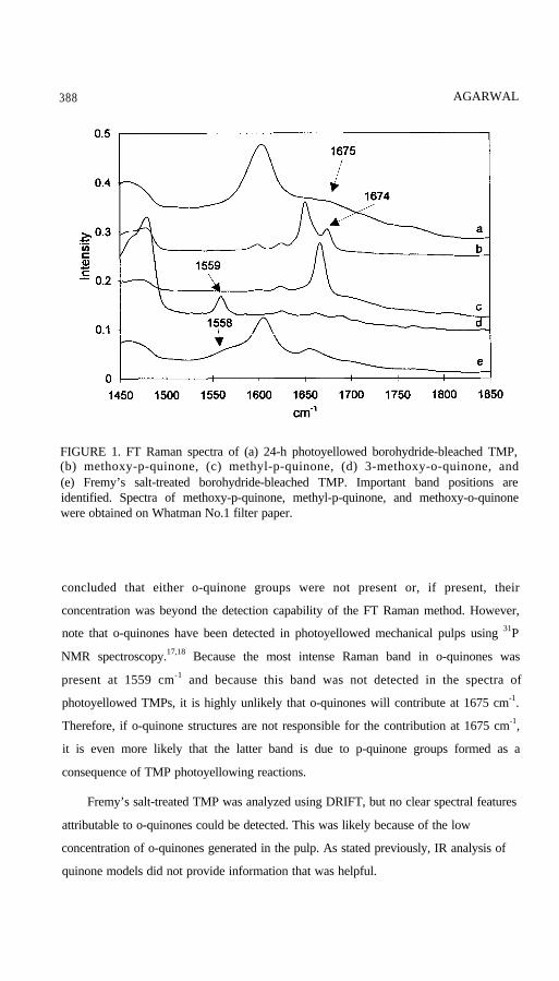
As is evident from the spectra (b) and (c) in Fig. 1, a band in 1640 to 1650 cm-1
region is only present in one of the two p-quinone models. The existence of this band
seems to be a function of the structure of a model. In contrast, a band near 1675 cm-1
was detected in both the p-quinone models. (Two other models analyzed in our
laboratory, 2,5-dihydroxy-p-benzoquinone and 2,3-dimethoxy-5-methyl-p-quinone, had
Raman bands, respectively, at 1672 and 1674 cm-1.) This suggests that for
photoyellowed pulps, even though no band is present at 1640 to 1650 cm-1, as long as a
band is present in the vicinity of 1675 cm-1, it is possible that p-quinone groups are
present. This observation is further supported by the reported carbonyl stretching
frequencies of p-quinones.14,15 If o-quinone groups were present in the photoyellowed
TMPs or their extracts, they should have had a Raman contribution at approximately
1559 cm-1. This was expected on the basis of the analysis of the spectra of both the
o-quinone model and Fremy’s salt-treated TMP (Fig. 1d, 1e); it is known that Fremy’s
salt treatment produces o-quinone groups in mechanical pulps.16,17 Indeed, the
spectrum of Fremy’s salt-treated TMP (Fig. 1e) showed a new feature at 1558 cm-1
indicative of formation of o-quinone groups. However, photoyellowed pulps and their
extracts did not show a discernible signal at 1559 cm-1. Therefore, it can be safely
388 AGARWAL
FIGURE 1. FT Raman spectra of (a) 24-h photoyellowed borohydride-bleached TMP,(b) methoxy-p-quinone, (c) methyl-p-quinone, (d) 3-methoxy-o-quinone, and(e) Fremy’s salt-treated borohydride-bleached TMP. Important band positions areidentified. Spectra of methoxy-p-quinone, methyl-p-quinone, and methoxy-o-quinonewere obtained on Whatman No.1 filter paper.
concluded that either o-quinone groups were not present or, if present, their
concentration was beyond the detection capability of the FT Raman method. However,
note that o-quinones have been detected in photoyellowed mechanical pulps using 31P
NMR spectroscopy.17,18 Because the most intense Raman band in o-quinones was
present at 1559 cm-1 and because this band was not detected in the spectra of
photoyellowed TMPs, it is highly unlikely that o-quinones will contribute at 1675 cm-1.
Therefore, if o-quinone structures are not responsible for the contribution at 1675 cm-1,
it is even more likely that the latter band is due to p-quinone groups formed as a
consequence of TMP photoyellowing reactions.
Fremy’s salt-treated TMP was analyzed using DRIFT, but no clear spectral features
attributable to o-quinones could be detected. This was likely because of the low
concentration of o-quinones generated in the pulp. As stated previously, IR analysis of
quinone models did not provide information that was helpful.

PHOTOYELLOWING 389
FIGURE 2. FT Raman spectra of (a) photoexposed methyl-hydroquinone, (b) authenticmethyl-p-quinone on filter paper, (c) methyl-hydroquinone, (d) photoexposed3-methoxy-catechol, and (e) 3-methoxy-catechol. Only spectral region 850 to 1850 cm-1
is shown, and spectra have been displaced with respect to each other (intensity axis) fordisplay purposes. Important band positions are identified.
Photooxidation of Methylhydroquinone and 3-Methoxy-catechol
To further investigate what happens when hydroquinone and catechol models are
exposed to light and to determine if the photoproducts have a Raman contribution near
1675 cm-1, solutions of methylhydroquinone and 3-methoxy-catechol were exposed to
light. Gas chromatographic analysis of the hydroquinone product mixture indicated that
methyl-p-quinone was one of the main products—photoexposure generated 4%
additional p-quinone. Analysis of hydroquinone showed that it had 0.68% impurity of
methyl-p-quinone. Because these experiments were performed under air, it is possible
that some percentage of the 4% increase in p-quinone may have been due to the
hydroquinone being air oxidized. However, based on the author’s experience, such air-
oxidation contribution is expected to be quite low. When the photooxidized mixture was
subjected to Raman analysis, a band at 1667 cm-1 was detected (Fig. 2; methyl-p-
quinone has a Raman band at 1667 cm-1). The corresponding hydroquinone (control) did
not have a band at 1667 cm-1. This indicated that it is likely that if hydroquinones are
390 AGARWAL
present in TMPs, upon light exposure they are converted to the corresponding
p-quinones. Similarly, when the Raman spectrum of photoexposed 3-methoxy-catechol
was recorded and analyzed, no contribution was detected near 1667 or 1675 cm-1.
However, weak contributions at 1561 and 1572 cm-1 were detected.
The photoyellowing behavior of methylhydroquinone was also studied in the
presence of polyethylene glycol, ascorbic acid, and thioglycerol (known yellowing
inhibitors). In all cases, the resultant yellowness was significantly less compared with
the control. This is reminiscent of the photobehavior of mechanical pulps in the presence
of these inhibitors. 19 Moreover, for the polyethylene glycol- and thioglycerol-containing
samples, the Raman band at 1667 cm-1 attributable to a p-quinone was not detected,
indicating that the amount of p-quinone produced was significantly reduced. (Ascorbic
acid has a band at 1666 cm-1; therefore, in its presence, detection of the p-quinone band
was not possible.)
Laser-Induced Fluorescence
It has been reported that when lignocellulosic and cellulosic materials are excited
by 514.5 nm laser light, a strong emission signal is obtained.20,21 This emission is
called laser-induced fluorescence (LIF). Also, it is known that the LIF of wood,
mechanical pulps, and milled wood lignin can be quenched by molecular oxygen13,29
and that the sensitivity of the LIF to oxygen is largely reversible, meaning that if oxygen
is replaced by air or nitrogen (whichever was the original sampling environment), the
LIF signal intensity is more or less recovered.20 For example, the oxygen-sensitive
nature of the fluorescence of methylhydroquinone (on filter paper) is shown in Fig. 3.
To date, the oxygen-sensitive aspect of the LIF has remained unexplained.29 In
addition, because only the LIF of lignin-containing samples were oxygen-sensitive, it
was decided that studying lignin models would be a good approach to understanding this
phenomenon.
When excited at 514.5 nm, behavior of lignin models (with respect to the issue of
oxygen sensitivity), varied quite significantly. Results of these experiments are
summarized in Table 1. Some of the models did not produce any LIF. Others produced
LIF that was not quenched by molecular oxygen (sometimes trace amount of impurities

PHOTOYELLOWING 391
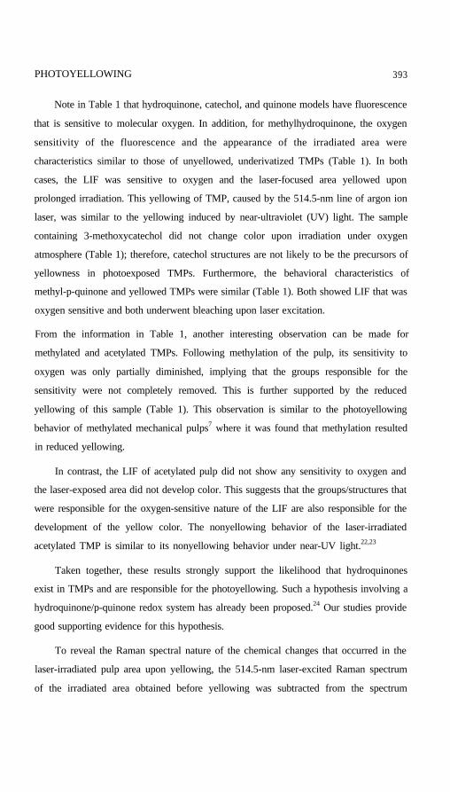
FIGURE 3. Laser-induced fluorescence (514.5 nm excited) of methyl-hydroquinone andits sensitivity to molecular oxygen: (a) spectrum of sample in air, (b) spectrum whennitrogen is flushed through the sample cell, (c) after 1 h nitrogen flushing, and(d) spectrum when nitrogen is being replaced by molecular oxygen. Molecular oxygenclearly quenches the fluorescence of the hydroquinone.
can give rise to LIF). However, there were still other lignin models, representing
quinones, hydroquinone, and catechol, that produced oxygen-sensitive LIF. Together
with the lignin models, different types of pulps and woods were examined for the
occurrence of this phenomenon. The sensitivity of LIF to molecular oxygen is included
in Table 1, column 3. For lignin models where LIF was found to be oxygen-sensitive, the
extent of quenching depended upon the concentration of the model. Therefore, the
sensitivity is reported only as “yes” instead of “+++” or “++” (wood, pulp, and lignin
section). Table 1 also indicates how the sample area appeared after extended (48 h or
more) irradiation in the presence of oxygen.
The yellowing reported in Table 1 was due to laser light, no thermal effects were
encountered. For a pulp-area that is being sampled, a temperature increase can be
measured by calculating the ratio of anti-Stokes to Stokes Raman lines. Using this
approach, the irradiated area was found to have remained at constant temperature.
392 AGARWAL
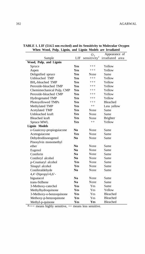
TABLE 1. LIF (514.5 nm excited) and its Sensitivity to Molecular OxygenWhen Wood, Pulp, Lignin, and Lignin Models are Irradiated
O 2 Appearance ofSample LIF sensitivitya irradiated area
Wood, Pulp, and LigninSpruceAspenDelignified spruceUnbleached TMPBH4-bleached TMPPeroxide-bleached TMPChemimechanical Pulp, CMPPeroxide-bleached CMPHydrogenated TMPPhotoyellowed TMPsMethylated TMPAcetylated TMPUnbleached kraftBleached kraftSpruce MWL
Lignin Modelsα-Guaicoxy-propioguiaconeAcetoguiaconeDehydrodiisoeugenolPinosylvin monomethyletherEugenolConiferinConiferyl alcoholp-Coumaryl alcoholSinapyl alcoholConiferaldehyde4,4’-Dipropyl-6,6’-biguaiacoltrans-Stilbene3-Methoxy-catecholMethylhydroquinone3-Methoxy-o-benzoquinoneMethoxy-p-benzoquinoneMethyl-p-quinone
NoNoYesYesYesYesYes
NoneNoneYesYesYesYes
SameSameSameYellowBleachedBleached
Yes Bleacheda+++ means highly sensitive, ++ means less sensitive.
YesYesYesYesYesYesYesYesYesYesYesYesYesYesYes
NoYesNo
NoNoNoNoYesYesNo
++++++
None+++++++++++++++++++++++
NoneNoneNone
++
NoneNoneNone
NoneNoneNoneNoneNoneNoneNone
YellowYellowSameYellowYellowYellowYellowYellowYellowBleachedLess yellowSameSameBrighterYellow
SameSameSame
SameSameSameSameSameSameSame
PHOTOYELLOWING 393
Note in Table 1 that hydroquinone, catechol, and quinone models have fluorescence
that is sensitive to molecular oxygen. In addition, for methylhydroquinone, the oxygen
sensitivity of the fluorescence and the appearance of the irradiated area were
characteristics similar to those of unyellowed, underivatized TMPs (Table 1). In both
cases, the LIF was sensitive to oxygen and the laser-focused area yellowed upon
prolonged irradiation. This yellowing of TMP, caused by the 514.5-nm line of argon ion
laser, was similar to the yellowing induced by near-ultraviolet (UV) light. The sample
containing 3-methoxycatechol did not change color upon irradiation under oxygen
atmosphere (Table 1); therefore, catechol structures are not likely to be the precursors of
yellowness in photoexposed TMPs. Furthermore, the behavioral characteristics of
methyl-p-quinone and yellowed TMPs were similar (Table 1). Both showed LIF that was
oxygen sensitive and both underwent bleaching upon laser excitation.
From the information in Table 1, another interesting observation can be made for
methylated and acetylated TMPs. Following methylation of the pulp, its sensitivity to
oxygen was only partially diminished, implying that the groups responsible for the
sensitivity were not completely removed. This is further supported by the reduced
yellowing of this sample (Table 1). This observation is similar to the photoyellowing
behavior of methylated mechanical pulps7 where it was found that methylation resulted
in reduced yellowing.
In contrast, the LIF of acetylated pulp did not show any sensitivity to oxygen and
the laser-exposed area did not develop color. This suggests that the groups/structures that
were responsible for the oxygen-sensitive nature of the LIF are also responsible for the
development of the yellow color. The nonyellowing behavior of the laser-irradiated
acetylated TMP is similar to its nonyellowing behavior under near-UV light.22,23
Taken together, these results strongly support the likelihood that hydroquinones
exist in TMPs and are responsible for the photoyellowing. Such a hypothesis involving a
hydroquinone/p-quinone redox system has already been proposed.24 Our studies provide
good supporting evidence for this hypothesis.
To reveal the Raman spectral nature of the chemical changes that occurred in the
laser-irradiated pulp area upon yellowing, the 514.5-nm laser-excited Raman spectrum
of the irradiated area obtained before yellowing was subtracted from the spectrum
394 AGARWAL
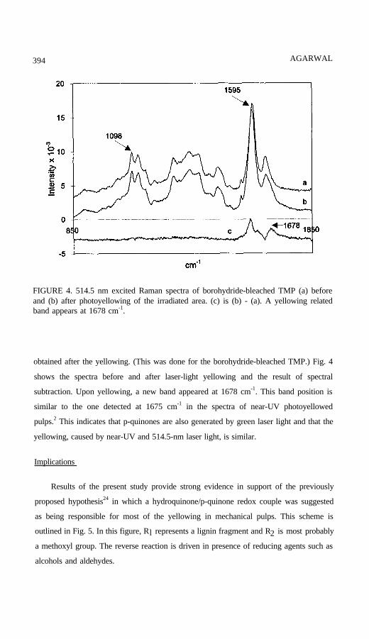
FIGURE 4. 514.5 nm excited Raman spectra of borohydride-bleached TMP (a) beforeand (b) after photoyellowing of the irradiated area. (c) is (b) - (a). A yellowing relatedband appears at 1678 cm-1.
obtained after the yellowing. (This was done for the borohydride-bleached TMP.) Fig. 4
shows the spectra before and after laser-light yellowing and the result of spectral
subtraction. Upon yellowing, a new band appeared at 1678 cm-1. This band position is
similar to the one detected at 1675 cm-1 in the spectra of near-UV photoyellowed
pulps.2 This indicates that p-quinones are also generated by green laser light and that the
yellowing, caused by near-UV and 514.5-nm laser light, is similar.
Implications
Results of the present study provide strong evidence in support of the previously
proposed hypothesis24 in which a hydroquinone/p-quinone redox couple was suggested
as being responsible for most of the yellowing in mechanical pulps. This scheme is
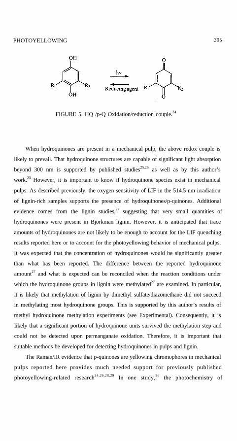
outlined in Fig. 5. In this figure, Rl represents a lignin fragment and R2 is most probably
a methoxyl group. The reverse reaction is driven in presence of reducing agents such as
alcohols and aldehydes.
PHOTOYELLOWING 395
FIGURE 5. HQ /p-Q Oxidation/reduction couple.24
When hydroquinones are present in a mechanical pulp, the above redox couple is
likely to prevail. That hydroquinone structures are capable of significant light absorption
beyond 300 nm is supported by published studies25,26 as well as by this author’s
work.23 However, it is important to know if hydroquinone species exist in mechanical
pulps. As described previously, the oxygen sensitivity of LIF in the 514.5-nm irradiation
of lignin-rich samples supports the presence of hydroquinones/p-quinones. Additional
evidence comes from the lignin studies,27 suggesting that very small quantities of
hydroquinones were present in Bjorkman lignin. However, it is anticipated that trace
amounts of hydroquinones are not likely to be enough to account for the LIF quenching
results reported here or to account for the photoyellowing behavior of mechanical pulps.
It was expected that the concentration of hydroquinones would be significantly greater
than what has been reported. The difference between the reported hydroquinone
amount27 and what is expected can be reconciled when the reaction conditions under
which the hydroquinone groups in lignin were methylated27 are examined. In particular,
it is likely that methylation of lignin by dimethyl sulfate/diazomethane did not succeed
in methylating most hydroquinone groups. This is supported by this author’s results of
methyl hydroquinone methylation experiments (see Experimental). Consequently, it is
likely that a significant portion of hydroquinone units survived the methylation step and
could not be detected upon permanganate oxidation. Therefore, it is important that
suitable methods be developed for detecting hydroquinones in pulps and lignin.
The Raman/IR evidence that p-quinones are yellowing chromophores in mechanical
pulps reported here provides much needed support for previously published
photoyellowing-related research24,26,28,29 In one study,26 the photochemistry of
396 AGARWAL
methoxy-p-benzoquinone and methoxy-hydroquinone in solid 2-hydroxy-propyl-
cellulose films and filter paper was studied. It was concluded that the quinone and
hydroquinone chromophores play a key role in the discoloration process of high-yield
pulps.
Results of yet another study28 indicated that the redox equilibrium between
hydroquinones and their corresponding p-quinones exist in pulps and, depending upon
the history of pulps (e.g., bleaching and acetylation), both hydroquinones and p-quinones
are capable of reactions that lead to yellowing. However, in this work,28 the hypothesis
of the authors that catechols and o-quinones are also responsible for such reactions is not
supported by our investigation, In this context, note that most o-quinones are red but
photoexposed pulps are yellow.
The photobehavior of methylated and acetylated mechanical pulps can be explained
on the basis of the present findings. It is known that methylation with dimethyl sulfate
and diazomethane does not render pulps completely photostable.7,8,30 Methylated pulps
yellow but at a lower rate. Although, to the best of our knowledge, no reports have been
published showing that hydroquinones are present in methylated pulps, based on the
finding that the LIF of a methylated pulp is oxygen-sensitive (Table 1), it is likely that
not all hydroquinone groups were methylated and that the remaining hydroquinone
groups were responsible for the yellowing of methylated pulp. The level of oxygen
sensitivity seems to be an indicator of the extent to which such groups remain
unmethylated. The difficulty with which hydroquinones undergo methylation was further
demonstrated when methyl hydroquinone was methylated using diazomethane and
methyl iodide (see Experimental). In both cases, only a very small amount of starting
material could be methylated. This being so in the case of a homogeneous reaction
mixture, it is expected that the reaction would be even less successful when the
hydroquinone groups are methylated in a heterogeneous lignocellulosic matrix. In
contrast, acetylation of TMP with pyridine/acetic anhydride easily acetylated
hydroquinone groups. This was evident both from the photostablity of the pulp23 and by
the lack of oxygen sensitivity of the LIF signal of the acetylated TMP (Table 1).
For methylated TMP, it has been estimated that the concentration of free phenolic
groups is about 0.76/100 C9 units.8 Although it is possible that these residual phenoxy
groups are of the hydroquinone type, it is not clear if the aminolysis and periodate
PHOTOYELLOWING 397
methods (used to detect phenoxy1 groups) can detect hydroquinone phenoxyls. If not,
then the concentration of hydroquinones could be even greater. In this context, it is
noteworthy that the periodate method fails to detect methoxyl-free phenolic and
hydroquinone units.31 Suitability of these methods for accurately detecting
hydroquinones needs to be established before phenoxy1 concentration data of methylated
pulps can be properly interpreted. Additional information in support of the possibility of
incomplete methylation of the hydroquinone groups comes from the analysis of
acetylated pulps. No free phenolic groups could be found in fully acetylated pulps.32
This suggests that acetylation is a better derivatizing agent for the hydroquinone
functionality.
The effect of yellowing inhibitors like polyethylene glycol, ascorbic acid, and
thioglycerol on the photoyellowing behavior of pulps can be explained by previously
suggested mechanisms,33-36 some of which are based on the chemistry of p-quinones/
hydroquinones.34 The present findings provide much needed evidence in support of
these reaction mechanisms, because the assumptions for the existence of p-quinones and
hydroquinones are no longer necessary.
It is a well recognized fact that after pulps are photoexposed, a band at about
1727 cm-1 is detected in their IR spectra.2,37 In the author’s work on spruce TMPs,2 the
intensity of this band was found to be linearly correlated with the post color number. It
was reported that this suggests that the sequence of chemical events leading to formation
of carbonyl groups (non-quinonoid) is related to the photoyellowing reactions. In this
author’s opinion, photoreduction of p-quinones in the presence of primary or secondary
alcohols, which act as hydrogen donors, can account for the formation of carbonyl
groups. In pulp, either carbohydrates or lignin can play the role of a hydrogen atom
donor. Such photochemical reactions are known to occur in p-quinones.38 In the
proposed hypothesis, shown in Fig. 6, a lignin fragment is indicated by the letter L, and
an OCH3 group is represented by R1. A primary or a secondary alcohol structure is
denoted by R2R3CHOH and is expected to be present in TMP.
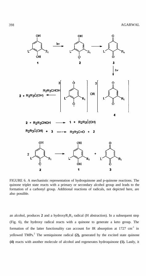
The scheme in Fig. 6 suggests that light drives both the hydroquinone-to-quinone
(forward) and the quinone-to-hydroquinone (reverse) reactions. Upon light exposure, the
former (1) is converted first to a semiquinone radical (2), then to 3, a quinone. The
quinone (3) absorbs light to produce a triplet state (4) or a diradical which, in presence of
398 AGARWAL
FIGURE 6. A mechanistic representation of hydroquinone and p-quinone reactions. Thequinone triplet state reacts with a primary or secondary alcohol group and leads to theformation of a carbonyl group. Additional reactions of radicals, not depicted here, arealso possible.
an alcohol, produces 2 and a hydroxyR2R3 radical (H abstraction). In a subsequent step
(Fig. 6), the hydroxy radical reacts with a quinone to generate a keto group. The
formation of the latter functionality can account for IR absorption at 1727 cm-1 in
yellowed TMPs.2 The semiquinone radical (2), generated by the excited state quinone
(4) reacts with another molecule of alcohol and regenerates hydroquinone (1). Lastly, it
PHOTOYELLOWING 399
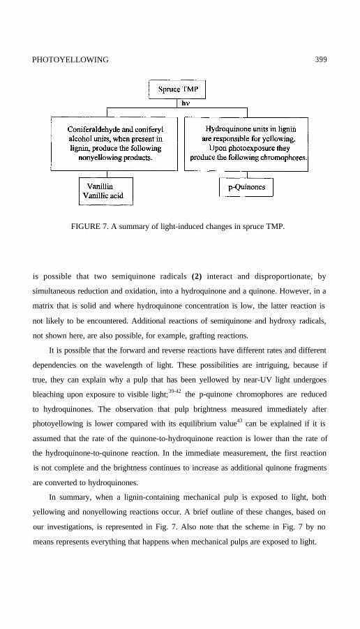
FIGURE 7. A summary of light-induced changes in spruce TMP.
is possible that two semiquinone radicals (2) interact and disproportionate, by
simultaneous reduction and oxidation, into a hydroquinone and a quinone. However, in a
matrix that is solid and where hydroquinone concentration is low, the latter reaction is
not likely to be encountered. Additional reactions of semiquinone and hydroxy radicals,
not shown here, are also possible, for example, grafting reactions.
It is possible that the forward and reverse reactions have different rates and different
dependencies on the wavelength of light. These possibilities are intriguing, because if
true, they can explain why a pulp that has been yellowed by near-UV light undergoes
bleaching upon exposure to visible light;39-42 the p-quinone chromophores are reduced
to hydroquinones. The observation that pulp brightness measured immediately after
photoyellowing is lower compared with its equilibrium value43 can be explained if it is
assumed that the rate of the quinone-to-hydroquinone reaction is lower than the rate of
the hydroquinone-to-quinone reaction. In the immediate measurement, the first reaction
is not complete and the brightness continues to increase as additional quinone fragments
are converted to hydroquinones.
In summary, when a lignin-containing mechanical pulp is exposed to light, both
yellowing and nonyellowing reactions occur. A brief outline of these changes, based on
our investigations, is represented in Fig. 7. Also note that the scheme in Fig. 7 by no
means represents everything that happens when mechanical pulps are exposed to light.
400 A G A R W A L
CONCLUSIONS
The Raman/IR band at 1675 cm-1 that was detected in photoyellowed TMP pulps
was assigned to p-quinone groups. It is suggested that such groups are formed from
corresponding hydroquinones present in pulps. Raman data did not provide support for
the possibility that o-quinone groups are generated upon photoyellowing.
For lignin-rich samples, using the molecular oxygen sensitivity of 514.5 nm excited
laser-induced fluorescence, information can be obtained to determine
if quinone/hydroquinone groups are present in the samples.
A hydroquinoneip-quinone redox system successfully accounted for a large number of
photoyellowing results.
ACKNOWLEDGMENTS
The author thanks Jim Bond, previously at Consolidated Papers, Wisconsin Rapids, WI,
and Ingegerd Forsskahl, Finnish Pulp and Paper Research Institute, Helsinki, Finland, for
providing mechanical pulps that were used in this study. The author is also grateful to
Sally Ralph for synthesizing some of the lignin models and methylating
methylhydroquinone. The assistance provided by John Ralph (USDA Dairy Forage
Laboratory, Madison, WI) in treating methylhydroquinone with diazomethane is much
appreciated. Finally, this author wishes to acknowledge contributions of Jim McSweeny
and Anita Cannon to the research reported here.
REFERENCES
PHOTOYELLOWING 401
402 AGARWAL


![© Copyright by ZPUE Silesia Sp. z o.o. 24kV.pdf · Depth of panel [mm] 1575 1675 1675 1575 1725 1600 1688 1675 Conformity with standards PN-EN 62271-200; PN-EN 62271-1 GOST 1516.3-96](https://img.pdfslide.us/doc/110x75/5f2d73def820c7329245cc02/-copyright-by-zpue-silesia-sp-z-oo-24kvpdf-depth-of-panel-mm-1575-1675.jpg)