Embed Size (px)
Citation preview
RESEARCH ARTICLE Open Access
Assessment of thoracic ultrasound incomplementary diagnosis and in follow upof community-acquired pneumonia (cap)Maria D’Amato1* , Gaetano Rea2, Vincenzo Carnevale3, Maria Arcangela Grimaldi4, Anna Rita Saponara5,Eric Rosenthal6, Michele Maria Maggi7, Lucia Dimitri8 and Marco Sperandeo9
Abstract
Background: Chest X-ray (CXR) is the primary diagnostic tool for community-acquired pneumonia (CAP). Someauthors recently proposed that thoracic ultrasound (TUS) could valuably flank or even reliably substitute CXR in thediagnosis and follow-up of CAP. We investigated the clinical utility of TUS in a large sample of patients with CAP, tochallenge the hypothesis that it may be a substitute for CXR.
Methods: Out of 645 consecutive patients with a CXR-confirmed CAP diagnosed in the emergency room of ourhospital over a three-years period, 510 were subsequently admitted to our department of Internal Medicine. Thesepatients were evaluated by TUS by a well-trained operator who was blinded of the initial diagnosis. TUS scans wereperformed both at admission and repeated at day 4-6th and 9-14th during stay.
Results: TUS identified 375/510 (73.5%) of CXR-confirmed lesions, mostly located in posterior-basal or mid-thoracicareas of the lungs. Pleural effusion was detected in 26.9% of patients by CXR and in 30.4% by TUS. TUS documentedthe change in size of the consolidated areas as follows: 6.3 ± 3.4 cm at time 0, 2.5 ± 1.8 at 4-6 d, 0.9 ± 1.4 at 9-14 d.Out of the 12 patients with delayed CAP healing, 7 of them turned out to have lung cancer.
Conclusions: TUS allowed to detect lung consolidations in over 70% of patients with CXR-confirmed CAP, but it gavefalse negative results in 26.5% of cases. Our longitudinal results confirm the role of TUS in the follow-up of detectablelesions. Thus, TUS should be regarded as a complementary and monitoring tool in pneumonia, instead of a primaryimaging modality.
Keywords: Community acquired pneumonia (CAP), Thoracic ultrasound (TUS), Complementary diagnostic tool, Follow-up
BackgroundCommunity-acquired pneumonia (CAP) is one of the mostcommon infectious diseases contributing to mortality andmorbidity worldwide [1]. Pneumonia exhibits a broad rangeof severity and induces many diagnostic and therapeuticchallenges [2]. According to the American College ofRadiology Appropriateness Criteria Expert Panel on Thor-acic Radiology, a chest X-ray (CXR) is usually appropriate inpatients with positive physical examination or risk factors forpneumonia [2, 3]. Standard CXR can identify pneumonia inalmost all areas of the lung, and also helps to define its
severity (multilobar or not) and the presence of complica-tions, such as cavitations [4], and co-morbidity (intrathoracicdiseases of the mediastinum and the heart). ComputedTomography (CT) is the golden standard for CAP diagnosis.However, it has a more limited role in daily clinical practiceand is mostly used in dubious cases or in the assessment ofcomplicated pneumonia, due to its higher cost and radiationexposure [2].Besides these traditional diagnostic tools, thoracic ultra-
sound (TUS) is gaining growing popularity as a possiblecomplementary tool for the diagnosis of pulmonary dis-eases. Some authors even went so far to say that TUScould represent an alternative tool for the diagnosis ofpulmonary diseases, due to its intrinsic characteristics.TUS is a non-invasive and radiation free method, readily
* Correspondence: [email protected] of Pneumology, “Federico II University”, AO “Dei Colli” MonaldiHospital, Via Domenico Fontana,134, Naples, ItalyFull list of author information is available at the end of the article
© The Author(s). 2017 Open Access This article is distributed under the terms of the Creative Commons Attribution 4.0International License (http://creativecommons.org/licenses/by/4.0/), which permits unrestricted use, distribution, andreproduction in any medium, provided you give appropriate credit to the original author(s) and the source, provide a link tothe Creative Commons license, and indicate if changes were made. The Creative Commons Public Domain Dedication waiver(http://creativecommons.org/publicdomain/zero/1.0/) applies to the data made available in this article, unless otherwise stated.
D’Amato et al. BMC Medical Imaging (2017) 17:52 DOI 10.1186/s12880-017-0225-5
available in many clinical departments and are alsosuitable for bedside exam in critical care settings. More-over, several studies by our and other groups showed thatTUS may provide useful information in different pleural-pulmonary diseases [5, 6] and particularly in CAP [7].Some authors even proposed TUS a possible substitutefor CXR for the diagnosis of CAP [8], at least in selectedgroups of patients as pregnant women, children, or when-ever radiation exposure should be limited. However, TUScannot visualize foci of pneumonia which are not adherentto the pleural surface or positioned where ultrasound can-not penetrate. It should also be stressed that until now thecurrent evidence-based guidelines on pneumonia diagno-sis and management do not include TUS [9]. On the otherhand, the TUS method could have a relevant complemen-tary role in CAP diagnosis and management. Consideringthat the roles of several putative biomarkers in the man-agement of patients with pneumonia are not definite, andcan not provide clues for the occurrence of superveningcomplications [3, 4, 9–11]. In this sense, TUS could be anoption to monitor the evolution of pneumonia focifollowing a CXR-confirmed diagnosis [12]. Despite thementioned results and the growing number of studies onthis matter, the debate on the role of TUS in the diagnosisand management of CAP is still ongoing.Our study was aimed to investigate the clinical perform-
ance of TUS in the primary diagnosis and in the manage-ment of CAP, as compared to standard CXR. We evaluatedthe following aspects: a) how many cases of CAP were con-firmed by TUS after clinical and CXR diagnosis, and b)how changes in TUS imaging appearances, from onset torecovery of CAP, could help identifying therapy failure orthe need to investigate an alternative diagnosis.
MethodsPatientsWe investigated all patients consecutively admitted toour department of Internal Medicine between Septem-ber 2013 and November 2016 with a CXR-confirmeddiagnosis of CAP. In the Emergency Room (ER) all pa-tients had undergone a conventional diagnostic work-up,including anamnesis, physical examination, laboratorytests and chest radiography. The diagnosis of CAP wasposed according to the American Thoracic Society(ATS) guidelines [13]. The CURB65 score [14] was usedto drive the allocation of patients as follows: severecases, with 3 or 4 criteria were referred to the intensivecare unit –ICU- or to our Internal Medicine department;non-severe cases, at moderate risk, with 1 or 2 criteriawere referred to ward or management as outpatients;non-severe cases, at low risk and with 0 criteria, werenot hospitalized [15]. All patients timely received empir-ical therapy, according to guidelines for the evaluationand treatment of CAP. Patients with either contingent
constraints or clinical conditions averting a completeTUS scan were conservatively excluded.All participants gave witnessed informed consent and
the study was approved by the ethics committee ofSUN-AO dei Colli- Naples-Italy.
Thoracic ultrasound examinationIn all patients admitted to our department, TUS was per-formed by a blinded operator, who was not aware of CXRresults, nor of the entire clinical-laboratory picture. Inorder to follow the evolution of CAP foci after therapy,TUS was performed in at least three repeated sittings: onday 0 (initial), between days 4 and 6 (intermediate), andbetween days 9 and 14 (final), according to a definedwork-up [12] (see Table 1 for details. TUS was performedat the bedside by a physician with at least 10 years ofultrasound experience. An Esaote Technos MPX, Twiceand My Lab30 Gold and Twice device (Genoa, Italy) usinga multi-frequency (3.5–5 MHz and 3–8 MHz) convexprobe and the pre-setting for thoracic ultrasound in Bmode was used (depth of images penetration: 7–14 cm;gain control: 40-50%; use of harmonic imaging; electronicfocus: pleural line). Each TUS was assessed for the num-ber, location, shape, size, and breath-dependent changes ofthe consolidation area attributable to pneumonia. Twomain sonographic patterns of lung consolidation weredefined: hypoechoic consolidation and mixed consolida-tion (hypoechoic and hyperechoic). The dimensions of theconsolidated areas are reported as the average betweenlongitudinal and transversal axes. Local and basal pleuraleffusions were also recorded. In addition, the presence ofspot and/or linear/arborescent hyperechoic images onTUS, improperly referred to as an air bronchogram, werealso recorded. The presence of artefacts (increased TUSB-line counts in the hemithorax with consolidation) wasnot considered in this study, because such artefacts are atbest a sensitive, but very non-specific sign of lung injury,common to many conditions [16, 17].
Table 1 TUS procedures
• Pulmonary thoracic assessment setting (including: tissue harmonicsimaging activation,the time gain compensation (TGC) should notexceed 50%, electronic imaging focus on the pleural line) usingmainly a 3.5-5 MHz convex probe EsaoteTechnosMpx, My Lab 30and Twice (Genova, Italy).
• Patients’ chests were examined posteriorly, lateral and anteriorly,while sitting. A few patients were examined in a semi-supine position,due to severe discomfort when sitting upright. Posteriorly, we opted forlongitudinal and transversal interscapular and paravertebral line scans.Anteriorly, the longitudinal and transversal interclavicular, parasternal lineand supraclavicular scans were used.Laterally, we used the longitudinaland transversal anterior, median and posterior line axillary views.
• The duration of ultrasound probe application in each site (posterior,lateral and anterior) was 4–5 min and overall time needed to completethe entire lung examination was 12–15 min.
D’Amato et al. BMC Medical Imaging (2017) 17:52 Page 2 of 8
The positive clinical evolution of CAP was detected byclinical assessment and CXR, and faced to the changesof TUS findings during stay and/or within 30 days on anoutpatient basis after discharge.
Assessment of inter-reader agreementVideo-clips recorded during TUS examinations (eachlasting a minimum of 3 min) were later reviewed by asecond examiner, who was blinded of all previous TUSfindings; clips for control assessment were randomlyassigned to one of two examiners with 20 years ofexperience in transthoracic ultrasound.
Statistical analysisThe results concerning the dimensions of TUS-detectable lesions are presented both as range and asmean ± SD. Inter-reader agreement was assessed usingSpearman’s coefficient for all parameters. The signifi-cance of changes in size of US-detectable lesions overtime was tested by Repeated Measures ANOVA. A pvalue of <0.05 was considered significant.In the clinical application phase of the study, a
repeated measurements ANOVA model (on basal, 4thday, and 8–10th day) was used to assess dimensionalchanges over time in lung consolidation areas and wascarried out via linear mixed models. Within-patientscorrelation was accounted for by an unstructured correl-ation type matrix [18]. Hochberg’s method was followedto obtain p-values corrected for multiple comparisons.P-values <0.05 were considered significant. All analyseswere performed using SAS Release 9.1 (SAS Institute,Cary, NC, USA). Inter-reader agreement was assessedusing Spearman’s coefficient for all parameters.
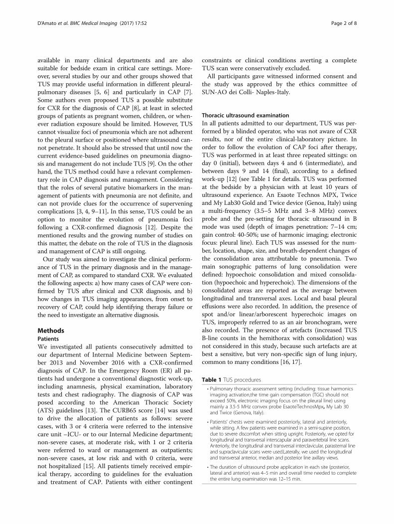
ResultsPatientsSeven hundred ninety-six consecutive adult patients pre-sented to the emergency room of the “Casa Sollievo dellaSofferenza” Hospital (San Giovanni Rotondo - Italy) withsymptoms and clinical/laboratory signs consistent with thediagnosis of CAP. Following the conventional diagnosticwork-up, in 736 of them the diagnosis was confirmed bychest X-ray (CXR), and 91 patients were discharged andmanaged as outpatients after the ER workup. Among the645 patients admitted to the hospital, 32 were referred tothe intensive care unit, and 613 to our department. Amongthe latter, 103 patients were excluded (see methods). Fivehundred ten patients admitted to our department were fi-nally studied (see Fig. 1). The demographic and clinical char-acteristics of investigated patients are reported in Table 2.
Initial TUS findingsThe topographic distribution of lung consolidation de-tected by TUS is indicated in Table 3. TUS imaging was
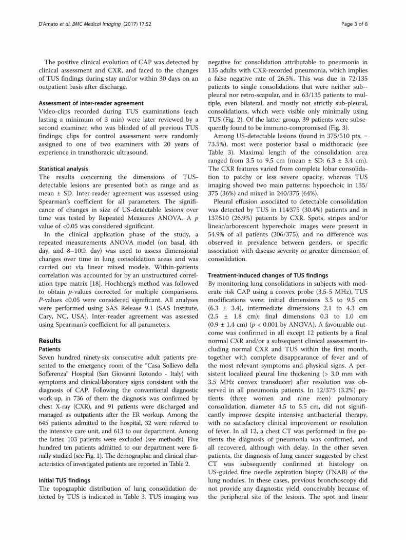
negative for consolidation attributable to pneumonia in135 adults with CXR-recorded pneumonia, which impliesa false negative rate of 26.5%. This was due in 72/135patients to single consolidations that were neither sub--pleural nor retro-scapular, and in 63/135 patients to mul-tiple, even bilateral, and mostly not strictly sub-pleural,consolidations, which were visible only minimally usingTUS (Fig. 2). Of the latter group, 39 patients were subse-quently found to be immuno-compromised (Fig. 3).Among US-detectable lesions (found in 375/510 pts. =
73.5%), most were posterior basal o midthoracic (seeTable 3). Maximal length of the consolidation arearanged from 3.5 to 9.5 cm (mean ± SD: 6.3 ± 3.4 cm).The CXR features varied from complete lobar consolida-tion to patchy or less severe opacity, whereas TUSimaging showed two main patterns: hypoechoic in 135/375 (36%) and mixed in 240/375 (64%).Pleural effusion associated to detectable consolidation
was detected by TUS in 114⁄375 (30.4%) patients and in137⁄510 (26.9%) patients by CXR. Spots, stripes and/orlinear/arborescent hyperechoic images were present in54.9% of all patients (206/375), and no difference wasobserved in prevalence between genders, or specificassociation with disease severity or greater dimension ofconsolidation.
Treatment-induced changes of TUS findingsBy monitoring lung consolidations in subjects with mod-erate risk CAP using a convex probe (3.5-5 MHz), TUSmodifications were: initial dimensions 3.5 to 9.5 cm(6.3 ± 3.4), intermediate dimensions 2.1 to 4.3 cm(2.5 ± 1.8 cm); final dimensions 0.3 to 1.0 cm(0.9 ± 1.4 cm) (p < 0.001 by ANOVA). A favourable out-come was confirmed in all except 12 patients by a finalnormal CXR and/or a subsequent clinical assessment in-cluding normal CXR and TUS within the first month,together with complete disappearance of fever and ofthe most relevant symptoms and physical signs. A per-sistent localized pleural line thickening (> 3.0 mm with3.5 MHz convex transducer) after resolution was ob-served in all pneumonia patients. In 12/375 (3.2%) pa-tients (three women and nine men) pulmonaryconsolidation, diameter 4.5 to 5.5 cm, did not signifi-cantly improve despite intensive antibacterial therapy,with no satisfactory clinical improvement or resolutionof fever. In all 12, a chest CT was performed: in five pa-tients the diagnosis of pneumonia was confirmed, andall recovered, although with delay. In the other sevenpatients, the diagnosis of lung cancer suggested by chestCT was subsequently confirmed at histology onUS-guided fine needle aspiration biopsy (FNAB) of thelung nodules. In these cases, previous bronchoscopy didnot provide any diagnostic yield, conceivably because ofthe peripheral site of the lesions. The spot and linear
D’Amato et al. BMC Medical Imaging (2017) 17:52 Page 3 of 8
Fig. 1 Flow-chart of the main results
Table 2 Characteristics of the included patients (n = 510)
Age (years), (mean ± SD) Range 32-78 (58.4 ± 14.7)
Gender (M⁄F) 281/229
CURB 65 2.4 ± 0.6
Mean hospital stay 8.9 ± 2.5 days
Consolidated areas identified by TUS 375/510
Size of Consolidated areas (cm) 6.3 ± 3.4
Comorbidity(more than one in 60 pts) 455/510 pts.(89.2%)
Diabetes mellitus 96 (18.8%)
COPD 107 (21%)
Pulmonary fibrosis 28 (5.5%)
Heart failure (III-IV NYA) 80 (15.7%)
Chronic kidney diseases 12(2.3%)
Oncological diseases 68 (13.3%)
Coronary disease 56(11%)
Table 3 Topographic distribution detected at lung ultrasoundexamination of pneumonia patients (n = 375)
Localization of pulmonary focus Number of patients (n = 375)
Posterior-basal 202 (54%)
Posterior mid-thoracic 60 (16%)
Posterior-lateral mid-thoracic 75 (20%)
Anterior mid-thoracic 15 (4%)
Para-cardiac 12 (3.2%)
Anterior apical 6 (1.6%)
Posterior apical 3 (0.8%)
Multiple consolidation 31 (8.3%)
D’Amato et al. BMC Medical Imaging (2017) 17:52 Page 4 of 8
hyperechoic images were observed in 5/7 of the patientswith a subsequent diagnosis of lung cancer.
Inter-reader agreementInter-reader agreement was excellent (Spearman’s coeffi-cient ≥ 0.90 for all parameters).
Positive TUS findings in patients with negative CXRimagingPneumonia that was not preliminarily CXR-proven butwas suggested by the clinical picture and by the finding ofTUS areas attributable to consolidation was identified inten patients. They showed small sub-pleural consolidationareas of 1.0 to 1.5 cm; these cases did not progress towardgreater extension of consolidation, and those considereddoubtful for pneumonia were excluded from the subse-quent data analysis of TUS imaging distribution.
DiscussionOur current results, obtained from the largest everinvestigated series, substantially confirm the previousones we obtained from an independent series of inpa-tients [12]. In most cases of CAP, TUS examination de-tects the sites of inflammation, which have typicalalthough not specific patterns. TUS allows the measure-ment of dimensions of the pulmonary focus before andafter medical therapy, which implies the possible use of
this tool to monitor treatment efficacy. The present dataalso confirm the higher sensibility of TUS in identifyingpleural effusion and its role to facilitate fluid drainage.Accordingly, in US-detectable cases TUS appears tovaluably integrate the diagnostic information obtainedfrom a CXR alone [19]. Due to these and other reasons,several authors went so far to even suggest that CXRcan be replaced by TUS in the clinic to identify CAP[20, 21]. However, our current results mandate extremecaution on this matter.Actually, most cases of CAP (around 80% of cases) are
subpleural (that is adherent to the 70% of pleura visual-ized by TUS) and begin peripherally. This explains whyCAP is also most often visible by TUS [13, 22]. In ourseries, 73.5% of CXR-positive lung consolidations due toCAP were also visible with TUS, being localized in sub-pleural areas (see details on location in Table 3). In thesesites, hypoechoic and mixed (hypoechoic and hypere-choic) lesions corresponded to the foci of pneumoniaidentified by CXR. This result confirms in a large cohortof patients the reliability of TUS imaging to corroboratethe diagnosis of CAP obtained from CXR. However, itshould also be stressed as more than one out of fourcases of CAP (26.5%) were not detectable by TUS, evenif performed with the highest methodological accuracy(standardized complete scan, technical accuracy, involve-ment of only highly experienced operators, and so on)
Fig. 2 Right lobar pneumonia. Lung consolidation is well defined by CXR (top left) and CT (bottom left); by TUS, pleural effusion is easily identified(top right) but only a very small density and adherent to pleura pneumonia is visible (blue arrows) (bottom right)
D’Amato et al. BMC Medical Imaging (2017) 17:52 Page 5 of 8
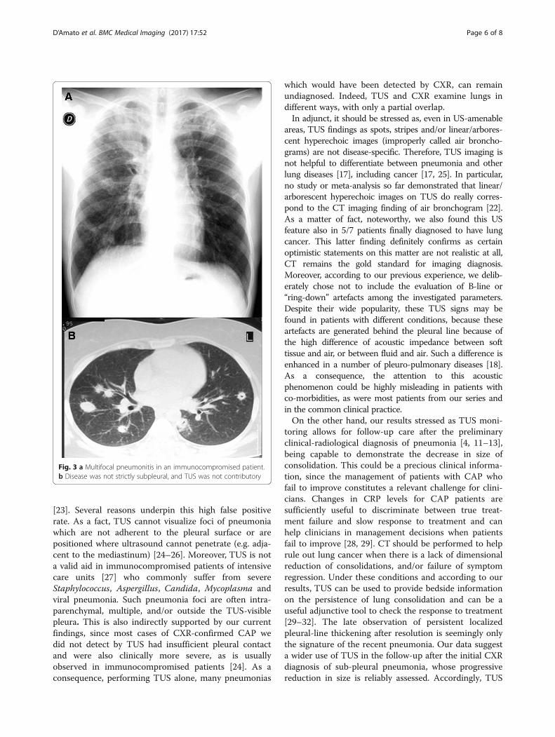
[23]. Several reasons underpin this high false positiverate. As a fact, TUS cannot visualize foci of pneumoniawhich are not adherent to the pleural surface or arepositioned where ultrasound cannot penetrate (e.g. adja-cent to the mediastinum) [24–26]. Moreover, TUS is nota valid aid in immunocompromised patients of intensivecare units [27] who commonly suffer from severeStaphylococcus, Aspergillus, Candida, Mycoplasma andviral pneumonia. Such pneumonia foci are often intra-parenchymal, multiple, and/or outside the TUS-visiblepleura. This is also indirectly supported by our currentfindings, since most cases of CXR-confirmed CAP wedid not detect by TUS had insufficient pleural contactand were also clinically more severe, as is usuallyobserved in immunocompromised patients [24]. As aconsequence, performing TUS alone, many pneumonias
which would have been detected by CXR, can remainundiagnosed. Indeed, TUS and CXR examine lungs indifferent ways, with only a partial overlap.In adjunct, it should be stressed as, even in US-amenable
areas, TUS findings as spots, stripes and/or linear/arbores-cent hyperechoic images (improperly called air broncho-grams) are not disease-specific. Therefore, TUS imaging isnot helpful to differentiate between pneumonia and otherlung diseases [17], including cancer [17, 25]. In particular,no study or meta-analysis so far demonstrated that linear/arborescent hyperechoic images on TUS do really corres-pond to the CT imaging finding of air bronchogram [22].As a matter of fact, noteworthy, we also found this USfeature also in 5/7 patients finally diagnosed to have lungcancer. This latter finding definitely confirms as certainoptimistic statements on this matter are not realistic at all,CT remains the gold standard for imaging diagnosis.Moreover, according to our previous experience, we delib-erately chose not to include the evaluation of B-line or“ring-down” artefacts among the investigated parameters.Despite their wide popularity, these TUS signs may befound in patients with different conditions, because theseartefacts are generated behind the pleural line because ofthe high difference of acoustic impedance between softtissue and air, or between fluid and air. Such a difference isenhanced in a number of pleuro-pulmonary diseases [18].As a consequence, the attention to this acousticphenomenon could be highly misleading in patients withco-morbidities, as were most patients from our series andin the common clinical practice.On the other hand, our results stressed as TUS moni-
toring allows for follow-up care after the preliminaryclinical-radiological diagnosis of pneumonia [4, 11–13],being capable to demonstrate the decrease in size ofconsolidation. This could be a precious clinical informa-tion, since the management of patients with CAP whofail to improve constitutes a relevant challenge for clini-cians. Changes in CRP levels for CAP patients aresufficiently useful to discriminate between true treat-ment failure and slow response to treatment and canhelp clinicians in management decisions when patientsfail to improve [28, 29]. CT should be performed to helprule out lung cancer when there is a lack of dimensionalreduction of consolidations, and/or failure of symptomregression. Under these conditions and according to ourresults, TUS can be used to provide bedside informationon the persistence of lung consolidation and can be auseful adjunctive tool to check the response to treatment[29–32]. The late observation of persistent localizedpleural-line thickening after resolution is seemingly onlythe signature of the recent pneumonia. Our data suggesta wider use of TUS in the follow-up after the initial CXRdiagnosis of sub-pleural pneumonia, whose progressivereduction in size is reliably assessed. Accordingly, TUS
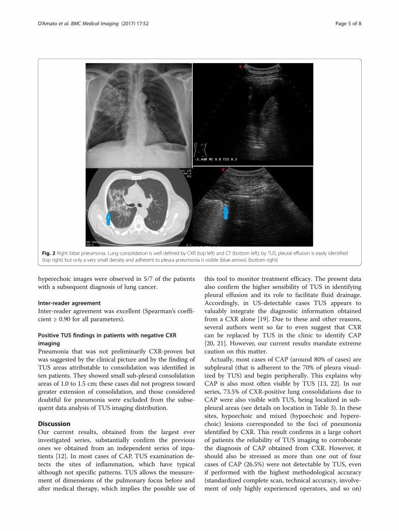
Fig. 3 a Multifocal pneumonitis in an immunocompromised patient.b Disease was not strictly subpleural, and TUS was not contributory
D’Amato et al. BMC Medical Imaging (2017) 17:52 Page 6 of 8
could also decrease the need to repeat radiologicalprocedures, particularly in the follow-up of pregnantpatients or in the follow-up of patients not requiringhospital admission. When scheduling follow-up on anoutpatient basis, TUS is seemingly a less expensiveprocedure and it is already successfully used to monitorother conditions and diseases [33].Discordant results have been reported on TUS-positive
and CXR–negative by other authors [2, 3, 13, 22]. Suchdiscrepancies may result from Rx–negative small lungconsolidations detected exclusively on TUS. Alternatively,they may stem from the different relationship betweenlung consolidations and the pleura, or from an alternativediagnosis (not CAP), or from the presence of sub-segmental lung focal areas of atelectasis beyond terminalbronchioles [23].Our study has several strengths. Firstly, we prospectively
validated our previous results by studying an independentlarge series of unselected patients. All patients of ourseries were managed in a substantially coherent way, atvariance with previous studies suffering from a widevariability in criteria of admission [31], management, anddischarge of pneumonia patients without follow up.Our study has some limits, too. In fact, we tried to mimic
the common practice through the unselective inclusion of allpatients coming to the emergency room of our hospital andbeing suitable for subsequent (repeated) observation in ourInternal Medicine department. Such a design was aimed toreduce possible observational bias, but this way we excludedpatients with an insufficient number of TUS assessments,which implied the exclusion of patients admitted to otherunits, including ICU. Accordingly, the number of recruitedpatients with more severe disease and with likely more sig-nificant co-morbidities and further complications was lower.On the other hand, the exclusion of those managed as out-patients also excluded less severe cases. In addition, all TUSwere performed by a highly trained staff and this couldundermine the generalizability of our results. Actually, TUSrequires a technically experienced operator and appropriatemachine settings [11, 12, 30]. The clinical assessment ofTUS consolidation mostly depends on the subjective expert-ise of the ultrasound operator, as in most sonographic diag-noses. Interpretation of TUS is not the easiest component ofany ultrasound course and has many pitfalls, mostly forfalse-negative results. This is a risk increased by over-confidence [21]. As a fact, the negative ethical and potentialmedico-legal implications of omitting a CXR (co-morbidityassociated, intraparenchimal not subpleural consolidationand therefore incorrect or incomplete diagnosis), particularlywhen addressing the therapeutic choices, are evident [34].
ConclusionIn conclusion, we exclude that TUS could reliably re-place CXR, and we confirm that the assessment of
physical signs, CXR, and biomarkers such as procalcito-nin and CRP, remain the pillars for the diagnosis ofpneumonia. On the other hand, TUS represents a highlyvaluable complementary imaging procedure, which canbe performed at bedside, and easily repeated after theinitial assessment. Therefore we recommend its use as acomplementary and monitoring tool.
AcknowledgmentsNot applicable.
FundingNot applicable.
Availability of data and materialsThe datasets used and/or analyzed during the current study are availablefrom the corresponding author on reasonable request.
Authors’ contributionsAll authors contributed to the conception and design of the study, as to theacquisition, analysis and interpretation of data. They also contributed indrafting and critically revising the manuscript, and read and approved thefinal manuscript, so take public responsibility of its content. All authors areresponsible for accuracy or integrity any part of the work. In particular theprominent contribution was as follows: MD and MS: wrote the manuscript.VC: made substantial contributions to conception and design. MS, MAG, GRmade substantial contribution in acquisition of data, and data analysis andinterpretation. ER, MMM: made substantial contributions to data analysis andinterpretation. AS and LD: was involved in critically revising the manuscriptfor intellectual content. MD, GR e MS: were involved in data acquisition anddatabase synthesis and cleaning. All authors read and approved the finalmanuscript.
Ethics approval and consent to participateThe study was approved by the ethics committee of SUN-AO dei Colli- Naples-Italy (protocol n°054-2015) and all participants gave witnessed informed consent.
Consent for publicationNot applicable.
Competing interestsThe authors declare that they have no competing interests.
Publisher’s NoteSpringer Nature remains neutral with regard to jurisdictional claims in publishedmaps and institutional affiliations.
Author details1Department of Pneumology, “Federico II University”, AO “Dei Colli” MonaldiHospital, Via Domenico Fontana,134, Naples, Italy. 2Department of Radiology,AO “Dei Colli” Monaldi Hospital, Naples, Italy. 3Unit of Internal Medicine,“Casa Sollievo della Sofferenza” Hospital, IRCCS, San Giovanni Rotondo (FG),Italy. 4Unit of Internal Medicine and Pneumology, “Casa Sollievo dellaSofferenza” Hospital, IRCCS, San Giovanni Rotondo (FG), Italy. 5Unit of InternalMedicine, Local Health Service, Potenza, Italy. 6Department of InternalMedicine, Hospital Archet 1, Nice, France. 7Unit of Emergency Medicine,“Casa Sollievo della Sofferenza” Hospital, IRCCS, San Giovanni Rotondo (FG),Italy. 8Unit of Pathology, “Casa Sollievo della Sofferenza” Hospital, IRCCS, SanGiovanni Rotondo (FG), Italy. 9Unit of Interventional and DiagnosticUltrasound of Internal Medicine, “Casa Sollievo della Sofferenza” Hospital,IRCCS, San Giovanni Rotondo (FG), Italy.
Received: 9 April 2017 Accepted: 23 August 2017
References1. Spellberg B. Community-acquired pneumonia. N Engl J Med. 2014;370:1861–2.2. Dean NC, Jones JP, Aronsky D, Brown S, Vines CG, Jones BE, Allen T.
Hospital admission decision for patients with community-acquired
D’Amato et al. BMC Medical Imaging (2017) 17:52 Page 7 of 8
pneumonia: variability among physicians in an emergency department. AnnEmerg Med. 2012;59:35–41.
3. Gibot S, Béné MC, Noel R, Massin F, Guy J, Cravoisy A, et al. Combinationbiomarkers to diagnose sepsis in the critically ill patient. Am J Respir CritCare Med. 2012;186:65–71.
4. Kirsch J, Ramirez J, Mohammed TL, Amorosa JK, Brown K, Dyer DS, et al.ACR appropriateness criteria® acute respiratory illness in immunocompetentpatients. J Thorac Imaging. 2011;26:W42–4.
5. Reissig A, Gorg C, Mathis G. Transthoracic sonography in the diagnosis ofpulmonary diseases: a systematic approach. Ultraschall Med. 2009;30:438–54.
6. Sartori S, Tombesi P. Emerging roles for transthoracic ultrasonography inpulmonary diseases. World J Radiol. 2010;2:203–14.
7. Gehmacher O, Mathis G, Kopf A, Scheier M. Ultrasound imaging ofpneumonia. Ultrasound Med Biol. 1995;21:1119–22.
8. Thomas Berlet Thoracic ultrasound for the diagnosis of pneumonia inadults: a meta-analysis. Respir Res. 2015;16:89.
9. Rothrock SG, Green SM, Fanelli JM, Cruzen E, Costanzo KA, Pagane J. Dopublished guidelines predict pneumonia in children presenting to an urbanED? Pediatr Emerg Care. 2001;17:240–3.
10. Wunderink RG, Waterer GW. Update in pulmonary infections 2010. Am JRespir Crit Care Med. 2011;184:186–90.
11. Sperandeo M, Filabozzi P, Varriale A, Carnevale V, Piattelli ML, Sperandeo G,Brunetti E, Decuzzi M. Role of thoracic ultrasound in the assessment ofpleural and pulmonary diseases. J Ultrasound. 2008;11:39–46.
12. Sperandeo M, Carnevale V, Muscarella S, Sperandeo G, Varriale A, Filabozzi P,Piattelli ML, D'Alessandro V, Copetti M, Pellegrini F, Dimitri L, Vendemiale G.Clinical application of transthoracic ultrasonography in inpatients withpneumonia. Eur J Clin Invest. 2011;41:1–7.
13. American Thoracic Society; Infectious Diseases Society of America.Guidelines for the management of adults with hospital-acquired, ventilator-associated, and healthcare-associated pneumonia. Am J Respir Crit CareMed. 2005;171:388–416.
14. Liu B, Yin Q, Chen YX, Zhao YZ, Li CS. Role of Presepsin (sCD14-ST) and theCURB65 scoring system in predicting severity and outcome of community-acquired pneumonia in an emergency department. Respir Med. 2014;108:1204–13.
15. Lim WS, van der Eerden MM, Laing R, Boersma WG, Karalus N, Town GI, et al.Defining community acquired pneumonia severity on presentation to hospital:an international derivation and validation study. Thorax. 2003;58:377–82.
16. Zanforlin A, Smargiassi A, Inchingolo R, Sher S, Ramazzina E, Corbo GM, et al. B-lines: to count or not to count? JACC Cardiovasc Imaging. 2014;7:635–6.
17. Trovato GM, Sperandeo M. Sounds, ultrasounds, and artifacts: which clinicalrole for lung imaging? Am J Respir Crit Care Med. 2013;187:780–1.
18. Sperandeo M, Varriale A, Sperandeo G, et al. Assessment of ultrasoundacoustic artifacts in patients with acute dyspnea: a multicenter study. ActaRadiol. 2012;53:885–92.
19. Miyashita N, Akaike H, Teranishi H, Nakano T, Ouchi K, Okimoto N. Chestcomputed tomography for the diagnosis of mycoplasma pneumoniaeinfection. Respirology. 2014;19:144–5.
20. Ye X, Xiao H, Chen B, Zhang S. Accuracy of lung ultrasonography versuschest radiography for the diagnosis of adult community-acquiredpneumonia: review of the literature and meta-analysis. PLoSOne. 2015;24:10.
21. Jones BP, Tay ET, Elikashvili I, Sanders JE, Paul AZ, Nelson BP, et al. Feasibilityand safety of substituting lung ultrasonography for chest radiography whendiagnosing pneumonia in children: a randomized controlled trial. Chest.2016;150:131–8.
22. Sperandeo M, Filabozzi P, Carnevale V. Ultrasound diagnosis of ventilator-associated pneumonia: a not-so-easy issue. Chest. 2016;149:1350–1.
23. Sperandeo M, Rea G, Santantonio A, Carnevale V. Lung ultrasound indiagnosis of transient tachypnea of the newborn: limitations and pitfalls.Chest. 2016;150:977–8.
24. Craven DE, Palladino R, McQuillen DP. Healthcare-associated pneumonia inadults: management principles to improve outcomes. Infect Dis Clin NorthAm. 2004;18:939–62.
25. Jeon KN, Bae K, Park MJ, Choi HC, Shin HS, Shin S, et al. US-guidedtransthoracic biopsy of peripheral lung lesions: pleural contact lengthinfluences diagnostic yield. Acta Radiol. 2014;55:295–301.
26. Zhang Y, Qiang JW, Shen Y, Ye JD, Zhang J, Zhu L. Using air bronchogramson multi-detector CT to predict the invasiveness of small lungadenocarcinoma. Eur J Radiol. 2016;85:571–7.
27. Piccoli M, Trambaiolo P, Salustri A, Cerquetani E, Posteraro A, Pastena G, etal. Bedside diagnosis and follow-up of patients with pleural effusion by a
hand-carried ultrasound device early after cardiac surgery. Chest. 2005;128:3413–20.
28. Liapikou A, Ferrer M, Polverino E, Balasso V, Esperatti M, Piñer R, et al. Severecommunity-acquired pneumonia: validation of the Infectious DiseasesSociety of America/American Thoracic Society guidelines to predict anintensive care unit admission. Clin Infect Dis. 2009;48:377–85.
29. Catalano D, Trovato G, Sperandeo M, Sacco MC. Lung ultrasound inpediatric pneumonia. The persistent need of chest X-rays. Pediatr Pulmonol.2014;49:617–8.
30. Catalano D, Trovato FM, Pirri C, Trovato GM. Outpatient diagnosis andtherapeutic units linked with ED referrals: a sustainable quality-centeredapproach. Am J Emerg Med. 2013;31:1612.
31. Sperandeo M, Rotondo A, Guglielmi G, Catalano D, Feragalli B, Trovato GM.Transthoracic ultrasound in the assessment of pleural and pulmonarydiseases: use and limitations. Radiol Med. 2014;119:729–40.
32. Ruiz-González A, Falguera M, Porcel JM, Martínez-Alonso M, Cabezas P, GeijoP, et al. C-reactive protein for discriminating treatment failure from slowresponding pneumonia. Eur J Intern Med. 2010;21:548–52.
33. Medford AR. Chest ultrasonography as a replacement for chest radiographyfor community-acquired pneumonia. Chest. 2013;143:877–8.
34. Goss CH, Rubenfeld GD, Park DR, Sherbin VL, Goodman MS, Root RK. Cost andincidence of social comorbidities in low-risk patients with community-acquiredpneumonia admitted to a public hospital. Chest. 2003;124:2148–55.
• We accept pre-submission inquiries
• Our selector tool helps you to find the most relevant journal
• We provide round the clock customer support
• Convenient online submission
• Thorough peer review
• Inclusion in PubMed and all major indexing services
• Maximum visibility for your research
Submit your manuscript atwww.biomedcentral.com/submit
Submit your next manuscript to BioMed Central and we will help you at every step:
D’Amato et al. BMC Medical Imaging (2017) 17:52 Page 8 of 8