Embed Size (px)
Citation preview
Original Article
Copyright © 2014 Revista Latino-Americana de EnfermagemThis is an Open Access article distributed under the terms of the Creative Commons Attribution Non-Commercial License (CC BY-NC).This license lets others distribute, remix, tweak, and build upon your work non-commercially, and although their new works must also acknowledge you and be non-commercial, they don’t have to license their derivative works on the same terms.
Rev. Latino-Am. Enfermagem2014 Mar.-Apr.;22(2):277-85DOI: 10.1590/0104-1169.3238.2413
www.eerp.usp.br/rlae
Corresponding Author:
Percy Nohama Pontifícia Universidade Católica do Paraná. Escola PolitécnicaRua Imaculada Conceição, 1155Bairro: Prado VelhoCEP: 80215-901, Curitiba, PR, BrasilE-mail: [email protected]
Anita Batista dos Santos Heberle1
Marcos Antônio Muniz de Moura2
Mauren Abreu de Souza3
Percy Nohama4
1 MSc, RN, Hospital Universitário Regional de Maringá, Universidade Estadual de Maringá, Maringá, PR, Brazil.2 Doctoral student, Universidade Tecnológica Federal do Paraná, Curitiba, PR, Brazil. Assitant Professor, Escola Politécnica, Pontifícia Universidade
Católica do Paraná, Curitiba, PR, Brazil.3 Post-doctoral fellow, Universidade Tecnológica Federal do Paraná, Curitiba, PR, Brasil. Assistant Professor, Escola de Educação e Humanidades,
Pontifícia Universidade Católica do Paraná, Curitiba, PR, Brazil.4 PhD, Full Professor, Escola Politécnica, Pontifícia Universidade Católica do Paraná, Curitiba, PR, Brazil.
Objective: to evaluate techniques of massage and pumping in the treatment of postpartum
breast engorgement through thermography. Method: the study was conducted in the Human
Milk Bank of a hospital in Curitiba, Brazil. We randomly selected 16 lactating women with
engorgement with the classification lobar, ampullary and glandular, moderate and intense. We
compared the differential patterns of temperature, before and after the treatment by means of
massage and pumping. Results: we found a negative gradient of 0.3°C of temperature between
the pre- and post-treatment in the experimental group. Breasts with intense engorgement
were 0.7°C warmer when compared with moderate engorgement. Conclusion: massage and
electromechanical pumping were superior to manual methods when evaluated by thermography.
REBEC: U1111-1136-9027.
Descriptors: Thermography; Lactation Disorders; Breast Feeding; Nursing.
Assessment of techniques of massage and pumping in the
treatment of breast engorgement by thermography

278
www.eerp.usp.br/rlae
Rev. Latino-Am. Enfermagem 2014 Mar.-Apr.;22(2):277-85.
Introduction
Mammary engorgement, in the breastfeeding
process, is the first symptom encountered by the
mother in the self-regulation of the physiology of
lactation. Sometimes, the breasts produce a quantity
of milk secretion greater than the demand of the child,
becoming so full and taut they are called “gable chest”(1),
one of the factors of interruption of the exclusive
breast feeding (EBF) in children under four months of
life(2). Breast engorgement arises from the increased
vascularity and accumulation of milk and, secondarily,
by lymphatic and vascular congestion(3). Signals arise
such as pain, interstitial edema, increase in the volume
of the breasts(4), shiny skin, flattened nipples, with or
without accompanying diffuse and reddened areas(3),
elevation of the body temperature including a febrile
state. After the breasts are emptied(5), a reduction in
temperature occurs.
Thermal analysis using images was described by
Lehmann, in 1877, and has been used in the health
area(6). Thermography is a technique widely used in
the industrial sector and still incipient in the medical
field even though it is innocuous. It is configured as
a method of capturing infrared radiation emitted
by a body, determining its temperature(7). Starting
from the thermographic inspection, differential
patterns of thermal distribution are observed,
providing information related to a particular process
that is occurring in the body(8), establishing itself
up to the present as an auxiliary component for
diagnosis(9).
The temperature of the breast undergoes changes
due to several factors, among which are the endogenous,
which can be studied by means of a thermogram(10). In
the research literature, temperature changes in breasts
of pregnant women have been observed(11), and hormone
dosage in lactating women with engorged breasts, in
which it was found that systems with high sensitivity
could provide early diagnosis, both of postpartum
breast discomfort as well as the evolution of breast
engorgement (BE)(12).
Both medicine and nursing use physical examination
as a link between art and science. Based on the biological
sciences, data are obtained from appropriate tools and
the techniques of inspection, palpation and auscultation.
Additionally, subjective information is derived from the
interview. Thus, the data collected subsidize clinical
reasoning in the formulation of the diagnosis(13). Although
well known, breast engorgement is understudied(14).
The changes in temperature of the breasts are clearly
evidenced, in the case of engorgement, and should be
considered for making the diagnosis and in possibilities
for treatment. Until now, the literature has not
pronounced the existence of this parameter in the study
of possible treatments of this condition that affects the
nursing mother.
Regarding the application of thermography in the
puerperal breast, the encountered studies were old(11-
12) and some factors contributed to the discrediting
of thermography in the 1970s, such as the lack of
controlled studies, indertemination of the sensitivity
and specificity of the method and the high rate of errors
in the interpretation of images. Starting in the 1990s,
with the technological advancement of computerized
infrared cameras, especially their thermal resolution
that can vary from 0.08mK to 0.03mK at 30ºC,
interest was once again aroused for this method in
the clinical area(6).
For breastfeeding women with BE, the physical
exam with palpation generally enables identificaton of
types of engorgement that most commonly affect these
women(15). However, the utilization of technological
resources overlaps the subjective nature of palpation in
the clnical exam(16).
Regarding the emptying of the breasts, according
to existing technical standards for human milk banks
(HMB), the Brazilian Network of Human Milk Banks
(RBBLH), the extraction should preferably be done
by manual pumping, accepting resources such as
suction pumps preceded by massage(17). Actually,
the most utilized techniques for treatment of breast
engorgement are primary, that is, manual techniques,
either by the lactating woman herself, a third party,
or someone specialized, such as the nurse. Such
a procedure is time consuming and stressful for the
person performing it. Generally, these professionals
remain seated and leaning in the same position for
a long time. The spine and the wrists are strained
while the hands are moving doing the massage. Also
the legs and arms become tired, aching and signs of
fatigue emerge(18).
Therefore, and targeting both to minimize
the ergonomic working conditions of the nursing
staff, as well as to bring about elements that denote
potential advantages in the use of new technologies,
this study aimed to evaluate the application of the
mechanical techniques of massaging and pumping
in the treatment for BE in lactation, by means of
thermography.
279
www.eerp.usp.br/rlae
Heberle ABS, Moura MAM, Souza MA, Nohama P.
Methodology
This research consisted of a clinical study conducted
in a controlled and randomized fashion, with a quanti-
qualitative approach. After bibliographical survey, it
was found that there were few reports about evaluation
methods of BE in lactation. It was then proposed, using
infrared thermography as an experimental technology to
assess the results produced by massage and mechanical
pumping, since this technique had not been applied in
any research similar to this.
The study was conducted in the HMB of the
Hospital Universitário Evangélico de Curitiba, Curitiba,
Brazil. We randomly selected 16 women (two groups
of eight), adults (aged ≥18), who sought care at the
HMB between the days of January 24 and February 23,
2011. Recruitment occurred after the routine approach
was performed by a nurse with extensive experience in
breastfeeding, at which time the inclusion and exclusion
criteria were identified.
The sample size was calculated based on the
population of women with BE treated at the HMB of
the hospital. Considering the margin of error of 7%, a
confidence level of 95% and initial estimated prevalence
of 50%, 196 volunteers were needed. However, the
sample was compromised due to the lack of availability
for use of the instrumentation, the acclimatization
period required for application of the thermography
protocol, and due to not signing the terms of free and
informed consent. The volunteers were divided into two
groups. By means of systematic random sorting, they
were alocated to the control or the intervention group.
To do this, the investigator flipped a coin. With the result
of “face”, the first lactating woman would be in the
control group and receive treatment with massage and
manual pumping. If it was “crown”, the lactating woman
belonged to the experimental group and received
massage and electromechanical pumping. Thus, the two
methods of treatment were alternated starting from the
initial random selection.
As inclusion criteria, the following were adopted:
volunteers between the third and tenth postpartum
Day; moderate and/or intense bilateral encorgement
and of any classification as to its location in the breast,
according to criteria described in the literature(19). The
degree of swelling of the breast were described between
the zero grade and the grade four. Thus, it is considered
zero grade, a cross, two crosses, three crosses and four
crosses progressively to the grades. The tender breast
was identified with the zero degree, and the grade four
was assigned to the firm, taut and swollen breasts. The
remaining degrees of swelling varied between these two
extremes(5). In this study, BE was classified according to
the intensity of the signs and symptoms: two crosses
(light), when the breast presented turgescence without
discomfort; three crosses (moderate) denoted weight
in the breast with absence of pain; and four crosses
(intense) for heavy breast, taut skin and accompanied by
pain. We also considered the location of engorgement,
which was: (1) lobular, when there was swelling in
one or more sparse points of the breast (2) lobar, with
stasis of milk accompanied by pain in one or more
sparse regions of the breast; (3 ) ampullar, when stasis
of milk is delimited to the areolar border, and may be
accompanied by pain; (4) glandular, with stasis of milk
throughout the mammary gland and pain sensations(15).
Exclusionary criteria included: women with a
history of mammoplasty and / or breast prosthesis;
use of synthetic oxytocin; use of analgesics in the six
hours before the study; use of cream or talc on the
breasts on the exam day; bath up until an hour before
the study; sunbathing or light in the last two hours
prior to the study; history of palpable breast lesion
(PBL) or non-palpable; previous history of lactational
mastitis; obstructive glandular engorgement; tissue
integrity impaired in any region of the breast; and, not
accepting the proposed method. The volunteers signed
the Terms of Free and Informed Consent and the study
was approved by the Ethics in Research Committee of
the Pontifícia Universidade Católica do Paraná, under
No. 5863.
In the evaluation, two methods were applied:
clinical exam and thermographic exam, conducted with
the lactating women seated, hands on thighs, facing
forwards, including both breasts in a single frontal
image. Following the recommendations of Internacional
Academy of Clinical Thermology(20), before conducting
the exam, the patients disrobed the region under analysis
and remained for 15 minutes in the air-conditioned
environment in order to have thermal stabilization. The
lactating women remained alone in a room of 18m3, at a
temperature between 23 and 24°C, 40 to 55% relative
humidity, monitored by a table thermo-hygrometer. In
the same way, subjects rested for 15 minutes before
the acquisition of the images, performed after the
application of the treatment techniques (massage and
pumping).
The instrument employed in this study was a
camera A 325 (FLIR Systems Inc.), with the infrared
radiation sensor, that captures the image of the object
280
www.eerp.usp.br/rlae
Rev. Latino-Am. Enfermagem 2014 Mar.-Apr.;22(2):277-85.
and exhibits with 16-bit resolution, in real time, on a
computer screen, at a rate of 60Hz.
It is worth mentioning that the reference
thermogram of the breast in normal lactation had
already been acquired by the authors in a previous
study. Thus, according to the criteria adopted for other
pathologies(21), thermography would be considered an
appropriate method after the treatment, the thermogram
presented altered color patterns in relation to normal
thermal distribution. Also, when the mean of the three
areas proposed for analysis in each breast presented
a temperature gradient greater than 0.3ºC. Similarly,
thermal asymmetry would be considered if there was
minimum difference of 0.3°C between the breasts.
The techniques adopted for the treatment of the
engorged breasts consisted of massage and pumping,
using manual techniques for the control group and
electromechanical for the experimental group. In
this, the vibro-therapeutic massager of the trademark
Physical, domestically manufactured, was applied; in
the mode of higher vibration and the milking pump of
the trademark Medela®, manufactured in Switzerland,
in maximum suction. For familiarization, the equipment
was first applied to the hands of the breastfeeding
women and, subsequently, to their breasts.
In the lactating women affected with glandular
engorgement, electromechanical (experimental group)
or manual (control group) massage was applied for one
minute. By means of palpation, we determined whether
softening occurred; in the positive cases, pumping was
performed prior to the next massage; in negative cases,
massage was applied again for another two minutes
and pumping took place before the next massage.
In cases of lobar, lobular and ampullary engorement,
massage was applied to all swollen areas until softening
of the region occurred, interspersed with the pumping
technique, respectively.
The data were stored in an Excel spreadsheet,
and for statistical analysis, SPSS 9.1 software and
the R software was used. To assess the effect of the
treatment on each group, the paired t-test was applied.
For comparison of the types of engorgement according
to the classification for intensity, we applied the F-test
of ANOVA for the analysis of variance. For all tests, we
fixed the value of 5% for statistical significance.
Analysis of the thermographic images
Image analysis began with the identification of the
most appropriate marker, and the palette that would
provide the best visualization of the areas of interest.
Thus, we selected the marker, Flying spotmeter, the
Medical Palette for the analysis of minimum temperature
(coldest pixel), and the Palettes Hainhi and Rain10 for
the final presentation of the image. For this analysis,
we considered the minimum temperatures captured in
the coldest pixels contained in the images. This was to
avoid the temperature measurement of the vascular
system from the surface, cited as an obstacle to the
investigation(6).
Results
We inspected the breasts of the volunteers, it was
observed that none of the lactating mothers presented
BE of the lobular type. Table 1 shows that the
subjects in the experimental group had 50% glandular
engorgement and the same proportion of lobar and
lobar / ampullary. In the control group, 62.5% of the
lactating mothers were affected by BE of the lobar and
lobar / ampullary areas.
Table 1 - Distribution of breasts according to the
breast engorgement by location, within each
group, (experimental and control). Curitiba, PR,
Brasil, 2011 (N=16)
Location of breast engorgement
BreastsExperimental
groupControlgroup
n % n %Glandular 4 50 3 37.5
Lobar and lobar/ampullary 4 50 5 62.5
Total 8 100 8 100
In this research, seven breastfeeding women were
affected by glandular engorgement, four of moderate
type and three of the intense type. In comparison of the
right breast with the left, according to the classification
of intensity of engorgement, before treatment, with the
intent of verifying if there were temperature differences
between these two types of engorgement. We applied
here the F-test of analysis of variance ANOVA, and the
results denoted a significant difference between breast
with moderate and intense BE (p=0,0119). When
comparing the left breast with the right, we did not
observe significant differences.
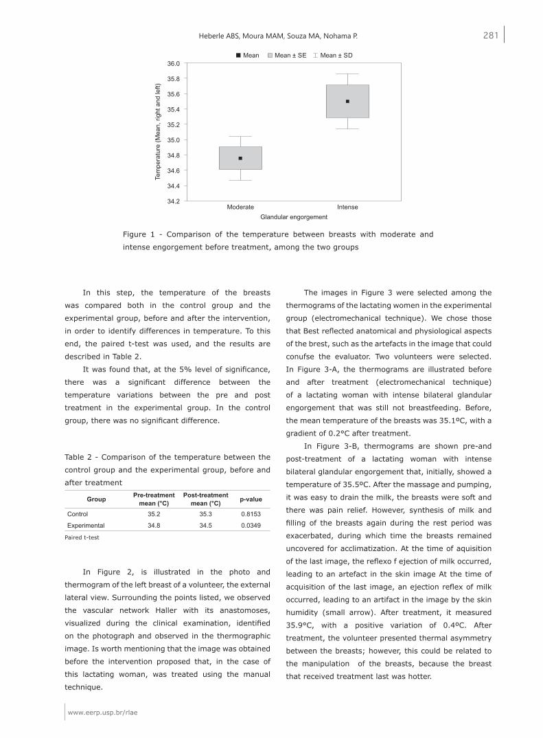
In Figure 1, it was verified that the breasts with
moderate engorgement had a mean temperature
of 34.8°C versus 35.5°C for those with intense
engorgement.
281
www.eerp.usp.br/rlae
Heberle ABS, Moura MAM, Souza MA, Nohama P.
Table 2 - Comparison of the temperature between the
control group and the experimental group, before and
after treatment
Paired t-test
Figure 1 - Comparison of the temperature between breasts with moderate and
intense engorgement before treatment, among the two groups
Group Pre-treatment mean (°C)
Post-treatment mean (°C) p-value
Control 35.2 35.3 0.8153
Experimental 34.8 34.5 0.0349
36.0
35.8
35.6
35.4
35.2
35.0
34.8
34.6
34.4
34.2
Tem
pera
ture
(Mea
n, ri
ght a
nd le
ft)
Moderate IntenseGlandular engorgement
Mean Mean ± SE Mean ± SD
In this step, the temperature of the breasts
was compared both in the control group and the
experimental group, before and after the intervention,
in order to identify differences in temperature. To this
end, the paired t-test was used, and the results are
described in Table 2.
It was found that, at the 5% level of significance,
there was a significant difference between the
temperature variations between the pre and post
treatment in the experimental group. In the control
group, there was no significant difference.
In Figure 2, is illustrated in the photo and
thermogram of the left breast of a volunteer, the external
lateral view. Surrounding the points listed, we observed
the vascular network Haller with its anastomoses,
visualized during the clinical examination, identified
on the photograph and observed in the thermographic
image. Is worth mentioning that the image was obtained
before the intervention proposed that, in the case of
this lactating woman, was treated using the manual
technique.
The images in Figure 3 were selected among the
thermograms of the lactating women in the experimental
group (electromechanical technique). We chose those
that Best reflected anatomical and physiological aspects
of the brest, such as the artefacts in the image that could
conufse the evaluator. Two volunteers were selected.
In Figure 3-A, the thermograms are illustrated before
and after treatment (electromechanical technique)
of a lactating woman with intense bilateral glandular
engorgement that was still not breastfeeding. Before,
the mean temperature of the breasts was 35.1ºC, with a
gradient of 0.2°C after treatment.
In Figure 3-B, thermograms are shown pre-and
post-treatment of a lactating woman with intense
bilateral glandular engorgement that, initially, showed a
temperature of 35.5ºC. After the massage and pumping,
it was easy to drain the milk, the breasts were soft and
there was pain relief. However, synthesis of milk and
filling of the breasts again during the rest period was
exacerbated, during which time the breasts remained
uncovered for acclimatization. At the time of aquisition
of the last image, the reflexo f ejection of milk occurred,
leading to an artefact in the skin image At the time of
acquisition of the last image, an ejection reflex of milk
occurred, leading to an artifact in the image by the skin
humidity (small arrow). After treatment, it measured
35.9°C, with a positive variation of 0.4ºC. After
treatment, the volunteer presented thermal asymmetry
between the breasts; however, this could be related to
the manipulation of the breasts, because the breast
that received treatment last was hotter.

282
www.eerp.usp.br/rlae
Rev. Latino-Am. Enfermagem 2014 Mar.-Apr.;22(2):277-85.
Figure 2 - Photograph (a) and thermogram (b) of the left breast of a lactating woman with
moderate glandular engorgement (palette Rain 900), showing the Haller vascular network
Figure 3 - In A, thermograms with intense globular BE. The images showed three tonalities of gray as follows:
dark gray (colder temperature), medium gray (intermediate temperature) and light gray (warmer temperature).
One observes the large thermal concentration in axillary and inframammary regions. In B, the breasts filled after
treatment and there was an ejection reflex (small arrow, below the white arrow). Already the nipple (white arrow),
presented itself with pallor, decreased perfusion and mild edema by use of the pumping machine
12 3
4
5
6 7
8
12 3
4
5
6 7
8
a) b)
Figura 3 - A ANTES DEPOIS
Figura 3 - B ANTES DEPOIS
Discussion
For this study, the application of a technological
instrument as a methodology became a challenge,
principally for obtaining quantitative data, related to
the physiological aspects of the breast. A new tool was
used, thermography, which provided a new modality
for imaging engorged breasts, which made it possible
283
www.eerp.usp.br/rlae
Heberle ABS, Moura MAM, Souza MA, Nohama P.
to monitor and evaluate the state of the breasts when
faced with the type of treatment applied.
In view of the special moment that breastfeeding
women experienced during the intervention, it was
feared that the application of the proposed protocol could
provoke stress in the volunteers, compared to the time
for acclimatization, succession of thermograms, and
electromechanical techniques for treatment. Changes
in mood, tearfulness, personal insecurity and hormonal
changes are common problems in this period(13). Thus,
we opted for only two images, one before and one after
treatment. The periods of 15 minutes of acclimatization
and post-treatment rest were used to orientate about
breastfeeding and to clarify doubts about this process.
In relation to location of the BE, two types
lobar and lobar/ampullary accounted for 50% of the
breastfeeding women in the control group, and 62.5% of
the experimental group, that is, accounting for 56.25%
of the sample. In another study(15), this type of BE was
also predominant and was encountered in 80% of the
cases. With regard to the intensity of the lactating
women with glandular BE, 42.85% were of the intense
type and 57.15% were moderate.
Among the lactating women that presented with
glandular BE, it was found that the breasts with intense
engorgement were significantly warmer than in those
with moderate BE. When comparing the left and right
breasts, the difference was 0.3°C and 0.2°C, respectively,
showing lower thermal variation. Among the lactating
women that presented with moderate engorgement, the
difference in temperature was 0.6ºC. But among those
with intense BE, the difference was 0.7ºC. Therefore, the
temperature difference between the breasts of the same
woman was lower when compared with the temperature
difference between women, as well as between the two
classifications of intensity. This was consistent with
the literature, because one of the known properties of
adipose tissue is to be a thermal insulator(22). Studies
showed a variation in the proportion of glandular and
adipose tissue, both among lactating women and those
not lactating, and of the amount of the glandular tissues.
It was also observed that there was no such variation
between the breasts of the same woman(23). It is worth
mentioning that an important thermal asymmetry
(2.3°C) was detected between the breasts of a unique
lactating woman, a fact that called for attention and
merits further investigation.
After the interventions, there was a significant
decrease in temperature in the experimental group.
In the control group, this thermal gradient was not
observed. This can be explained by the use of hands
in the application of massage, which may have
promoted greater transfer of kinetic energy. Although
the experimental group experienced a reduction of
temperature, in one of the lactating women (Figure
3-B) a temperature increase occurred after treatment
using electromechanical techniques. In these two cases
illustrated, although both had intense BE, the breast
engorgement of volunteer 2 (Figure 3-B) was more
severe and the greater in its intensity, a longer in time
and greater in the intensity of the massage was offered
to solve the problem and, consequently, the higher the
glandular temperature. Thus, it is believed that the 15
minutes of rest were insufficient for thermal stabilization
of the breast, in addition to the re-filling that occurred
during the rest period. This event suggests that favor
empty breasts milk production in lactating women, with
milk production exacerbated.
Intrinsic and extrinsic factors exist that can interfere
with the analysis of the thermograms. In this study, skin
humidity provoked by the milk ejection reflex, as well as
the Haller network, could confound the evaluator. The
vasuclar Haller network is described as an anastomotic
circle around the auréolo-papillary complex, formed
by the superficial veins which dilate intensely in the
puerperal process(24). It was observed that the superficial
vascular network covers the entire mammary gland and
inspection by physical examination became essential
to its identification, as it helped in the interpretation of
images in the regions of interest, namely, cooler spots
of the breast. The Haller network is quite expressive
in the puerperal breast, requiring a careful inspection
of the thermographic image and familiarization of the
evaluator with the vascular anatomy, because large
vessels “saturate” the images and are obstacles in the
investigation(6).
Conclusions
In her professional practice, the nurse has
care permeated by technology. She also has the
opportunity to contribute with research, propose and
develop innovative actions to improve care for people,
adding technical subsidy of optimization to nursing
care, specifically in the prevention and treatment of
lactational BE.
It was verified that thermography was a
comfortable, innocuous and safe technique, but requires
an environment suitable for the study. It provided greater
physiological knowledge of the puerperal breast, and was
284
www.eerp.usp.br/rlae
Rev. Latino-Am. Enfermagem 2014 Mar.-Apr.;22(2):277-85.
able to differentiate breasts with moderate and intense,
glandular BE, namely, it showed that the greater the
intensity of swelling, increased also the temperature of
the breast. The thermal asymmetry between the breasts
and the temperature gradients at points of interest
indicated possible pathologies, dysfunctions, perfusions,
or functional abnormalities, which were mostly
imperceptible on physical examination. In this work,
thermography was used as a new technology in the Field
of medical imaging, in which the primary advantage was
the measure of the temperature gradients in various
points of interest, as well as to verify the thermal
symmetries that were produced by the treatment
techniques for BE of massage and electromechanical
pumping. The adopted method provided reliable data
and subsidized clinical examination in the methodological
process of this research.
Compared to a standard method, the use of cold
packs in the treatment of BE, such steps to decrease
edema, vascularization and pain, there is evidence
that treatment by electromechanical techniques also
reduced the temperature of the breasts, promoting the
decreased glandular vascularization and consequently
the production of excess milk, providing comfort and
pain relief in lactating women.
Finally, electromechanical techniques proposed and
thermography, within their limitations in this pioneering
study, showed promising results in the investigation
of treatment of breast engorgement during lactation.
Additionally, thermography may also indicate clues for
other mammary problems such as cancer, circulation in
vessels and peripheral arteries, and internal inflammation.
References
1. Almeida JAG. Amamentação: um híbrido natureza-
cultura. Rio de Janeiro: Fiocruz; 1999.
2. Carvalhaes MABL, Parada CMGL, Costa MP. Fatores
associados à condição do aleitamento materno exclusivo
em crianças menores de 4 meses, em Botucatu, SP. Rev.
Latino-Am. Enfermagem. 2007;15:62-9.
3. Giugliani ERJ. Problemas comuns na lactação e seu
manejo. J Pediatr. 2004;80(5)suppl: 147-54.
4. Book OR, Guralnik L, Keidar Z, Gaitini DE, Engel A.
Pitfalls of the lactating breast on computed tomography.
J Comput Assist Tomogr. 2004;28(5):647-9.
5. Shimo AKK. Mama Puerperal: aspectos preventivos
e curativos do ingurgitamento mamário. [dissertação].
Ribeirão Preto: Escola de Enfermagem de Ribeirão Preto
da Universidade de São Paulo; 1983. 163 p.
6. Balbinot LF. Termografia computadorizada na
identificação de trigger points miofasciais. [Dissertação
Mestrado em Ciências do Movimento Humano –
Biomecânica]. Florianópolis: UDESC; 2006. 126 p.
7. Nunes LAO, Andrade ACC Filho, Sartori JL. Diagnóstico
de Diversas Patologias com Tecnologia Brasileira. Rev Micro
e Nanotecnol do Pólo Industrial de Manaus. [Internet].
2007;(10) [acesso 27 abril 2013]. Disponível em: http://
www.suframa.gov.br/minapim/news/visArtigo.cfm?
Ident=419&Lang=BR
8. Bezerra LA, Lima RCF, Lyra PRM, Araújo MC, Santos
FGC, Bezerra KM. Uma comparação entre temperaturas
de mamas obtidas pelo método dos volumes finitos em
malhas não-estruturadas e aquelas adquiridas através de
termogramas de pacientes de hospital público localizado
em clima tropical. 8º Congresso Iberoamericano de
Engenharia Mecânica; outu 2007. [acesso 31 março
2012]. Disponível em: <http://congreso.pucp.edu.pe/
cibim8/pdf/03/03-29.pdf
9. Rustemeyer J, Radtke J, Bremerich A. Thermography
and thermoregulation of the face. Head & Face Medicine.
[Internet]. 2007, 3:17 [acesso 20 fev 2010]. Disponível
em: http://www.head-face-med.com/content/ 3/1/17
10. Nyirjesy I. Breast Thermography. Clinl Obstet
Gynecol. 1982;25(2):401-8.
11. Birnbaum SJ. Breast temperature as a test for
pregnancy. Obstet Gynecol. 1966;27(3): 378-80.
12. Menczer J, Eskin BA. Evaluation of post-partum breast
by thermography. Obstet Gynecol. 1969;33(2):260-3.
13. Barros SM. Enfermagem no Ciclo Gravídico-
Puerperal. Barueri: Manole; 2006. 231 p.
14. Walker M. Breastfeeding and engorgement.
Breastfeeding Abstract. 2000;20(2):11-2. [Internet]. La
Leche League Internacional [atualizado 31 ago 2006].
[acesso 8 ago 2012]. Disponível em: http://www.llli.
org/ba/nov00.html
15. Silva IA. Inovação no tratamento do ingurgitamento
mamário - o uso do vibrador elétrico para massagens.
Acta Paul Enferm. 1996;9(1):61-70.
16. Gomes MB, Guimarães FC, Guimarães SMR, Claro
NAC. Limiar de dor à pressão em pacientes com cefaléia
tensional e disfunção temporomandibular. Cienc Odontol
Bras. 2006; 9(4):84-91.
17. Agência Nacional de Vigilância Sanitária (BR). Banco
de Leite Humano: funcionamento, prevenção e controle
de riscos. Brasília: Agência Nacional de Vigilância
Sanitária; 2008. 160 p.
18. Margato MP, Lucio CC, Heberle ABS, Nohama P.
Pesquisa e Desenvolvimento de Produto para Auxiliar no
Tratamento de Ingurgitamento Mamário em Lactantes.
285
www.eerp.usp.br/rlae
Heberle ABS, Moura MAM, Souza MA, Nohama P.
Received: Apr. 14th 2013
Accepted: Dec. 10th 2013
Anais do Congresso Internacional de Ergonomia e
Usabilidade de Interfaces Humano-Tecnologia: Produto,
Informações, Ambiente Construído e Transporte, 2010.
Rio de Janeiro; 2010.
19. Vinha VHP. O livro da amamentação. 2. ed. São
Paulo: CLR Balieiro; 2002. 91 p.
20. IACT, Thermography Guidelines: Standards and
Protocols in Clinical Thermographic Imaging [Internet].
Internacional Academy of Clinical Thermology; Sept
2002. [acesso 20 junho 2012]. Disponível em: http://
www.iact-org.org/professionals/thermog-guidelines.html
21. Brioshi ML, Cheren AJ, Ruiz RC, Sardá Júnior JJ, Silva
FMRM. O uso da termografia infravermelha na avaliação
do retorno ao trabalho em programa de reabilitação
ampliado (PRA). Acta Fisiatr. 2009;16(2):87-92.
22. Fonseca-Alaniz MH, Takada J, Alonso-Vale MIC, Lima
FB. O tecido adiposo como ógão endócrino: da teoria à
prática. J Pediatr. 2007;83(5):suppl S192-S203
23. Geddes DT: Ultrasound imaging of the lactating
breast. Methodology and application. Int Breastfeed J.
2009;4:4.
24. Rolim LMO, Figueiredo ALM. Anatomia da mama e
fisiologia da lactação. Bol SOPER. [Internet]. [acesso 5
out 2010]. Disponível em: http://www.soperj.org.br/
revista/detalhes.asp?id=22