Embed Size (px)
Citation preview

*Corresponding Author Address: Dr.Muruganand Email: [email protected]
International Journal of Dental and Health Sciences
Volume 02,Issue 06
Original Article
ASSESSMENT OF RELATIONSHIP OF MANDIBULAR
THIRD MOLAR ROOT TO INFERIOR ALVEOLAR
NERVE CANAL USING PHOTOSTIMULABLE
PHOSPHOR PLATE
D.Prabhu Shankar1,P Manodh 2, V.G.Muruganand 3, Aparna Murugan4
1.Associate professor,Dental college 2.Associate professor, Dental college 3.PG trainee, Dental college 4.PG trainee, Dental college
ABSTRACT:
The extraction of impacted mandibular third molars has long been associated with the dreaded and rarely, grave and irreversible complications. The oral and maxillofacial surgeons’ dogma strongly links most complications with the proximity of inferior alveolar canal and the impacted teeth, the relationship of which is still a topic of debate among surgeons. In this randomized, prospective case study, involving 114 patients, we have attempted to evaluate the relationship between impacted mandibular third molar root and the inferior alveolar canal with intra oral periapical radiographs using photo stimulable phosphor plates and the incidence of inferior alveolar nerve injury following removal of impacted mandibular third molar. Preoperative assessment based on Rood and Shehab Criteria revealed 38.6% cases with an interruption of white line, 25.4% had darkening of root and 11.4% cases with diversion of the canal and 7% of cases revealed an inferior alveolar nerve deficit corresponding with the radiographic signs of “darkening of the third molar root” and “interruption of white line” by the third molar root. In comparison with the other imaging modalities Photo Stimulable Phosphor plate consumes less time with higher image resolution and magnification to gain better knowledge of the inferior alveolar canal and root relationship Key words: Impaction - Mandibular molar- Photostimulable plate – radiographic signs -Inferior alveolar nerve INTRODUCTION:
Impacted teeth can be defined as those
teeth that are prevented from reaching
its normal eruption by adjacent tooth,
overlying bone, or soft tissue, or other
impediments. The impacted mandibular
third molar removal is the most common
surgical procedure in oral and
maxillofacial surgery and it is widely
acknowledged to be useful due to its
prevention of future problems. [3] The
major complications associated with the
surgical removal are the neurosensory
deficits post-operatively with an
incidence of 0.26 to 8.4 % of inferior
alveolar nerve. [2,16] The degree and the
incidence of nerve injury is multi-
factorial which includes surgeon’s
experience, age and sex of the patient
and most importantly the anatomical
relationship between the mandibular

Shankar P.et al, Int J Dent Health Sci 2015; 2(6):1506-1518
1507
canal and impacted mandibular third
molar root. According to Rood and
Shehab[36] there are seven radio
graphical signs which suggest a close
relationship between mandibular third
molar and inferior alveolar canal, four of
these signs are seen in the root of the
tooth and three signs are seen on the
inferior alveolar canal.
For precise identification of the
proximity of mandibular canal and third
molar root pre-operatively and to avoid
complications and upgrade the surgical
planning , various intra oral and extra
oral radiographs like Orthopantomogram
(OPG), intraoral periapical radiograph
(IOPA), computed tomography (CT),
cone beam computer tomography
(CBCT), Photo Stimulable Phosphor plate
(PSP) images are available, with Photo
Stimulable Phosphor plates being latest
technology incorporated by oral and
maxillofacial surgeons worldwide. [3,9]
The purpose of this study is to
evaluate the relationship between
mandibular third molars and the
inferior alveolar canal with intra oral
periapical radiographs using a
photostimulable phosphor plate and the
incidence of inferior alveolar nerve injury
following removal of impacted
mandibular third molar
MATERIALS AND METHODS:
114 patients, who reported to the
department of Oral and Maxillofacial
Surgery, Meenakshi Ammal Dental
College, for the Transalveolar extraction
of impacted mandibular third molars,
were selected for this study based on the
inclusion criteria of presence of one or
two Rood and Shehab Criteria. An
Impacted mandibular third molar with
periapical infection and cysts, no
involvement of third molar root to
inferior alveolar canal or radiographs
with more than two radiographic signs
were excluded. Radiographs of included
patients were taken using
Photostimulable Phosphor plate (fig I, II,
III) IOPAR with paralleling cone
technique and assessed for Rood and
Shehab criteria.
Preoperative assessment: The details of
the patients and third molar status were
recorded according to a standardized
protocol, including age and gender,
impaction pattern, and the presence of 1
or more of the 7 radiographic signs listed
below (fig IV).
A) Darkening of the root
B) Deflected root
C) Narrowing of the root
D) Dark and bifid root
E) Interruption of the white line
F) Diversion of the inferior alveolar canal
G) Narrowing of the inferior alveolar
canal.
A Standard operative technique for
transalveolar extraction of mandibular
third molar was performed.
Postoperative assessment: All the
patients were examined post operatively

Shankar P.et al, Int J Dent Health Sci 2015; 2(6):1506-1518
1508
on the 7th day at the time of suture
removal for the presence of any injury to
the inferior alveolar nerve. The patients
were questioned on paresthesia
/anesthesia in the operated side of the
lip and chin region. Neurosensory tests
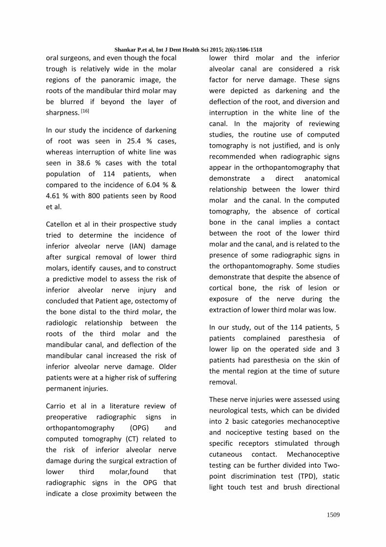
like static light touch (fig V), two point
discrimination test (figVI) and pin prick
test (fig VII) were performed for
confirmation. Patients with
neurosensory changes were followed up
for a period of 6 months. If recovery was
not complete after 6 months, the injury
was considered permanent or delayed.
RESULTS:
114 patients (68 (59.6 %) male and 46
(40.35%) female) who reported to
Meenakshi Ammal Dental College,
Department of Oral and Maxillofacial
Surgery with an age range of 18-35 years
were included in the study (Fig- VIII).
(TABLE – 1) Out of the 114 cases 44
(38.6%) cases presented with
interruption of white line as radiographic
marker. 29 (25.4%) had darkening of
root, 13 (11.4%) cases presented with
diversion of the canal, 7 (6.1%) cases
presented with narrowing of the canal, 5
(4.4%) cases had Diversion of root, 4
(3.5%) cases presented with dark bifid
apex, 3 (2.6%) cases had narrowing of
root and 9 patients (7.9%) had 2
radiographic signs- darkening of the root
and interruption of the white line (fig-IX).
The radiographic signs “darkening of the
third molar root” and “interruption of
white line by the third molar root” had
an inferior alveolar nerve deficit in 4
(3.5%) and 3 (2.6%) cases respectively
after impaction surgery and showed
statistically significant P value (TABLE 3).
Out of the 4 cases with darkening of
root, 1 case (0.87%) had an inferior
alveolar defect for a period of more than
6 months and the rest 3 cases (2.63%)
had a temporary nerve deficit and
recovered within a period of 3 months.
All the 3 cases seen with interruption of
the white line with temporary nerve
deficit recovered within a period of 3
months.
Out of the 9 radiographs with 2
radiographical signs 1 case (0.87%) had a
neurosensory deficit, which was
temporary (TABLE 2). Out of the 114
cases, 8 cases revealed an inferior
alveolar nerve deficit, of which 7 cases
showed grade 1 nerve injury
(neuropraxia), while the 1 case showed
grade III nerve injury (axonotmesis) (fig-
X). Pinprick test with two point
discrimination of the lower lip were
negative in these patients with absence
of response to light touch.
DISCUSSION:
Photo-Stimulable Phosphor (PSP)
imaging is a digital technique for intra-
oral radiography, which commercially
came into use in the late 1980s.
Although many studies have assessed
extra-oral radiographic methods for
assessment of mandibular third molars
before removal, no study seems to have
analyzed the use of digital intraoral
radiography [15], which makes this study
the first of its kind. In addition, the
intraoral image is sharper than the
panoramic image which is used by many
Shankar P.et al, Int J Dent Health Sci 2015; 2(6):1506-1518
1509
oral surgeons, and even though the focal
trough is relatively wide in the molar
regions of the panoramic image, the
roots of the mandibular third molar may
be blurred if beyond the layer of
sharpness. [16]
In our study the incidence of darkening
of root was seen in 25.4 % cases,
whereas interruption of white line was
seen in 38.6 % cases with the total
population of 114 patients, when
compared to the incidence of 6.04 % &
4.61 % with 800 patients seen by Rood
et al.
Catellon et al in their prospective study
tried to determine the incidence of
inferior alveolar nerve (IAN) damage
after surgical removal of lower third
molars, identify causes, and to construct
a predictive model to assess the risk of
inferior alveolar nerve injury and
concluded that Patient age, ostectomy of
the bone distal to the third molar, the
radiologic relationship between the
roots of the third molar and the
mandibular canal, and deflection of the
mandibular canal increased the risk of
inferior alveolar nerve damage. Older
patients were at a higher risk of suffering
permanent injuries.
Carrio et al in a literature review of
preoperative radiographic signs in
orthopantomography (OPG) and
computed tomography (CT) related to
the risk of inferior alveolar nerve
damage during the surgical extraction of
lower third molar,found that
radiographic signs in the OPG that
indicate a close proximity between the
lower third molar and the inferior
alveolar canal are considered a risk
factor for nerve damage. These signs
were depicted as darkening and the
deflection of the root, and diversion and
interruption in the white line of the
canal. In the majority of reviewing
studies, the routine use of computed
tomography is not justified, and is only
recommended when radiographic signs
appear in the orthopantomography that
demonstrate a direct anatomical
relationship between the lower third
molar and the canal. In the computed
tomography, the absence of cortical
bone in the canal implies a contact
between the root of the lower third
molar and the canal, and is related to the
presence of some radiographic signs in
the orthopantomography. Some studies
demonstrate that despite the absence of
cortical bone, the risk of lesion or
exposure of the nerve during the
extraction of lower third molar was low.
In our study, out of the 114 patients, 5
patients complained paresthesia of
lower lip on the operated side and 3
patients had paresthesia on the skin of
the mental region at the time of suture
removal.
These nerve injuries were assessed using
neurological tests, which can be divided
into 2 basic categories mechanoceptive
and nociceptive testing based on the
specific receptors stimulated through
cutaneous contact. Mechanoceptive
testing can be further divided into Two-
point discrimination test (TPD), static
light touch test and brush directional

Shankar P.et al, Int J Dent Health Sci 2015; 2(6):1506-1518
1510
stroke test. Nociceptive testing is
subdivided into pinprick test and thermal
discrimination test.
In our study, we have used two-point
discrimination test, static light touch test
and pin prick test for patients with signs
of inferior alveolar nerve deficit,
according to the protocol given by A. R.
Loescher and K. G. Smith. Patients who
participated in the study were unable to
discriminate between two points on the
operated side initially till the third month
postoperatively.
A classification of neurological injuries
based on mechanism of injury has been
described by Sunderland.35 These
include compression injury
(neuropraxia), severe compression,
stretch injury (axonotmesis), complete
section of a nerve trunk (neurotmesis),
and other injuries (perineural
inflammation). The damaged nerve will
react by going through stages of
Wallerian degeneration and an attempt
of axon regeneration, but the altered
sensation is likely to remain permanent
if there is no change after one to two
years. In contrast, some authors claim
that persistently symptomatic patients
are classified as having a permanent
nerve injury if there has been no change
after 3 months1.The Permanent altered
sensation is more likely to occur where
the nerve was severed (neurotmesis) or
crushed as a result of sectioning with a
rotary instrument or crushing of the
nerve as a result of displacement of root
tips into the inferior alveolar canal [35].
According to Robert et al 5 inferior
alveolar nerve injury was found in 4 out
of 1000 patients, in our study, we had a
nerve injury in 8 patients out of 114
patients, which was higher compared to
Robert et al study. Out of the 8 patients,
7 patients had neuroprexia and 1 case
had axontmesis, none of the patients
had neurotmesis as per Sunderland
classification. And according to Seddon
classification, it was grade 1 injury in 7
patients and grade III injury in 1 patient.
The patients who had neurosensory
deficit were managed medically with
capsule Renerve plus once daily for 3
weeks and reviewed periodically for
sensory changes, which was done in
accordance with Gintaraset al 14 Of the 8
cases which showed a sensory nerve
deficit, 7 cases showed a complete
recovery at the end of 3 months,
whereas 1 case had a prolonged
prophesy for a period of more than 6
months.
CONCLUSION:
This is a pioneering study done using
Photo-Stimulable Phosphor plate
periapical radiograph to evaluate the
relationship between impacted
mandibular third molar root apex to the
inferior alveolar canal. Significant
statistical and clinical co- relation was
observed between radiographic signs
like darkening of root, interruption of
white line and nerve injury. 7.01 % of
inferior alveolar nerve injury present in
our study are similar to the previous
studies done using conventional intra
oral periapical radiograph,
orthopantomogram (OPG), cone beam

Shankar P.et al, Int J Dent Health Sci 2015; 2(6):1506-1518
1511
computer tomography (CBCT) and
computer tomography (CT). In
comparison with the other imaging
modalities Photo-Stimulable Phosphor
plates are cost effective and less time
consuming. It revealed higher image
resolution and magnification which
helped us to gain better knowledge of
the inferior alveolar canal and root
relation enabling us to carry out the
surgical procedure with greater safety.
REFERENCES:
1. Nguyen, Edward, Dragan Grubor, and ArunChandu. Risk Factors for Permanent Injury of Inferior Alveolar and Lingual Nerves During Third Molar Surgery. Journal of Oral and Maxillofacial Surgery 72, no. 12 (2014): 2394-2401.
2. Xu, Guang-zhou, Chi Yang, Xin-Dong Fan, Chuang-Qi Yu, Xie-Yi Cai, Yong Wang, and DongMei He. Anatomic relationship between impacted third mandibular molar and the mandibular canal as the risk factor of inferior alveolar nerve injury. British Journal of Oral and Maxillofacial Surgery 51, no. 8 (2013): e215-e219.
3. Hasegawa, T., S. RI, T. Shigeta, M. Akashi, Y. Imai, Y. Kakei, Y. Shibuya, and T. Komori. Risk factors associated with inferior alveolar nerve injury after extraction of the mandibular third molar a comparative study of preoperative images by panoramic radiography and computed tomography. International journal of oral and maxillofacial surgery 42, no. 7 (2013): 843-851.
4. Selvi, Firat, Thomas B. Dodson, Anders Nattestad, Kevin Robertson, and Len Tolstunov. Factors that are associated with injury to the inferior alveolar nerve in high-risk patients after removal of third molars. British Journal of Oral and Maxillofacial Surgery 51, no. 8 (2013): 868-873.
5. Juan M.Cespedes-Sanchez, Raul Ayuso-montero, Antoni Mari-Roig. The importance of a good evalution in order to prevent oral nerve injuries-A review.ActaOdontolgicaScandinavica 72, no. 3 (2014): 161-167.
6. Meshram, VikasSukhadeo, PriyatamaVikasMeshram, and PravinLambade. Assessment of Nerve Injuries after Surgical Removal of Mandibular Third Molar: A Prospective Study. Asian Journal of Neuroscience 2013 (2013).
7. Kim, Jin-Woo, In-Ho Cha, Sun-Jong Kim, and Myung-Rae Kim. Which risk factors are associated with neurosensory deficits of inferior alveolar nerve after mandibular third molar extraction? Journal of Oral and Maxillofacial Surgery70, no. 11 (2012): 2508-2514.
8. Soğur, E., B. G. Baksı, and A. Mert. The effect of delayed scanning of storage phosphor plates on occlusal caries detection.Dentomaxillofacial Radiology (2012) 41, 309-315.
9. Akcicek, G., S. Uysal, N. Avcu, and O. Kansu. Comparison of different imaging techniques for the evaluation of proximity between molars and the mandibular canal.Clinical Dentistry and Research 36, no. 1 (2012): 2-7.
10. Szalma, József, Edina Lempel, SáraJeges, and Lajos Olasz. Darkening of third molar roots: panoramic radiographic associations with inferior alveolar nerve exposure. Journal of Oral and Maxillofacial Surgery 69, no. 6 (2011): 1544-1549.

Shankar P.et al, Int J Dent Health Sci 2015; 2(6):1506-1518
1512
11. Leung, Yiu Yan, and Lim Kwong Cheung. Correlation of radiographic signs, inferior dental nerve exposure, and deficit in third molar surgery. Journal of Oral and Maxillofacial Surgery 69, no. 7 (2011): 1873-1879.
12. Tolstunov, Len, Bahram Javid, Lance Keyes, and Anders Nattestad. Pericoronalostectomy: an alternative surgical technique for management of mandibular third molars in close proximity to the inferior alveolar nerve. Journal of Oral and Maxillofacial Surgery 69, no. 7 (2011): 1858-1866.
13. Cilasun, Ulkem, TulinYildirim, EsraGuzeldemir, and ZaferOzgurPektas. Coronectomy in patients with high risk of inferior alveolar nerve injury diagnosed by computed tomography. Journal of Oral and Maxillofacial Surgery 69, no. 6 (2011): 1557-1561.
14. Juodzbalys, Gintaras, Hom-Lay Wang, and GintautasSabalys. Injury of the inferior alveolar nerve during implant placement: a literature review. Journal of oral & maxillofacial research 2, no. 1 (2011).
15. Jerjes, Waseem, TahwinderUpile, Priya Shah, FaraiNhembe, DipaliGudka, Panagiotis Kafas, Eileen McCarthy et al. Risk factors associated with injury to the inferior alveolar and lingual nerves following third molar surgery revisited. Oral Surgery, Oral Medicine, Oral Pathology, Oral Radiology, and Endodontology 109, no. 3 (2010): 335-345.
16. Cheung, Lim K., Y. Y. Leung, L. K. Chow, M. C. M. Wong, E. K. K. Chan, and Y. H. Fok. Incidence of neurosensory deficits and recovery after lower third molar surgery: a prospective clinical study of 4338 cases. International journal of oral and maxillofacial surgery 39, no. 4 (2010): 320-326.
17. Poort, Lucas J., Johan W. van Neck, and Karel GH van der Wal. Sensory testing of inferior alveolar nerve injuries: a review of methods used in prospective studies. Journal of Oral and Maxillofacial Surgery 67, no. 2 (2009): 292-300.
18. Matzen, Louise Hauge, Jennifer Christensen, and Ann Wenzel. Patient discomfort and retakes in periapical examination of mandibular third molars using digital receptors and film. Oral Surgery, Oral Medicine, Oral Pathology, Oral Radiology, and Endodontology 107, no. 4 (2009): 566-572.
19. Ergün, S. P. Güneri, D. Ilgüy, M. Ilgüy, and H. Boyacıoğlu. How many times can we use a phosphor plate? A preliminary study. (2014).
20. Hillerup, Søren. Iatrogenic injury to the inferior alveolar nerve: etiology, signs and symptoms, and observations on recovery. International journal of oral and maxillofacial surgery 37, no. 8 (2008): 704-709.
21. Chiu, Hui-Lin, Shui-Hui Lin, Chia-Hui Chen, Wen-Chen Wang, Jin-Yi Chen, Yuk-Kwan Chen, and Li-Min Lin. Analysis of photostimulable phosphor plate image artifacts in an oral and maxillofacial radiology department. Oral Surgery, Oral Medicine, Oral Pathology, Oral Radiology, and Endodontology 106, no. 5 (2008): 749-756.
22. Almendros-Marqués, Nieves, Leonardo Berini-Aytés, and Cosme Gay-Escoda. Influence of lower third molar position on the incidence of preoperative complications. Oral Surgery, Oral Medicine, Oral Pathology, Oral Radiology, and Endodontology 102, no. 6 (2006): 725-732.
23. Hull, Donald J., Daniel A. Shugars, Raymond P. White Jr, and Ceib Phillips. Proximity of a lower third molar to the inferior alveolar canal as a predictor of delayedrecovery. Journal of oral and

Shankar P.et al, Int J Dent Health Sci 2015; 2(6):1506-1518
1513
maxillofacial surgery 64, no. 9 (2006): 1371-1376.
24. Koong, B., M. J. Pharoah, M. Bulsara, and M. Tennant. Methods of determining the relationship of the mandibular canal and third molars: a survey of Australian oral and maxillofacial surgeons. Australian dental journal 51, no. 1 (2006): 64-68.
25. Tsuchida, Ryoko, Kazuyuki Araki, Atsushi Endo, ItsuoFunahashi, and Tomohiro Okano. Physical properties and ease of operation of a wireless intraoral x-ray sensor. Oral Surgery, Oral Medicine, Oral Pathology, Oral Radiology, and Endodontology 100, no. 5 (2005): 603-608.
26. Mahasantipiya, Phattaranant May, N. W. Savage, P. A. J. Monsour, and R. J. Wilson. Narrowing of the inferior dental canal in relation to the lower third molars. (2014).
27. Miloro, Michael, and Jeffrey DaBell. Radiographic proximity of the mandibular third molar to the inferior alveolar canal. Oral Surgery, Oral Medicine, Oral Pathology, Oral Radiology, and Endodontology 100, no. 5 (2005): 545-549.
28. Tay, Andrew Ban Guan, and Wee Ser Go. Effect of exposed inferior alveolar neurovascular bundle during surgical removal of impacted lower third molars. Journal of oral and maxillofacial surgery 62, no. 5 (2004): 592-600.
29. Pogrel, M. Anthony, J. S. Lee, and D. F. Muff. Coronectomy: a technique to protect the inferior alveolar nerve. Journal of oral and maxillofacial surgery 62, no. 12 (2004): 1447-1452.
30. Susarla, Srinivas M., Bart F. Blaeser, and Daniel Magalnick. Third molar surgery and associated complications. Oral and maxillofacial surgery clinics of North America 15, no. 2 (2003): 177-186.
31. Drage, Nicholas A., and Tara Renton. Inferior alveolar nerve injury related to mandibular third molar surgery: an unusual case presentation. Oral Surgery, Oral Medicine, Oral Pathology, Oral Radiology, and Endodontology 93, no. 3 (2002): 358-361.
32. Valmaseda-Castellón, Eduard, Leonardo Berini-Aytés, and Cosme Gay-Escoda. Inferior alveolar nerve damage after lower third molar surgical extraction: a prospective study of 1117 surgical extractions. Oral Surgery, Oral Medicine, Oral Pathology, Oral Radiology, and Endodontology 92, no. 4 (2001): 377-383.
33. Couture, Rex A., and Charles Hildebolt. Quantitative dental radiography with a new photostimulable phosphor system. Oral Surgery, Oral Medicine, Oral Pathology, Oral Radiology, and Endodontology 89, no. 4 (2000): 498-508.
34. Huda, Walter, Lynn N. Rill, Douglas K. Benn, and James C. Pettigrew. Comparison of a photostimulable phosphor system with film for dental radiology. Oral Surgery, Oral Medicine, Oral Pathology, Oral Radiology, and Endodontology 83, no. 6 (1997): 725-731.
35. Schultze-Mosgau, S., and R. H. Reich. Assessment of inferior alveolar and lingual nerve disturbances after dentoalveolar surgery, and of recovery of sensitivity. International journal of oral and maxillofacial surgery 22, no. 4 (1993): 214-217.
36. Rood, J. P., and B. A. A. NooraldeenShehab. The radiological prediction of inferior alveolar nerve injury during third molar surgery. British Journal of Oral and Maxillofacial Surgery 28, no. 1 (1990): 20-25.
37. Rood, J. P. Degrees of injury to the inferior alveolar nerve sustained during
Shankar P.et al, Int J Dent Health Sci 2015; 2(6):1506-1518
1514
the removal of impacted mandibular third molars by the lingual split technique.British Journal of Oral Surgery 21, no. 2 (1983): 103-116.
38. Azaz, Badri, ArieShteyer, and Moshe Piamenta. Radiographic and clinical manifestations of the impacted mandibular third molar. International journal of oral surgery 5, no. 4 (1976): 153-160.
39. Duinkerke, A. S. H., A. C. M. Van de Poel, F. P. G. M. Van der Linden, W. H. Doesburg, and W. A. J. G. Lemmens. Evaluation of a technique for standardized periapical radiographs. Oral Surgery, Oral Medicine, Oral Pathology 44, no. 4 (1977): 646-651.
40. Q.A collectible sponsored by CRCPD’s committee on quality Assurance in Diagnostic X-ray (H-7).
41. Loescher, A. R., K. G. Smith, and P. P. Robinson. Nerve damage and third molar removal. Dent Update 30, no. 7 (2003): 375-82.
42. Palma-Carrió C1, García-Mira B, Larrazabal-Morón C, Peñarrocha-Diago M. Radiographic signs associated with inferior alveolar nerve damage following lower third molar extraction Med Oral Patol Oral Cir Bucal. 2010 Nov 1;15(6):e886-90
TABLES:
TABLE -1:Incidence of seven radio graphical signs
RADIOGRAPHIC SIGNS SIGN APPEARENCE PERCENT OF POPULATION (TOTAL 114 CASES)
Darkening of root 29 25.4
Interruption of white line 44 38.6
Diversion of canal 13 11.4
Narrowing of canal 7 6.1
Diversion of root 5 4.4
Dark & bifid apex 4 3.5
Narrowing of root 3 2.6
Darkening & interruption of line
9 7.9

Shankar P.et al, Int J Dent Health Sci 2015; 2(6):1506-1518
1515
TABLE - 2: Incidence of paresthesia seen
RADIOGRAPHIC SIGN PARESTHESIA SEEN PERCENT OF POPULATION (TOTAL 114CASES)
Darkening of root 4 3.5
Interruption of white line 3 2.63
Diversion of canal 0 NA
Narrowing of canal 0 NA
Diversion of root 0 NA
Dark & bifid apex 0 NA
Narrowing of root 0 NA
Darkening of root & interruption of white line
1 0.87
TABLE - 3:
Darkening of root Interruption of whiteline Darkening&Interruption of white line
15.207a
32.818b
5.444a 1 1 1
.000 .000 .020
FIGURES:
fig I : Photostimulable plate fig II: Photostimulable cover

Shankar P.et al, Int J Dent Health Sci 2015; 2(6):1506-1518
1516
fig III: Photostimulable plate holder
fig IV:
A) Darkening of the root B) Deflected root C) Narrowing of the root D) Dark and Bifid root E) Interruption of the white line F) Diversion of the inferior alveolar canal G) Narrowing of the inferior alveolar canal
Shankar P.et al, Int J Dent Health Sci 2015; 2(6):1506-1518
1517
fig V: Light Touch Assessment (lt)
fig VI: Two - Point Discrimination test (tpd)
fig VII: Pinprick Test (pp)
fig : VIII
60%
40%MALE
FEMALE

Shankar P.et al, Int J Dent Health Sci 2015; 2(6):1506-1518
1518
Fig IX: Radiographic signs present in total population
Fig X: Neurosensory Deficit
total population
darkening of root
interuption of white line
diversion of canal
narrowing of canal
dark and bifid apex
diversion of root
narrowing of root
diversion+interuption
total population
parasthesia seen
neuropraxia
axonotmesis