Embed Size (px)
Citation preview

ASSESSMENT OF PROTEINURIA IN PEDIATRIC PRACTICE
CAROLYN ABITBOL, M.D.UNIVERSITY OF MIAMI MILLER SCHOOL OF MEDICINE
/ HOLTZ CHILDREN’S HOSPITAL

OBJECTIVES
• Define proteinuria in pediatric health and disease
• Examine the mechanisms of proteinuria and distinguish sites of origin
• Interpret the clinical prognostic determinants of proteinuria in the context of pediatric practice
• Provide a paradigm for the assessment of proteinuria in pediatric practice
• Consider treatment strategies for proteinuria in children and adolescents

PROTEINURIA: PREVALENCE
• Up to 10% of children have proteinuria in a single urine sample (NKF Concensus Panel: PARADE)
• Only 0.1% have persistant proteinuria after 4 samples (Vehaskari et al 1982)
• AAP no longer advises the use of screening urinalysis at any age
DEFINITIONS
• Dipstick (albumin only):trace: <15 mg/dl; 1+ ≈ 30 mg/dl; 2+ ≈100 mg/dl; 3+ ≈300 mg/dl; 4+ ≈>2000mg/dl
• Random Urine pr/cr (mg/mg)Normal <100 mg/m2 /day = 0.1Intermediate Range >100<1000 mg/ m2 /day = >0.1<1.0Nephrotic Range > 1000 mg/ m2 /day = >1.0
• Microalbumin (µAlb)<30 µgrams/ mg creatinine

URINARY DIPSTICK
• Negative
• Trace = 15- 30 mg/dl
• 1+= 30-100 mg/dl
• 2+= 100 – 300 mg/dl
• 3+=300-1000 mg/dl
• 4+=>1000 mg/dl
Only >/= 1+ is abnormal
Colorimetric reaction between tetrabromophenol blue and albumin (only)
Not sensitive for low molecular weight proteins which include IgG, transferrin, uromodulin
False Positives•Alkaline urine pH>8•Concentrated urine SG>1.025•Gross Hematuria•Pyuria•Bacteriuria•Iodinated radioconstrast dyes•Dipstick held in urinary stream•High-dose PCNs, Ceph, or Sulfa

TYPES OF PROTEINURIA• Orthostatic (Postural) Proteinuria
• Incidental Proteinuria
• Fever
• Exercise
• Overflow proteinuria
• Pathologic Proteinuria
• Hyperfiltration (Diabetic; Low Nephron Mass; HT; Chronic Kidney Disease)
• Inflammation (Glomerulonephritides)
• Nephrotic Syndrome

MECHANISMS OF PROTEINURIA
• Loss of “net negative charge barrier”.• Loss of fenestrated “pore barrier”.• Filtration/generation of cytokines & noxious
chemokines provoke interstitial cell proliferation and activation.
• “Overload proteinuria” from filtered albumin causes disruption of endosomal reabsorption of albumin and other proteins
• Tubular proteinuria from ischemic/ interstitial injury injury

THIS PARADIGM SHIFT IMPLIES THAT EXCRETED PROTEIN REPRESENTS THE NET EFFECT OF THE INTERPLAY OF GLOMERULAR PERMEABILITY ALTERATIONS AND THE SATURABLE REABSORPTIVE CAPACITY OF THE PROXIMAL TUBULE.

MECHANISMS OF PROTEINURIALoss of “net negative charge” and pore size barrier
Loss of podocyte number and integrity
Loss of endosomal reclammation and degradation of filtered albumin

TUBULAR REABSORPTION OF FILTERED PROTEINS• Complexes with cubulin or megalin
•Endocytic invagination with formation of endosomes and lysosomes
• Degradation of filtered proteins and reabsorption of amino acids

CHARACTERIZATION OF PROTEINURIA• If microalbumin characterizes renal disease
progression in adults; can we draw parallels with adults?
• Glomerular proteinuria is characterized by albuminuria.
• Tubular proteinuria is characterized by microglobulins (the “non-albumin component of proteinuria)
• Fractionation of Proteinuria• Microalbumin/Total protein ratio (µAlb/TP < 15%)• “Tubular Proteinuria” (microglobulins): 35%• Tamm Horsfall (uromodulin): 50%
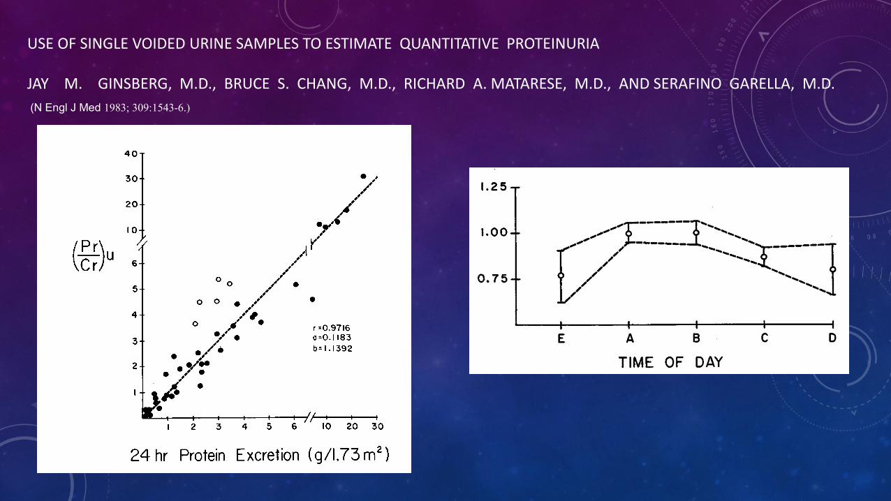
USE OF SINGLE VOIDED URINE SAMPLES TO ESTIMATE QUANTITATIVE PROTEINURIA
JAY M. GINSBERG, M.D., BRUCE S. CHANG, M.D., RICHARD A. MATARESE, M.D., AND SERAFINO GARELLA, M.D.(N Engl J Med 1983; 309:1543-6.)

Similar prognostic ability for each of the three methods to quantify proteinuria for characterizing a >50% decline in GFR or need for RRT based on clinically meaningful cutoffs of UP/C (Kaplan–Meier, dashed lines and Generalized Gamma, solid lines).
Dana Y. Fuhrman et al. CJASN 2017;12:912-920

Collinearity between the three methods to quantify proteinuria at the index study visit, n=751.
Dana Y. Fuhrman et al. CJASN 2017;12:912-920
©2017 by American Society of Nephrology

Profiling proteinuria in pediatric patientsCarolyn L. Abitbol . Jayanthi Chandar . Ali Mirza Onder . Obioma Nwobi . Brenda Montané . Gastón Zilleruelo
Infants 1-67-12
>12-210
10
20
30
40
50
60
70
80Tubular Disease
Glomerular Disease
Patient Disease Category by Age Distribution
Num
ber o
f Pat
ient
s
All0
1
2
3
4
5
Glomerular Tubular Infants
43%
58%
26%
18%
µAlb Fraction
Urine Protein Profiles by Disease Category
Prot
einu
ria
Up/
c (m
g/m
g) w
ithµ A
lbum
in F
ract
ion

CALCULATIONS FOR PROFILING RANDOM SPECIMENS
• Upr/cr=mg/mg Normal ≤ 0.2 (?)• Microalbumin (µAlb)/cr
mg/mg*1000=µg/mg Cr Normal < 30 µg/mg Cr
• % µAlb = µAlb/Pr*100 = Glomerular proteinuria• 1- µAlb/Pr*100 = Tubular Proteinuria• Nephrotic Range Upr/cr >2.0 (1 gram/m2/day)


ORTHOSTATIC PROTEINURIA DUE TO RENAL VEIN ENTRAPMENT
CONCLUSIONS
• Proteinuria is an important clinical marker of renal injury.• Quantitating and “Profiling” proteinuria may allow:
• Predicting the need for surgical intervention• Assessing response to surgical and medical treatment
• Further collaborative trials in chronic kidney injury in children should includeassessment and profiling proteinuria.
ORTHOSTATIC PROTEINURIA:FOLLOW-UP
Although orthostatic proteinuria does not generally persistbeyond the third decade of life, testing for proteinuria onan annual basis is prudent, especially because bothpathologic and physiologic proteinuria (ie, the smallamount of protein normally present in urine) also has anorthostatic component. If the first-voided morningspecimen has a 1+ or greater reaction for protein, furtherstudies are indicated.


CONCLUSIONS
• Persistent proteinuria predicts renal disease progression in children.
• Proteinuria should be screened quantitatively in infants & children “at risk” including those with potential low renal mass and/or excess BMI.
• diabetes mellitus (Type 1 or 2)• Obesity• preterm birth• history of familial kidney disease
• Early “reno-protection” is advocated in proteinuric kidney disease

![HIGHLIGHTS OF PRESCRIBING INFORMATION Proteinuria: …€¦ · Proteinuria [see Warnings and Precautions (5.6)] 2 g or greater proteinuria in 24 hours Withhold until less than or](https://img.pdfslide.us/doc/110x75/5f0775a37e708231d41d16a8/highlights-of-prescribing-information-proteinuria-proteinuria-see-warnings-and.jpg)