Embed Size (px)
Citation preview

Assessment of Pressure Ulcers
Presented byJeri Ann Lundgren, RN, BSN, PHN, CWS, CWCNPathway Health Services

Training Objectives• Describe etiologies of Pressure Ulcers • Discuss how to properly assess and
describe a pressure ulcer• Demonstrate how to properly stage a
pressure ulcer
Pressure Ulcers• A pressure ulcer is localized injury to the
skin and/or underlying tissue usually over a bony prominence, as a result of pressure, or pressure in combination with shear and/or friction
• Copyright: NPUAP 2007
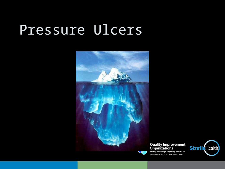
Pressure Ulcers
ASSESSMENT• When a pressure ulcer is present, daily
monitoring should include:– An evaluation of the ulcer, if no dressing
present– An evaluation of the status of the dressing,
if present– The presence of complications– Whether pain, if present, is being
adequately controlled
ASSESSMENT• Wounds should be assessed/documented on a weekly
basis, however when there is a complication or change identified daily monitoring/documentation maybe necessary, until resolved. However, the amount of observation of wound bed possible will depend upon the type of dressing that is used (many dressings are meant to stay in place for several days).
• A clean pressure ulcer with adequate blood supply & innervation should show evidence of stabilization or some healing within 2-4 weeks.
• Nurse Notes should reflect progress of wound only.
Wound Bed Assessment
• Describe the tissue in the wound bed using professional terms– Necrotic/eschar– Slough– Granulation– Epithelial
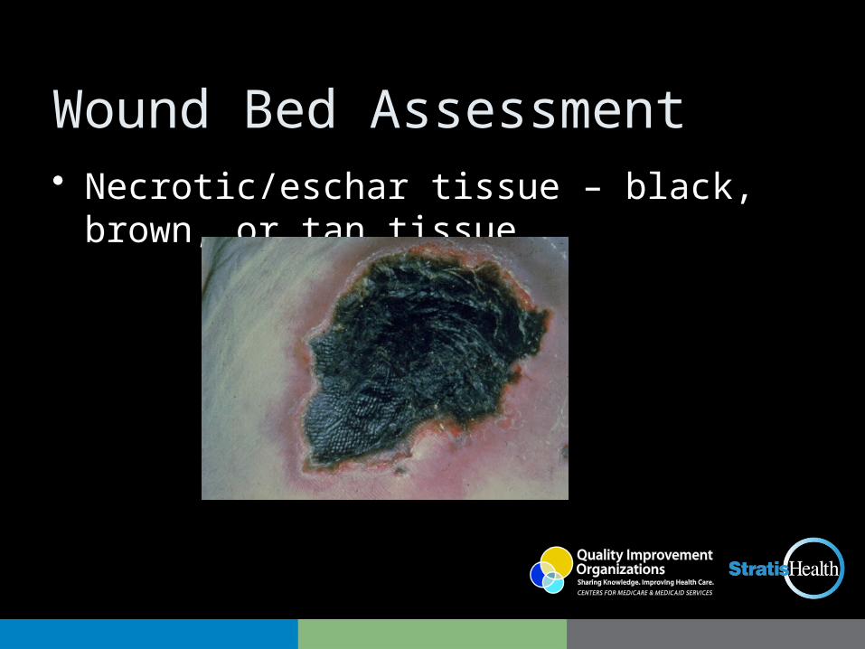
Wound Bed Assessment• Necrotic/eschar tissue – black, brown, or tan
tissue
Wound Bed Assessment
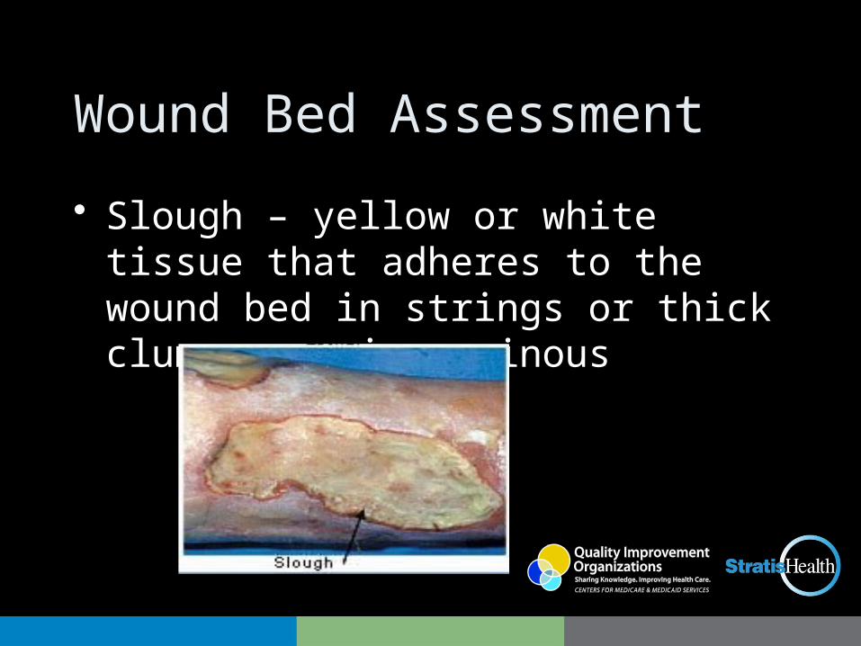
• Slough – yellow or white tissue that adheres to the wound bed in strings or thick clumps, or is mucinous
Wound Bed Assessment
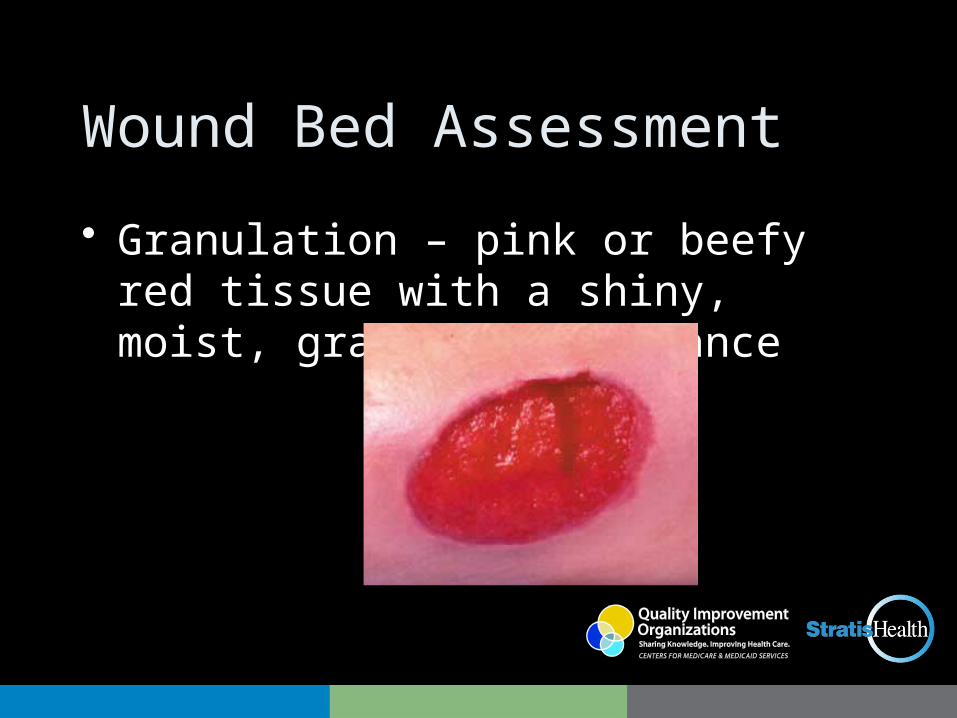
• Granulation – pink or beefy red tissue with a shiny, moist, granular appearance
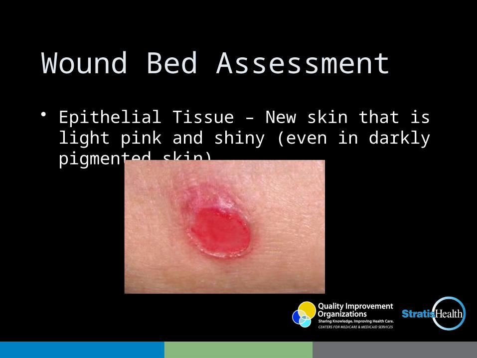
Wound Bed Assessment
• Epithelial Tissue – New skin that is light pink and shiny (even in darkly pigmented skin)
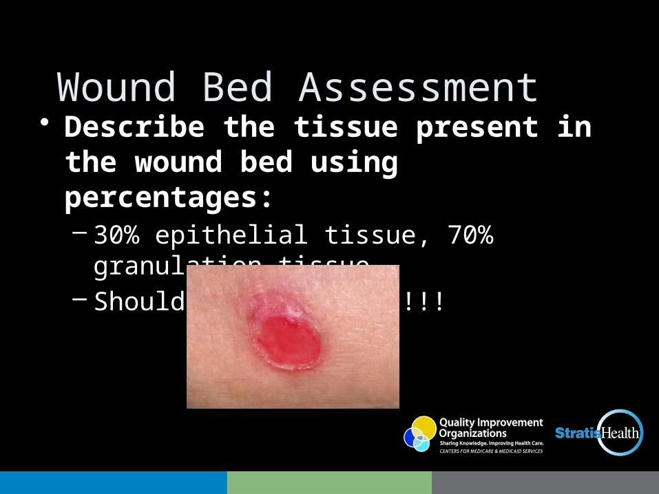
Wound Bed Assessment• Describe the tissue present in the
wound bed using percentages:– 30% epithelial tissue, 70% granulation tissue– Should equal 100%!!!!!!
Stage I Pressure Ulcer• Stage I:
Intact skin with non-blanchable redness of a localized area usually over a bony prominence. Darkly pigmented skin may not have visible blanching; its color may differ from the surrounding area.Further description: The area may be painful, firm, soft, warmer or cooler as compared to adjacent tissue. Stage I may be difficult to detect in individuals with dark skin tones. May indicate "at risk" persons (a heralding sign of risk)
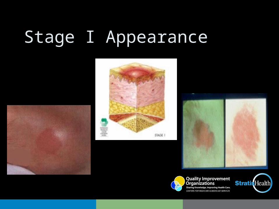
Stage I Appearance
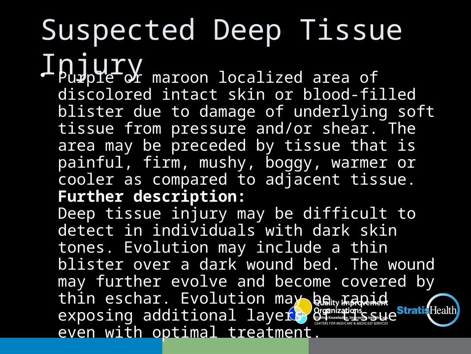
Suspected Deep Tissue Injury• Purple or maroon localized area of discolored intact
skin or blood-filled blister due to damage of underlying soft tissue from pressure and/or shear. The area may be preceded by tissue that is painful, firm, mushy, boggy, warmer or cooler as compared to adjacent tissue. Further description: Deep tissue injury may be difficult to detect in individuals with dark skin tones. Evolution may include a thin blister over a dark wound bed. The wound may further evolve and become covered by thin eschar. Evolution may be rapid exposing additional layers of tissue even with optimal treatment.
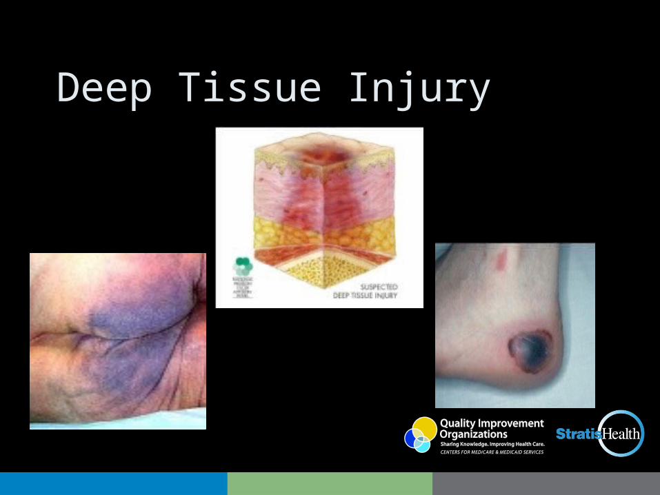
Deep Tissue Injury
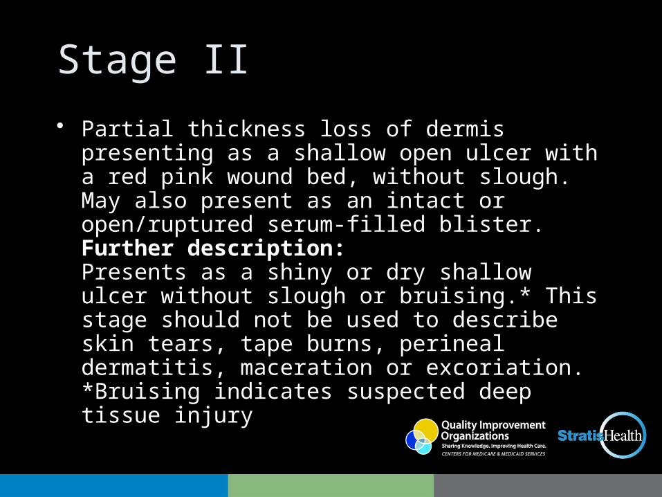
Stage II
• Partial thickness loss of dermis presenting as a shallow open ulcer with a red pink wound bed, without slough. May also present as an intact or open/ruptured serum-filled blister. Further description: Presents as a shiny or dry shallow ulcer without slough or bruising.* This stage should not be used to describe skin tears, tape burns, perineal dermatitis, maceration or excoriation. *Bruising indicates suspected deep tissue injury
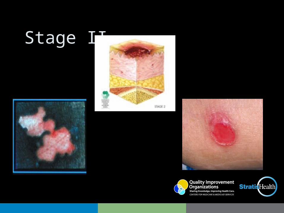
Stage II
Stage III• Full thickness tissue loss. Subcutaneous fat may be
visible but bone, tendon or muscle are not exposed. Slough may be present but does not obscure the depth of tissue loss. May include undermining and tunneling. Further description: The depth of a stage III pressure ulcer varies by anatomical location. The bridge of the nose, ear, occiput and malleolus do not have subcutaneous tissue and stage III ulcers can be shallow. In contrast, areas of significant adiposity can develop extremely deep stage III pressure ulcers. Bone/tendon is not visible or directly palpable.
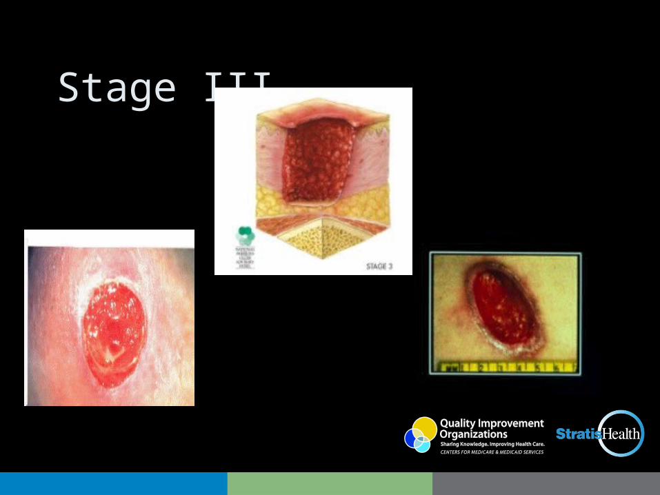
Stage III
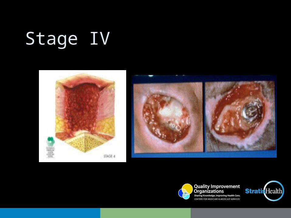
Stage IV• Full thickness tissue loss with exposed bone, tendon
or muscle. Slough or eschar may be present on some parts of the wound bed. Often include undermining and tunneling.Further description: The depth of a stage IV pressure ulcer varies by anatomical location. The bridge of the nose, ear, occiput and malleolus do not have subcutaneous tissue and these ulcers can be shallow. Stage IV ulcers can extend into muscle and/or supporting structures (e.g., fascia, tendon or joint capsule) making osteomyelitis possible. Exposed bone/tendon is visible or directly palpable.
Stage IV
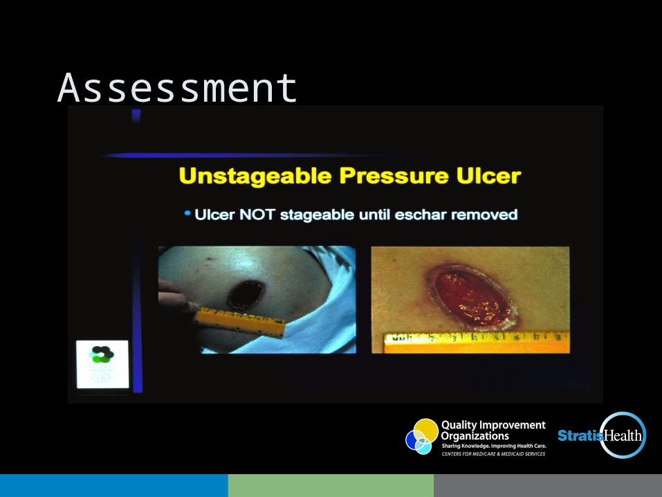
Unstageable• Full thickness tissue loss in which the base of the
ulcer is covered by slough (yellow, tan, gray, green or brown) and/or eschar (tan, brown or black) in the wound bed.Further description: Until enough slough and/or eschar is removed to expose the base of the wound, the true depth, and therefore stage, cannot be determined. Stable (dry, adherent, intact without erythema or fluctuance) eschar on the heels serves as "the body's natural (biological) cover" and should not be removed.
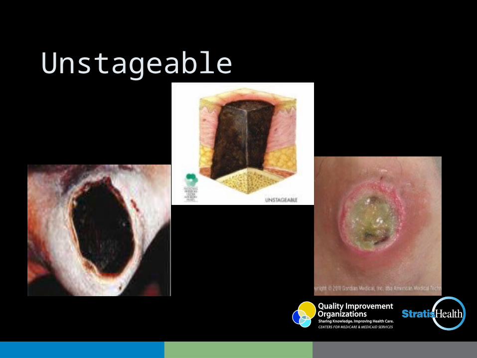
Unstageable
Assessment
Pressure Ulcer Assessment
• Purpose of staging is for consistent communication of depth of tissue destruction
• Once staged, the ulcer should not be back staged, rather the wound should be described in terms of size, shape, color, drainage, and odor using one of the wound assessment measures (www.npuap.com)
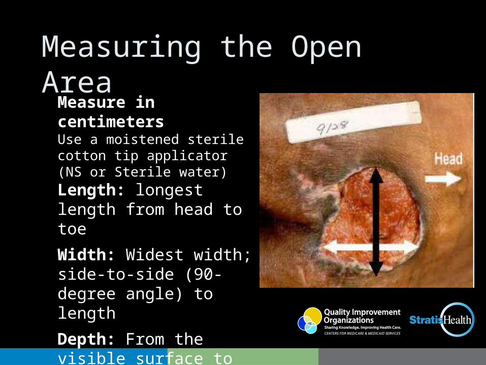
Measuring the Open AreaMeasure in centimetersUse a moistened sterile cotton tip applicator (NS or Sterile water)
Length: longest length from head to toe
Width: Widest width; side-to-side (90-degree angle) to length
Depth: From the visible surface to the deepest area
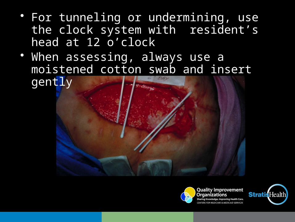
• For tunneling or undermining, use the clock system with resident’s head at 12 o’clock
• When assessing, always use a moistened cotton swab and insert gently
Assessment
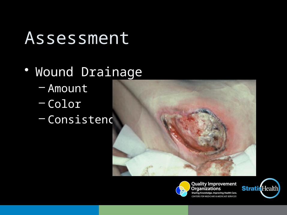
• Wound Drainage– Amount – Color– Consistency
Assessment
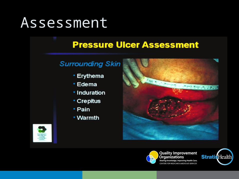
Pressure Ulcer Assessment• Odor if it is present (assess odor only after
wound is irrigated)• Pain – nature, frequency and management• Signs or symptoms of infection• One tool that can be used to monitor changing
status of a pressure ulcer is the PUSH tool (www.npuap.org). However, it is not a comprehensive assessment of the wound

Resources
• Available Resources and Web Sites:– www.wocn.org (Wound, Ostomy & Continence
Nurse Society)– www.ahrq.gov (Agency for Health Care Research
and Quality, formally AHCPR)– www.aawm.org (American Academy of Wound
Management)– www.npuap.org (National Pressure Ulcer Advisory
Panel)– www.woundsource.com (Great source to find
wound care products)

Thanks for your participation!!!
Jeri Lundgren, RN, BSN, PHN, CWS, CWCN
Pathway Health Services
Cell: 612-805-9703
This material was prepared by Stratis Health, the Quality Improvement Organization for Minnesota, under a contract with the Centers for Medicare & Medicaid Services (CMS), an agency of the US Department of Health and Human Services. The contents presented do not necessarily reflect CMS policy. 10SOW-MN-C7-11-40 011012


![Lundgren [Skrivskyddad]](https://img.pdfslide.us/doc/110x75/61fb60d12e268c58cd5d75e9/lundgren-skrivskyddad.jpg)