Embed Size (px)
Citation preview
Assessment of Assessment of AdenomasAdenomas
Geraint WilliamsPathology Department
Cardiff University
The great majority of lesions in the Screening Programme are
small adenomas and hyperplastic polyps
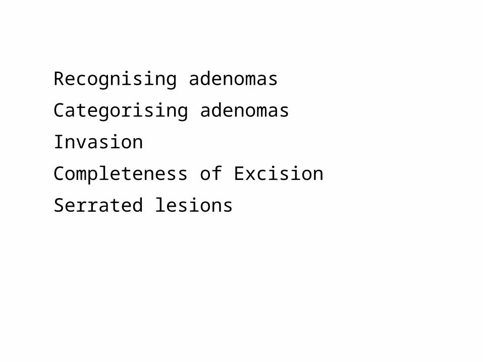
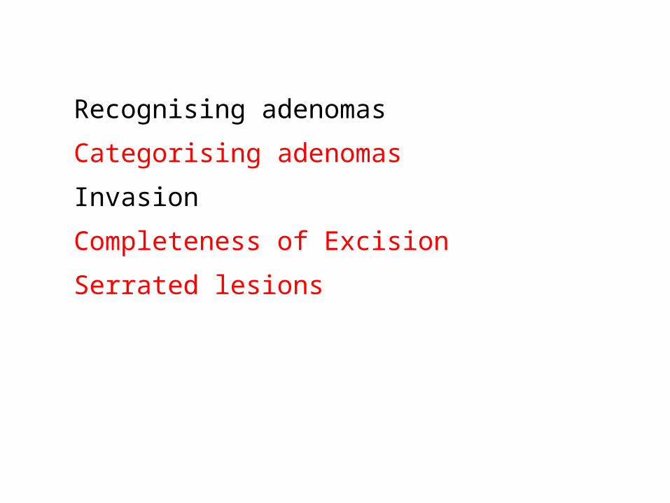
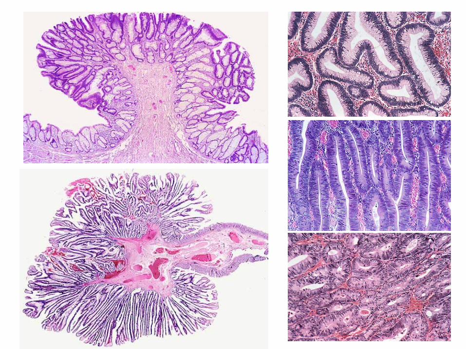
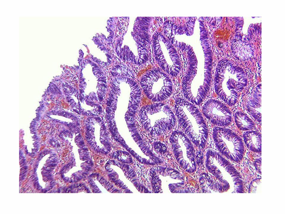
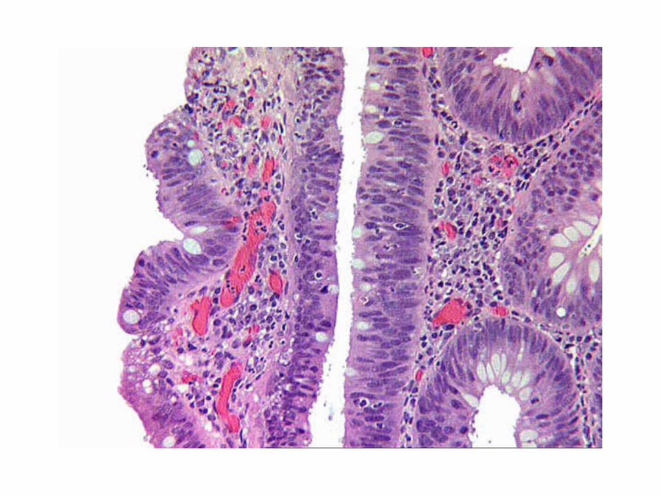
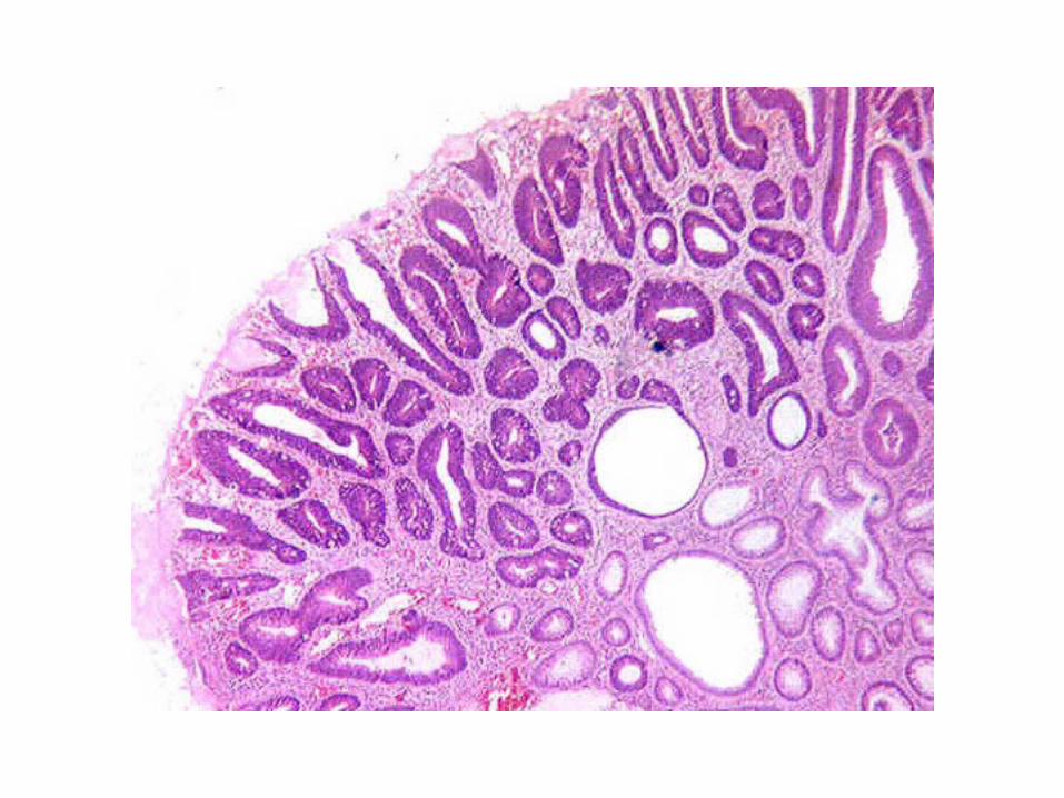
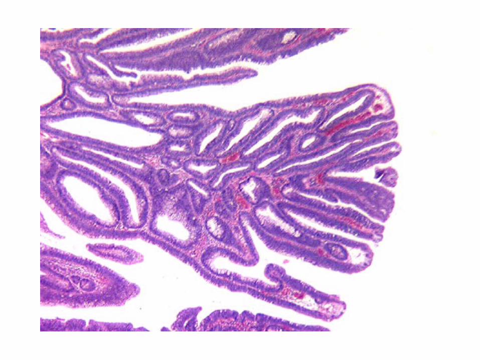
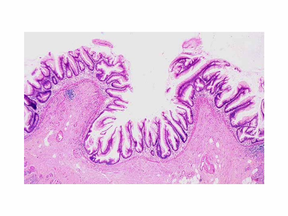
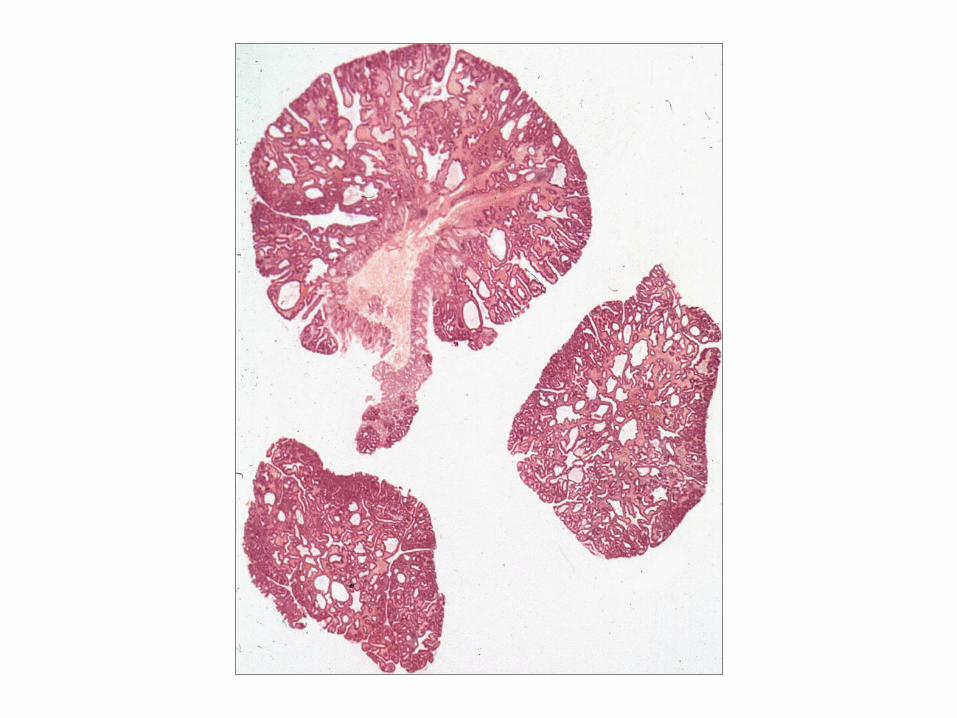
Recognising adenomas
Categorising adenomas
Invasion
Completeness of Excision
Serrated lesions
Recognising adenomas
Categorising adenomas
Invasion
Completeness of Excision
Serrated lesions
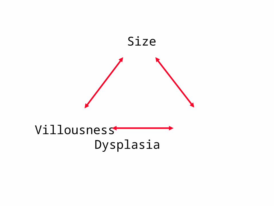
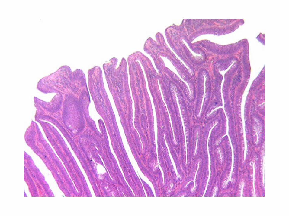
Size
Villousness Dysplasia
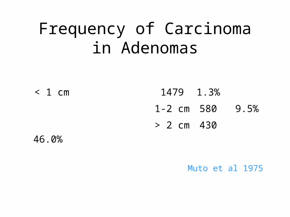
Frequency of Carcinoma in Adenomas
< 1 cm 1479 1.3%
1-2 cm 580 9.5%
> 2 cm 430 46.0%
Muto et al 1975
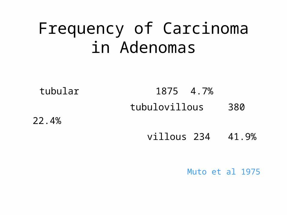
Frequency of Carcinoma in Adenomas
tubular 1875 4.7%
tubulovillous 380 22.4%
villous 234 41.9%
Muto et al 1975
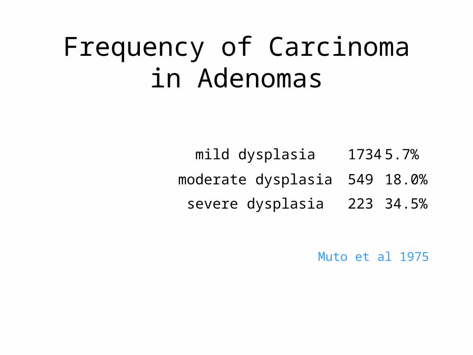
Frequency of Carcinoma in Adenomas
mild dysplasia 1734 5.7%
moderate dysplasia 549 18.0%
severe dysplasia 223 34.5%
Muto et al 1975
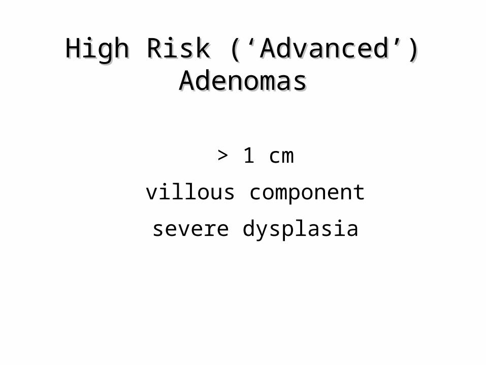
High Risk (‘Advanced’) High Risk (‘Advanced’) AdenomasAdenomas
> 1 cm
villous component
severe dysplasia
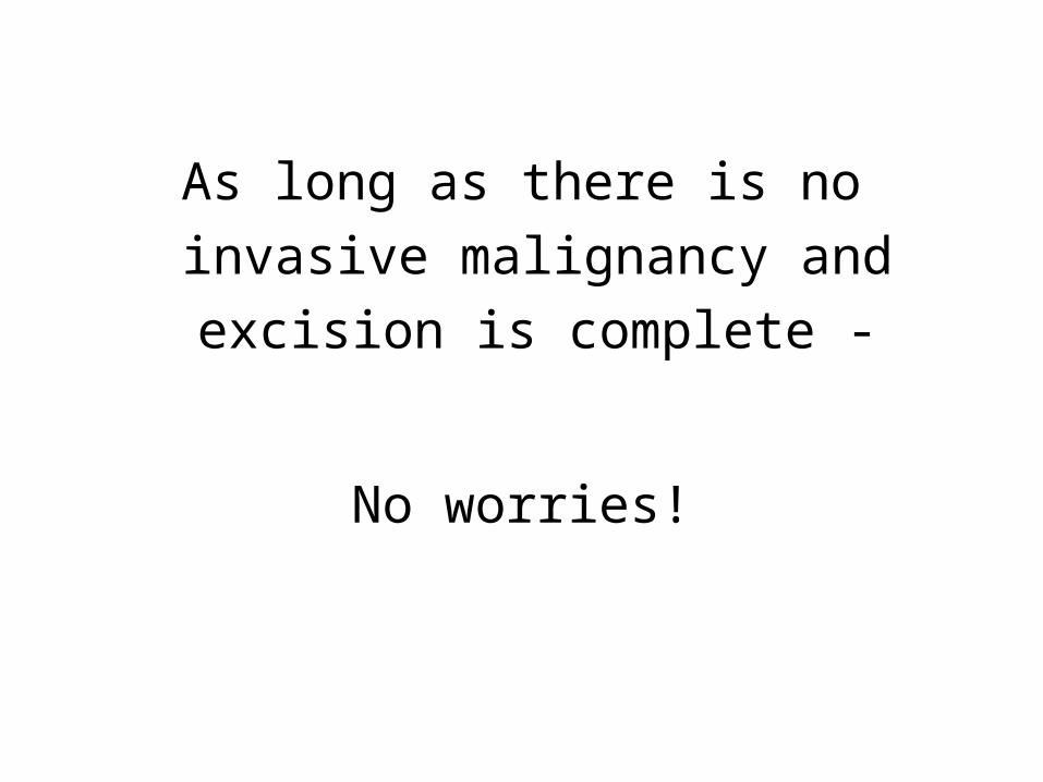
As long as there is no invasive malignancy and excision is
complete -
No worries!
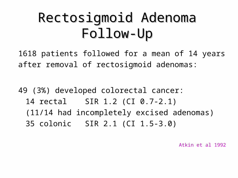
Rectosigmoid Adenoma Follow-Rectosigmoid Adenoma Follow-UpUp
1618 patients followed for a mean of 14 years after removal of rectosigmoid adenomas:
49 (3%) developed colorectal cancer:14 rectal SIR 1.2 (CI 0.7-2.1) (11/14 had incompletely excised adenomas)35 colonic SIR 2.1 (CI 1.5-3.0)
Atkin et al 1992
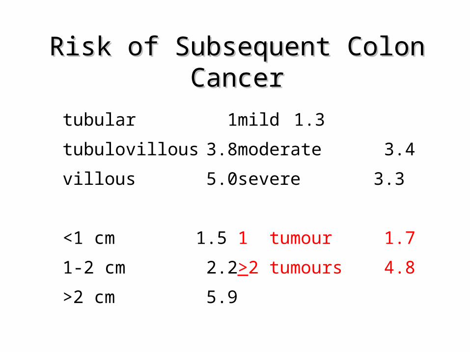
Risk of Subsequent Colon Risk of Subsequent Colon CancerCancer
tubular 1 mild 1.3
tubulovillous 3.8 moderate 3.4
villous 5.0 severe 3.3
<1 cm 1.5 1 tumour 1.7
1-2 cm 2.2 >2 tumours 4.8
>2 cm 5.9
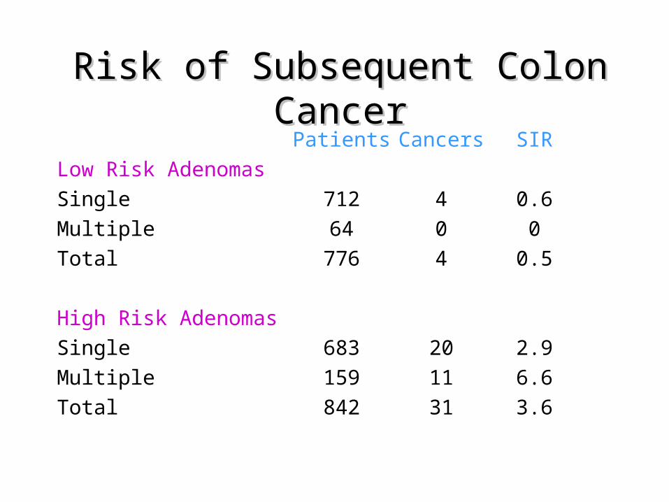
Risk of Subsequent Colon Risk of Subsequent Colon CancerCancer
Patients Cancers SIRLow Risk AdenomasSingle 712 4 0.6Multiple 64 0 0Total 776 4 0.5
High Risk AdenomasSingle 683 20 2.9Multiple 159 11 6.6Total 842 31 3.6
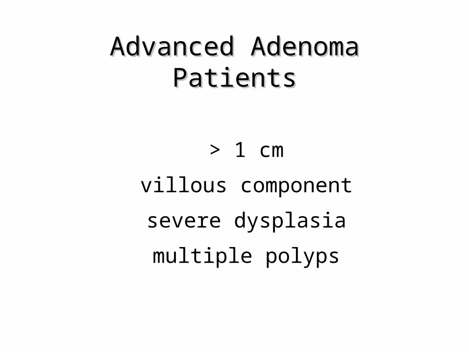
Advanced Adenoma PatientsAdvanced Adenoma Patients
> 1 cm
villous component
severe dysplasia
multiple polyps
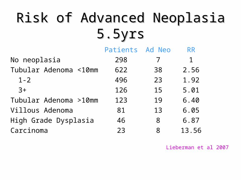
Risk of Advanced Neoplasia Risk of Advanced Neoplasia 5.5yrs5.5yrs
Patients Ad Neo RRNo neoplasia 298 7 1Tubular Adenoma <10mm 622 38 2.56
1-2 496 23 1.923+ 126 15 5.01
Tubular Adenoma >10mm 123 19 6.40Villous Adenoma 81 13 6.05High Grade Dysplasia 46 8 6.87Carcinoma 23 8 13.56
Lieberman et al 2007
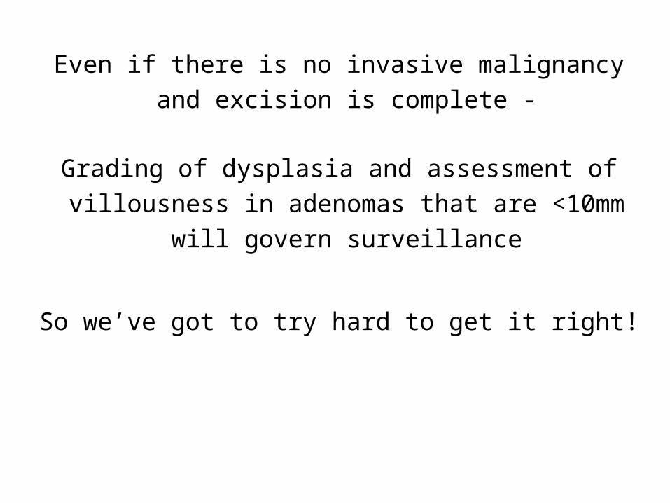
Even if there is no invasive malignancy and excision is complete -
Grading of dysplasia and assessment of villousness in adenomas that are <10mm will
govern surveillance
So we’ve got to try hard to get it right!
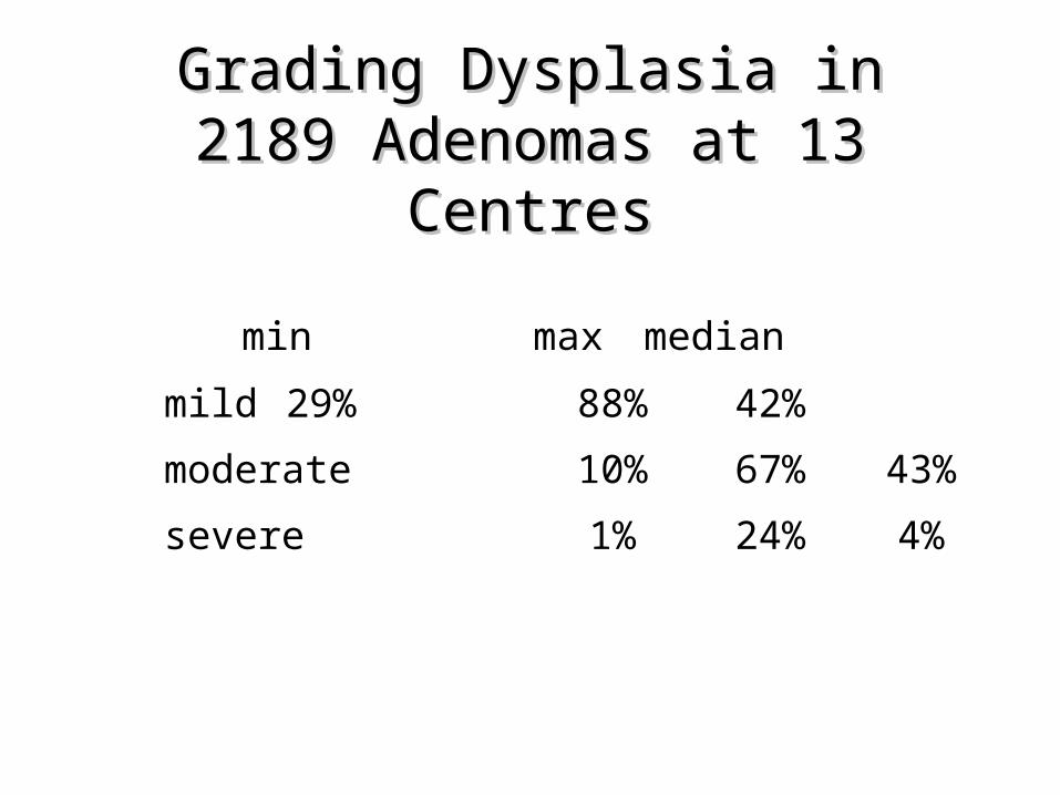
Grading Dysplasia in 2189 Grading Dysplasia in 2189 Adenomas at 13 CentresAdenomas at 13 Centres
min max median
mild 29% 88% 42%
moderate 10% 67%43%
severe 1% 24% 4%

Low grade and high grade
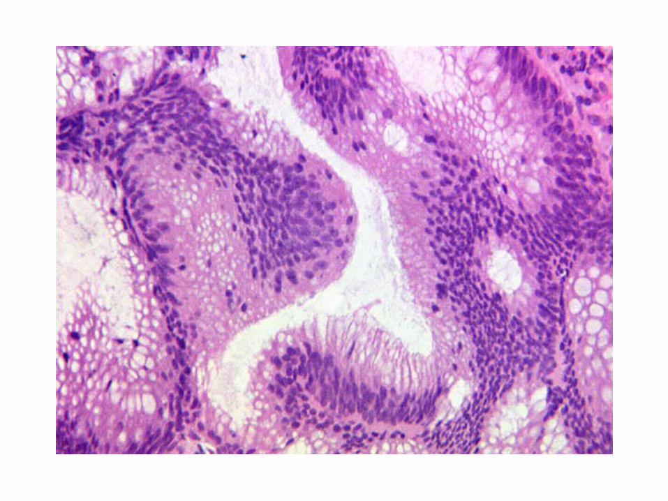
High Grade DysplasiaHigh Grade Dysplasia
Expected in <5% of all adenomas
Equates to ‘intramucosal adenocarcinoma’
Involves more than 1-2 glands
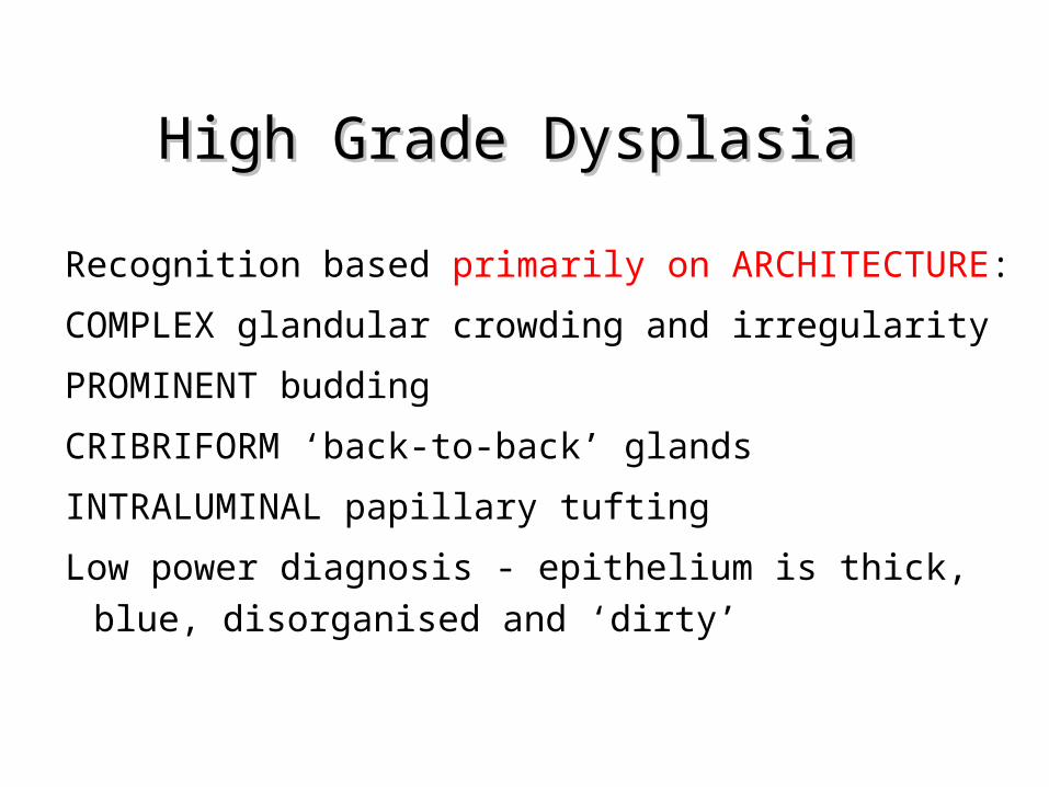
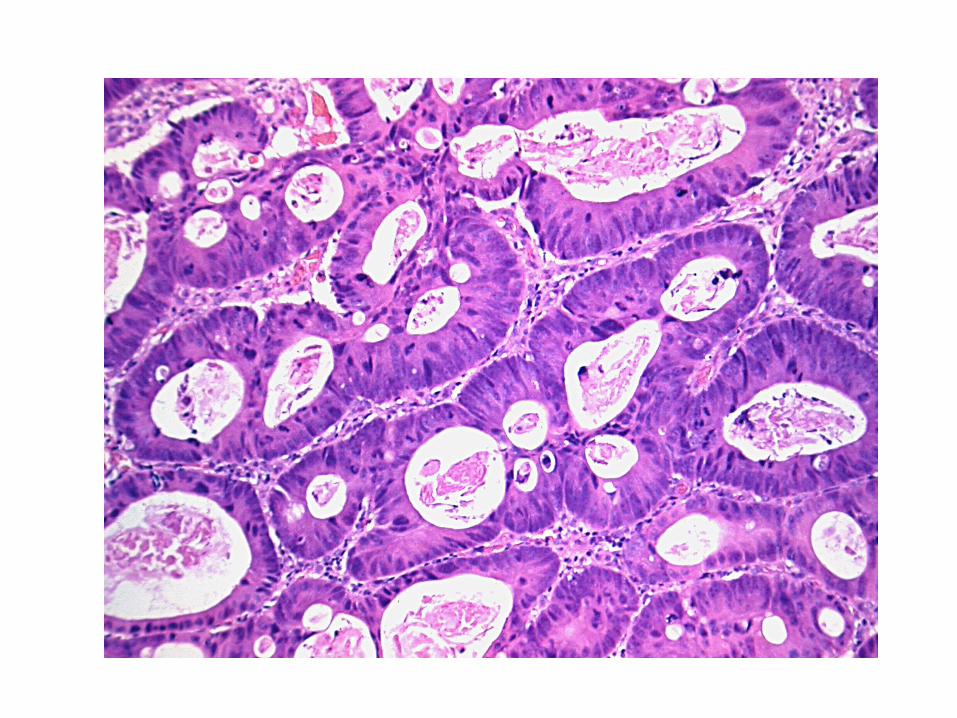
High Grade DysplasiaHigh Grade Dysplasia
Recognition based primarily on ARCHITECTURE:
COMPLEX glandular crowding and irregularity
PROMINENT budding
CRIBRIFORM ‘back-to-back’ glands
INTRALUMINAL papillary tufting
Low power diagnosis - epithelium is thick, blue, disorganised and ‘dirty’
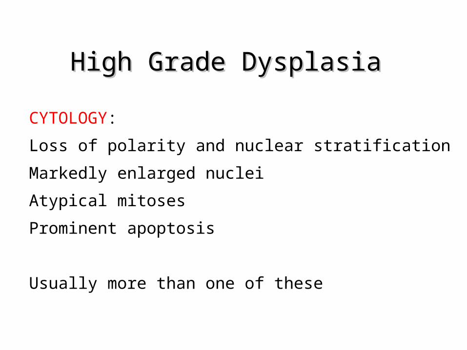
High Grade DysplasiaHigh Grade Dysplasia
CYTOLOGY:
Loss of polarity and nuclear stratification
Markedly enlarged nuclei
Atypical mitoses
Prominent apoptosis
Usually more than one of these
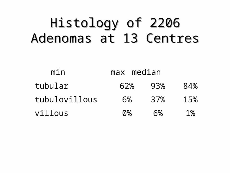
Histology of 2206 Adenomas Histology of 2206 Adenomas at 13 Centresat 13 Centres
min max median
tubular 62% 93% 84%
tubulovillous 6% 37% 15%
villous 0% 6% 1%

Reproducibility of Identifying Reproducibility of Identifying VillousnessVillousness
– 3 observers– Overall agreement 61%
Jensen et al 1995

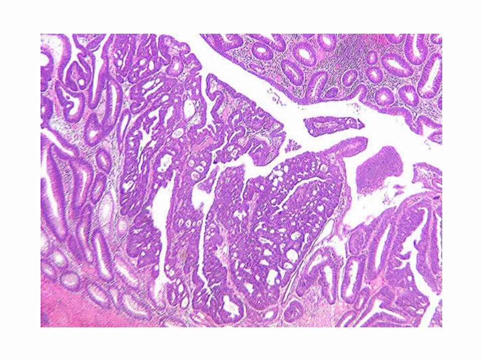
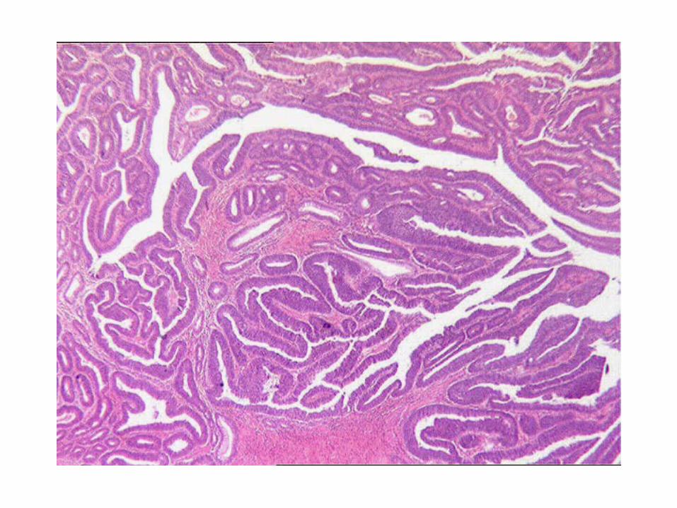
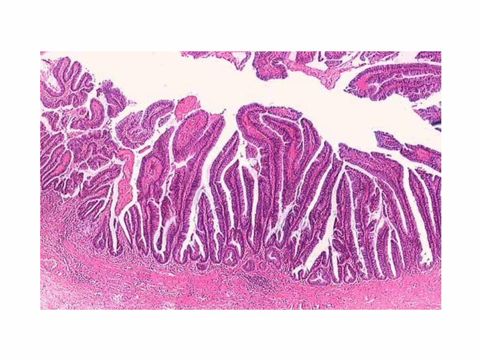
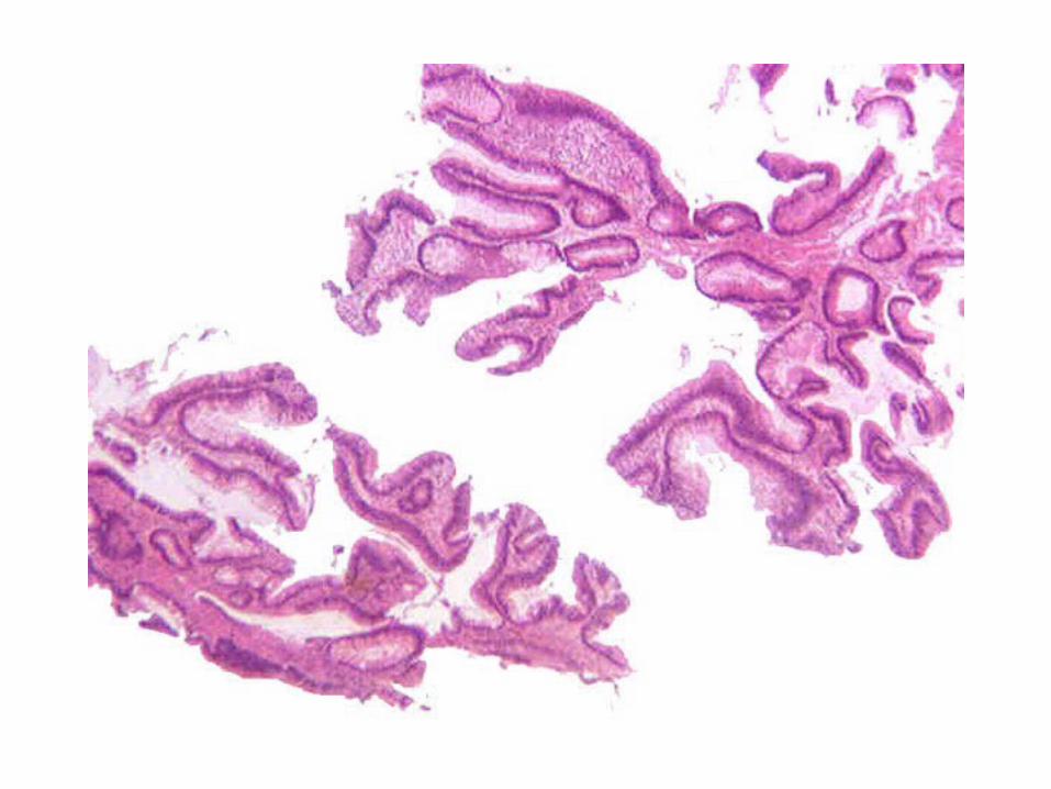
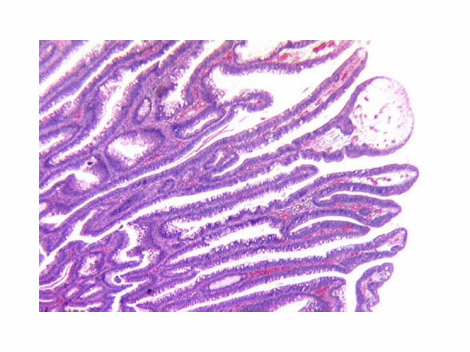
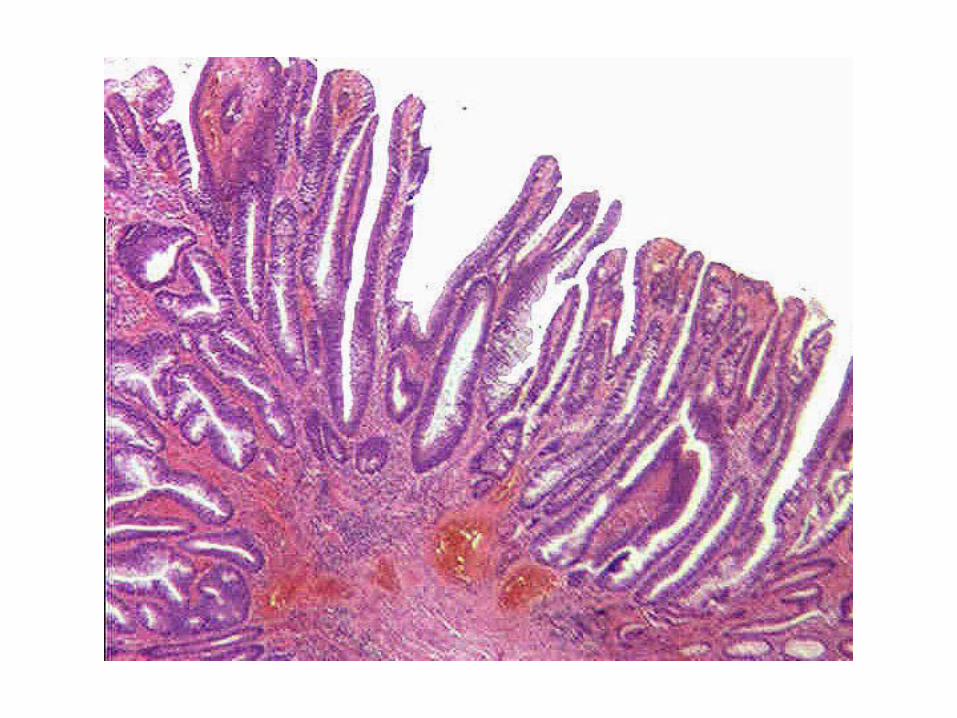
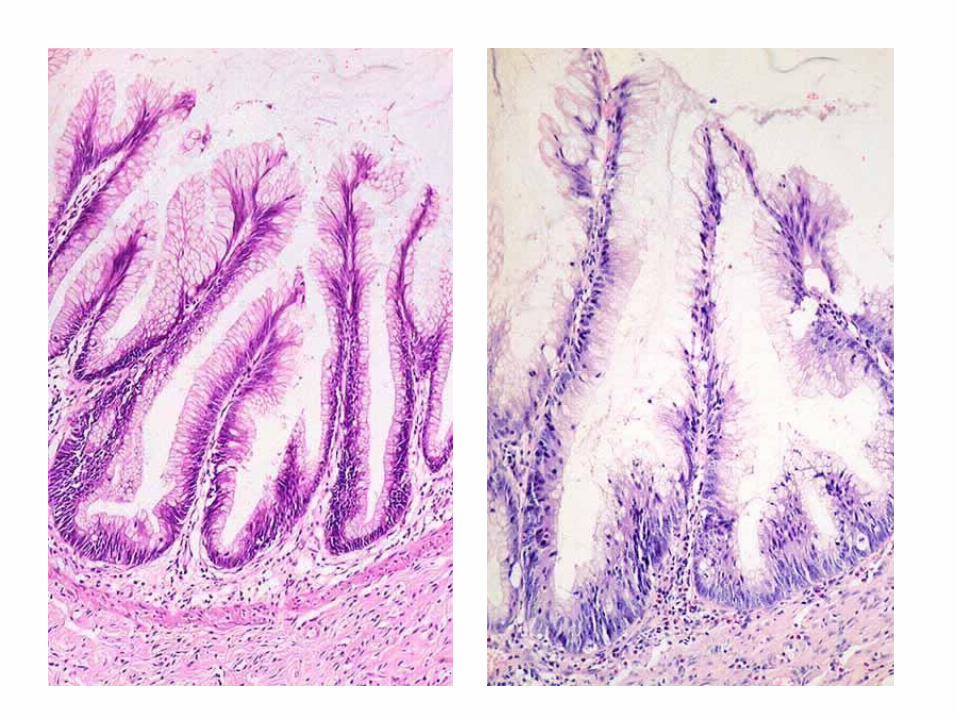
Tubulovillous AdenomasTubulovillous Adenomas
The 20% Rule
Neoplastic VilliNeoplastic Villi
Classical
Palmate
Foreshortened
May have prominent low grade mucinous epithelium
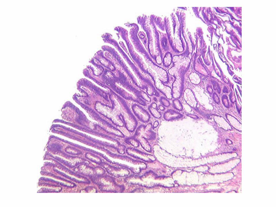
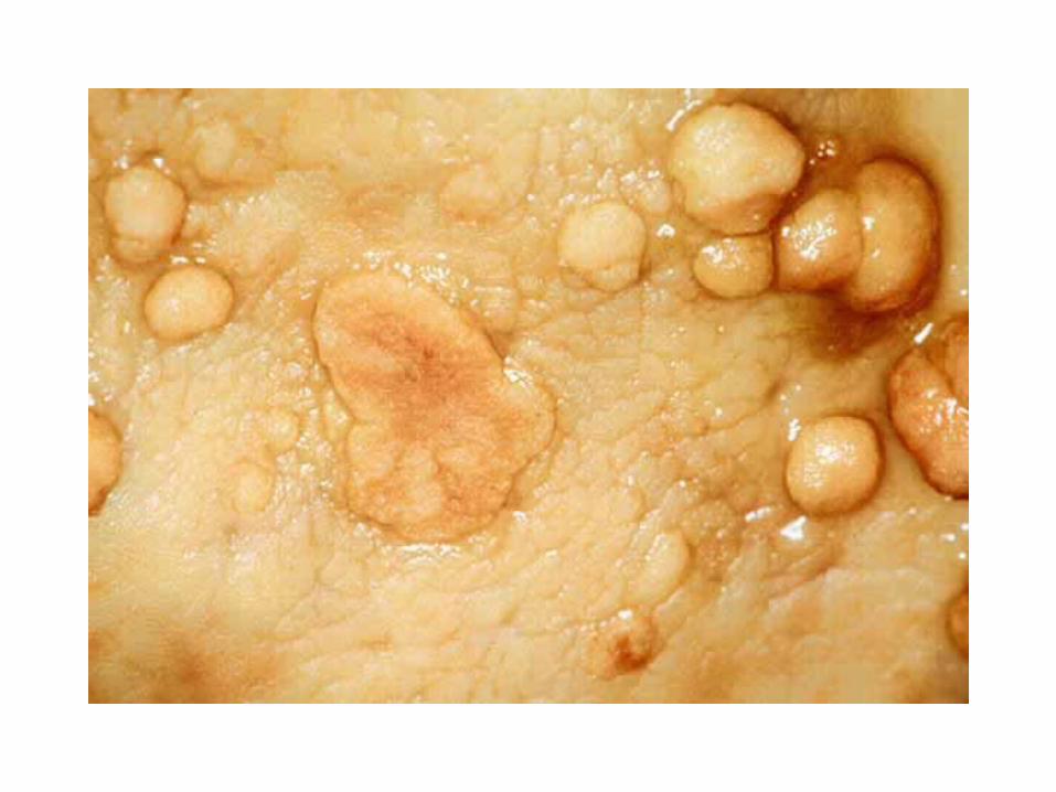
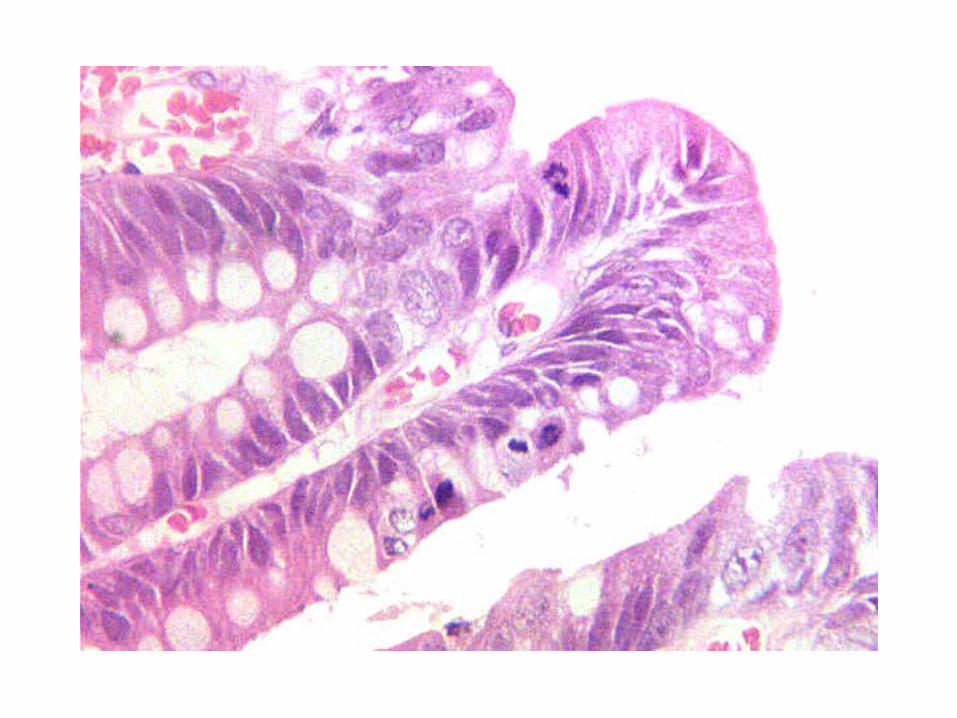
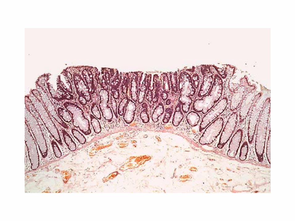
Flat AdenomasFlat Adenomas
– thickness does not exceed twice that of adjacent mucosa
– more often right sided– usually small (<1cm) with tubular growth
pattern– more often high grade dysplasia– 40% contain carcinoma– uncommon because no chromoendoscopy
Muto et al 1985
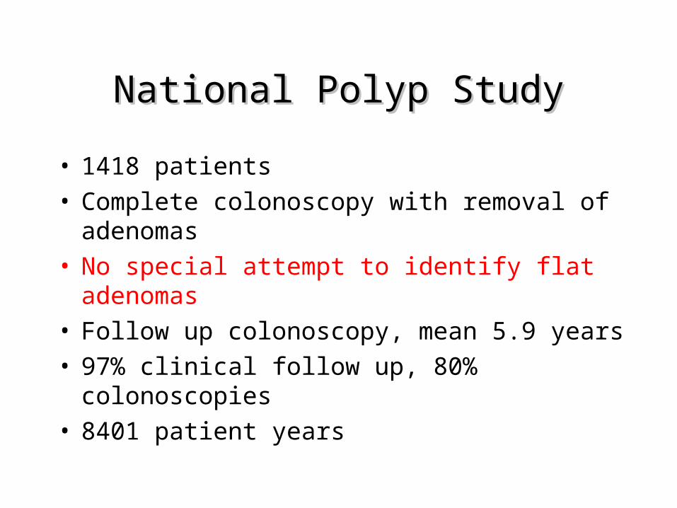
National Polyp StudyNational Polyp Study
• 1418 patients• Complete colonoscopy with removal of
adenomas• No special attempt to identify flat
adenomas• Follow up colonoscopy, mean 5.9 years• 97% clinical follow up, 80% colonoscopies• 8401 patient years
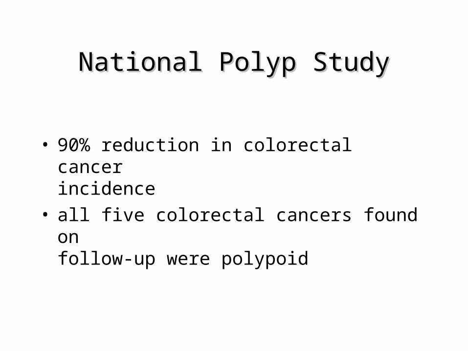
National Polyp StudyNational Polyp Study
• 90% reduction in colorectal cancer incidence
• all five colorectal cancers found on follow-up were polypoid
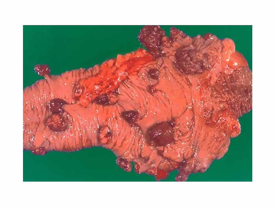
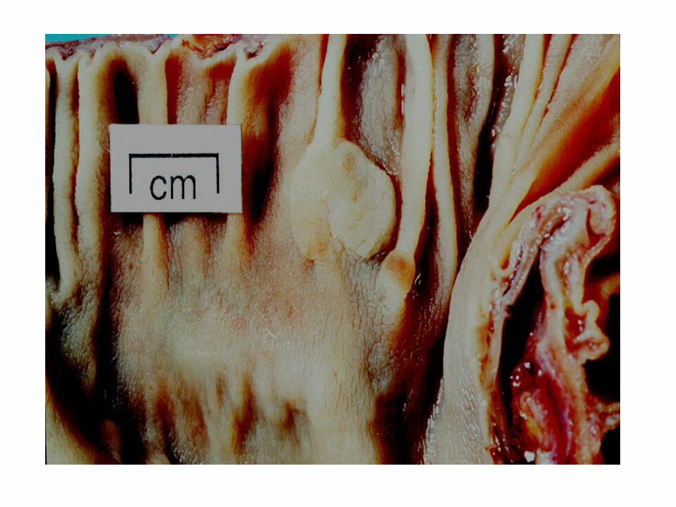
Macroscopic Examination & Macroscopic Examination & Trimming of PolypsTrimming of Polyps
• Size - to nearest millimetre in formalin fixed specimen (whole polyps)
• Polypoid lesions• Fixed intact• Bisect through stalk if <10mm• If larger, trim to leave central intact stalk• At least three levels of stalk
• Sessile lesions pinned out and all-embedded after inking margins
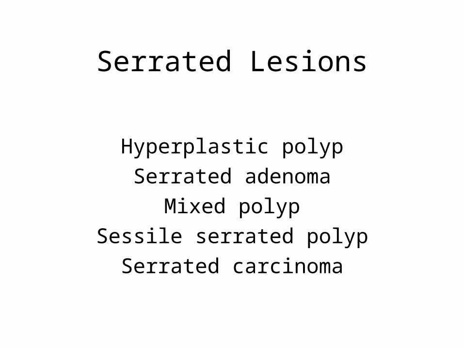
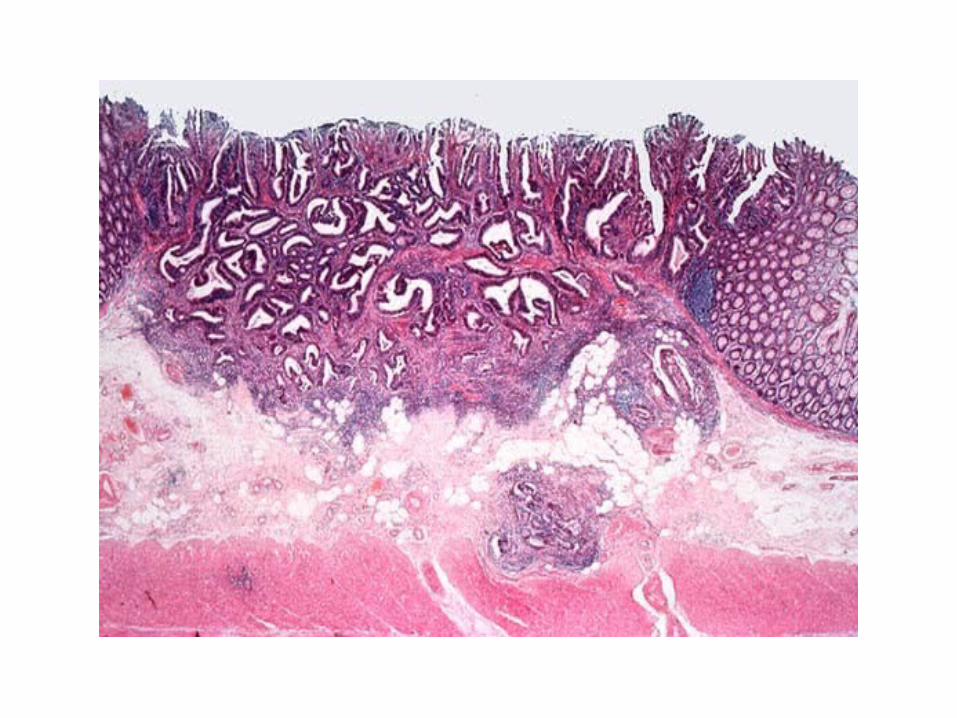
Serrated Lesions
Hyperplastic polypSerrated adenoma
Mixed polypSessile serrated polypSerrated carcinoma
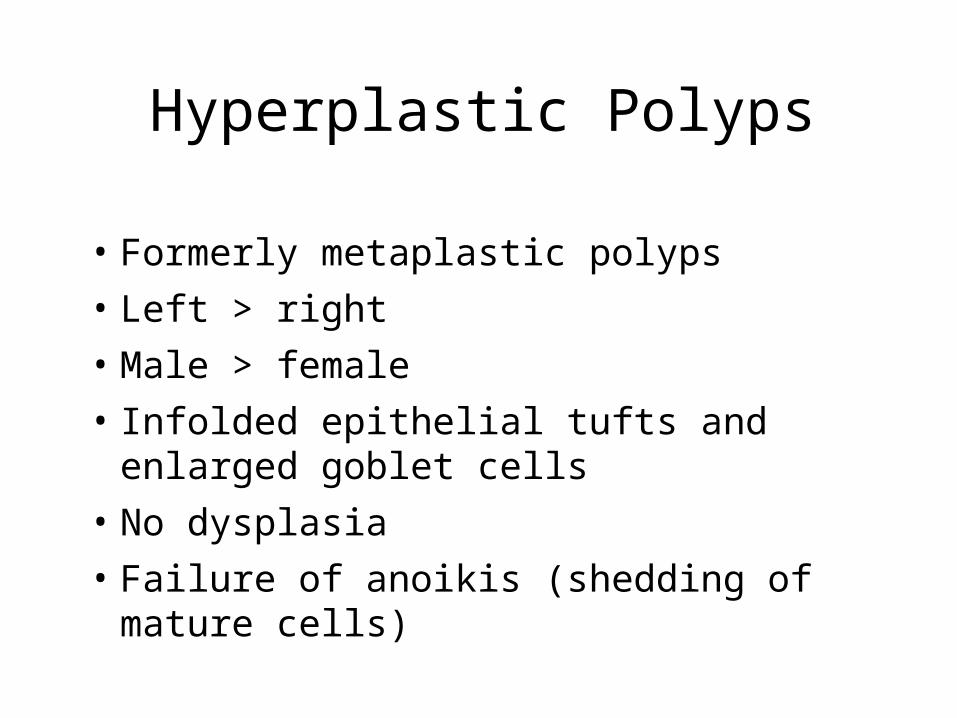
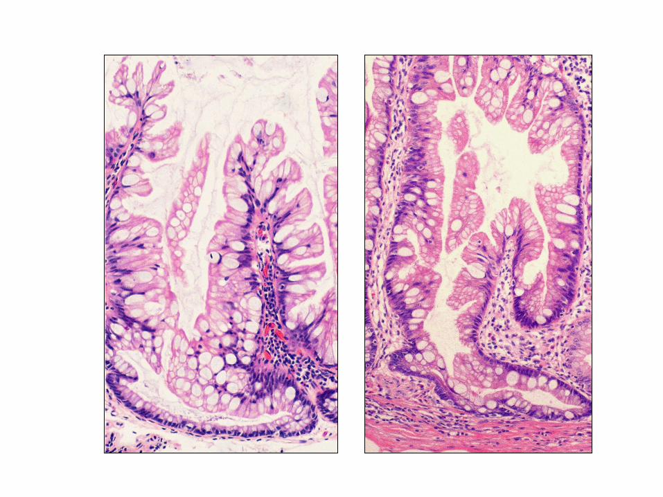
Hyperplastic Polyps
• Formerly metaplastic polyps• Left > right• Male > female • Infolded epithelial tufts and enlarged
goblet cells• No dysplasia• Failure of anoikis (shedding of mature
cells)
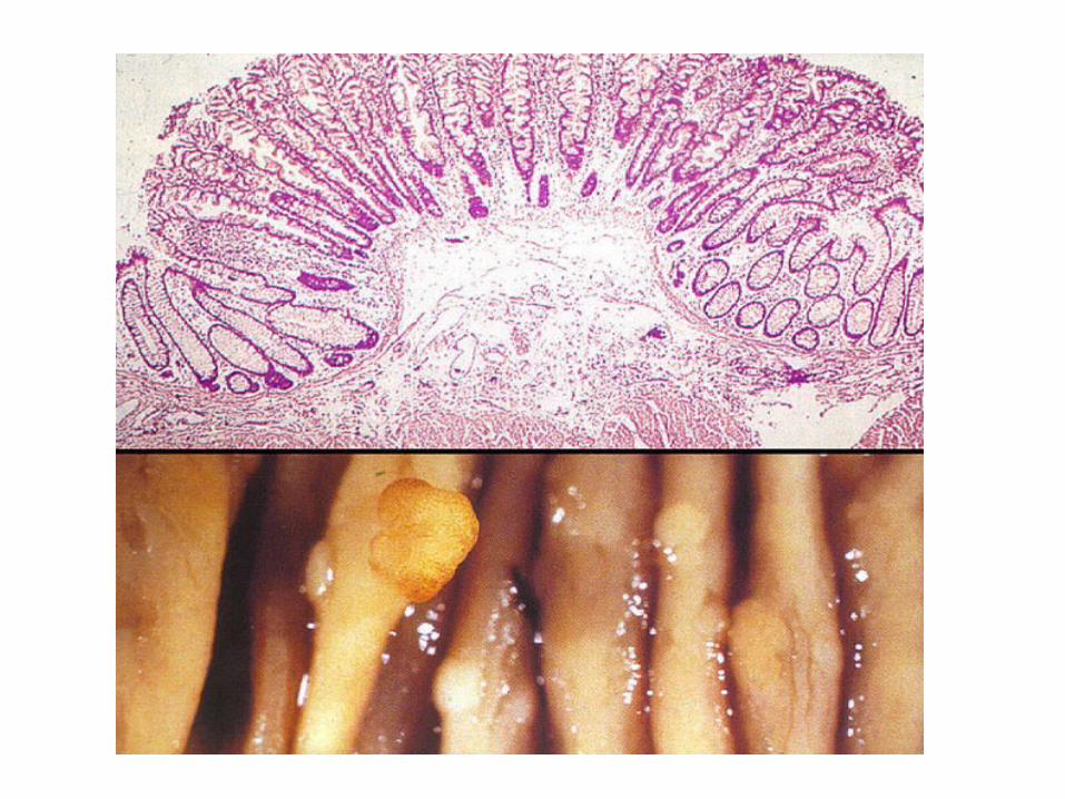
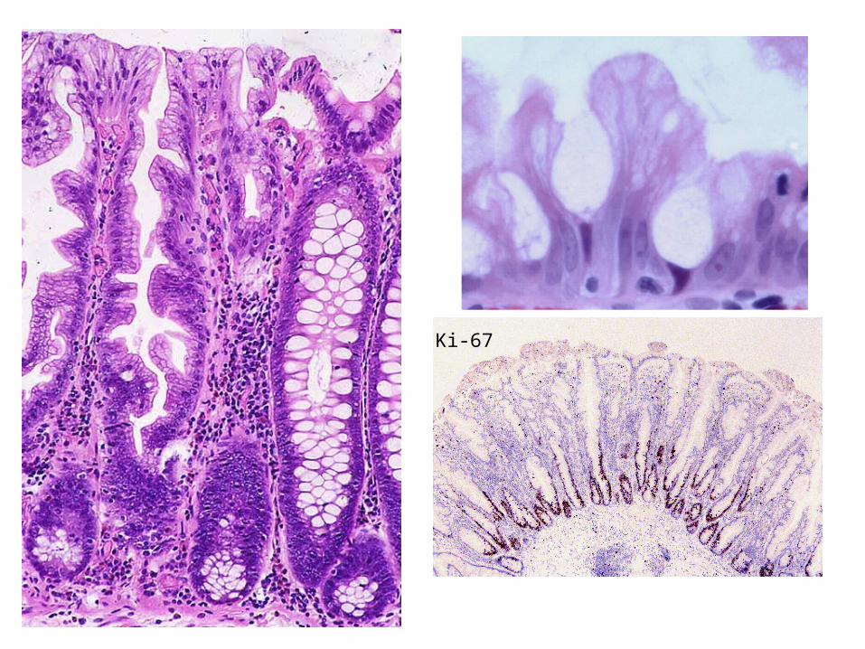
Ki-67

Hyperplastic Polyp
Increase in frequency with age
17 times commoner in colons with carcinoma
Similar dietary and lifestyle risk factors to CRC
K-ras mutation common
Clonal
Monocryptal?
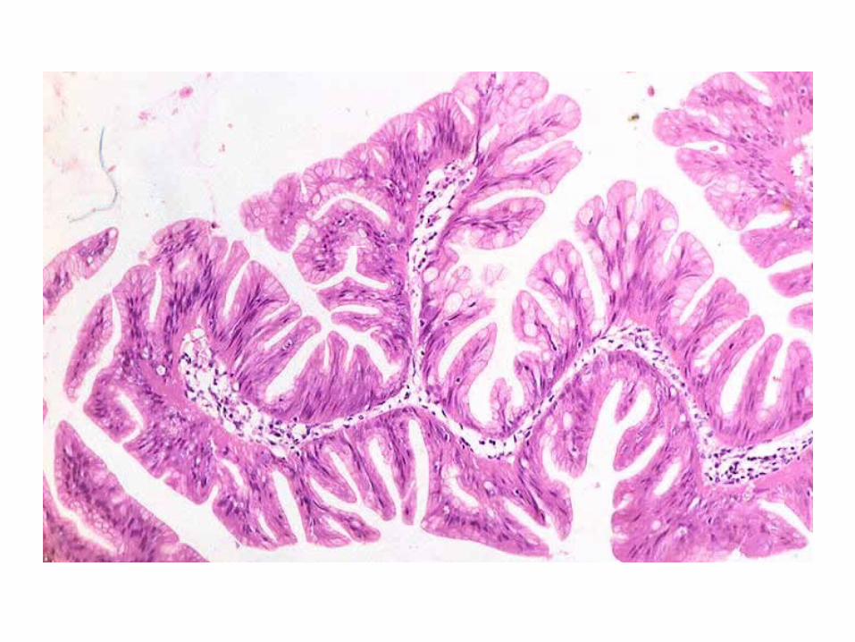
Serrated Adenoma
Dysplasia by definition
Eosinophilic cytoplasm
Pseudostratified, ‘pencillate’ nuclei
May be tubular, tubulovillous or villous
Invade to give serrated carcinoma
Longacre & Fenoglio-Preiser 1990
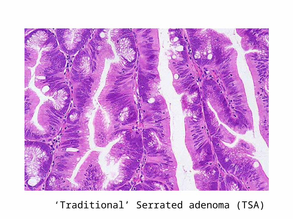
‘Traditional’ Serrated adenoma (TSA)
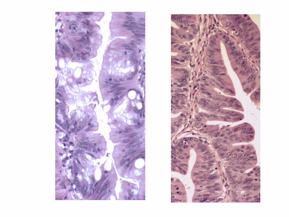
Mixed Polyps
Collision between hyperplastic polyp and adenoma
Dysplasia in Hyperplastic Polyp
Longacre & Fenoglio-Preiser 1990
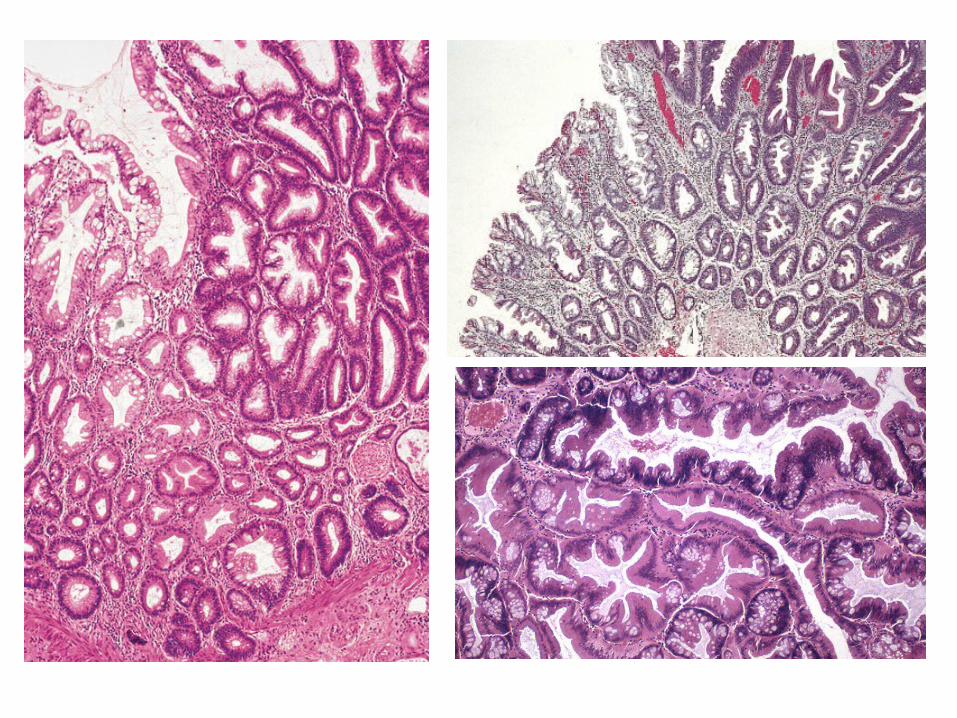
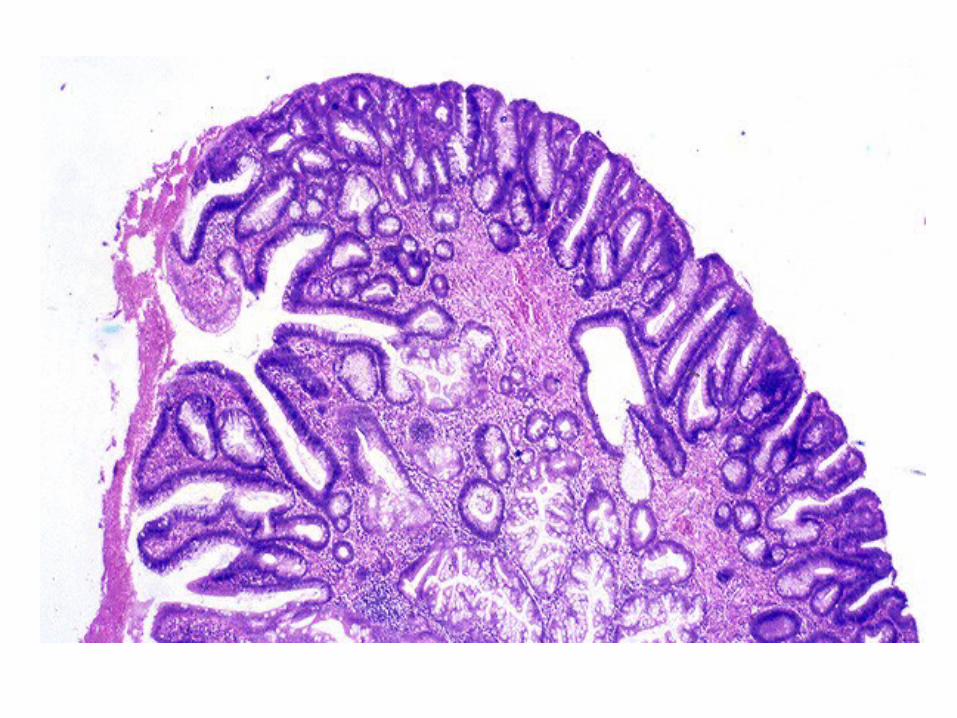
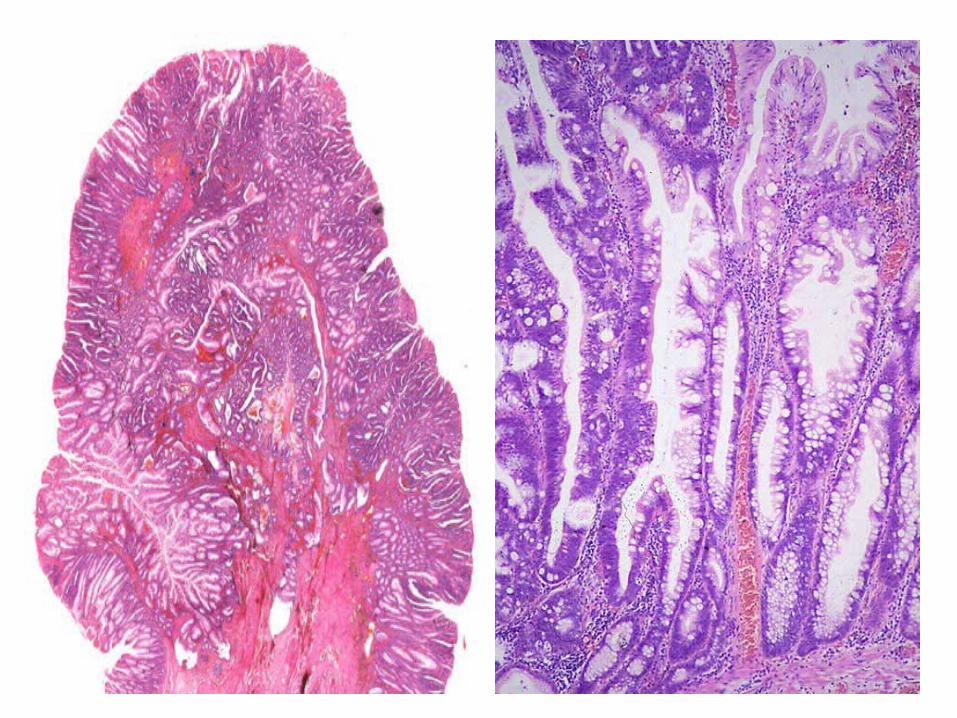
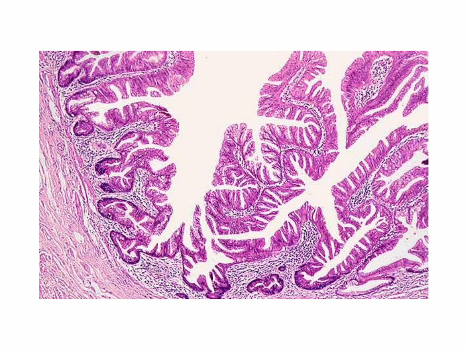
Sessile Serrated Polyp (Adenoma)
• Serrated polyps with unusual architectural features
• No conventional dysplasia but may have ‘nuclear atypia’ or ‘hypermucinous’ change
• Right colon
• Females > males
• Large sessile, poorly defined
Torlakovic & Snover 1996
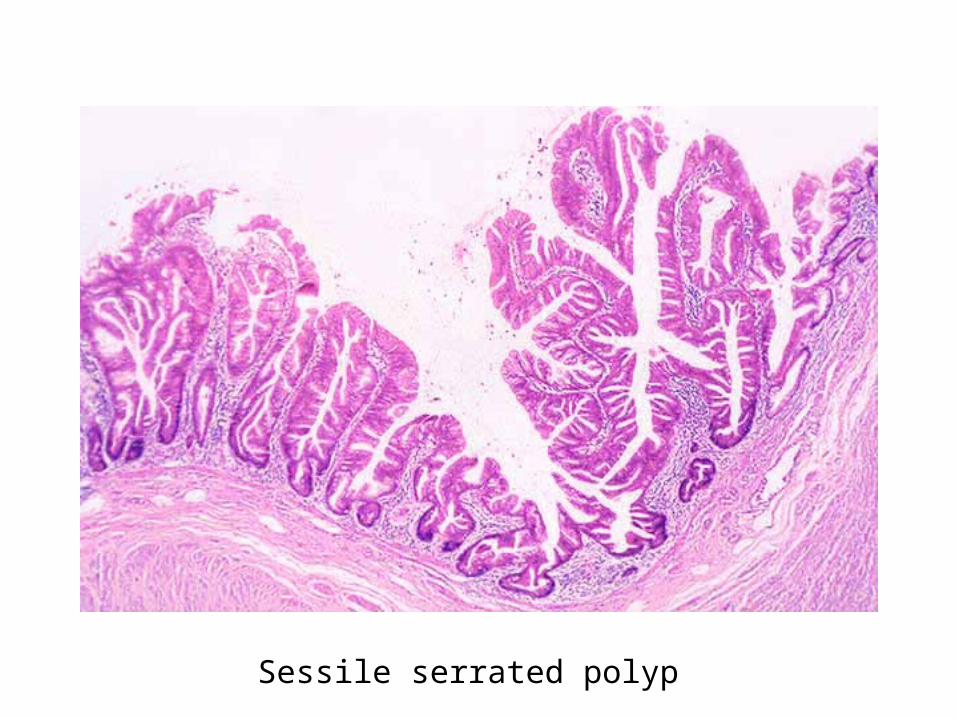
Sessile serrated polyp
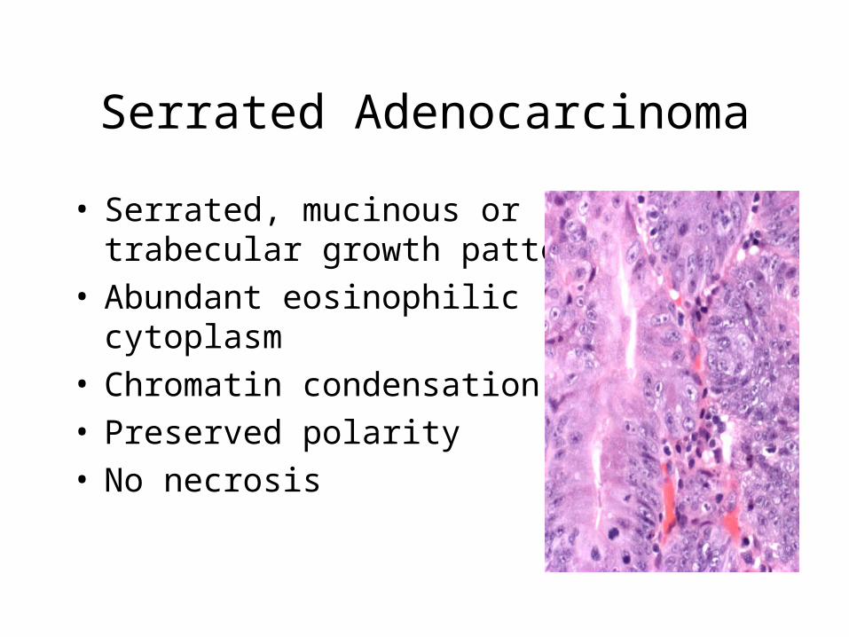
Serrated Adenocarcinoma
• Serrated, mucinous or trabecular growth pattern
• Abundant eosinophilic cytoplasm
• Chromatin condensation• Preserved polarity• No necrosis
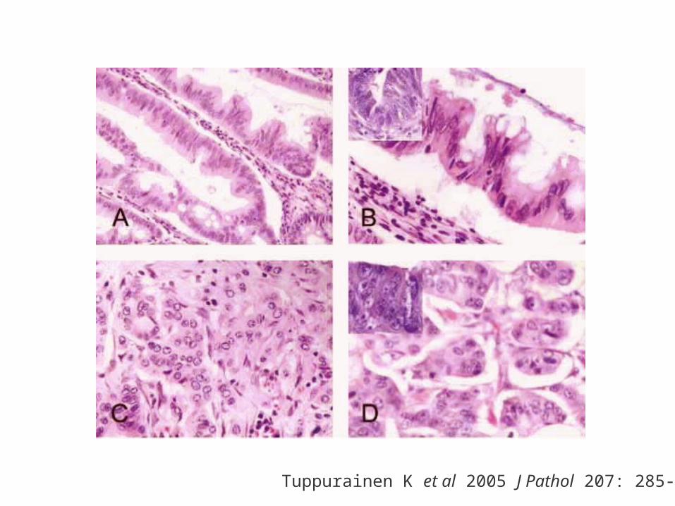
Tuppurainen K et al 2005 J Pathol 207: 285-94
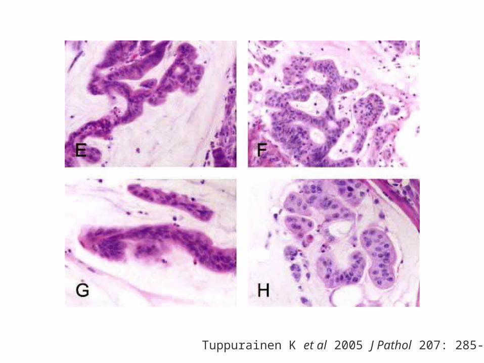
Tuppurainen K et al 2005 J Pathol 207: 285-94
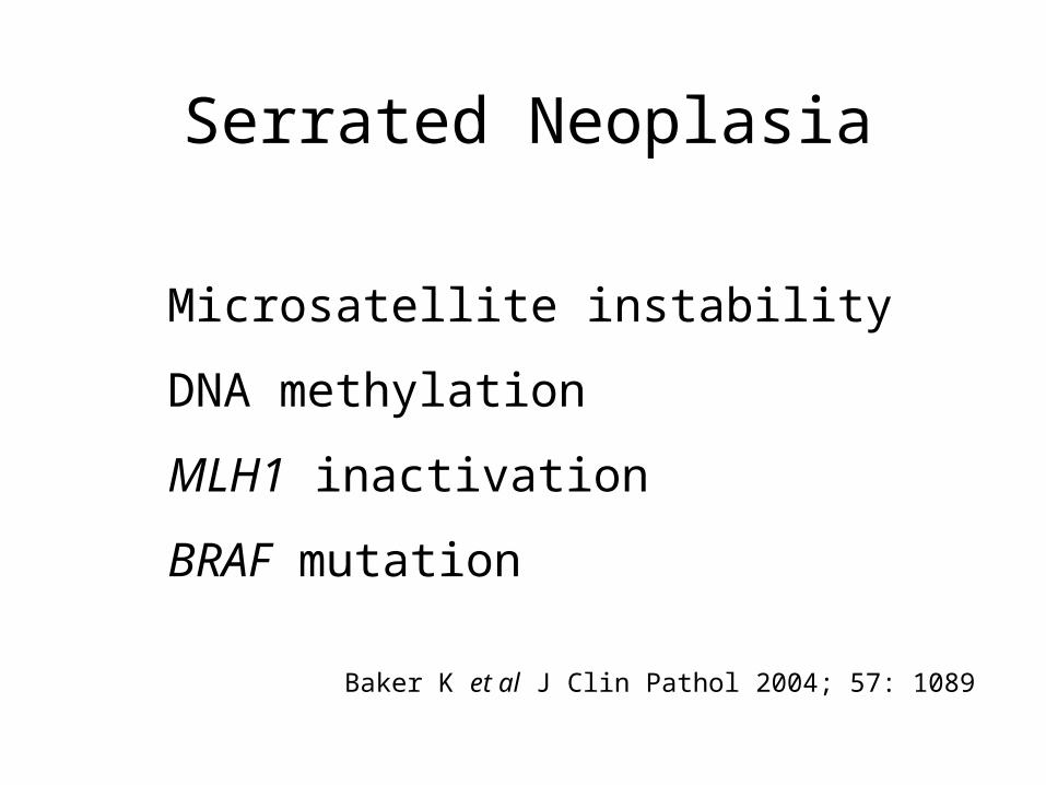
Serrated Neoplasia
Microsatellite instability
DNA methylation
MLH1 inactivation
BRAF mutation
Baker K et al J Clin Pathol 2004; 57: 1089
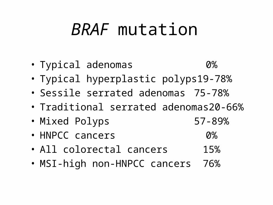
BRAF mutation
• Typical adenomas 0%• Typical hyperplastic polyps 19-78%• Sessile serrated adenomas 75-78%• Traditional serrated adenomas 20-66%• Mixed Polyps 57-89%
• HNPCC cancers 0%• All colorectal cancers 15%• MSI-high non-HNPCC cancers 76%
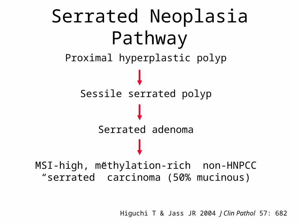
Serrated Neoplasia Pathway
Proximal hyperplastic polyp
Sessile serrated polyp
Serrated adenoma
MSI-high, methylation-rich non-HNPCC “serrated” carcinoma (50% mucinous)
Higuchi T & Jass JR 2004 J Clin Pathol 57: 682
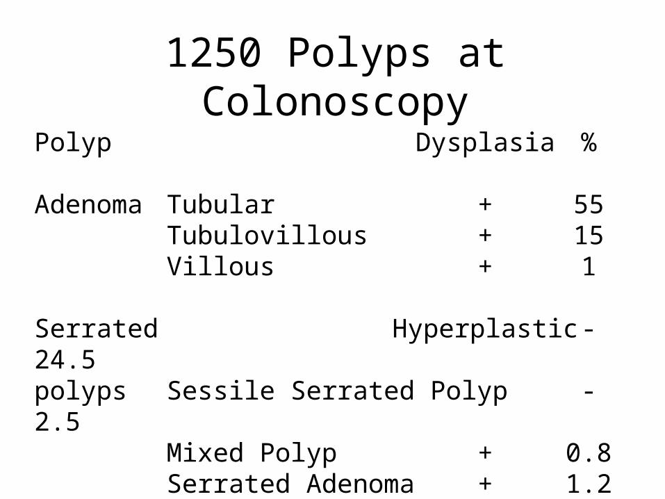
1250 Polyps at Colonoscopy
Polyp Dysplasia %
Adenoma Tubular + 55 Tubulovillous + 15
Villous + 1
Serrated Hyperplastic - 24.5polyps Sessile Serrated Polyp - 2.5
Mixed Polyp + 0.8Serrated Adenoma + 1.2
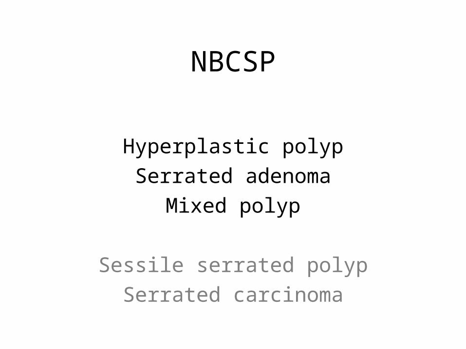
NBCSP
Hyperplastic polypSerrated adenoma
Mixed polyp
Sessile serrated polypSerrated carcinoma