Embed Size (px)
Citation preview

ASSESSMENT AND DIAGNOSIS

Overview
Pain: Underreported, Underdiagnosed and Undertreated
• Ongoing pain has been underreported, underdiagnosed, and undertreated in nearly all health care settings
• Individuals with pain that reduces quality of life should be encouraged to seek help
• Comprehensive assessment and treatments likely to produce best results
Institute of Medicine. Relieving Pain in America: A Blueprint for Transforming Prevention, Care, Education, and Research. The National Academies Press; Washington, DC: 2011.
Importance of Pain Assessment
Pain is a significant predictor of morbidity and mortality.
• Screen for red flags requiring immediate investigation and/or referral
• Identify underlying cause – Pain is better managed if the underlying causes are determined
and addressed
• Recognize type of pain to help guide selection of appropriate therapies for treatment of pain
• Determine baseline pain intensity to future enable assessment of efficacy of treatment
Forde G, Stanos S. J Fam Pract 2007; 56(8 Suppl Hot Topics):S21-30; Sokka T, Pincus T. Poster presentation at ACR 2005.
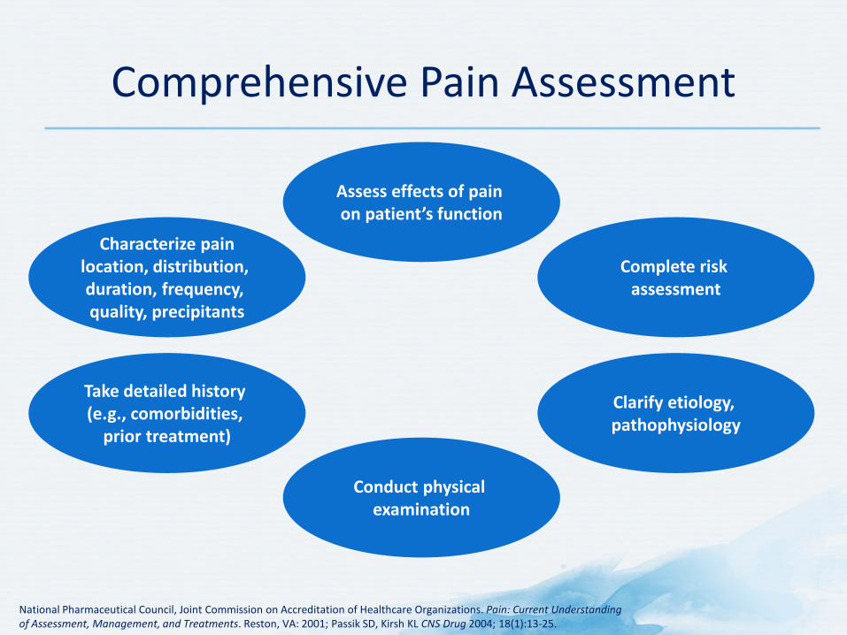
Comprehensive Pain Assessment
National Pharmaceutical Council, Joint Commission on Accreditation of Healthcare Organizations. Pain: Current Understanding of Assessment, Management, and Treatments. Reston, VA: 2001; Passik SD, Kirsh KL CNS Drug 2004; 18(1):13-25.
Characterize pain location, distribution, duration, frequency, quality, precipitants
Take detailed history (e.g., comorbidities,
prior treatment)
Conduct physical examination
Clarify etiology, pathophysiology
Complete risk assessment
Assess effects of pain on patient’s function
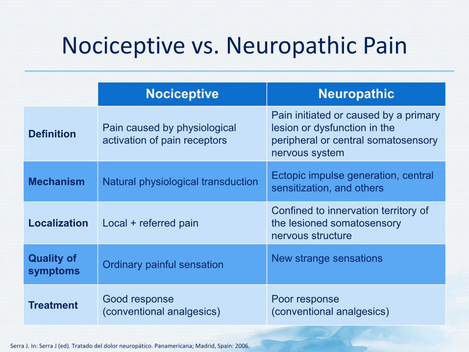
Nociceptive vs. Neuropathic Pain
Nociceptive Neuropathic
Definition Pain caused by physiological activation of pain receptors
Pain initiated or caused by a primary lesion or dysfunction in the peripheral or central somatosensory nervous system
Mechanism Natural physiological transduction Ectopic impulse generation, central sensitization, and others
Localization Local + referred pain Confined to innervation territory of the lesioned somatosensory nervous structure
Quality of symptoms Ordinary painful sensation New strange sensations
Treatment Good response (conventional analgesics)
Poor response (conventional analgesics)
Serra J. In: Serra J (ed). Tratado del dolor neuropático. Panamericana; Madrid, Spain: 2006.
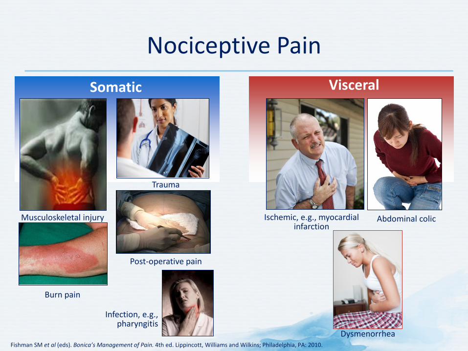
Nociceptive Pain
Fishman SM et al (eds). Bonica’s Management of Pain. 4th ed. Lippincott, Williams and Wilkins; Philadelphia, PA: 2010.
Trauma
Burn pain
Musculoskeletal injury
Post-operative pain
Infection, e.g., pharyngitis
Ischemic, e.g., myocardial infarction
Abdominal colic
Dysmenorrhea
Somatic Visceral
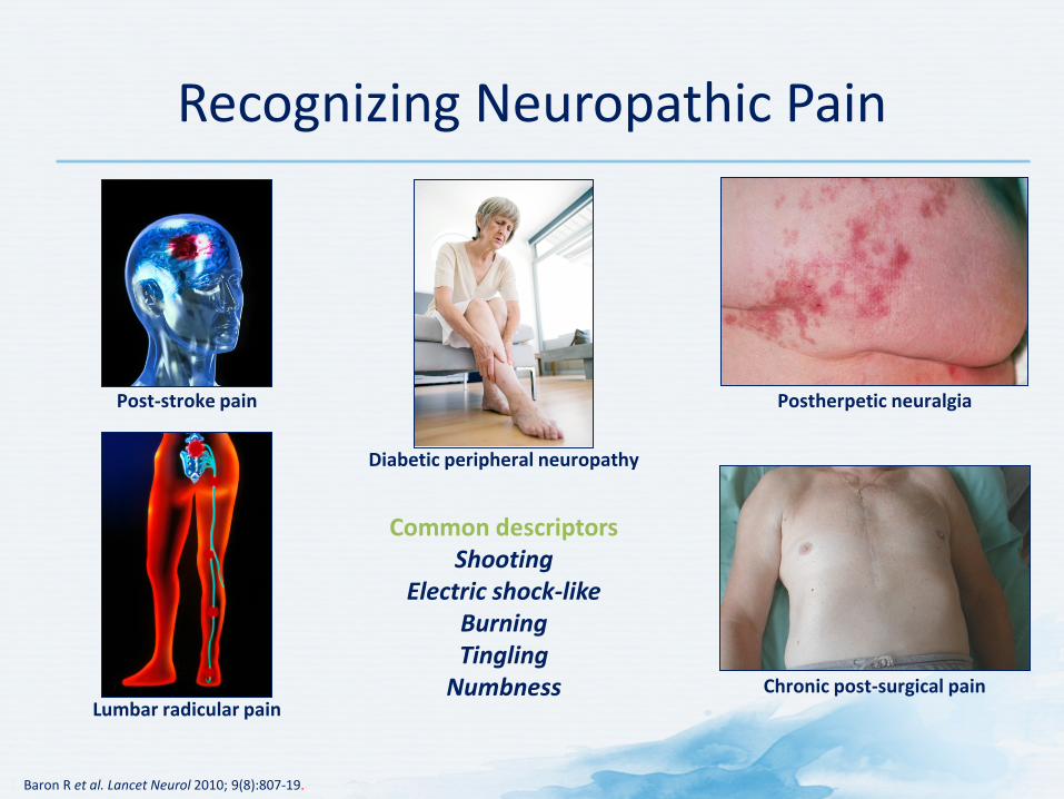
Recognizing Neuropathic Pain
1. Baron R et al. Lancet Neurol 2010; 9(8):807-19.
Common descriptors
Shooting Electric shock-like
Burning Tingling
Numbness
Postherpetic neuralgia
Lumbar radicular pain Chronic post-surgical pain
Post-stroke pain
Diabetic peripheral neuropathy

History
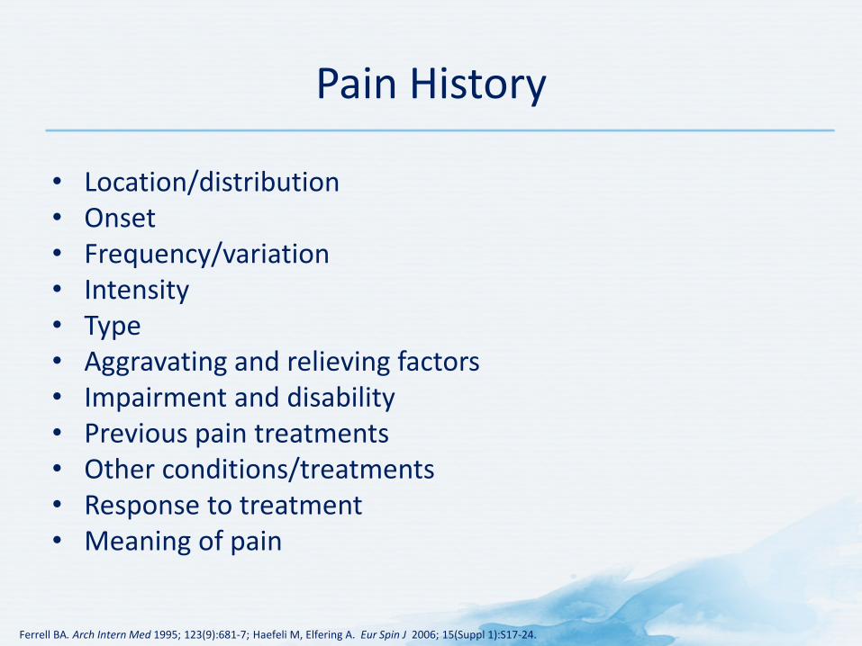
Pain History
• Location/distribution • Onset • Frequency/variation • Intensity • Type • Aggravating and relieving factors • Impairment and disability • Previous pain treatments • Other conditions/treatments • Response to treatment • Meaning of pain
Ferrell BA. Arch Intern Med 1995; 123(9):681-7; Haefeli M, Elfering A. Eur Spin J 2006; 15(Suppl 1):S17-24.
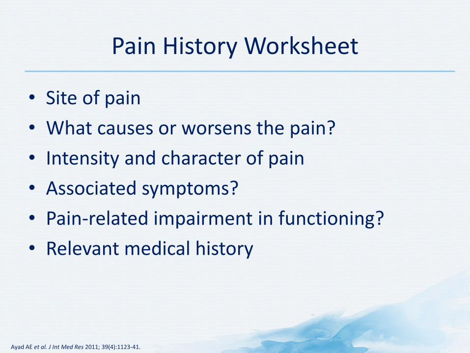
Pain History Worksheet
• Site of pain • What causes or worsens the pain? • Intensity and character of pain • Associated symptoms? • Pain-related impairment in functioning? • Relevant medical history
Ayad AE et al. J Int Med Res 2011; 39(4):1123-41.
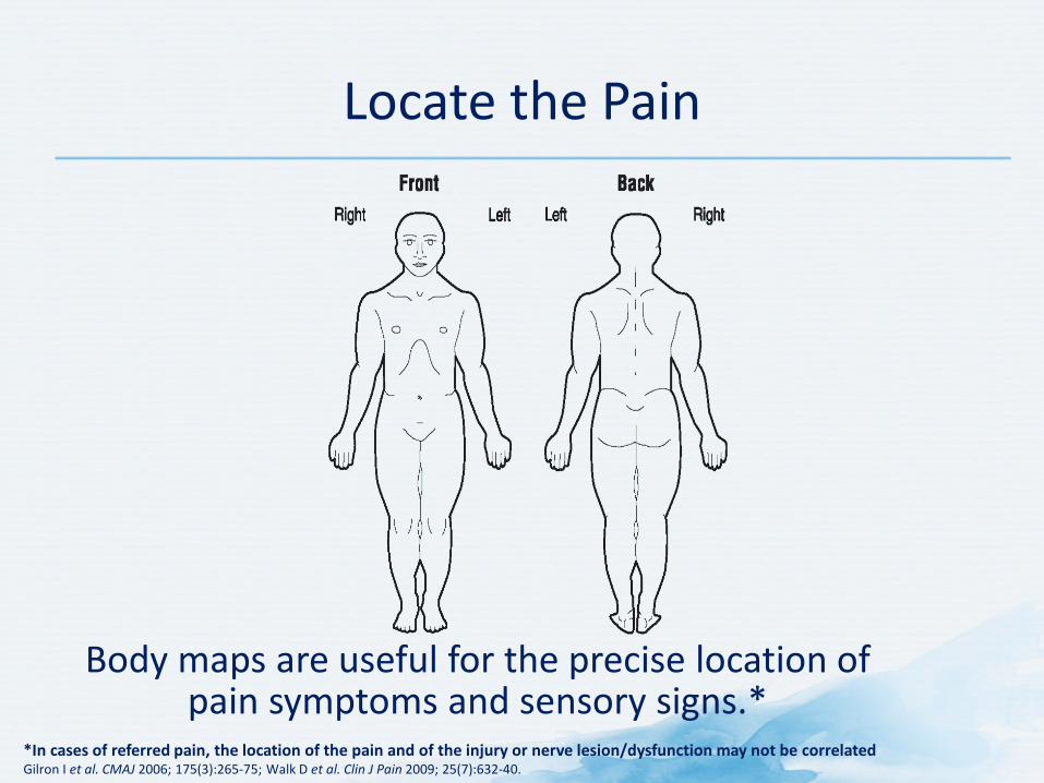
Locate the Pain
Body maps are useful for the precise location of pain symptoms and sensory signs.*
*In cases of referred pain, the location of the pain and of the injury or nerve lesion/dysfunction may not be correlated Gilron I et al. CMAJ 2006; 175(3):265-75; Walk D et al. Clin J Pain 2009; 25(7):632-40.
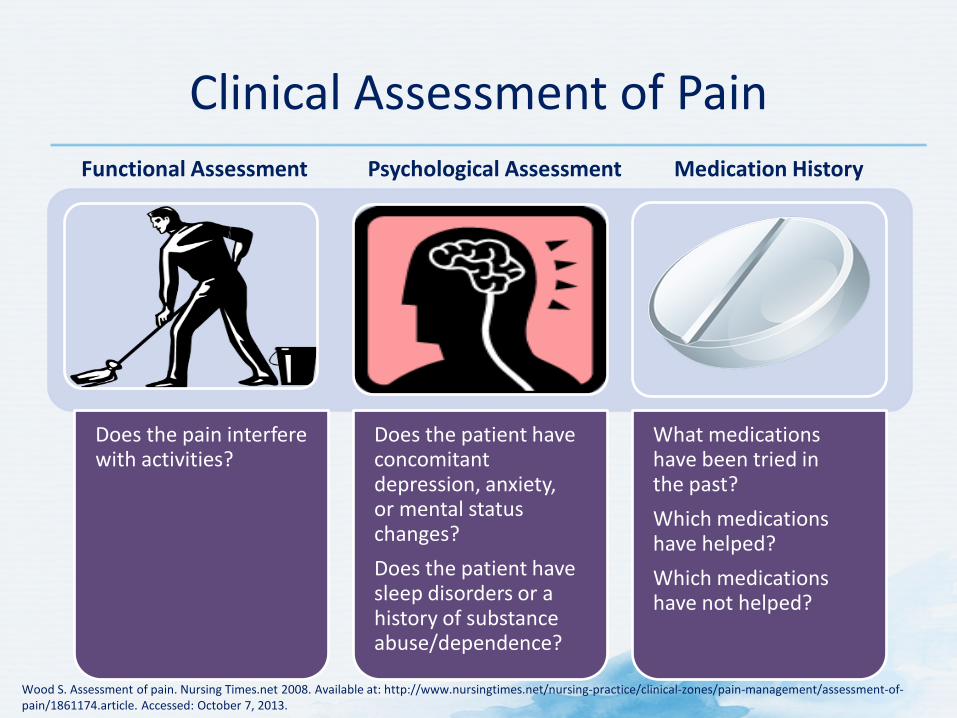
Clinical Assessment of Pain
Wood S. Assessment of pain. Nursing Times.net 2008. Available at: http://www.nursingtimes.net/nursing-practice/clinical-zones/pain-management/assessment-of-pain/1861174.article. Accessed: October 7, 2013.
Does the pain interfere with activities?
Does the patient have concomitant depression, anxiety, or mental status changes? Does the patient have sleep disorders or a history of substance abuse/dependence?
What medications have been tried in the past? Which medications have helped? Which medications have not helped?
Functional Assessment Psychological Assessment Medication History
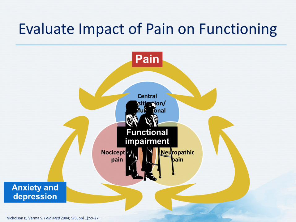
Central sensitization/ dysfunctional
pain
Neuropathic pain
Nociceptive pain
Nicholson B, Verma S. Pain Med 2004; 5(Suppl 1):S9-27.
Evaluate Impact of Pain on Functioning
Anxiety and depression
Pain
Functional impairment
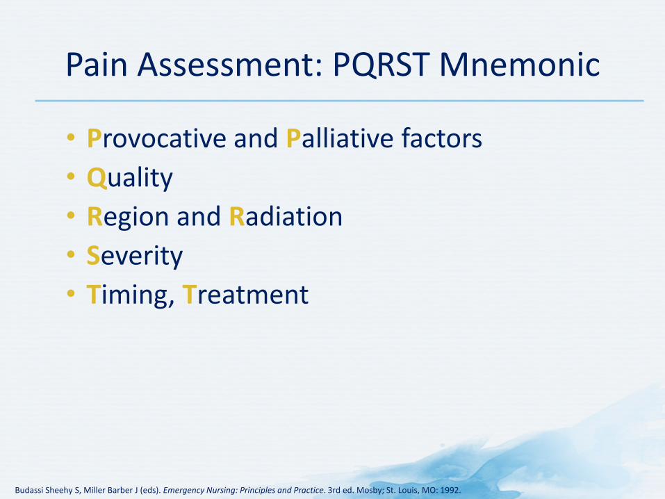
Pain Assessment: PQRST Mnemonic
• Provocative and Palliative factors • Quality • Region and Radiation • Severity • Timing, Treatment
Budassi Sheehy S, Miller Barber J (eds). Emergency Nursing: Principles and Practice. 3rd ed. Mosby; St. Louis, MO: 1992.
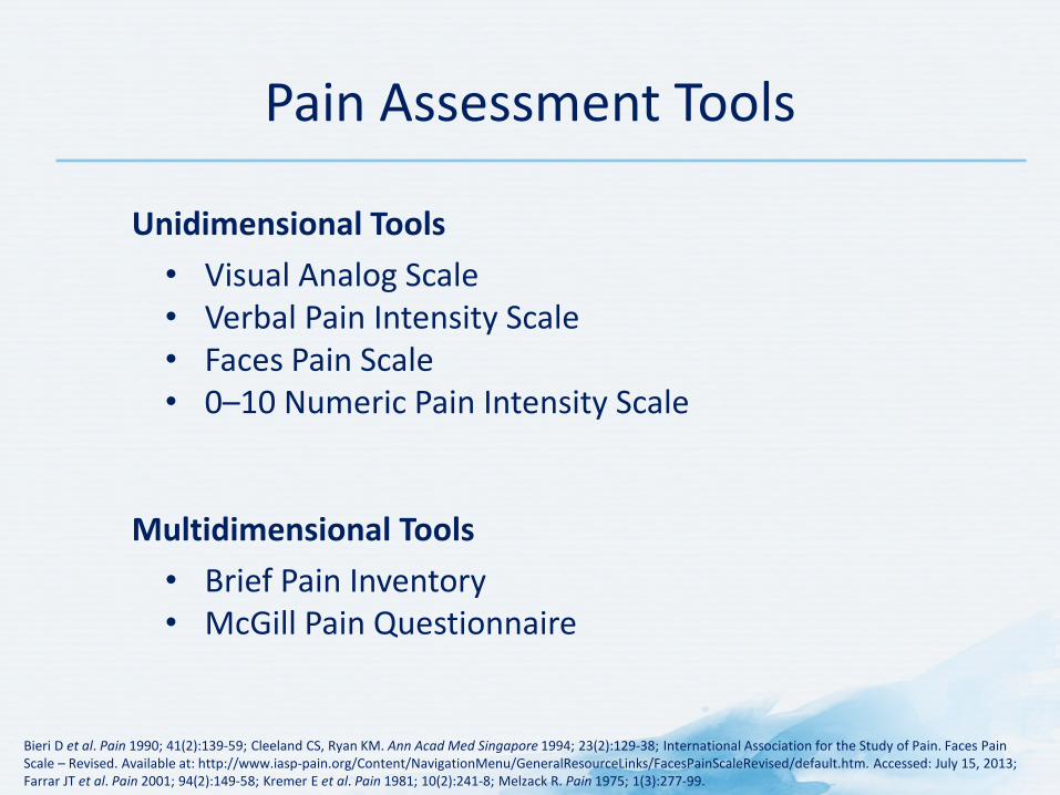
Pain Assessment Tools
Bieri D et al. Pain 1990; 41(2):139-59; Cleeland CS, Ryan KM. Ann Acad Med Singapore 1994; 23(2):129-38; International Association for the Study of Pain. Faces Pain Scale – Revised. Available at: http://www.iasp-pain.org/Content/NavigationMenu/GeneralResourceLinks/FacesPainScaleRevised/default.htm. Accessed: July 15, 2013; Farrar JT et al. Pain 2001; 94(2):149-58; Kremer E et al. Pain 1981; 10(2):241-8; Melzack R. Pain 1975; 1(3):277-99.
Unidimensional Tools • Visual Analog Scale • Verbal Pain Intensity Scale • Faces Pain Scale • 0–10 Numeric Pain Intensity Scale
Multidimensional Tools • Brief Pain Inventory • McGill Pain Questionnaire
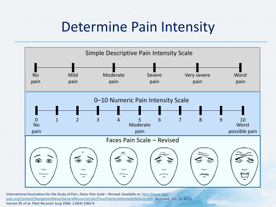
Determine Pain Intensity
International Association for the Study of Pain. Faces Pain Scale – Revised. Available at: http://www.iasp-pain.org/Content/NavigationMenu/GeneralResourceLinks/FacesPainScaleRevised/default.htm. Accessed: July 15, 2013; Iverson RE et al. Plast Reconstr Surg 2006; 118(4):1060-9.
0
0–10 Numeric Pain Intensity Scale
No pain
1 2 3 4 5 6 7 8 9 10 Moderate
pain Worst
possible pain
Simple Descriptive Pain Intensity Scale
No pain
Mild pain
Moderate pain
Severe pain
Very severe pain
Worst pain
Faces Pain Scale – Revised
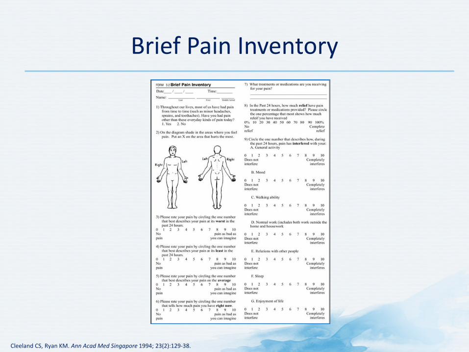
Brief Pain Inventory
Cleeland CS, Ryan KM. Ann Acad Med Singapore 1994; 23(2):129-38.
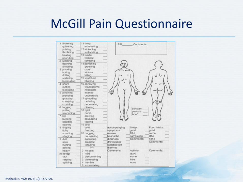
McGill Pain Questionnaire
Melzack R. Pain 1975; 1(3):277-99.
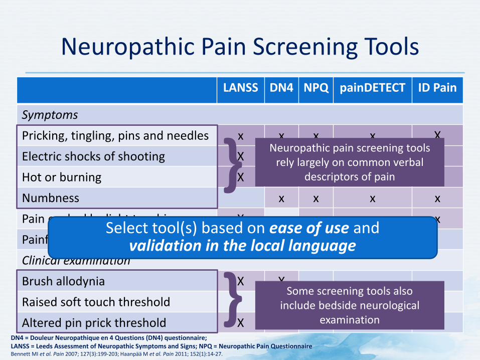
Neuropathic Pain Screening Tools LANSS DN4 NPQ painDETECT ID Pain
Symptoms Pricking, tingling, pins and needles x x x x X Electric shocks of shooting X x x x x Hot or burning X x x x x Numbness x x x x Pain evoked by light touching X x x x Painful cold or freezing pain x X Clinical examination Brush allodynia X X Raised soft touch threshold X Altered pin prick threshold X X
DN4 = Douleur Neuropathique en 4 Questions (DN4) questionnaire; LANSS = Leeds Assessment of Neuropathic Symptoms and Signs; NPQ = Neuropathic Pain Questionnaire Bennett MI et al. Pain 2007; 127(3):199-203; Haanpää M et al. Pain 2011; 152(1):14-27.
Neuropathic pain screening tools rely largely on common verbal
descriptors of pain }
} Some screening tools also include bedside neurological
examination
Select tool(s) based on ease of use and validation in the local language
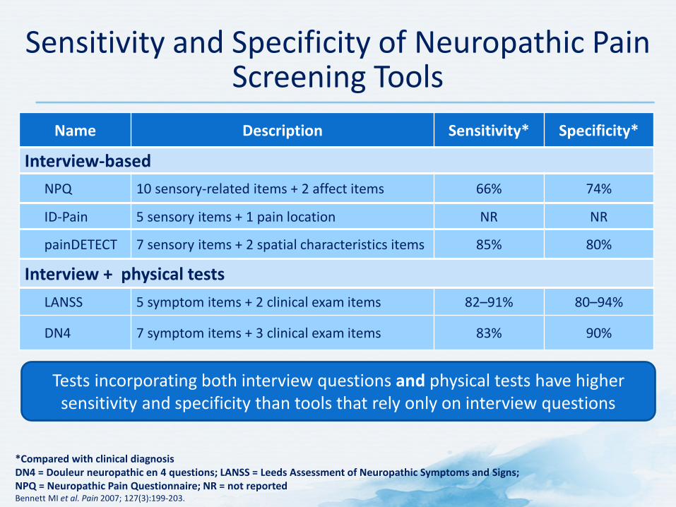
Name Description Sensitivity* Specificity*
Interview-based NPQ 10 sensory-related items + 2 affect items 66% 74%
ID-Pain 5 sensory items + 1 pain location NR NR
painDETECT 7 sensory items + 2 spatial characteristics items 85% 80%
Interview + physical tests LANSS 5 symptom items + 2 clinical exam items 82–91% 80–94%
DN4 7 symptom items + 3 clinical exam items 83% 90%
Sensitivity and Specificity of Neuropathic Pain Screening Tools
*Compared with clinical diagnosis DN4 = Douleur neuropathic en 4 questions; LANSS = Leeds Assessment of Neuropathic Symptoms and Signs; NPQ = Neuropathic Pain Questionnaire; NR = not reported Bennett MI et al. Pain 2007; 127(3):199-203.
Tests incorporating both interview questions and physical tests have higher sensitivity and specificity than tools that rely only on interview questions
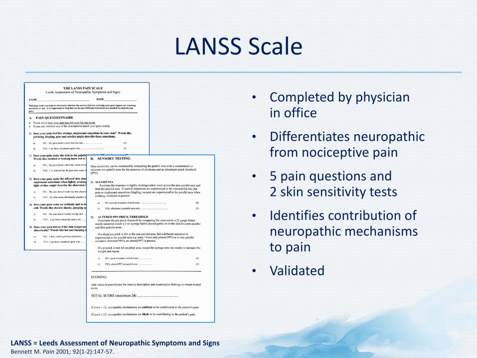
LANSS Scale
• Completed by physician in office
• Differentiates neuropathic from nociceptive pain
• 5 pain questions and 2 skin sensitivity tests
• Identifies contribution of neuropathic mechanisms to pain
• Validated
LANSS = Leeds Assessment of Neuropathic Symptoms and Signs Bennett M. Pain 2001; 92(1-2):147-57.
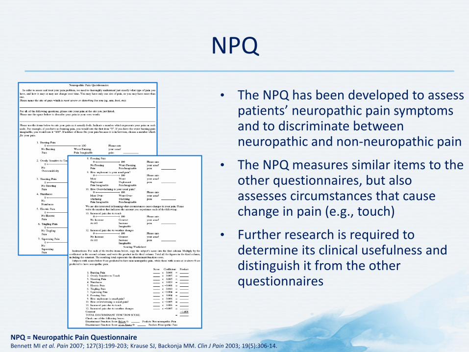
NPQ
• The NPQ has been developed to assess patients’ neuropathic pain symptoms and to discriminate between neuropathic and non-neuropathic pain
• The NPQ measures similar items to the other questionnaires, but also assesses circumstances that cause change in pain (e.g., touch)
• Further research is required to determine its clinical usefulness and distinguish it from the other questionnaires
NPQ = Neuropathic Pain Questionnaire Bennett MI et al. Pain 2007; 127(3):199-203; Krause SJ, Backonja MM. Clin J Pain 2003; 19(5):306-14.
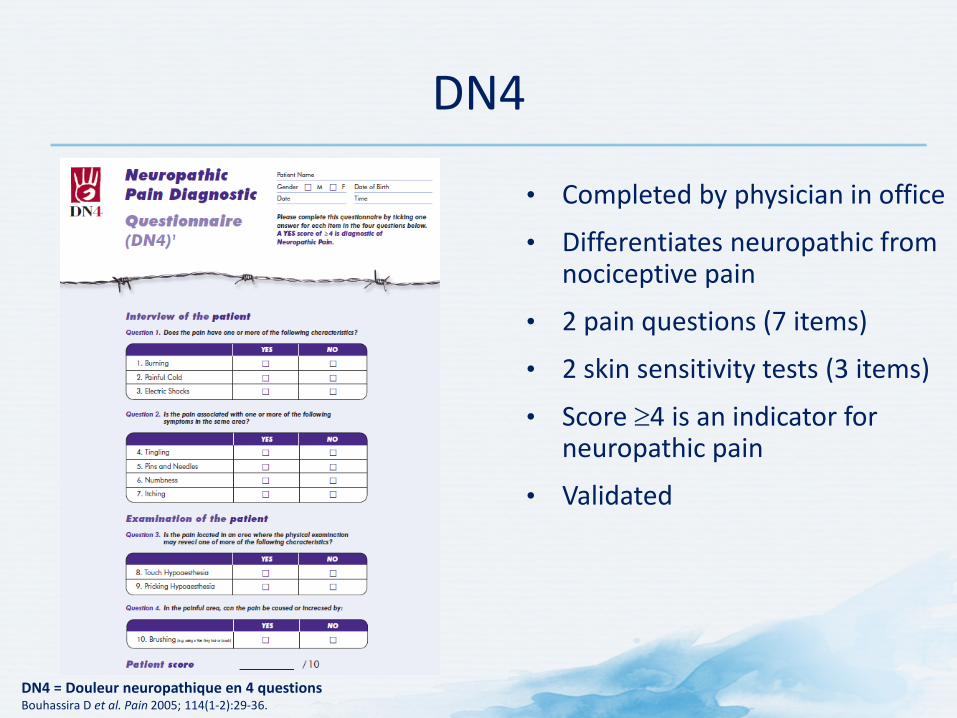
DN4
• Completed by physician in office
• Differentiates neuropathic from nociceptive pain
• 2 pain questions (7 items)
• 2 skin sensitivity tests (3 items)
• Score ≥4 is an indicator for neuropathic pain
• Validated
DN4 = Douleur neuropathique en 4 questions Bouhassira D et al. Pain 2005; 114(1-2):29-36.
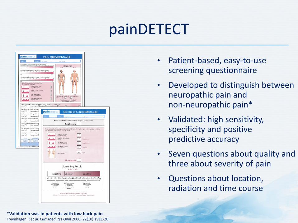
painDETECT
• Patient-based, easy-to-use screening questionnaire
• Developed to distinguish between neuropathic pain and non-neuropathic pain*
• Validated: high sensitivity, specificity and positive predictive accuracy
• Seven questions about quality and three about severity of pain
• Questions about location, radiation and time course
*Validation was in patients with low back pain Freynhagen R et al. Curr Med Res Opin 2006; 22(10):1911-20.
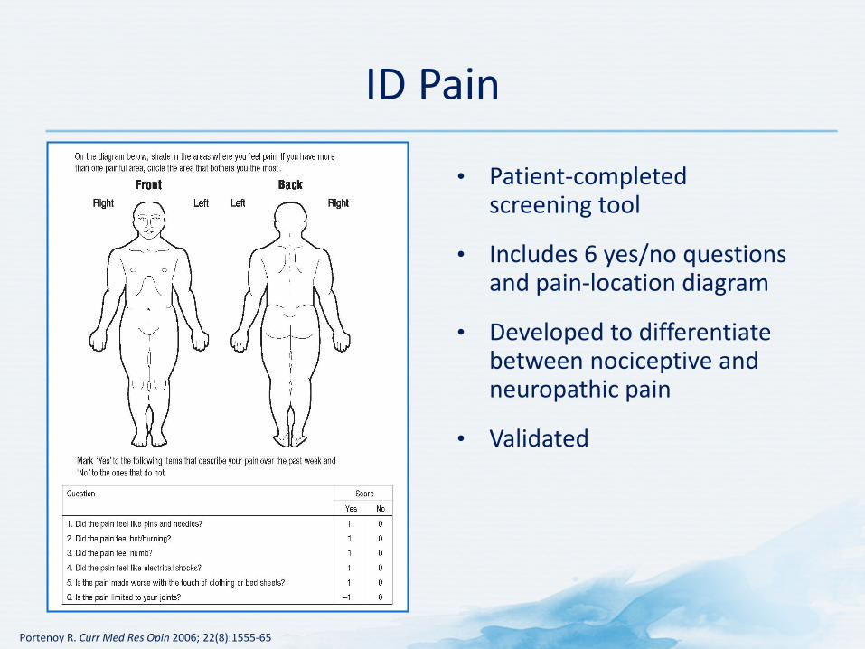
ID Pain
• Patient-completed screening tool
• Includes 6 yes/no questions and pain-location diagram
• Developed to differentiate between nociceptive and neuropathic pain
• Validated
Portenoy R. Curr Med Res Opin 2006; 22(8):1555-65

Physical Examination
Comprehensive Physical Examination Is Important
• Conduct comprehensive physical and neurological exams when evaluating and identifying patient’s subjective complaints of pain1
– Should serve to verify preliminary impression from history and guide the selection of laboratory and imaging studies2
• Confirm or exclude underlying causes
1. American Society of Anesthesiologists Task Force on Pain Management, Chronic Pain Section. Anesthesiology 1997; 86(4):995-1004; 2. Brunton S. J Fam Pract 2004; 53(10 suppl):S3-10.
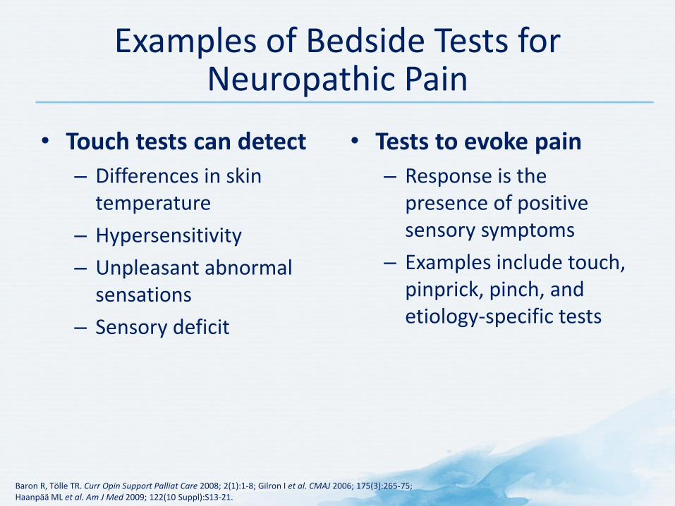
Examples of Bedside Tests for Neuropathic Pain
• Touch tests can detect – Differences in skin
temperature – Hypersensitivity – Unpleasant abnormal
sensations – Sensory deficit
• Tests to evoke pain – Response is the
presence of positive sensory symptoms
– Examples include touch, pinprick, pinch, and etiology-specific tests
Baron R, Tölle TR. Curr Opin Support Palliat Care 2008; 2(1):1-8; Gilron I et al. CMAJ 2006; 175(3):265-75; Haanpää ML et al. Am J Med 2009; 122(10 Suppl):S13-21.

Look: Simple Bedside Tests
Baron R. Clin J Pain 2000; 16(2 Suppl):S12-20; Jensen TS, Baron R. Pain 2003; 102(1-2):1-8.
Light manual pinprick with safety pin or sharp stick
Very sharp, superficial pain
Stroke skin with brush, cotton or apply acetone
Sharp, burning superficial pain ALLODYNIA
HYPERALGESIA

Imaging and Other Tests
Pain Diagnostics
• Plain X-rays with multiple views • MRI • CT • CT myelogram • Nerve conduction velocity • Electromyography
CT = computed tomography; MRI = magnetic resonance imaging Brunton S. J Fam Pract 2004; 53(10 Suppl):S3-S10.
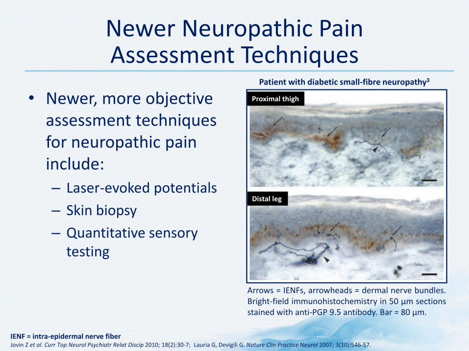
IENF = intra-epidermal nerve fiber Jovin Z et al. Curr Top Neurol Psychiatr Relat Discip 2010; 18(2):30-7; Lauria G, Devigili G. Nature Clin Practice Neurol 2007; 3(10):546-57.
Arrows = IENFs, arrowheads = dermal nerve bundles. Bright-field immunohistochemistry in 50 µm sections stained with anti-PGP 9.5 antibody. Bar = 80 µm.
Patient with diabetic small-fibre neuropathy3
Proximal thigh
Distal leg
Newer Neuropathic Pain Assessment Techniques
• Newer, more objective assessment techniques for neuropathic pain include: – Laser-evoked potentials – Skin biopsy – Quantitative sensory
testing
EFNS = European Federation of Neurological Societies 1. Cruccu G et al. Eur J Neurol 2010; 17(8):1010-8; Garcia-Larrea L, Godinho F. Eur Neurolog Disease 2007; 2:39-41; 2. Truini A et al. Clin Neurophysiol 2005; 116(4):821-6; Garcia-Larrea L et al. Brain 2002; 125(Pt 12):2766-81.
Laser-Evoked Potentials
How They Work • Detect dysfunction of pain and
temperature pathways, which are the basis of neuropathic pain development2
• Laser-generated radiant heat pulses selectively excite free nerve endings in the superficial skin layers3
• Brain responses are recorded4
• Late laser evoked potentials reflect activity of Aδ nerve endings in superficial skin layers1
• Laser evoked potential magnitudes may accurately gauge subjective experience of pain4
Potential Place in Practice • Easiest, most reliable, and most
sensitive neurophysiological way to assess the function of nociceptive pathways1
• EFNS has recommended the use of laser evoked potentials as an ancillary tool in the evaluation of neuropathic pain2
• Use in diagnosis currently limited by availability of equipment2
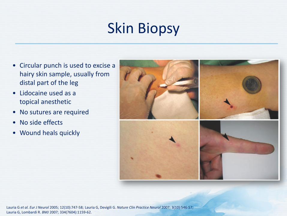
• Circular punch is used to excise a hairy skin sample, usually from distal part of the leg
• Lidocaine used as a topical anesthetic
• No sutures are required
• No side effects
• Wound heals quickly
Lauria G et al. Eur J Neurol 2005; 12(10):747-58; Lauria G, Devigili G. Nature Clin Practice Neurol 2007; 3(10):546-57; Lauria G, Lombardi R. BMJ 2007; 334(7604):1159-62.
Skin Biopsy
1. Rolke R et al. Pain 2006; 123(3):231-43; 2. Hansson P et al. Pain 2007; 129(3):256-9; 3. Jovin Z et al. Curr Top Neurol Psychiatr Relat Discip 2010; 18(2):30-7; 4. Cruccu G, Truini A. Neurol Sci 2006; 27(Suppl 4):S288-90.
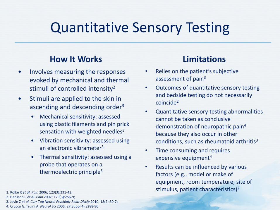
Quantitative Sensory Testing
How It Works • Involves measuring the responses
evoked by mechanical and thermal stimuli of controlled intensity2
• Stimuli are applied to the skin in ascending and descending order3
• Mechanical sensitivity: assessed using plastic filaments and pin prick sensation with weighted needles3
• Vibration sensitivity: assessed using an electronic vibrameter3
• Thermal sensitivity: assessed using a probe that operates on a thermoelectric principle3
Limitations • Relies on the patient’s subjective
assessment of pain3
• Outcomes of quantitative sensory testing and bedside testing do not necessarily coincide2
• Quantitative sensory testing abnormalities cannot be taken as conclusive demonstration of neuropathic pain4 because they also occur in other conditions, such as rheumatoid arthritis3
• Time consuming and requires expensive equipment4
• Results can be influenced by various factors (e.g., model or make of equipment, room temperature, site of stimulus, patient characteristics)2

Diagnosis
Pain Diagnosis
• Confirm or exclude underlying causes • There is no single diagnostic test for pain • Multiple tests may not be helpful
American Society of Anesthesiologists Task Force on Pain Management, Chronic Pain Section. Anesthesiology 1997; 86(4):995-1004; Brunton S. J Fam Pract 2004; 53(10 Suppl):S3-10.
Whenever possible, it is important to identify and treat the underlying
cause of pain!
Forde G, Stanos S. J Fam Pract 2007; 56(8 Suppl Hot Topics):S21-30.
Identify and Treat Underlying Cause
Evaluate for patients presenting with pain the
presence of red flags!
Initiate appropriate investigations/ management or refer to specialist
Littlejohn GO. J R Coll Physicians Edinb 2005; 35(4):340-4.
Be Alert for Red Flags

Summary
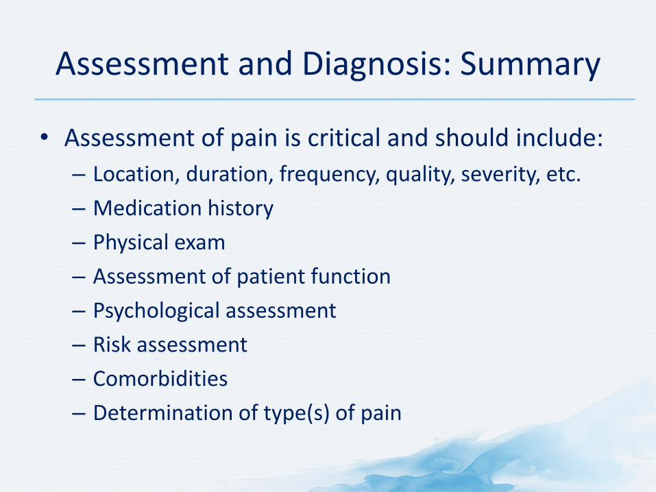
Assessment and Diagnosis: Summary
• Assessment of pain is critical and should include: – Location, duration, frequency, quality, severity, etc. – Medication history – Physical exam – Assessment of patient function – Psychological assessment – Risk assessment – Comorbidities – Determination of type(s) of pain