Embed Size (px)
Citation preview

Assessing Tumor-infiltrating Lymphocytes in Solid Tumors:A Practical Review for Pathologists and Proposal for a
Standardized Method From the InternationalImmunooncology Biomarkers Working Group: Part 1:
Assessing the Host Immune Response, TILs in Invasive BreastCarcinoma and Ductal Carcinoma In Situ, Metastatic Tumor
Deposits and Areas for Further Research
Shona Hendry, MBBS,*w Roberto Salgado, MD,zy Thomas Gevaert, MD,8zPrudence A. Russell, MBBS,#** Tom John, PhD,wwzzyy Bibhusal Thapa, MBBS,MS,zz88 Michael Christie, MBBS, PhD,zz Koen van de Vijver, MD, PhD,##M.V. Estrada, MD,*** Paula I. Gonzalez-Ericsson, MD,www Melinda Sanders,MD,zzz Benjamin Solomon, MBBS, PhD,yyy Cinzia Solinas, MD,888 Gert
G.G.M. Van den Eynden, MD, PhD,888zzz Yves Allory, MD, PhD,###****wwwwMatthias Preusser, MD,zzzz Johannes Hainfellner, MD,yyyy Giancarlo Pruneri,MD,8888zzzz Andrea Vingiani, MD,8888zzzz Sandra Demaria, MD,####Fraser Symmans, MBChB,***** Paolo Nuciforo, MD,wwwww Laura Comerma,MD, PhD,wwwww E.A. Thompson, PhD,zzzzz Sunil Lakhani, BSc, MBBS,
PhD,yyyyy88888 Seong-Rim Kim, MD,zzzzz Stuart Schnitt, MD,#####******Cecile Colpaert, MD, PhD,wwwwww Christos Sotiriou, MD, PhD,zzzzzz Stefan J.
Scherer, MD, PhD,yyyyyy Michail Ignatiadis, MD, PhD,zzzzzz Sunil Badve,MBBS,888888 Robert H. Pierce, MD,zzzzzz Giuseppe Viale, MD,######
Nicolas Sirtaine, MD,******* Frederique Penault-Llorca, MD,PhD,wwwwwwwzzzzzzz Tomohagu Sugie, MD,yyyyyyy Susan Fineberg,
MD,8888888zzzzzzz Soonmyung Paik, MD,zzzzz####### Ashok Srinivasan,MD,zzzzz Andrea Richardson, MD, PhD,**************wwwwwwww Yihong
Wang, MD, PhD,zzzzzzzzyyyyyyyy Ewa Chmielik, MD,88888888zzzzzzzz JaneBrock, MBBS, PhD,************** Douglas B. Johnson,
MD,#########********* Justin Balko, PharmD, PhD,#########*********Stephan Wienert, DMedSci,wwwwwwwwwzzzzzzzzz Veerle Bossuyt, MD,yyyyyyyyyStefan Michiels, PhD,888888888 Nils Ternes, PhD,888888888 Nicole Burchardi,
PhD,zzzzzzzzz Stephen J. Luen, MBChB,wyyy Peter Savas, MBBS,wyyyFrederick Klauschen, MD, PhD, MSc,wwwwwwwww Peter H. Watson,
MBBChir,#########********** Brad H. Nelson,PhD,**********wwwwwwwwwwzzzzzzzzzz Carmen Criscitiello, MD, PhD,8888
Sandra O’Toole, MD, PhD,yyyyyyyyyyzzzzzzzzzz Denis Larsimont, MD,*******Roland de Wind, MD,******* Giuseppe Curigliano, MD, PhD,8888 Fabrice
Andre, MD, PhD,zzzzzzzzz########## Magali Lacroix-Triki,MD,zzzzzzzzzz Mark van de Vijver, MD, PhD,*********** Federico Rojo,MD,wwwwwwwwwww Giuseppe Floris, MD, PhD,zzzzzzzzzzz Shahinaz Bedri,MBBS,yyyyyyyyyyy Joseph Sparano, MD,88888888888 David Rimm, MD,PhD,yyyyyyyyy Torsten Nielsen, MD, PhD,zzzzzzzzzzz Zuzana Kos,
MD,########### Stephen Hewitt, MD, PhD,************ Baljit Singh,MD,wwwwwwwwwwww Gelareh Farshid, MBBS, MD, MPH,zzzzzzzzzzzzyyyyyyyyyyyySibylle Loibl, MD, PhD,zzzzzzzzz Kimberly H. Allison, MD,888888888888
Nadine Tung, MD,zzzzzzzzzzzz Sylvia Adams,MD,############************* Karen Willard-Gallo, PhD,888 Hugo M.
Horlings, MD, PhD,wwwwwwwwwwwww Leena Gandhi, MD,PhD,*************zzzzzzzzzzzzz Andre Moreira, MD, PhD,yyyyyyyyyyyyy Fred
Hirsch, MD, PhD,8888888888888 Maria V. Dieci,
REVIEW ARTICLE
Adv Anat Pathol � Volume 24, Number 5, September 2017 www.anatomicpathology.com | 235
Copyright r 2017 Wolters Kluwer Health, Inc. All rights reserved.

MD,zzzzzzzzzzzzz############# Maria Urbanowicz,MD,************** Iva Brcic, MD, PhD,wwwwwwwwwwwwww Konstanty Korski,
MD, PhD,zzzzzzzzzzzzzz Fabien Gaire, PhD,zzzzzzzzzzzzzz Hartmut Koeppen,MD, PhD,yyyyyyyyyyyyyy Amy Lo, MD, MS,yyyyyyyyyyyyyy88888888888888
Jennifer Giltnane, MD, PhD,yyyyyyyyyyyyyyMarlon C. Rebelatto, PhD,zzzzzzzzzzzzzzKeith E. Steele, PhD,zzzzzzzzzzzzzz Jiping Zha, MD, PhD,zzzzzzzzzzzzzz
Kenneth Emancipator, MD,############## Jonathan W. Juco,MD,############## Carsten Denkert, MD, PhD,wwwwwwwww Jorge Reis-Filho, MD,
PhD,*************** Sherene Loi, MD, PhD,yyyand Stephen B. Fox, BSc, MBChB, DPhil*w
From the Departments of *Pathology; yyyMedical Oncology, Peter MacCallum Cancer Centre, Melbourne; wThe Sir Peter MacCallum Departmentof Oncology; Departments of **Pathology; 88Medicine, University of Melbourne; zzDepartment of Anatomical Pathology, Royal MelbourneHospital, Parkville; #Department of Anatomical Pathology, St Vincent’s Hospital Melbourne, Fitzroy; wwDepartment of Medical Oncology,Austin Health; zzOlivia Newton-John Cancer Research Institute, Heidelberg; yySchool of Cancer Medicine, La Trobe University, Bundoora;yyyyyCentre for Clinical Research and School of Medicine, The University of Queensland; 88888Pathology Queensland, Royal Brisbane andWomen’s Hospital, Brisbane; yyyyyyyyyyThe Cancer Research Program, Garvan Institute of Medical Research, Darlinghurst; 8888888888Aus-tralian Clinical Labs, Bella Vista; zzzzzzzzzzzzDirectorate of Surgical Pathology, SA Pathology; yyyyyyyyyyyyDiscipline of Medicine, AdelaideUniversity, Adelaide, Australia; ***********Department of Surgical Oncology, Netherlands Cancer Institute; wwwwwwwwwwwwwDepartment ofPathology; ##Divisions of Diagnostic Oncology & Molecular Pathology, Netherlands Cancer Institute-Antoni van Leeuwenhoek, Amsterdam,The Netherlands; ###Universite Paris-Est; ****INSERM, UMR 955; wwwwDepartement de pathologie, APHP, Hopital Henri-Mondor, Creteil;888888888Service de Biostatistique et d’Epidemiologie, Gustave Roussy, CESP, Inserm U1018, Universite-Paris Sud, Universite Paris-Saclay;zzzzzzzzzzINSERM Unit U981, and Department of Medical Oncology, Gustave Roussy, Villejuif; ##########Faculte de Medecine, Uni-versite Paris Sud, Kremlin-Bicetre; wwwwwwwDepartment of Surgical Pathology and Biopathology, Jean Perrin Comprehensive Cancer Centre;zzzzzzzUniversity of Auvergne UMR1240, Clermont-Ferrand, France; zzzzDepartment of Medicine, Clinical Division of Oncology; yyyyIn-stitute of Neurology, Comprehensive Cancer Centre Vienna, Medical University of Vienna, Vienna; wwwwwwwwwwwwwwInstitute of Pathology,Medical University of Graz, Austria; 8888European Institute of Oncology; zzzzSchool of Medicine; ######Department of Pathology, IstitutoEuropeo di Oncologia, University of Milan, Milan; zzzzzzzzzzzzzDepartment of Surgery, Oncology and Gastroenterology, University ofPadova; #############Medical Oncology 2, Veneto Institute of Oncology IOV-IRCCS, Padua, Italy; wwwwwMolecular Oncology Group, Valld’Hebron Institute of Oncology, Barcelona; wwwwwwwwwwwPathology Department, IIS-Fundacion Jimenez Diaz, UAM, Madrid, Spain; yDe-partment of Pathology and TCRU, GZA; zzzDepartment of Pathology, GZA Ziekenhuizen, Antwerp; 8Laboratory of Experimental Urology,Department of Development and Regeneration, KU Leuven; zzzzzzzzzzzDepartment of Pathology, University Hospital Leuven, Leuven,Belgium; zDepartment of Pathology, AZ Klina, Brasschaat; wwwwwwDepartment of Pathology, GZA Ziekenhuizen, Sint-Augustinus, Wilrijk;888Molecular Immunology Unit; zzzzzzDepartment of Medical Oncology, Institut Jules Bordet, Universite Libre de Bruxelles; zBreast CancerTranslational Research Laboratory/Breast International Group, Institut Jules Bordet; **************European Organisation for Research andTreatment of Cancer (EORTC) Headquarters; *******Department of Pathology, Institut Jules Bordet, Universite Libre de Bruxelles, Brussels,Belgium; yyyyyyyDepartment of Surgery, Kansai Medical School, Hirakata, Japan; #######Severance Biomedical Science Institute andDepartment of Medical Oncology, Yonsei University College of Medicine, Seoul, South Korea; 88888888Tumor Pathology Department, MariaSklodowska-Curie Memorial Cancer Center; zzzzzzzzInstitute of Oncology, Gliwice Branch, Gliwice, Poland; zzzzzzzzzzzzzzPathology andTissue Analytics, Roche Innovation Centre Munich, Penzberg; wwwwwwwwwInstitute of Pathology, Charite Universitatsmedizin Berlin;zzzzzzzzzVMscope GmbH, Berlin; zzzzzzzzzGerman Breast Group GmbH, Neu-Isenburg, Germany; **********Trev & Joyce DeeleyResearch Centre, British Columbia Cancer Agency; wwwwwwwwwwDepartment of Biochemistry and Microbiology, University of Victoria, Victoria;Departments of zzzzzzzzzzMedical Genetics; #########Pathology and Laboratory Medicine; zzzzzzzzzzzDepartment of Pathology andLaboratory Medicine, Genetic Pathology Evaluation Centre, University of British Columbia, Vancouver, BC; ###########Department ofPathology and Laboratory Medicine, University of Ottawa, Ottawa, Canada; yyyyyyyyyyyDepartment of Pathology and Laboratory Medicine,Weill Cornell Medical College, Doha, Qatar; zzzzzzzzDepartment of Pathology and Laboratory Medicine, Rhode Island Hospital and LifespanMedical Center; yyyyyyyyWarren Alpert Medical School of Brown University, Providence; zzzzzNational Surgical Adjuvant Breast and BowelProject Operations Center/NRG Oncology, Pittsburgh, PA; wwwBreast Cancer Research Program, Vanderbilt Ingram Cancer Center, VanderbiltUniversity; Departments of zzzPathology, Microbiology and Immunology; ########Department of Medicine, Vanderbilt University MedicalCentre; *********Vanderbilt Ingram Cancer Center, Nashville; yyyyyyyyyDepartment of Pathology, Yale University School of Medicine, NewHaven; 88888888888Department of Oncology, Montefiore Medical Centre, Albert Einstein College of Medicine; 8888888Montefiore MedicalCenter; zzzzzzzThe Albert Einstein College of Medicine, Bronx, NY; ********Department of Pathology, Brigham and Women’s Hospital;#####Cancer Research Institute and Department of Pathology, Beth Israel Deaconess Cancer Center; ******Harvard Medical School;zzzzzzzzzzzzDivision of Hematology-Oncology, Beth Israel Deaconess Medical Center; wwwwwwwwDepartment of Cancer Biology;zzzzzzzzzzzzzDana-Farber Cancer Institute, Boston, MA; 8888888888888Department of Medicine, Division of Medical Oncology, Universityof Colorado Anschutz Medical Campus, Aurora, CO; zzzzzDepartment of Cancer Biology, Mayo Clinic, Jacksonville, FL; 888888Departmentof Pathology and Laboratory Medicine, Indiana University, Indianapolis, IN; zzzzzzCancer Immunotherapy Trials Network, Central Labo-ratory and Program in Immunology, Fred Hutchinson Cancer Research Center, Seattle, WA; wwwwwwwwwwwwDepartment of Pathology, NewYork University Langone Medical Centre; ############New York University Medical School; *************Perlmutter Cancer Center;yyyyyyyyyyyyyPulmonary Pathology, New York University Center for Biospecimen Research and Development, New York University;***************Department of Pathology, Memorial Sloan-Kettering Cancer Center; ####Departments of Radiation Oncology and Pathol-ogy, Weill Cornell Medicine, New York, NY; *****Department of Pathology, University of Texas M.D. Anderson Cancer Center, Houston, TX;888888888888Pathology Department, Stanford University Medical Centre, Stanford; 88888888888888Department of Pathology, StanfordUniversity, Palo Alto; ***Department of Pathology, School of Medicine, University of California, San Diego; yyyyyyyyyyyyyyResearch Pathology,Genentech Inc., South San Francisco, CA; *************Laboratory of Pathology, Center for Cancer Research, National Cancer Institute,National Institutes of Health, Bethesda; zzzzzzzzzzzzzzTranslational Sciences, MedImmune, Gaithersberg, MD; yyyyyyAcademic MedicalInnovation, Novartis Pharmaceuticals Corporation, East Hanover; and ##############Translational Medicine, Merck & Co. Inc., Kenil-worth, NJ.
Supported by a grant from the Breast Cancer Research Foundation (BRCF) (R.S.); ISCiii/FEDER (CIBERONCO, PI15/00934) (F.R.); the NIH(NCI K23 CA204726) (D.B.J.).
The authors have no conflicts of interest to disclose.Reprints: Roberto Salgado, MD, Department of Pathology, Oosterveldlaan 24, 2610 Wilrijk, Belgium (e-mail: [email protected]).Supplemental Digital Content is available for this article. Direct URL citations appear in the printed text and are provided in the HTML and PDF
versions of this article on the journal’s Website, www.anatomicpathology.com.All figures can be viewed online in color at www.anatomicpathology.com.Copyright r 2017 Wolters Kluwer Health, Inc. All rights reserved.
Hendry et al Adv Anat Pathol � Volume 24, Number 5, September 2017
236 | www.anatomicpathology.com Copyright r 2017 Wolters Kluwer Health, Inc. All rights reserved.
Copyright r 2017 Wolters Kluwer Health, Inc. All rights reserved.

Abstract: Assessment of tumor-infiltrating lymphocytes (TILs) inhistopathologic specimens can provide important prognosticinformation in diverse solid tumor types, and may also be of valuein predicting response to treatments. However, implementation as aroutine clinical biomarker has not yet been achieved. As successfuluse of immune checkpoint inhibitors and other forms of immu-notherapy become a clinical reality, the need for widely applicable,accessible, and reliable immunooncology biomarkers is clear. Inpart 1 of this review we briefly discuss the host immune response totumors and different approaches to TIL assessment. We propose astandardized methodology to assess TILs in solid tumors onhematoxylin and eosin sections, in both primary and metastaticsettings, based on the International Immuno-Oncology BiomarkerWorking Group guidelines for TIL assessment in invasive breastcarcinoma. A review of the literature regarding the value of TILassessment in different solid tumor types follows in part 2. Themethod we propose is reproducible, affordable, easily applied, andhas demonstrated prognostic and predictive significance in invasivebreast carcinoma. This standardized methodology may be used as areference against which other methods are compared, and shouldbe evaluated for clinical validity and utility. Standardization of TILassessment will help to improve consistency and reproducibility inthis field, enrich both the quality and quantity of comparable evi-dence, and help to thoroughly evaluate the utility of TILs assess-ment in this era of immunotherapy.
Key Words: lymphocytes, tumor-infiltrating, biomarkers, cancer,
immunotherapy, pathology
(Adv Anat Pathol 2017;24:235–251)
Pathologists have long recognized the stroma, immuneinfiltrate, nerves, and vasculature as integral parts of
the tumor microenvironment, which often provideimportant information regarding tumor behavior, prog-nosis, and response to treatment. It is well established thattumors are antigenic and can induce an immune response,due in part to altered protein products that may be rec-ognized as foreign by the host immune system.1,2 Agrowing body of research has shown that the extent andcomposition of the host immune response to the tumor hasprognostic and predictive significance in many solidmalignancies (reviewed in3). The assessment of immuneinfiltrate in tumors, most commonly referred to as tumor-infiltrating lymphocytes (TILs), is also gaining importancein the current quest for optimal biomarkers to selectpatients with the highest likelihood of responding toimmunotherapeutic agents. Therefore, TIL assessment hasbeen proposed as a biomarker for inclusion in routinehistopathologic reporting.4,5 Current TIL scoring systemsused in research and proposed for different tumor typesvary widely in detail, scope, accuracy, and time andresource requirements.
Development of prognostic and predictive biomarkersin oncology requires robust assessment of the test’s ana-lytical validity, clinical validity, and clinical utility.6,7 Evi-dence is accumulating to support the use of TILs scoring asa prognostic biomarker in various solid tumors and evi-dence for the predictive benefit of TILs is being investigatedat present. Different methods of assessing TILs will havedifferent preanalytical, analytical, and postanalytical chal-lenges. For example, semiquantitative hematoxylin andeosin (H&E)-based scores may suffer from low precisionand poor interobserver reproducibility if no clear guidanceexists, whereas digital quantification of immunohis-tochemistry (IHC)-stained sections may produce different
results due to inaccurate measurement of the test variablewithout controlled calibration. Testing of the clinical val-idity of biomarkers involves determining the extent towhich the biomarker predicts the clinical outcome ofinterest, that is, patient prognosis or response to treatment.7
Assessment of the clinical validity of TILs scoring requiresa standardized, reproducible method, which can be vali-dated preferably in several independent populations. Manybiomarker studies are observational, retrospective studiesin which the study population is selected solely by theavailability of samples.8,9 Although prospective controlledstudies designed to test biomarkers are rare and unlikely tobe performed on a large scale, prospective-retrospectivestudies may offer a comparable level of evidence.8 Theseprospective-retrospective studies involve use of samplescollected during a prospective randomized clinical trial, andallow high-quality evaluation of the biomarker of interestprovided the study design meets certain criteria and resultscan be replicated in an independent population.8 Guidelinesfor the reporting of biomarker studies are available9,10 andshould be considered when evaluating the TILs literature.
In part 1 of this review, we aim to briefly describe thehost immune response to tumors and approaches used toassess this in the current context of immunotherapy. Wepropose a standardized methodology for TIL assessment insolid tumors, based on the International Immuno-Oncology Biomarkers Working Group guidelines for TILassessment in invasive breast carcinoma, which may beadapted to different tumor types. We then discuss the lit-erature and our experiences in the areas of invasive breastcancer, ductal carcinoma in situ (DCIS), and metastatictumor deposits, then conclude with a discussion of openquestions and areas for further research. In part 2 of thisreview, the literature surrounding methods of TIL assess-ment and the prognostic and potentially predictive sig-nificance of TILs in different solid tumors is discussed,including carcinomas of the lung, colon, upper gastro-intestinal tract, head and neck, genitourinary tract, andgynecological organs, as well as mesothelioma, melanoma,and primary brain tumors. Ways in which the proposedmethodology can be adapted to different tumors are sug-gested, based on available evidence and expert opinion.Standardization of TIL assessment will allow direct com-parison of different studies, highlight areas for furtherresearch, and form the basis of TIL assessment in routinehistologic practice.
THE HOST IMMUNE RESPONSEAltered protein products caused by the genetic muta-
tions in cancer cells can function as neoantigens, eliciting animmune response against a perceived “foreign” cell.2 Inaddition, the inflammatory, hypoxic, and often necroticmicroenvironment of tumors sends concomitant dangersignals to the host immune system.11 Infiltrating immunecells can function to control tumor growth and progression,but can also help to create an immunosuppressive envi-ronment in which the tumor can thrive.12 CD8+ cytotoxicT cells, T-helper 1 (Th1) cells producing interferon-g, andnatural killer cells are generally associated with favorableantitumor immune responses, along with macrophagespolarized to an M1 phenotype and dendritic cells showing aDC1 phenotype. Immunosuppressive effects are seen withTh2 cells, M2 macrophages, DC2 dendritic cells, myeloid-derived suppressor cells, and FOXP3+ regulatory T (Treg)
Adv Anat Pathol � Volume 24, Number 5, September 2017 TIL Assessment in Solid Tumors, Part 1
Copyright r 2017 Wolters Kluwer Health, Inc. All rights reserved. www.anatomicpathology.com | 237
Copyright r 2017 Wolters Kluwer Health, Inc. All rights reserved.

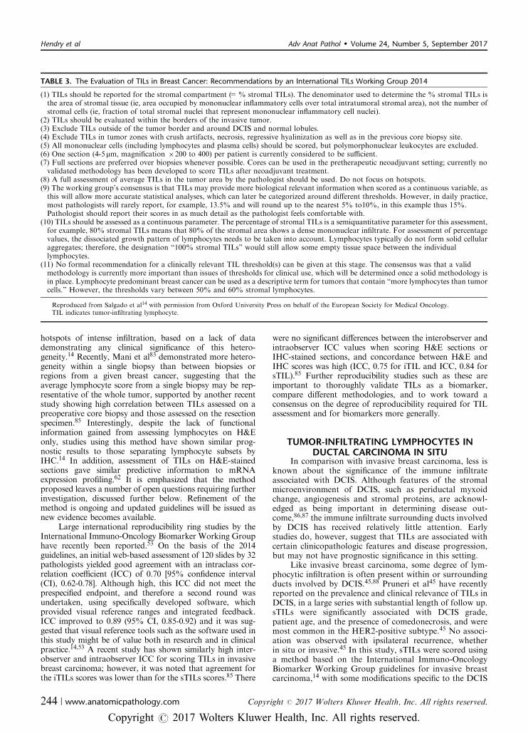
cells producing interleukin-10 and transforming growthfactor-b. B cells and plasma cells can also adopt eithereffector or regulatory phenotypes, and hence cancarry positive or negative antitumor associations dependingon contextual factors. This balance of the cellularconstituents of the immune response is illustratedin Figure 1. The presence of tertiary lymphoid structures,aggregates which recapitulate the components and archi-tecture of a lymph node, in the tumor microenvironment iscorrelated with better prognosis in different types of solidtumors (reviewed in13). The exact composition of theimmune infiltrate can vary widely within and betweentumors and clearly modulate the effectiveness of theantitumor response.
In research settings, many different methods are beingused to investigate the host immune response to tumors.Many clinical studies have found significant results using anassessment of H&E-stained sections by trained patholo-gists, with qualitative or semiquantitative scoring systemswhich vary according to tumor type.14–16 IHC allows defi-nition of the majority of immune cell subsets that can berefined by combinations of markers, including CD8+
cytotoxic T cells, CD4+ T-helper cells, FOXP3+ Tregs, Bcells, macrophages, and dendritic cells. Other cell types suchas myeloid-derived suppressor cells require multiple cellsurface markers for definition and are challenging to iden-tify on serial IHC sections. Digital image analysis has beenvalidated in multiple studies and can provide accuratequantitation of immune cell infiltrates in IHC-stainedsections.17,18 Multiplexed fluorescent IHC with multi-spectral imaging is a recent development that allows in situ
identification of different immune cell subsets on the samesection, providing quantitative information on thedistribution and composition of the immune infiltrate onformalin fixed, paraffin embedded (FFPE) tissue.19–21 Thistechnology requires a significant investment in initialoptimization, has complex data analysis and storagerequirements, and is yet to become routine. Recently, anovel approach to multiplexed IHC was described utilizingNanoString NCounter fluorescent barcodes to identifybound antibodies, allowing quantitation of multiple pro-teins in situ on an FFPE slide.22 FFPE tissue can also beused for matrix-assisted laser desorption/ionization-imag-ing mass spectrometry (MALDI-IMS), a proteomic tech-nique that can identify hundreds proteins in situ withoutthe need for specific antibodies.23,24 These exciting newtechnologies, termed “molecular histology” combine thespatial and architectural information from traditional his-tology approaches with detailed molecular profiling, whichis likely to be particularly relevant in describing the immunemicroenvironment of tumors.
Flow cytometry is a common approach to immune cellprofiling and has many benefits including the character-ization of immune cell subsets by multiple markers, quan-titative data acquisition, wide availability, and the ability toexamine small subpopulations of interest.25 However, freshtissue is required and no information is provided on thedistribution or organization of the immune infiltrate orrelationship to other microenvironmental structures. Nev-ertheless, recent study of TILs in invasive breast carcinomafound a significant positive correlation between fresh tumortissue analyzed by flow cytometry and IHC-stained sections
CD8 T cell
Th17cell
NKTcell
Tumor Suppression Tumor Progression
NKcell
N1 cell
N2 cell
Th1cell
IFNγ
IFNγ
B cell
Ig’s
TfhcellIL21
Tregcell
Th2cell
IL10TGFβ
DC1
DC2
M2
M1
MDSC
FIGURE 1. The cellular constituents of the host immune response to tumors can control tumor growth or contribute to an immuno-suppressive environment that promotes tumor progression. Breg indicates regulatory B cell; DC, dendritic cell; GC B cell, germinalcenter B cell; IFNg, interferon g; Ig’s, immunoglobulins; IL10, interleukin-10; IL21, interleukin-21; M, macrophage; MDSC, myeloid-derived suppressor cell; N, neutrophil; NK, natural killer cell; NKT, natural killer T cell; Tfh, follicular B helper T cells; Th, helper CD4+ Tcell; TGFb, transforming growth factor b; Treg, regulatory T cell.
Hendry et al Adv Anat Pathol � Volume 24, Number 5, September 2017
238 | www.anatomicpathology.com Copyright r 2017 Wolters Kluwer Health, Inc. All rights reserved.
Copyright r 2017 Wolters Kluwer Health, Inc. All rights reserved.

scored by a pathologist.26 Messenger RNA (mRNA)profiling of tumor tissue can detect “immune gene sig-natures,” using the level of expression of immune-relatedgenes to describe the composition and functional status ofthe immune infiltrate.27 Again, no information is providedon the distribution of the infiltrate and this resource-intensive technology is currently largely restricted to aresearch setting. These more complex methods are by theirnature more difficult to implement in large, multicenterclinical trials, which ultimately are required for the vali-dation of potential biomarkers. The costs of such detailedtechniques must also be weighed against the additionalinformation that may be derived regarding the compositionand functional status of the immune infiltrate.
The host immune response to tumors is currently ofgreat interest to oncologists and researchers followingimpressive early results of immune checkpoint inhibitortherapy. An important mechanism of tumor immune eva-sion is the expression of immune checkpoint molecules suchas cytotoxic T lymphocyte antigen-4 and programmeddeath ligand-1 (PD-L1), both on tumor cells and on infil-trating immune cells.28 By blocking these signaling path-ways, immune checkpoint inhibitors can reactivate the hostimmune system to recognize and control the tumor.28
Clinical trials have demonstrated often durable responses indifferent tumor types including melanoma,29,30 urothelialcarcinoma,31 Hodgkin lymphoma,32 non–small cell lungcarcinoma,33–35 renal cell carcinoma,36 and squamous cellcarcinoma of the head and neck.37 However, responseswithin tumor types vary widely and the selection of patientslikely to respond remains problematic.38 Despite FederalDrug Administration approval as companion and comple-mentary diagnostics for the use of anti-PD-1 therapy innon–small cell lung carcinoma,39,40 immunohistochemicalidentification of PD-L1 expression on tumor cells and/orimmune cells is an imperfect biomarker40 and a significantresearch effort is ongoing to identify reliable, broadlyapplicable, and clinically valid biomarkers. T-cell infiltra-tion into tumors is critical to the success of immunecheckpoint blockade,41 and tumors with high levels ofinfiltrating effector T cells, as measured by gene expressionprofiling, appear to show improved responses.42–44 Assess-ment of TILs in this context is a highly active area ofresearch and guidance for a standardized methodology istherefore timely.
PROPOSAL FOR A STANDARDIZEDMETHODOLOGY FOR SCORING
TUMOR-INFILTRATING LYMPHOCYTES INSOLID TUMORS, IN BOTH PRIMARY AND
METASTATIC SETTINGSMuch research has been performed to establish the
prognostic and predictive significance of TILs in differentsolid tumors. However, further work is needed to ensurethat the valuable information that could be obtained fromTILs assessment is not lost due to issues of resource com-mitments, methodology, or lack of standardization. It isour view that a semiquantitative H&E-based TILs assess-ment provides clinically relevant information in a formatthat is applicable to large-scale randomized clinical trials, topreclinical and clinical research projects, and to everydaypathology practice, whether it be in high, middle, or low-income countries. It needs to be emphasized that the
sophisticated tools mentioned above are resource-intensiveand may be difficult to implement in middle or low-incomesettings. A biomarker based on a plain H&E-stained sectionis affordable and accessible, and will not add to the often-restrictive costs of accessing therapy. This is an importantconsideration in the current era of immunotherapeutics.
Over the past few years, Roberto Salgado, ShereneLoi, and Carsten Denkert have developed the InternationalImmuno-Oncology Biomarker Working Group on BreastCancer, with members including important clinical researchgroups, pathologists, clinicians, and statisticians worldwidecurrently knowledgeable in the field of immunooncologybiomarkers. The purpose of this Working Group is todevelop, in a timely manner, standards on the assessment ofimmunooncology biomarkers to aid pathologists, clini-cians, and researchers in their research and daily practice.The group has, for example, developed the first Interna-tional Guidelines on TIL Assessment in Breast Cancer14
and other guideline papers are in development. In additionto the breast cancer experts already included, academicexpert groups from other fields and biomarker expertgroups from industry were contacted and enthusiasticallyagreed to be a part of this initiative. We include worldwiderepresentatives of known clinical research groups fromexpert centers across all continents (United States, Europe,Australia, the Middle East, and Japan) and the member listis growing.
For these papers, a panel of pathologists, medicaloncologists, biostatisticians, and translational researchersfrom different expert groups conducted a systematic reviewof the literature. Panel members have had experience in TILassessment in preclinical research, clinical trials, or areinvolved in translational research focused on the inter-actions between immunology and cancer. There are noexisting guidelines on TIL assessment in solid tumorsavailable for comparison; neither are there proficiencytesting data available from international organizations. Nospecific funding was obtained for this project. Themethodology we propose is based on the InternationalGuidelines on TIL Assessment in Breast Cancer,14 follow-ing robust evidence from prospective-retrospective phaseIII trials in breast cancer. Reviewed studies were not limitedto randomized trials, but also included consecutive andretrospective series and in-press publications. Ways inwhich this methodology could be adapted to differenttumor types, while remaining as standardized as possible,were suggested, reviewed, and discussed by members of theauthor group expert in the particular field, to reach theconsensus opinions presented here. Where strong publishedevidence was lacking, the panel undertook a formal expertconsensus-based process by regular mail and face-to-facemeetings at the San Antonio Breast Cancer Symposium2016. Further updates and revisions are planned as moreevidence becomes available and the field progresses.
We propose a method for TIL assessment that wouldbe applicable to all solid tumors (Table 1 and Fig. 2). As forany proposed scoring system, this method should beevaluated for clinical validity and utility, as well as inter-observer reproducibility. This method may be used as areference against which other systems, such as IHC-baseddigital quantification or mRNA profiling, are compared, toestablish a sensible balance between simplicity and infor-mation yield. Individual tumor types have particular issuessurrounding TILs that may require further investigation toresolve, and this method may be adapted to incorporate
Adv Anat Pathol � Volume 24, Number 5, September 2017 TIL Assessment in Solid Tumors, Part 1
Copyright r 2017 Wolters Kluwer Health, Inc. All rights reserved. www.anatomicpathology.com | 239
Copyright r 2017 Wolters Kluwer Health, Inc. All rights reserved.

those particularities (Table 2). However, most of the issuesraised are common to many solid tumor types, such as therelative importance of stromal TILs (sTILs) and intra-tumoral TILs (iTILs) or the delineation of the centraltumor and the invasive margin, and we feel that the methodoutlined in Table 1 should be able to be applied in mostresearch and clinical settings. Pragmatic definitions of theinvasive margin and central tumor areas are discussedfurther in part 2 of this review, but are illustratedin Figure 3 and summarized as follows: the invasive marginis defined as a 1-mm-wide zone centered on the border ofthe malignant cells with the host tissue, and the centraltumor is defined as the central tumor tissue surrounded bythis zone. Tutorials illustrating TIL assessment in differenttumor types and reference scoring sheets illustrating den-sities of sTILs are available as supplementary materialonline, Supplemental Digital Content 1 http://links.lww.com/PAP/A13, Supplemental Digital Content 2, http://links.lww.com/PAP/A14, Supplemental Digital Content 3,http://links.lww.com/PAP/A15, Supplemental DigitalContent 4, http://links.lww.com/PAP/A16. Having a ref-erence method to score TILs will help to ensure thatpotential future studies investigating TILs in each tumortype can be compared. Many potentially useful biomarkerssuch as the Ki67 proliferative index in breast cancer sufferfrom a plethora of noncomparable methods, which affectsthe level of evidence that can be obtained and preventsuniform clinical implementation. International efforts atstandardization are also being explored for this importantbiomarker.46
TUMOR-INFILTRATING LYMPHOCYTES ININVASIVE BREAST CARCINOMA
The wide variation in the immune infiltrate seen inbreast carcinomas is well known to pathologists, with anearly description of medullary breast carcinoma by Mooreand Foote in 194947 highlighting the lymphocytic infiltrateas a key diagnostic criterion and linking it to good prog-nosis. This lymphocytic infiltrate was suggested to representa form of host resistance to the tumor; however, conflictingresults regarding the prognostic significance of TILs werefound in subsequent studies.48–51 In addition, it was rec-ognized that a prominent lymphocytic infiltrate may befound in tumors not meeting the other strict criteria formedullary breast carcinoma.51 Most breast carcinomasshow some degree of lymphocytic infiltrate, with higherrates of “lymphocyte predominant breast carcinoma,”variably defined as >50% or >60% of tumor area occu-pied by TILs, seen in triple negative breast carcinoma(20%) and HER2-positive breast carcinoma (16%) com-pared with estrogen receptor positive breast carcinoma(6%).52 Recently, through large-scale international efforts,evidence has accumulated to support the prognostic andpotential predictive impact of TILs, both in triple negativeand HER2-positive cancers. The clinical utility of assessingTILs in breast cancer relates to risk prediction models,adjuvant, and neoadjuvant chemotherapy decisions, andthe growing potential of immunotherapy.53
Several large systematic reviews and meta-analyseshave confirmed that high levels of TILs are associated withbetter disease-free survival and overall survival only in
TABLE 1. Proposed Guidelines for the Assessment of TILs in Solid Tumors: Recommendations by an International Immuno-OnoclogyBiomarker Working Group
(1) TILs should be reported separately for the stromal compartment (= % stromal TILs) and the tumor cell compartment (=%intratumoral TILs). The reasons are (1) in many tumors the TIL density in both compartments is different, and (2) if the TILs areevaluated simply per tumor area, the density and growth pattern of tumor cells (= a nonimmune parameter) will affect the TIL count. Thedenominator used to determine the % stromal TILs is the area of stromal tissue (ie, area occupied by mononuclear inflammatory cells overtotal stromal area), not the number of stromal cells (ie, fraction of total stromal nuclei that represent mononuclear inflammatory cellnuclei). Similarly, for intratumoral TILs the tumor cell area is the denominator. In some tumor types, for example, breast cancer, it mightbe decided to evaluate only the stromal TILs.
(2) TILs should be evaluated within the borders of the invasive tumor, including both “central tumor” and “invasive margin.” These areasmay be reported separately when required.
(3) The “invasive margin” is defined as a 1-mm region centered on the border separating the malignant cell nests from the host tissue. The“central tumor” represents the remaining tumor area.
(4) Exclude TILs at a distance outside of the tumor borders. TILs immediately adjacent to the invasive margin, that is, “peritumoral TILs,”may be evaluated when required.
(5) Exclude TILs in tumor zones with crush artifacts, necrosis, and regressive hyalinization, as well as in previous biopsy sites.(6) All mononuclear cells (including lymphocytes and plasma cells) should be scored, but polymorphonuclear leukocytes (neutrophils)should be excluded.
(7) One section (4-5mm, magnification �200 to 400) per patient can be considered to be sufficient for practical purposes. However, assessingadditional sections for each case whenever possible and reporting the number of sections reviewed per case specifically in the manuscript isrecommended as the extent of heterogeneity for different tumor types is unknown.
(8) Full sections are preferred over biopsies whenever possible. Cores can be used in the pretherapeutic neoadjuvant setting; currently novalidated methodology has been developed to score TILs after neoadjuvant treatment.
(9) A full assessment of average TILs in the tumor area (central tumor and invasive margin) should be used. Do not focus on hotspots.(10) TILs should be assessed as a continuous variable, as this may provide more biologically relevant information and allow more accuratestatistical analyses. However, in daily practice most pathologists will report discrete estimates, for example, 13.5% will be rounded to15%. Pathologists should report their scores in as much detail as the pathologist feels comfortable with.
(11) For assessment of percentage values, the dissociated growth pattern of lymphocytes needs to be taken into account. The percentage ofstromal TILs is a semiquantitative parameter for this assessment, for example, 80% stromal TILs means that 80% of the stromal areashows a dense mononuclear infiltrate. Lymphocytes typically do not form solid cellular aggregates, therefore the designation “100%stromal TILs” would still allow some empty tissue space between the individual lymphocytes.
(12) No formal recommendation for a clinically relevant TIL threshold(s) can be given at this stage. A valid methodology is currently moreimportant than issues of thresholds for clinical use, which will be determined once a solid methodology is in place.
TIL indicates tumor-infiltrating lymphocyte.
Hendry et al Adv Anat Pathol � Volume 24, Number 5, September 2017
240 | www.anatomicpathology.com Copyright r 2017 Wolters Kluwer Health, Inc. All rights reserved.
Copyright r 2017 Wolters Kluwer Health, Inc. All rights reserved.

triple negative and HER2-positive subtypes, with no sig-nificant benefit seen in estrogen receptor positive breastcarcinoma.54–56 Pooled hazard ratios for overall survivalwith variably defined “high TILs” ranged from 0.71 to
0.88.54–56 A recent pooled analysis of 5 large clinical trialsof anthracycline chemotherapy in triple negative breastcarcinoma presented similar results, with an adjusted rela-tive risk reduction of 12% for disease-free and overall
Standardized approach for TILs evaluation in solid tumors
Include area within tumor borders
Do not include immune
infiltrate outside of the tumor
TLS
Stromal TILs (sTILs)
Intra-tumoral TILs (iTILs)
Do not includegranulocytesin necrotic
areas
MononuclearstromalTILs
infiltrate
20-40% stromal TILs 0-10% stromal TILs 50-90% stromal TILs
For intermediate
group evaluate different areas at higher
magnification.
Step 1: Select tumor area
Step 2: Define stromal and intra-tumoral areas
Step 3: Scan at low magnification
Step 4: Determine type of inflammatory infiltrate
Step 5: Assess the percentage TILs
FIGURE 2. Guidelines for a standardized approach to TILs evaluation in solid tumors. Modified from Salgado et al14 with permissionfrom Oxford University Press on behalf of the European Society for Medical Oncology. TIL indicates tumor-infiltrating lymphocyte.Adaptations are themselves works protected by copyright. So in order to publish this adaptation, authorization must be obtained bothfrom the owner of the copyright in the original work and from the owner of copyright in the translation or adaptation. Please see thisimage in color online.
Adv Anat Pathol � Volume 24, Number 5, September 2017 TIL Assessment in Solid Tumors, Part 1
Copyright r 2017 Wolters Kluwer Health, Inc. All rights reserved. www.anatomicpathology.com | 241
Copyright r 2017 Wolters Kluwer Health, Inc. All rights reserved.

survival for every 10% increment in sTILs.57 Both iTILsand sTILs gave similar results when separatelyassessed54,56–58; however, iTILs did not appear to addprognostic information to sTILs in a multivariate model.57
The majority of these data come from large prospective-retrospective studies, associated with randomized clinicaltrials14,57,59–66; however, smaller retrospective studies werealso included in the meta-analyses. Although many of theincluded studies follow the TIL scoring method used byDenkert et al in 2010,61 there is variation with some studiesusing H&E assessment,14,59,60,62,63,66 some using IHC,64,65
and others using mRNA expression profiling.64 Con-clusions from some recent meta-analyses in this field67,68
indicate that care must be taken when interpreting theresults of studies of prognostic biomarkers. For example, it issuggested that FOXP3+ Tregs correlate with poor prognosisin breast cancer67,68; however, Treg levels also correlate withother poor prognostic factors such as estrogen receptor-negativity, HER2-positivity, lymph node metastasis, and highhistologic grade.69–72 Within the estrogen receptor-negativesubgroup, high levels of FOXP3+ Tregs may actually corre-late with improved prognosis as markers of an active immuneresponse.69,71 Both primary studies and meta-analyses shouldreport the results of multivariate analyses to confirm theprognostic value of the biomarker in question, independentof other known prognostic factors.10 Despite these
TABLE 2. Additional Points for Attention When Assessing TILs in Different Tumor Types
Invasive breast carcinomaRefer to Salgado et al.14 Consensus guidelines are reproduced in Table 3.
Ductal carcinoma in situ and other premalignant lesionsRefer to Pruneri et al.45
Stromal area is defined as the specialized stroma surrounding the ducts involved by in situ carcinoma, or when this is not clear, an areasurrounding the ducts within 2 high-power fields (B1mm).
Any type of circumferential infiltrate should be taken into account, including minimal, partial, subtotal, and total circumferential TILs.Exclude TILs that are in continuity between the invasive tumor and the in situ lesions with no clear distinction as to whether these areTILs associated with the invasive or in situ component.
MelanomaCurrently only iTILs are scored in the clinical setting. sTILs and/or peritumoral TILs may be reported separately in research settings.Only the vertical growth phase of the primary tumor is evaluated.Further research may determine what % iTILs corresponds to the traditional categorization of brisk/nonbrisk/absent.
Colorectal carcinomaSeparately reporting invasive margin and central tumor TILs is recommended. Invasive margin TILs seem to have the most prognosticsignificance in this setting.
Upper gastrointestinal tract carcinomasEarly evidence supports evaluating only sTILs in gastric carcinoma, due to a lack of prognostic significance of iTILs. This finding requiresfurther validation.
There are insufficient data on pancreatic ductal adenocarcinoma and hepatocellular carcinoma to make specific recommendations.Non–small cell lung carcinomasTILs and iTILs should be separately reported in the research setting at present. Insufficient evidence is available to support evaluatingonly sTILs over a combined assessment.
Do not include areas with pure intra-alveolar tumor spread (aerogenic spread) or with pure lepidic growth (no desmoplastic reaction).Do not include alveolar macrophages.
Ovarian carcinomaBoth the central tumor and invasive margin should be included, but it is not currently recommended to report these regions separately.iTILs and sTILs should be separately reported at present, as both compartments seem to have prognostic relevance. Further research maydetermine the relative importance of the different compartments.
Include TILs in the stroma pertaining to fibrovascular cores of papillary structures.Include the invasive margin of superficial peritoneal or ovarian deposits.
Head and neck squamous cell carcinomaCare should be taken to exclude preexisting lymphoid stroma from the assessment in oropharyngeal (ie, tonsillar and base of tongue)tumors. If a desmoplastic stroma is present, sTILs can be scored in this reactive stroma (as for lymph node metastases).
Genitourinary carcinomasSeparate reporting of iTILs and sTILs is recommended—this is important in the context of immune checkpoint inhibitor therapy withatezolizumab in urothelial carcinoma, where the PD-L1 “immune cell” score is derived from the sTILs score.
Care should be taken to avoid areas of diathermy/coagulation artifact, a common finding in bladder tumor specimens.Noninvasive papillary structures are not currently included in the assessment.There are insufficient data on prostate carcinoma and renal cell carcinoma to make specific recommendations.
Primary brain tumorsNo evidence-based recommendations on the optimal method for TIL quantification can be made at present.Immunohistochemistry may be required to clearly identify TILs and distinguish immune cells from preexisting or neoplastic glial andneuronal cells.
Consideration should be given to separately reporting TILs in the central tumor, perivascular areas, perinecrotic areas, and the invasivemargin.
Metastatic tumor depositsIn metastatic deposits within lymph nodes, if a desmoplastic stroma is present, sTILs and iTILs can be scored as for the primary lesion. Ifa desmoplastic stroma is not present, focus only on iTILs. Exclude the preexisting lymphoid stroma.
Other sites are evaluated as for the primary lesion.Future research should focus on the relative clinical utility of evaluating TILs in the primary tumor or in the most recent tumor tissue available.
iTIL indicates intratumoral TIL; PD-L1, programmed death ligand-1; sTILs, stromal TILs; TILs, tumor-infiltrating lymphocytes.
Hendry et al Adv Anat Pathol � Volume 24, Number 5, September 2017
242 | www.anatomicpathology.com Copyright r 2017 Wolters Kluwer Health, Inc. All rights reserved.
Copyright r 2017 Wolters Kluwer Health, Inc. All rights reserved.

inconsistencies, there seems to be a robust prognostic benefitof a high lymphocytic infiltrate in triple negative and HER2-positive breast cancer, irrespective of the exact methodologyor scoring system used.
TILs may also have value in predicting benefit fromneoadjuvant chemotherapy in subsets of breast cancerpatients. Both cytotoxic chemotherapy and endocrine ther-apy may partly function through immunomodulatoryeffects. Anthracyclines such as doxorubicin induce immu-nogenic cell death and depend on adaptive and innateimmunity for therapeutic efficacy,73,74 and it has been sug-gested that treatment with the aromatase inhibitor letrozole,with or without cytotoxic chemotherapy, may counteractsome of the immunosuppressive effects of estrogen signalingthrough reduction in tumoral Tregs.
75 Higher levels of TILs,both CD8+ T cells and FOXP3+ Tregs, in pretreatmentbiopsies correlated with higher rates of pathologic completeresponse to neoadjuvant chemotherapy in triple negativeand HER2-positive breast carcinoma.14,61–64,76 A meta-analysis of 6 large clinical trials found high sTILs were astrong predictive maker for response to neoadjuvant che-motherapy in triple negative, HER2-positive and luminalsubtypes; however, an overall survival benefit was not seenin the luminal cancers.77 This association is strongest forlymphocyte predominant breast carcinoma61,63,77 and seemsto be independent of the type of chemotherapy used.53,76
Indeed, in the NeoALLTO trial of neoadjuvant chemo-therapy in HER2-positive breast carcinoma, high levels ofTILs at diagnosis provided prognostic information
independent of pathologic complete response.14 Early datasuggest that iTILs may be of particular importance in thissetting, with iTILs but not sTILs significantly associatedwith pathologic complete response in triple negative breastcarcinoma.78 In addition, the presence of TILs in post-treatment specimens with residual disease appears to confera better prognosis than those showing an absence ofTILs.79,80 Guidelines from the International Immuno-Oncology Biomarker Working Group for assessing TILsin the residual disease setting will soon be available.
Recently published consensus guidelines providedetailed instructions and illustrations to aid pathologists inthe assessment of TILs in breast cancer.14 These guidelineswere developed at a meeting of major international breastcancer research teams (the International TILs WorkingGroup, now known as the International Immuno-OncologyBiomarker Working Group) in 2013, where it was recog-nized that standardized methodology and scoring systemsare critical to the integration of information about TILsinto future research and eventually into routine diagnosticpractice.14 The proposed methodology is based on that usedby Denkert et al in 2010,61 and requires assessment of only1 H&E slide per specimen, as detailed in Table 3. Briefly,TILs are reported as an overall percentage of the stromalarea within the borders of the invasive tumor that is cov-ered by mononuclear immune cells. Care is taken to excludeany lymphocytic infiltrate around normal lobules, in theprevious biopsy site or in areas of diathermy or crushartifact. It is stressed that no recommendation can currentlybe made for a clinically relevant cut off, and that TILsshould be reported as a continuous variable, in as muchdetail as is comfortable to the reporting pathologist.14 Atutorial outlining this methodology to score TILs in inva-sive breast cancer is available online in Supplementary File1, Supplemental Digital Content 1 http://links.lww.com/PAP/A13, and a reference scoring sheet to illustrate dif-ferent levels of sTILs in available in Supplementary File 2,Supplemental Digital Content 2, http://links.lww.com/PAP/A14.
In this method, only sTILs are assessed. Similarprognostic associations have been identified with bothsTILs and iTILs, defined as TILs directly interacting withcarcinoma cells with no intervening stroma.66,79 The tumorstroma and the immune cells within it are integral parts ofthe tumor microenvironment, thus “stromal TILs” withinthe borders of the invasive tumor can be considered trueTILs. It was felt that the assessment of iTILs in addition tosTILs would be unlikely to add value currently as they canbe challenging to detect on H&E, are present in fewer casesand are dependent on the size and distribution of tumornests.14 However, due to the difficulties recognizing iTILson an H&E, their importance may well be underestimated.Efforts are ongoing to identify and characterize iTILs usingIHC and/or digital analysis tools, enabling a more accurateassessment of the potential clinical significance of TILs inthis compartment. For example, CD103 is a marker of asubset of CD8+ TILs that are most prevalent amongstiTILs and may reflect CD8+ T cells that have beenengaged in an adaptive immune response.81 Whereas TILassessment on H&E was of borderline significance in a smallcohort of basal-like invasive breast tumors, dual CD103+
CD8+ TIL status was strongly prognostic for relapse-freeand overall survival.82 The intensity and distribution of TILsin breast cancers is often heterogenous83,84 and it is recom-mended to give an overall assessment rather than focusing on
Central tumor (CT)
Edge of malignant cell
nests
1mm wide
invasive margin
(IM)
Peri-tumor (PT)
FIGURE 3. The “invasive margin” (IM) is defined as the regioncentered on the border separating the host tissue from themalignant nests, with an extent of 1 mm. “Central tumor” (CT)corresponds to all the tissue inside the IM, and “peritumor” (PT)to tissue outside of the IM. Please see this image in color online.
Adv Anat Pathol � Volume 24, Number 5, September 2017 TIL Assessment in Solid Tumors, Part 1
Copyright r 2017 Wolters Kluwer Health, Inc. All rights reserved. www.anatomicpathology.com | 243
Copyright r 2017 Wolters Kluwer Health, Inc. All rights reserved.
hotspots of intense infiltration, based on a lack of datademonstrating any clinical significance of this hetero-geneity.14 Recently, Mani et al83 demonstrated more hetero-geneity within a single biopsy than between biopsies orregions from a given breast cancer, suggesting that theaverage lymphocyte score from a single biopsy may be rep-resentative of the whole tumor, supported by another recentstudy showing high correlation between TILs assessed on apreoperative core biopsy and those assessed on the resectionspecimen.85 Interestingly, despite the lack of functionalinformation gained from assessing lymphocytes on H&Eonly, studies using this method have shown similar prog-nostic results to those separating lymphocyte subsets byIHC.14 In addition, assessment of TILs on H&E-stainedsections gave similar predictive information to mRNAexpression profiling.62 It is emphasized that the methodproposed leaves a number of open questions requiring furtherinvestigation, discussed further below. Refinement of themethod is ongoing and updated guidelines will be issued asnew evidence becomes available.
Large international reproducibility ring studies by theInternational Immuno-Oncology Biomarker Working Grouphave recently been reported.53 On the basis of the 2014guidelines, an initial web-based assessment of 120 slides by 32pathologists yielded good agreement with an intraclass cor-relation coefficient (ICC) of 0.70 [95% confidence interval(CI), 0.62-0.78]. Although high, this ICC did not meet theprespecified endpoint, and therefore a second round wasundertaken, using specifically developed software, whichprovided visual reference ranges and integrated feedback.ICC improved to 0.89 (95% CI, 0.85-0.92) and it was sug-gested that visual reference tools such as the software used inthis study might be of value both in research and in clinicalpractice.14,53 A recent study has shown similarly high inter-observer and intraobserver ICC for scoring TILs in invasivebreast carcinoma; however, it was noted that agreement forthe iTILs scores was lower than for the sTILs scores.85 There
were no significant differences between the interobserver andintraobserver ICC values when scoring H&E sections orIHC-stained sections, and concordance between H&E andIHC scores was high (ICC, 0.75 for iTIL and ICC, 0.84 forsTIL).85 Further reproducibility studies such as these areimportant to thoroughly validate TILs as a biomarker,compare different methodologies, and to work toward aconsensus on the degree of reproducibility required for TILassessment and for biomarkers more generally.
TUMOR-INFILTRATING LYMPHOCYTES INDUCTAL CARCINOMA IN SITU
In comparison with invasive breast carcinoma, less isknown about the significance of the immune infiltrateassociated with DCIS. Although features of the stromalmicroenvironment of DCIS, such as periductal myxoidchange, angiogenesis and stromal proteins, are acknowl-edged as being important in determining disease out-come,86,87 the immune infiltrate surrounding ducts involvedby DCIS has received relatively little attention. Earlystudies do, however, suggest that TILs are associated withcertain clinicopathologic features and disease progression,but may not have prognostic significance in this setting.
Like invasive breast carcinoma, some degree of lym-phocytic infiltration is often present within or surroundingducts involved by DCIS.45,88 Pruneri et al45 have recentlyreported on the prevalence and clinical relevance of TILs inDCIS, in a large series with substantial length of follow up.sTILs were significantly associated with DCIS grade,patient age, and the presence of comedonecrosis, and weremost common in the HER2-positive subtype.45 No associ-ation was observed with ipsilateral recurrence, whetherin situ or invasive.45 In this study, sTILs were scored usinga method based on the International Immuno-OncologyBiomarker Working Group guidelines for invasive breastcarcinoma,14 with some modifications specific to the DCIS
TABLE 3. The Evaluation of TILs in Breast Cancer: Recommendations by an International TILs Working Group 2014
(1) TILs should be reported for the stromal compartment (=% stromal TILs). The denominator used to determine the % stromal TILs isthe area of stromal tissue (ie, area occupied by mononuclear inflammatory cells over total intratumoral stromal area), not the number ofstromal cells (ie, fraction of total stromal nuclei that represent mononuclear inflammatory cell nuclei).
(2) TILs should be evaluated within the borders of the invasive tumor.(3) Exclude TILs outside of the tumor border and around DCIS and normal lobules.(4) Exclude TILs in tumor zones with crush artifacts, necrosis, regressive hyalinization as well as in the previous core biopsy site.(5) All mononuclear cells (including lymphocytes and plasma cells) should be scored, but polymorphonuclear leukocytes are excluded.(6) One section (4-5mm, magnification �200 to 400) per patient is currently considered to be sufficient.(7) Full sections are preferred over biopsies whenever possible. Cores can be used in the pretherapeutic neoadjuvant setting; currently novalidated methodology has been developed to score TILs after neoadjuvant treatment.
(8) A full assessment of average TILs in the tumor area by the pathologist should be used. Do not focus on hotspots.(9) The working group’s consensus is that TILs may provide more biological relevant information when scored as a continuous variable, asthis will allow more accurate statistical analyses, which can later be categorized around different thresholds. However, in daily practice,most pathologists will rarely report, for example, 13.5% and will round up to the nearest 5% to10%, in this example thus 15%.Pathologist should report their scores in as much detail as the pathologist feels comfortable with.
(10) TILs should be assessed as a continuous parameter. The percentage of stromal TILs is a semiquantitative parameter for this assessment,for example, 80% stromal TILs means that 80% of the stromal area shows a dense mononuclear infiltrate. For assessment of percentagevalues, the dissociated growth pattern of lymphocytes needs to be taken into account. Lymphocytes typically do not form solid cellularaggregates; therefore, the designation “100% stromal TILs” would still allow some empty tissue space between the individuallymphocytes.
(11) No formal recommendation for a clinically relevant TIL threshold(s) can be given at this stage. The consensus was that a validmethodology is currently more important than issues of thresholds for clinical use, which will be determined once a solid methodology isin place. Lymphocyte predominant breast cancer can be used as a descriptive term for tumors that contain “more lymphocytes than tumorcells.” However, the thresholds vary between 50% and 60% stromal lymphocytes.
Reproduced from Salgado et al14 with permission from Oxford University Press on behalf of the European Society for Medical Oncology.TIL indicates tumor-infiltrating lymphocyte.
Hendry et al Adv Anat Pathol � Volume 24, Number 5, September 2017
244 | www.anatomicpathology.com Copyright r 2017 Wolters Kluwer Health, Inc. All rights reserved.
Copyright r 2017 Wolters Kluwer Health, Inc. All rights reserved.

setting (Table 2). A reference scoring sheet for DCISdeveloped by Pruneri et al45 is available in SupplementaryFile 3, Supplemental Digital Content 3, http://links.lww.com/PAP/A15. This method was highly reproducible withan ICC of 0.96 (95% CI, 0.95-0.97). Thompson et al88
performed a semiquantitative H&E assessment of TILs ontissue microarray sections, followed by a comprehensiveimmunohistochemical examination of the lymphocytesubsets involved. No significant associations were foundbetween the H&E-based TILs score and other clin-icopathologic features such as hormonal status, age, orconcurrent invasive breast carcinoma; however, smallnumbers precluded complete assessment.88 Campbell et al89
used both manual H&E TIL counts and digital imageanalysis of IHC-stained sections to comprehensively studythe immune microenvironment of DCIS. High-grade DCIScases showed higher overall TILs, CD4+ T cells, CD20+
B cells, FOXP3+ Tregs, and CD68+ macrophages thanlow or intermediate-grade DCIS cases.89 An exploratoryclassification system identified CD8+HLADR+ T cells,CD8+HLADR� T cells, and CD115+ macrophages to beassociated with recurrence.89 These findings suggest thatspecific TIL subsets may be of importance in DCIS, andthese subsets cannot be differentiated on H&E-stainedsections. Further research is required to evaluate thispotential effect of TIL subsets on recurrence in DCIS.
The host immune response to tumors impacts diseaseprogression,12 and this may be identifiable in preinvasivelesions such as DCIS. On the basis of immunohistochemicalquantitation, Hussein and Hassan90 described an increasein stromal lymphocyte subsets through progression fromproliferative breast disease and DCIS to invasive carci-noma. Bates et al reported that the density of FOXP3+
Tregs in DCIS was higher than that seen in normal breastbut less than in invasive carcinoma,72 with similar findingsreported in 2013 by Lal et al.91 DCIS with higher Treg levelshad a higher risk of recurrence.72 Interestingly, similarfindings have also been demonstrated in pancreatic lesions,with higher numbers of Tregs found in the stroma of inva-sive adenocarcinomas than around premalignant lesions,which showed a higher infiltrate than non-neoplasticlesions.92 Opposite findings were noted for CD8+ cyto-toxic T cells, which decreased with malignant progression.92
Higher levels of Tregs may reflect evasion of the hostimmune response as the tumor progresses. Challengesremain in assessing TILs in preinvasive lesions, includingdefinition of the tumor area and delineation of reactive and“bystander” lymphocytes. Pruneri et al45 defined the tumorarea as the specialized intralobular stroma surrounding theducts involved by DCIS (within two high-power micro-scopic fields). The stroma surrounding ducts involved byDCIS is known to be altered relative to normal breastintralobular stroma86,87; however, the functional sig-nificance of reactive TILs may be diluted by preexistinglymphocytes in this region. Further study of preinvasivelesions such as DCIS may provide insights into the processof immunoediting and immune evasion as invasive malig-nancies develop and progress.
TUMOR-INFILTRATING LYMPHOCYTES IN THEMETASTATIC SETTING
The immune response to metastatic tumor deposits aswell as the primary tumor is of clear importance to immu-notherapies, which are largely being studied and approved in
advanced settings. Important differences in the immuneinfiltrate may exist between the primary tumor and metastaticdeposits, which may reflect changes in the immune responseto the tumor over time or in specific anatomic locations. Insmall studies of paired primary and metastatic breast carci-nomas, lower levels of TILs were observed in the meta-stasis,93–95 a finding also demonstrated in renal cell carci-noma96 and non–small cell lung carcinoma,97 supporting theconcepts of immunoediting and immune evasion. In addition,Baine et al96 demonstrated a lower CD8/FOXP3 ratio inmetastatic deposits of renal cell carcinoma than in the pri-mary tumor, suggesting that the TILs in the metastasis maybe more polarized toward an immunosuppressive phenotype.The immune response to metastases may be site dependent;both in melanoma98 and in HER2-positive breast carci-noma,99 metastases in the lung contained the highest numbersof TILs, although an even distribution of TILs betweenmetastatic sites was found in renal cell carcinoma.96 Despitethe immune-privileged status of the central nervous sys-tem,100 brain metastases often demonstrate an immuneinfiltrate, which may be associated with improved overallsurvival.101,102 As discussed in part 2 of this review, TILs inmelanoma metastases have been shown to have similarprognostic importance to those in the primary lesion.103–106
TILs in liver and lung metastases of colorectal carcinomaalso show prognostic significance.107–109 An interestingobservation comparing primary breast tumors and later in-breast relapses, based on a very small cohort, is that higherlevels of TILs may be associated with true recurrence typerelapses while new primary type relapses show lower TILlevels.110 This is consistent with the notion that new primariescan only develop through immune evasion in the face of aprimed immune system, whereas true recurrences developdespite the immune response. Early data suggest an inter-esting interaction between TILs in metastatic breast cancerand tumor phenotype, namely high TILs in the metastatic orrecurrent lesion were associated with improved prognosis intriple negative breast carcinoma but with worse prognosis inHER2-positive breast carcinoma.111 Importantly, highernumbers of TILs at the invasive margin of melanomametastases correlated with improved response to immunecheckpoint inhibition.112
Evaluating TILs in the most recent tumor sampleavailable, including metastatic deposits, may be more rele-vant in describing the current immunologic status of thepatient than relying solely on the primary tumor. Thispractice is advocated in the assessment of other oncologicalbiomarkers such as epidermal growth factor receptormutation status in non–small cell lung carcinoma.113 Thestandardized methodology we propose (Table 1) should beable to be easily adapted to the metastatic setting, asmetastatic tumor deposits share many histopathologic fea-tures with primary tumors. A tutorial illustrating applica-tion of this method to breast carcinoma metastasis isavailable online in Supplementary File 4, SupplementalDigital Content 4, http://links.lww.com/PAP/A16. Theconsensus method previously reported for invasive breastcarcinoma has been successfully used to investigate theclinical relevance of TILs in breast cancer metastases in asubset of a large prospective-retrospective study.99 Evalu-ating TILs in metastatic deposits in lymph nodes is com-plicated by the presence of a preexisting lymphoid stroma;this issue is discussed further below. As in primary tumors,our proposed method should be thoroughly validated andshown to be reproducible in metastatic lesions. This will
Adv Anat Pathol � Volume 24, Number 5, September 2017 TIL Assessment in Solid Tumors, Part 1
Copyright r 2017 Wolters Kluwer Health, Inc. All rights reserved. www.anatomicpathology.com | 245
Copyright r 2017 Wolters Kluwer Health, Inc. All rights reserved.

allow for valid comparison between primary and metastatictumors, between different tumor types or between differentmetastatic sites, which may be of particular relevance in thecurrent era of immunotherapy.
AREAS OF UNCERTAINTY AND OPENQUESTIONS FOR FURTHER INVESTIGATIONDespite the evidence that is accumulating to support the
prognostic and potential predictive value of TILs assessment, anumber of areas of uncertainty remain. These open questionsencompass a range of practical issues encountered by patholo-gists when scoring TILs, as well as areas of incomplete under-standing in the fundamentals of tumor immunology. AssessingTILs in metastatic tumor deposits is clearly an important areafor future study and early evidence suggests that assessing TILsin lymph nodes can demonstrate prognostic value,99,103,114
despite the obvious difficulties in distinguishing lymphocytestruly related to the tumor from those preexisting “bystander”lymphocytes populating the node. Tumor draining lymph nodeshave important immunologic and structural differences fromnodes unrelated to the tumor, even before the arrival of meta-static tumor cells.115 In addition, preclinical evidence suggeststhat systemic immunity is required for tumor control anderadication,116 not only an effective local immune micro-environment. To further investigate this question andacknowledging the lack of supporting evidence, we propose apragmatic method for scoring TILs in lymph node metastaseson H&E sections illustrated in Figure 4. Metastatic tumordeposits often induce a desmoplastic stroma within the lymphnode, in which case the sTILs may be scored within thisreactive stroma (Figs. 4 A–D), and the iTILs scored as thoselymphocytes within the tumor nests, analogous to the primarylesion. In cases where the metastatic tumor deposit does notinduce a desmoplastic stroma (Figs. 4E, F), we suggest limitingthe evaluation to iTILs, that is, lymphocytes infiltrating anddisrupting tumor cells nests. The preexisting backgroundlymphoid stroma should be excluded from the assessment.This method would be most applicable where the full lymphnode is available for assessment. TILs may not be able to bereliably scored using this proposed method in core biopsies ofinvolved nodes, particularly in the absence of a desmoplasticstroma. Scoring iTILs on H&E sections is admittedly chal-lenging, and this may be an area in which interobserverreproducibility requires particular attention. These methodsare suggested as preliminary guidelines that should be vali-dated for reproducibility and clinical validity.
How best to describe regional heterogeneity, variationbetween multifocal tumors, or biphasic lesions with clear mor-phologic differences is also not yet well defined. An averagewhole section assessment may better capture any heterogeneitythan a hotspot or random sampling methods; however, furtherevidence is required to rigorously compare these approaches.Although core biopsies may be used in the neoadjuvant setting,whole sections are preferred. Evidence to support any recom-mendations on the minimum amount of tumor tissue that reli-ably represents the immune microenvironment of the wholetumor is not yet available. Whether TILs could potentially beassessed on cytology specimens is also a controversial butimportant area, particularly in the setting of lung carcinoma inwhich these are often the only tumor samples available. It is alsouncertain how to assess mixed inflammatory infiltrates, forexample neutrophilic, eosinophilic, and histiocytic infiltrates notrelated to necrosis, or how to approach lymphoid infiltrates in
adjacent normal tissue or adjacent noninvasive neoplasticlesions.
It must be emphasized that the margin between tumorand normal tissue, the “invasive margin,” as we havedefined it, may represent different growth patterns andbiologies in both primary and metastatic settings. Thismargin may represent an infiltrative front, but could alsorepresent a pushing front, or a mixture of the two. Cur-rently, research is ongoing to determine whether the generalimmunologic and biological context, and TIL infiltrationpatterns in particular, have different clinical meaningsassociated with specific growth patterns at the tumor mar-gin. Our definition of the invasive margin, based on thatproposed by the research group of Jerome Galon,117 is apragmatic one, which we hope will be of value in stand-ardizing future research in this clearly important area of theinterface between the tumor and the host.
As further study is undertaken and evidence becomesavailable, these (and other) issues may be resolved andincorporated into future guidelines. The methods we proposeshould be considered a tool for further investigation, to beadopted, rejected, or improved upon as the field progresses.
DISCUSSIONIt is clear that the host immune response to cancer can
alter tumor biology and response to treatment, a feature com-mon to many different solid tumors, as discussed further in part2 of this review. Many questions regarding the origin, deter-minants, and clinical relevance of the immune response incancer remain, and limitations to our proposed method areacknowledged. Importantly, no information regarding theimmune cell subsets or functional status is obtained fromassessment of an H&E section. The balance of immune cells andsoluble factors providing effective antitumor responses to thosewith immunosuppressive effects may be better described withmore sophisticated techniques such as gene expression profil-ing.27 Clearly not all mononuclear immune cells are T cells, andthe relative proportions of macrophages, dendritic cells,myeloid-derived suppressor cells and plasma cells in theimmune infiltrate may also provide important information.3
TILs may be exhausted or rendered inactive through immunecheckpoint pathways such as PD-1:PD-L1 signaling, or lackof immune stimulatory pathways such as OX-40:OX-40Lsignaling. Expression of these checkpoint molecules isinduced by a once-active immune response,28,118 which maybe reactivated through immunotherapy, chemotherapy orradiotherapy. High levels of TILs as defined on H&E maynot correspond to high levels of active antitumor TILs andthis functional suppression may explain, at least in part, whysome patients with high levels of TILs do not show improvedprognosis. It is not yet established whether an H&E-basedassessment can provide sufficient information for clinicaldecision making in the context of immunotherapy. However,we feel that this prospect should be thoroughly investigatedand not discarded prematurely due to the perceived simplicityof this approach.
The organization and distribution of the immuneresponse may not be fully described by obtaining an aver-age TILs score across the section. The importance of ter-tiary lymphoid structures, which represent an organizedimmune infiltrate with all the features of a lymph node tofacilitate antigen presentation, lymphocyte education, andeffector cell proliferation, has been described in differenttumor types.13 A recent study suggests that IHC is required
Hendry et al Adv Anat Pathol � Volume 24, Number 5, September 2017
246 | www.anatomicpathology.com Copyright r 2017 Wolters Kluwer Health, Inc. All rights reserved.
Copyright r 2017 Wolters Kluwer Health, Inc. All rights reserved.

Exclude pre-existing lymphoid stroma
Include metastatic tumor deposit
iTILs
Include metastatic tumourdeposit
sTILs
sTILs
Exclude pre-existing lymphoid stroma
Exclude pre-existing lymphoid stroma
Include metastatic tumor deposit
iTILs
No desmoplastic stroma– assess iTILs only
Desmoplastic stroma present– sTILs can be assessed
sTILs
Desmoplastic stroma present –sTILs can be assessed
A
C
E
D
B
F
FIGURE 4. Assessing TILs in metastatic tumor deposits in lymph nodes. Cases showing a desmoplastic stroma can be scored as for theprimary lesion, that is, sTILs may be scored within this reactive stroma (A–D). A, B, An example of a metastatic tumor deposit with clearlydistinguishable border between preexisting lymphoid tissue and tumoral sTILs. C, D, An example with very few sTILs in the stroma of thetumor deposit. E, F, In cases without a desmoplastic stroma, TILs are scored only within tumor nests, that is, iTILs only. The preexistinglymphoid stroma is excluded from the evaluation. iTIL indicates intratumoral TIL; sTILs, stromal TILs; TILs, tumor-infiltrating lympho-cytes. Please see this image in color online.
Adv Anat Pathol � Volume 24, Number 5, September 2017 TIL Assessment in Solid Tumors, Part 1
Copyright r 2017 Wolters Kluwer Health, Inc. All rights reserved. www.anatomicpathology.com | 247
Copyright r 2017 Wolters Kluwer Health, Inc. All rights reserved.

to reliably identify tertiary lymphoid structures85; however,consensus on the most appropriate marker or combinationof markers for this task is not yet apparent. Whether fur-ther descriptive analysis of the immune infiltrate in tumors,incorporating features such as the density and distributionof TILs, organization into tertiary lymphoid structures, orthe presence of hotspots, adds prognostic information to anoverall TILs assessment remains to be determined.
Why some tumors have high levels of TILs and othersdo not is an ongoing area of fascinating research. It hasbeen hypothesized that a higher mutational burden willcorrespond to a greater potential for neoantigen formationand a higher immune infiltrate.119 The tumor types withhigh average mutation load such as melanoma and non–small cell lung carcinoma, as well as the most highlymutated individual tumors within these types, seemto respond better to immunotherapy120,121; however, themutational load does not necessarily correlate withthe expression of immune-related genes in the tumor or theeffector T-cell infiltrate.122–124 Specific mutational sig-natures such as homologous recombination pathwaydefects,125 POLE mutations, or microsatellite instabilitymay prove to be important. Alternatively, activation ofspecific oncogenic pathways in tumor cells, such asWNT/b-catenin, has been implicated in preventing immune cellinfiltration.123,126 Access of the immune cells to the tumorvia the blood vessels and tumor stroma is also a criticalconsideration.127 It is clear that a standardized approach toscoring TILs will be of benefit when investigating the originand determinants of the immune infiltrate.
It is hoped that use of a standardized methodology forTILs assessment as a reference standard will help to resolvemany issues associated with interpreting future studies.Comparison of, for example, the value of a Th1 geneexpression profile with that of a simple H&E assessment, orthe effect of mutational load versus mutational signature, willbe informative to preclinical and clinical research. As evi-dence accumulates for the clinical utility of TILs as a prog-nostic and predictive biomarker, and as the search for reliableimmunotherapy biomarkers continues, pathologists’ role inassessing and quantifying immune infiltrates will increase.Technologies will advance, and detailed in situ assessment ofthe functional status of the immune infiltrate may soon bepossible in an inexpensive and widely available manner.However, it is imperative that we develop a pragmaticmethodology of generating reliable data on immune infil-trates, that can be applied in large-scale clinical trials and indaily histopathology practice, even in resource poor settings.The method we propose (Table 1) has shown high reprodu-cibility in the context of invasive breast carcinoma,53 andshould be able to be applied, with modifications as required,to all solid tumor types. These pragmatic recommendationsmay form the basis for further definitive guidelines for dif-ferent solid tumors as more evidence becomes available. It ishoped that a standardized methodology will be of value toclinicians, researchers, and practicing pathologists, to developrelevant cut-offs and conclusively demonstrate the clinicalutility of this simple biomarker.
ACKNOWLEDGMENT
The authors apologize to those authors whose workcould not be quoted due to space limitations.
REFERENCES
1. Gajewski TF, Schreiber H, Fu YX. Innate and adaptiveimmune cells in the tumor microenvironment. Nat Immunol.2013;14:1014–1022.
2. Coulie PG, Van den Eynde BJ, van der Bruggen P, et al.Tumour antigens recognized by T lymphocytes: at the core ofcancer immunotherapy. Nat Rev Cancer. 2014;14:135–146.
3. Fridman WH, Pages F, Sautes-Fridman C, et al. The immunecontexture in human tumours: impact on clinical outcome.Nat Rev Cancer. 2012;12:298–306.
4. Galon J, Mlecnik B, Bindea G, et al. Towards theintroduction of the ‘Immunoscore’ in the classification ofmalignant tumours. J Pathol. 2014;232:199–209.
5. Donnem T, Kilvaer TK, Andersen S, et al. Strategies forclinical implementation of TNM-immunoscore in resectednonsmall-cell lung cancer. Ann Oncol. 2016;27:225–232.
6. Masucci GV, Cesano A, Hawtin R, et al. Validation ofbiomarkers to predict response to immunotherapy in cancer:volume I—pre-analytical and analytical validation. J Immun-other Cancer. 2016;4:76.
7. Dobbin KK, Cesano A, Alvarez J, et al. Validation ofbiomarkers to predict response to immunotherapy in cancer:volume II—clinical validation and regulatory considerations.J Immunother Cancer. 2016;4:77.
8. Simon RM, Paik S, Hayes DF. Use of archived specimens inevaluation of prognostic and predictive biomarkers. J NatlCancer Inst. 2009;101:1446–1452.
9. Altman DG, McShane LM, Sauerbrei W, et al. Reportingrecommendations for tumor marker prognostic studies(REMARK): explanation and elaboration. PLoS Med.2012;9:e1001216.
10. McShane LM, Altman DG, Sauerbrei W, et al. REportingrecommendations for tumour MARKer prognostic studies(REMARK). Br J Cancer. 2005;93:387–391.
11. Crusz SM, Balkwill FR. Inflammation and cancer: advancesand new agents. Nat Rev Clin Oncol. 2015;12:584–596.
12. Schreiber RD, Old LJ, Smyth MJ. Cancer immunoediting:integrating immunity’s role in cancer suppression andpromotion. Science. 2011;331:1565–1570.
13. Dieu-Nosjean MC, Goc J, Giraldo NA, et al. Tertiarylymphoid structures in cancer and beyond. Trends Immunol.2014;35:571–580.
14. Salgado R, Denkert C, Demaria S, et al. The evaluation oftumor-infiltrating lymphocytes (TILs) in breast cancer:recommendations by an International TILs Working Group2014. Ann Oncol. 2015;26:259–271.
15. Schatton T, Scolyer RA, Thompson JF, et al. Tumor-infiltrating lymphocytes and their significance in melanomaprognosis. Methods Mol Biol. 2014;1102:287–324.
16. Klintrup K, Makinen JM, Kauppila S, et al. Inflammationand prognosis in colorectal cancer. Eur J Cancer. 2005;41:2645–2654.
17. Vayrynen JP, Vornanen JO, Sajanti S, et al. An improvedimage analysis method for cell counting lends credibility tothe prognostic significance of T cells in colorectal cancer.Virchows Arch. 2012;460:455–465.
18. Pages F, Berger A, Camus M, et al. Effector memory T cells,early metastasis, and survival in colorectal cancer. N Engl JMed. 2005;353:2654–2666.
19. Stack EC, Wang C, Roman KA, et al. Multiplexed immuno-histochemistry, imaging, and quantitation: a review, with anassessment of tyramide signal amplification, multispectralimaging and multiplex analysis. Methods. 2014;70:46–58.
20. Stack EC, Foukas PG, Lee PP. Multiplexed tissue biomarkerimaging. J Immunother Cancer. 2016;4:9.
21. Carvajal-Hausdorf DE, Schalper KA, Neumeister VM, et al.Quantitative measurement of cancer tissue biomarkers in thelab and in the clinic. Lab Invest. 2015;95:385–396.
22. Cesano A, Beecham J, Webster P, et al. Spatially-resolved,multiplexed digital characterization of protein distributionand abundance in FFPE tissue sections (abstract).
Hendry et al Adv Anat Pathol � Volume 24, Number 5, September 2017
248 | www.anatomicpathology.com Copyright r 2017 Wolters Kluwer Health, Inc. All rights reserved.
Copyright r 2017 Wolters Kluwer Health, Inc. All rights reserved.

Proceedings of the 107th Annual Meeting of the AmericanAssociation for Cancer Research. 2016;76: Abstract no. 1371.
23. Longuespee R, Fleron M, Pottier C, et al. Tissue proteomicsfor the next decade? Towards a molecular dimension inhistology. OMICS. 2014;18:539–552.
24. Balluff B, Schone C, Hofler H, et al. MALDI imaging massspectrometry for direct tissue analysis: technological advancementsand recent applications. Histochem Cell Biol. 2011;136:227–244.
25. Young YK, Bolt AM, Ahn R, et al. Analyzing the tumormicroenvironment by flow cytometry. Methods Mol Biol.2016;1458:95–110.
26. Buisseret L, Garaud S, de Wind A, et al. Tumor-infiltratinglymphocyte composition, organization and PD-1/PD-L1expression are linked in breast cancer. Oncoimmunology.2017;6:e1257452.
27. Gentles AJ, Newman AM, Liu CL, et al. The prognosticlandscape of genes and infiltrating immune cells acrosshuman cancers. Nat Med. 2015;21:938–945.
28. Pardoll DM. The blockade of immune checkpoints in cancerimmunotherapy. Nat Rev Cancer. 2012;12:252–264.
29. Hodi FS, O’Day SJ, McDermott DF, et al. Improved survivalwith ipilimumab in patients with metastatic melanoma. NEngl J Med. 2010;363:711–723.
30. Robert C, Long GV, Brady B, et al. Nivolumab in previouslyuntreated melanoma without BRAF mutation. N Engl J Med.2015;372:320–330.
31. Rosenberg JE, Hoffman-Censits J, Powles T, et al. Atezoli-zumab in patients with locally advanced and metastaticurothelial carcinoma who have progressed following treat-ment with platinum-based chemotherapy: a single-arm,multicentre, phase 2 trial. Lancet. 2016;387:1909–1920.
32. Ansell SM, Lesokhin AM, Borrello I, et al. PD-1 blockadewith nivolumab in relapsed or refractory Hodgkin’s lym-phoma. N Engl J Med. 2015;372:311–319.
33. Borghaei H, Paz-Ares L, Horn L, et al. Nivolumab versusdocetaxel in advanced nonsquamous non-small-cell lungcancer. N Engl J Med. 2015;373:1627–1639.
34. Brahmer JR, Reckamp KL, Baas P, et al. Nivolumab versusdocetaxel in advanced squamous-cell non–small-cell lungcancer. N Engl J Med. 2015;373:123–135.
35. Garon EB, Rizvi NA, Hui R, et al. Pembrolizumab for thetreatment of non-small-cell lung cancer. N Engl J Med.2015;372:2018–2028.
36. Motzer RJ, Escudier B, McDermott DF, et al. Nivolumabversus everolimus in advanced renal-cell carcinoma. N Engl JMed. 2015;373:1803–1813.
37. Seiwert TY, Burtness B, Mehra R, et al. Safety and clinicalactivity of pembrolizumab for treatment of recurrent ormetastatic squamous cell carcinoma of the head and neck(KEYNOTE-012): an open-label, multicentre, phase 1b trial.Lancet Oncol. 2016;17:956–965.
38. Topalian SL, Taube JM, Anders RA, et al. Mechanism-driven biomarkers to guide immune checkpoint blockade incancer therapy. Nat Rev Cancer. 2016;16:275–287.
39. Garber K. Predictive biomarkers for checkpoints, first testsapproved. Nat Biotechnol. 2015;33:1217–1218.
40. Kerr KM, Hirsch FR. Programmed Death Ligand-1 Immu-nohistochemistry: Friend or Foe? Arch Pathol Lab Med.2016;140:326–331.
41. Tang H, Wang Y, Chlewicki LK, et al. Facilitating T cellinfiltration in tumor microenvironment overcomes resistanceto PD-L1 blockade. Cancer Cell. 2016;29:285–296.
42. Ji RR, Chasalow SD, Wang L, et al. An immune-active tumormicroenvironment favors clinical response to ipilimumab.Cancer Immunol Immunother. 2012;61:1019–1031.
43. Herbst RS, Soria JC, Kowanetz M, et al. Predictive correlatesof response to the anti-PD-L1 antibody MPDL3280A incancer patients. Nature. 2014;515:563–567.
44. Fehrenbacher L, Spira A, Ballinger M, et al. Atezolizumabversus docetaxel for patients with previously treated non-small-cell lung cancer (POPLAR): a multicentre, open-label, phase 2randomised controlled trial. Lancet. 2016;387:1837–1846.
45. Pruneri G, Lazzeroni M, Bagnardi V, et al. The prevalenceand clinical relevance of tumor-infiltrating lymphocytes(TILs) in ductal carcinoma in situ of the breast. Ann Oncol.2017;28:321–328.
46. Leung SCY, Nielsen TO, Zabaglo L, et al. Analyticalvalidation of a standardized scoring protocol for Ki67: phase3 of an international multicenter collaboration. NPJ BreastCancer. 2016;2:16014.
47. Moore OS, Foote FW. The relatively favorable prognosisof medullary carcinoma of the breast. Cancer. 1949;2:635–640.
48. Richardson WW. Medullary carcinoma of the breast: adistinctive tumour type with a relatively good prognosisfollowing mastectomy. Br J Cancer. 1956;10:415–423.
49. Ridolfi RL, Rosen PP, Port A, et al. Medullary carcinoma ofthe breast: a clinicopathologic study with 10 year follow-up.Cancer. 1977;40:1365–1385.
50. Bloom HJG, Richardson WW, Field JR. Host resistance andsurvival in carcinoma of breast: a study of 104 cases ofmedullary carcinoma in a series of 1,411 cases of breast cancerfollowed for 20 years. BMJ. 1970;3:181–188.
51. Rapin V, Contesso G, Mouriesse H, et al. Medullary breastcarcinoma: a reevaluation of 95 cases of breast cancer withinflammatory stroma. Cancer. 1988;61:2503–2510.
52. Stanton SE, Adams S, Disis ML. Variation in the incidenceand magnitude of tumor-infiltrating lymphocytes in breastcancer subtypes: a systematic review. JAMA Oncol.2016;2:1354–1360.
53. Savas P, Salgado R, Denkert C, et al. Clinical relevance ofhost immunity in breast cancer: from TILs to the clinic. NatRev Clin Oncol. 2016;13:228–241.
54. Mao Y, Qu Q, Chen X, et al. The prognostic value of tumor-infiltrating lymphocytes in breast cancer: a systematic reviewand meta-analysis. PLoS One. 2016;11:e0152500.
55. Ibrahim EM, Al-Foheidi ME, Al-Mansour MM, et al. Theprognostic and predicting roles of tumor-infiltrating lympho-cytes in breast cancer: a meta-analysis. Open Breast Cancer J.2014;6:9–19.
56. Yu X, Zhang Z, Wang Z, et al. Prognostic and predictivevalue of tumor-infiltrating lymphocytes in breast cancer: asystematic review and meta-analysis. Clin Transl Oncol. 2016;18:497–506.
57. Loi S, Drubay D, Adams S, et al. Abstract S1-03: pooledindividual patient data analysis of tumor infiltrating lympho-cytes (TILs) in primary triple negative breast cancer (TNBC)treated with anthracycline-based chemotherapy. Cancer Res.2016;76:S1–03.
58. Ibrahim EM, Al-Foheidi ME, Al-Mansour MM, et al. Theprognostic value of tumor-infiltrating lymphocytes in triple-negative breast cancer: a meta-analysis. Breast Cancer ResTreat. 2014;148:467–476.
59. Loi S, Sirtaine N, Piette F, et al. Prognostic and predictivevalue of tumor-infiltrating lymphocytes in a phase IIIrandomized adjuvant breast cancer trial in node-positivebreast cancer comparing the addition of docetaxel todoxorubicin with doxorubicin-based chemotherapy: BIG 02-98. J Clin Oncol. 2013;31:860–867.
60. Loi S, Michiels S, Salgado R, et al. Tumor infiltratinglymphocytes are prognostic in triple negative breast cancerand predictive for trastuzumab benefit in early breast cancer:results from the FinHER trial. Ann Oncol. 2014;25:1544–1550.
61. Denkert C, Loibl S, Noske A, et al. Tumor-associatedlymphocytes as an independent predictor of response toneoadjuvant chemotherapy in breast cancer. J Clin Oncol.2010;28:105–113.
62. Denkert C, Von Minckwitz G, Brase JC, et al. Tumor-infiltrating lymphocytes and response to neoadjuvant chemo-therapy with or without carboplatin in human epidermalgrowth factor receptor 2–positive and triple-negative primarybreast cancers. J Clin Oncol. 2015;33:983–991.
63. Issa-Nummer Y, Darb-Esfahani S, Loibl S, et al. Prospectivevalidation of immunological infiltrate for prediction of
Adv Anat Pathol � Volume 24, Number 5, September 2017 TIL Assessment in Solid Tumors, Part 1
Copyright r 2017 Wolters Kluwer Health, Inc. All rights reserved. www.anatomicpathology.com | 249
Copyright r 2017 Wolters Kluwer Health, Inc. All rights reserved.

response to neoadjuvant chemotherapy in HER2-negativebreast cancer—a substudy of the Neoadjuvant GeparQuintoTrial. PLoS One. 2013;8:e79775.
64. West NR, Milne K, Truong PT, et al. Tumor-infiltratinglymphocytes predict response to anthracycline-based chemo-therapy in estrogen receptor-negative breast cancer. BreastCancer Res. 2011;13:R126.
65. Ali HR, Provenzano E, Dawson SJ, et al. Associationbetween CD8+ T-cell infiltration and breast cancer survivalin 12,439 patients. Ann Oncol. 2014;25:1536–1543.
66. Adams S, Gray RJ, Demaria S, et al. Prognostic value oftumor-infiltrating lymphocytes in triple-negative breast can-cers from two phase III randomized adjuvant breast cancertrials: ECOG 2197 and ECOG 1199. J Clin Oncol.2014;32:2959–2966.
67. Shou J, Zhang Z, Lai Y, et al. Worse outcome in breastcancer with higher tumor-infiltrating FOXP3+ Tregs: asystematic review and meta-analysis. BMC Cancer. 2016;16:687.
68. Jiang D, Gao Z, Cai Z, et al. Clinicopathological andprognostic significance of FOXP3+ tumor infiltratinglymphocytes in patients with breast cancer: a meta-analysis.BMC Cancer. 2015;15:727.
69. West NR, Kost SE, Martin SD, et al. Tumour-infiltratingFOXP3(+) lymphocytes are associated with cytotoxic immuneresponses and good clinical outcome in oestrogen receptor-negative breast cancer. Br J Cancer. 2013;108:155–162.
70. Bohling SD, Allison KH. Immunosuppressive regulatory Tcells are associated with aggressive breast cancer phenotypes:a potential therapeutic target. Mod Pathol. 2008;21:1527–1532.
71. Liu S, Foulkes WD, Leung SCY, et al. Prognostic significanceof FOXP3+ tumor-infiltrating lymphocytes in breast cancerdepends on estrogen receptor and human epidermal growthfactor receptor-2 expression status and concurrent cytotoxicT-cell infiltration. Breast Cancer Res. 2014;16:432.
72. Bates GJ, Fox SB, Han C, et al. Quantification of regulatoryT cells enables the identification of high-risk breast cancerpatients and those at risk of late relapse. J Clin Oncol.2006;24:5373–5380.
73. Obeid M, Tesniere A, Ghiringhelli F, et al. Calreticulinexposure dictates the immunogenicity of cancer cell death.Nat Med. 2007;13:54–61.
74. Mattarollo SR, Loi S, Duret H, et al. Pivotal role of innateand adaptive immunity in anthracycline chemotherapy ofestablished tumors. Cancer Res. 2011;71:4809–4820.
75. Generali D, Bates G, Berruti A, et al. Immunomodulation ofFOXP3+ regulatory T cells by the aromatase inhibitorletrozole in breast cancer patients. Clin Cancer Res.2009;15:1046–1051.
76. Mao Y, Qu Q, Zhang Y, et al. The value of tumor infiltratinglymphocytes (TILs) for predicting response to neoadjuvantchemotherapy in breast cancer: a systematic review and meta-analysis. PLoS One. 2014;9:e115103.
77. Denkert C, von Minckwitz G, Darb-Esfahani S, et al.Abstract S1-09: evaluation of tumor-infiltrating lymphocytes(TILs) as predictive and prognostic biomarker in differentsubtypes of breast cancer treated with neoadjuvant therapy—a metaanalysis of 3771 patients. Cancer Res. 2017;77:S1–09.
78. Vinayak S, Gray RJ, Adams S, et al. Association of increasedtumor-infiltrating lymphocytes (TILs) with immunomodula-tory (IM) triple-negative breast cancer (TNBC) subtype andresponse to neoadjuvant platinum-based therapy in PrE-COG0105. J Clin Oncol. 2014;32:1000.
79. Dieci MV, Criscitiello C, Goubar A, et al. Prognostic value oftumor-infiltrating lymphocytes on residual disease afterprimary chemotherapy for triple-negative breast cancer: aretrospective multicenter study. Ann Oncol. 2014;25:611–618.
80. Garcia-Martinez E, Gil GL, Benito AC, et al. Tumor-infiltrating immune cell profiles and their change afterneoadjuvant chemotherapy predict response and prognosisof breast cancer. Breast Cancer Res. 2014;16:488.
81. Workel HH, Komdeur FL, Wouters MC, et al. CD103defines intraepithelial CD8+ PD1+ tumour-infiltratinglymphocytes of prognostic significance in endometrial adeno-carcinoma. Eur J Cancer. 2016;60:1–11.
82. Wang ZQ, Milne K, Derocher H, et al. CD103 andintratumoral immune response in breast cancer. Clin CancerRes. 2016;22:6290–6297.
83. Mani NL, Schalper KA, Hatzis C, et al. Quantitativeassessment of the spatial heterogeneity of tumor-infiltratinglymphocytes in breast cancer. Breast Cancer Res. 2016;18:78.
84. Nawaz S, Heindl A, Koelble K, et al. Beyond immune density:critical role of spatial heterogeneity in estrogen receptor-negative breast cancer. Mod Pathol. 2015;28:766–777.
85. Buisseret L, Desmedt C, Garaud S, et al. Reliability of tumorinfiltrating lymphocyte and tertiary lymphoid structureassessment in human breast cancer. Mod Pathol. 2017. [Epubahead of print]. doi: 10.1038/modpathol.2017.43.
86. Pang JM, Gorringe KL, Fox SB. Ductal carcinoma in situ—update on risk assessment and management. Histopathology.2016;68:96–109.
87. Van Bockstal M, Libbrecht L, Floris G, et al. The Baader-Meinhof phenomenon in ductal carcinoma in situ of thebreast. Histopathology. 2016;69:522–523.
88. Thompson E, Taube JM, Elwood H, et al. The immunemicroenvironment of breast ductal carcinoma in situ. ModPathol. 2016;29:249–258.
89. Campbell MJ, Baehner F, O’Meara T, et al. Characterizingthe immune microenvironment in high-risk ductal carcinomain situ of the breast. Breast Cancer Res Treat. 2017;161:17–28.
90. Hussein MR, Hassan HI. Analysis of the mononuclearinflammatory cell infiltrate in the normal breast, benignproliferative breast disease, in situ and infiltrating ductalbreast carcinomas: preliminary observations. J Clin Pathol.2006;59:972–977.
91. Lal A, Chan L, Devries S, et al. FOXP3-positive regulatory Tlymphocytes and epithelial FOXP3 expression in synchronousnormal, ductal carcinoma in situ, and invasive cancer of thebreast. Breast Cancer Res Treat. 2013;139:381–390.
92. Hiraoka N, Onozato K, Kosuge T, et al. Prevalence ofFOXP3+ regulatory T cells increases during the progressionof pancreatic ductal adenocarcinoma and its premalignantlesions. Clin Cancer Res. 2006;12:5423–5434.
93. Cimino-Mathews A, Ye X, Meeker A, et al. Metastatic triple-negative breast cancers at first relapse have fewer tumor-infiltrating lymphocytes than their matched primary breasttumors: a pilot study. Hum Pathol. 2013;44:2055–2063.
94. Ogiya R, Niikura N, Kumaki N, et al. Comparison of tumor-infiltrating lymphocytes between primary and metastatictumors in breast cancer patients. Cancer Sci. 2016;107:1730–1735.
95. Sobottka B, Pestalozzi B, Fink D, et al. Similar lymphocyticinfiltration pattern in primary breast cancer and their corre-sponding distant metastases. Oncoimmunology. 2016;5:e1153208.
96. Baine MK, Turcu G, Zito CR, et al. Characterization of tumorinfiltrating lymphocytes in paired primary and metastatic renal cellcarcinoma specimens. Oncotarget. 2015;6:24990–25002.
97. Mansfield AS, Aubry MC, Moser JC, et al. Temporal andspatial discordance of programmed cell death-ligand 1expression and lymphocyte tumor infiltration between pairedprimary lesions and brain metastases in lung cancer. AnnOncol. 2016;27:1953–1958.
98. Ben-Avi R, TItzhaki O, Simansky D, et al. Metastatic lung lesionsas a preferred resection site for immunotherapy with tumorinfiltrating lymphocytes. J Immunother. 2016;39:218–222.
99. Luen S, Salgado R, Fox SB, et al. Tumour-infiltratinglymphocytes in advanced HER2-positive breast cancertreated with pertuzumab or placebo in addition to trastuzu-mab and docetaxel: a retrospective analysis of the CLEOPA-TRA study. Lancet Oncology. 2017;18:52–62.
100. Mellor AL, Munn DH. Creating immune privilege: activelocal suppression that benefits friends, but protects foes. NatRev Immunol. 2008;8:74–80.
Hendry et al Adv Anat Pathol � Volume 24, Number 5, September 2017
250 | www.anatomicpathology.com Copyright r 2017 Wolters Kluwer Health, Inc. All rights reserved.
Copyright r 2017 Wolters Kluwer Health, Inc. All rights reserved.

101. Bienkowski M, Preusser M. Prognostic role of tumour-infiltrating inflammatory cells in brain tumours: literaturereview. Curr Opin Neurol. 2015;28:647–658.
102. Berghoff AS, Fuchs E, Ricken G, et al. Density of tumor-infiltrating lymphocytes correlates with extent of brain edemaand overall survival time in patients with brain metastases.Oncoimmunology. 2016;5:e1057388.
103. Mihm MC Jr, Clemente CG, Cascinelli N. Tumor infiltratinglymphocytes in lymph node melanoma metastases: a histo-pathologic prognostic indicator and an expression of localimmune response. Lab Invest. 1996;74:43–47.
104. The Cancer Genome Atlas Network. Genomic classificationof cutaneous melanoma. Cell. 2015;161:1681–1696.
105. Kakavand H, Vilain RE, Wilmott JS, et al. Tumor PD-L1expression, immune cell correlates and PD-1+ lymphocytesin sentinel lymph node melanoma metastases. Mod Pathol.2015;28:1535–1544.
106. Bogunovic D, O’Neill DW, Belitskaya-Levy I, et al. Immuneprofile and mitotic index of metastatic melanoma lesionsenhance clinical staging in predicting patient survival. ProcNatl Acad Sci U S A. 2009;106:20429–20434.
107. Schweiger T, Berghoff AS, Glogner C, et al. Tumor-infiltrating lymphocyte subsets and tertiary lymphoidstructures in pulmonary metastases from colorectal cancer.Clin Exp Metastasis. 2016;33:727–739.
108. Halama N, Michel S, Kloor M, et al. Localization and densityof immune cells in the invasive margin of human colorectalcancer liver metastases are prognostic for response tochemotherapy. Cancer Res. 2011;71:5670–5677.
109. Kwak Y, Koh J, Kim D-W, et al. Immunoscore encompass-ing CD3+ and CD8+ T cell densities in distant metastasisis a robust prognostic marker for advanced colorectal cancer.Oncotarget. 2016;7:81778–81790.
110. West NR, Panet-Raymond V, Truong PT, et al. Intratumoralimmune responses can distinguish new primary and truerecurrence types of ipsilateral breast tumor recurrences(IBTR). Breast Cancer (Auckl). 2011;5:105–115.
111. Dieci MV, Giaratano T, Miglietta F, et al. Abstract P2-05-20:tumor infiltrating lymphocytes in recurrent HER2+ andtriple negative breast cancer: prognostic value according totumor phenotype. Cancer Res. 2016;77:P2–05-20.
112. Tumeh PC, Harview CL, Yearley JH, et al. PD-1 blockadeinduces responses by inhibiting adaptive immune resistance.Nature. 2014;515:568–571.
113. Lindeman NI, Cagle PT, Beasley MB, et al. Molecular testingguideline for selection of lung cancer patients for EGFR andALK tyrosine kinase inhibitors: guideline from the College ofAmerican Pathologists, International Association for the
Study of Lung Cancer, and Association for MolecularPathology. Arch Pathol Lab Med. 2013;137:828–860.
114. Kilvaer TK, Paulsen EE, Khanehkenari MR, et al. Thepresence of intraepithelial CD45RO+ cells in resected lymphnodes with metastases from NSCLC patients is an independ-ent predictor of disease-specific survival. Br J Cancer.2016;114:1145–1151.
115. Psaila B, Lyden D. The metastatic niche: adapting the foreignsoil. Nat Rev Cancer. 2009;9:285–293.
116. Spitzer MH, Carmi Y, Reticker-Flynn NE, et al. Systemicimmunity is required for effective cancer immunotherapy.Cell. 2017;168:487–502. e415.
117. Mlecnik B, Bindea G, Kirilovsky A, et al. The tumormicroenvironment and immunoscore are critical determinantsof dissemination to distant metastasis. Sci Transl Med.2015;8:327ra326.
118. Spranger S, Spaapen RM, Zha Y, et al. Up-regulation of PD-L1, IDO, and Tregs in the melanoma tumor microenvironmentis driven by CD8+ T cells. Sci Transl Med. 2013;5:ra116.
119. Rooney MS, Shukla SA, Wu CJ, et al. Molecular and geneticproperties of tumors associated with local immune cytolyticactivity. Cell. 2015;160:48–61.
120. Rizvi N, Hellmann MD, Snyder A, et al. Mutationallandscape determines sensitivity to PD-1 blockade in non–small cell lung cancer. Science. 2015;348:124–128.
121. Johnson DB, Frampton GM, Rioth MJ, et al. Targeted nextgeneration sequencing identifies markers of response to PD-1blockade. Cancer Immunol Res. 2016;4:959–967.
122. Spranger S, Luke JJ, Bao R, et al. Density of immunogenicantigens does not explain the presence or absence of theT-cell-inflamed tumor microenvironment in melanoma.Proc Natl Acad Sci U S A. 2016;113:E7759–E7768.
123. Sweis RF, Spranger S, Bao R, et al. Molecular drivers of thenon-T-cell-inflamed tumor microenvironment in urothelialbladder cancer. Cancer Immunol Res. 2016;4:563–568.
124. Danilova L, Wang H, Sunshine J, et al. Association of PD-1/PD-L axis expression with cytolytic activity, mutational load,and prognosis in melanoma and other solid tumors. Proc NatlAcad Sci U S A. 2016;113:E7769–E7777.
125. Smid M, Rodriguez-Gonzalez FG, Sieuwerts AM, et al.Breast cancer genome and transcriptome integration impli-cates specific mutational signatures with immune cell infiltra-tion. Nat Commun. 2016;7:12910.
126. Spranger S, Bao R, Gajewski TF. Melanoma-intrinsic beta-catenin signalling prevents anti-tumour immunity. Nature.2015;523:231–235.
127. Joyce JA, Fearon DT. T cell exclusion, immune privilege, andthe tumor microenvironment. Science. 2015;348:74–80.
Adv Anat Pathol � Volume 24, Number 5, September 2017 TIL Assessment in Solid Tumors, Part 1
Copyright r 2017 Wolters Kluwer Health, Inc. All rights reserved. www.anatomicpathology.com | 251
Copyright r 2017 Wolters Kluwer Health, Inc. All rights reserved.


![Residual tumor micro-foci and overwhelming regulatory T ... tumor.pdf[12, 13] among tumor infiltrating lymphocytes (TIL) were associated with a better prognosis. Hence, IFNγ+ and](https://img.pdfslide.us/doc/110x75/5fbd8f5fb794822b78252cb7/residual-tumor-micro-foci-and-overwhelming-regulatory-t-tumorpdf-12-13-among.jpg)