Embed Size (px)
Citation preview
Assessing Acute Abdomen
Wendy Blount, DVM
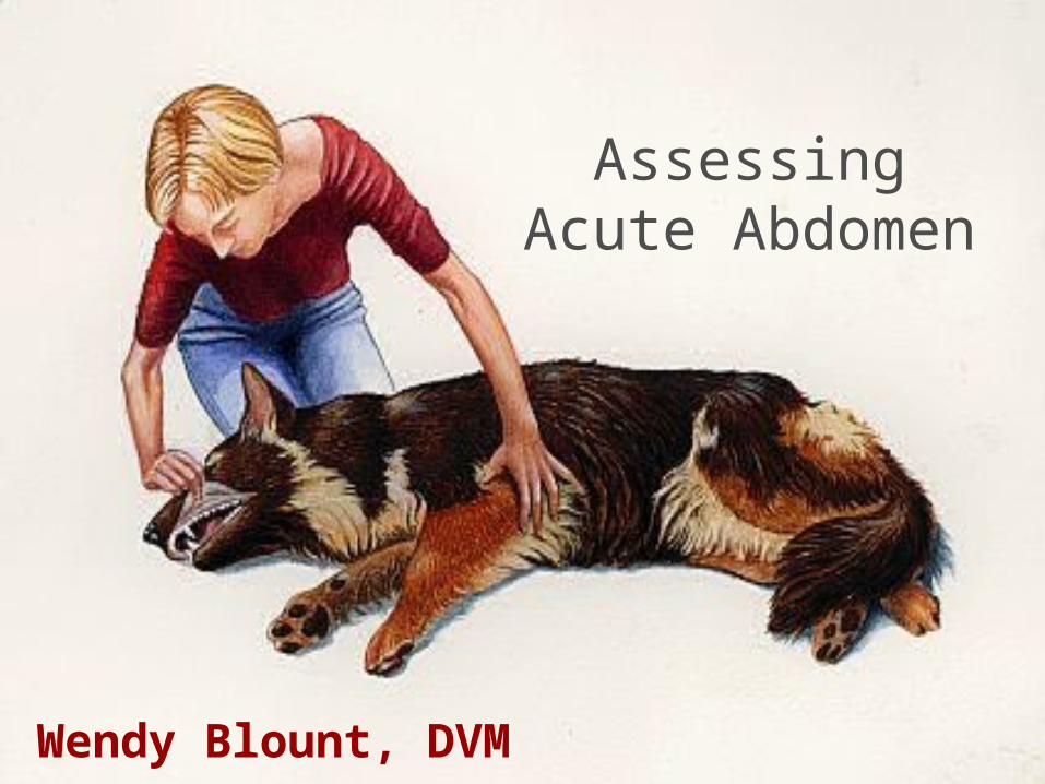
DDx Acute Abdomen
Acute Abdomen• Rapid onset of abdominal pain• Acute onset of clinical signs related to abdominal
pathology• Potentially life threatening
Determine quickly whether abdominal surgery is necessary
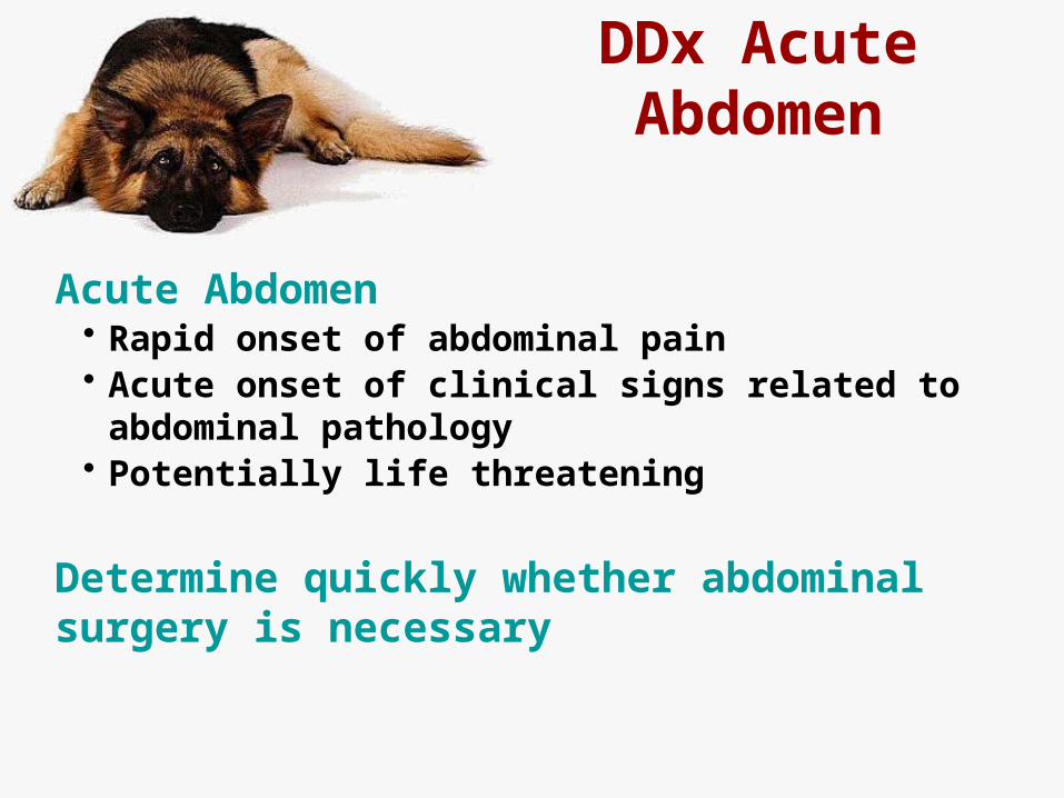
DDx Acute Abdomen
GI TractUrogenital TractHeptaobiliary SystemSpleen & LymphaticOther causesThings that may mimic acute abdomen
DDx Acute Abdomen
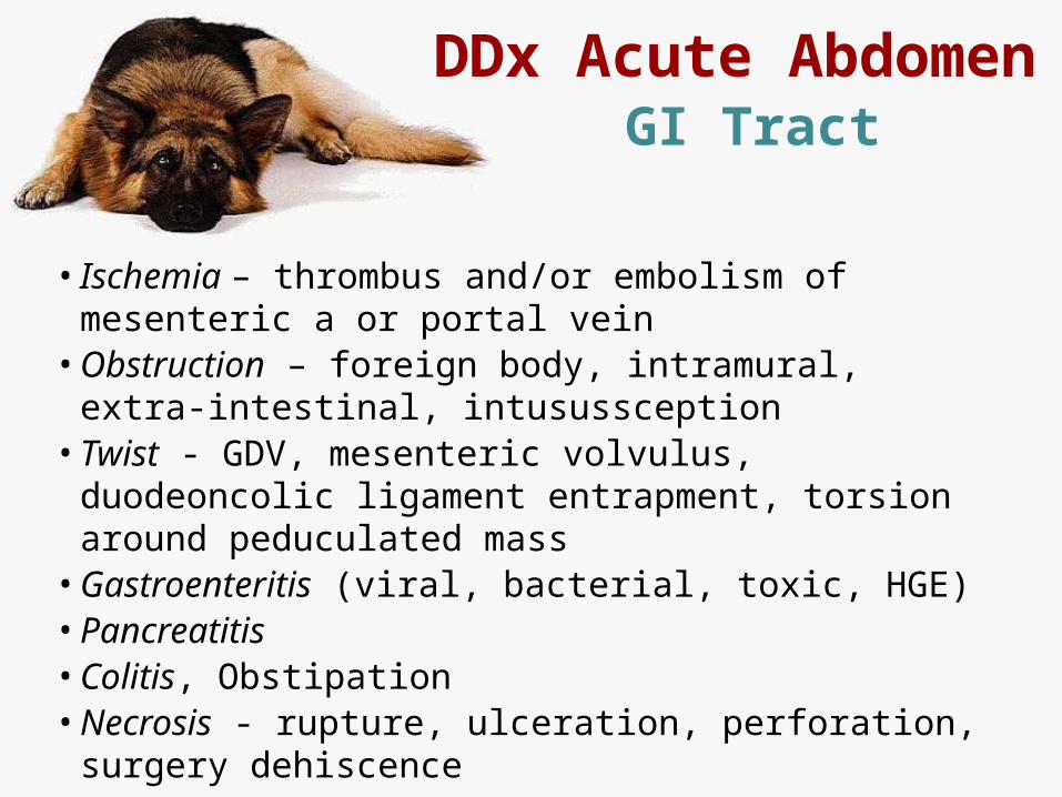
• Ischemia – thrombus and/or embolism of mesenteric a or portal vein
• Obstruction – foreign body, intramural, extra-intestinal, intusussception
• Twist - GDV, mesenteric volvulus, duodeoncolic ligament entrapment, torsion around peduculated mass
• Gastroenteritis (viral, bacterial, toxic, HGE)• Pancreatitis• Colitis, Obstipation• Necrosis - rupture, ulceration, perforation, surgery
dehiscence
GI Tract
DDx Acute Abdomen
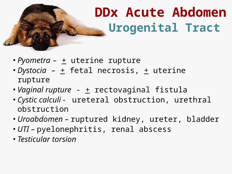
• Pyometra – + uterine rupture• Dystocia – + fetal necrosis, + uterine rupture• Vaginal rupture - + rectovaginal fistula• Cystic calculi - ureteral obstruction, urethral obstruction• Uroabdomen – ruptured kidney, ureter, bladder• UTI – pyelonephritis, renal abscess• Testicular torsion
Urogenital Tract
DDx Acute Abdomen
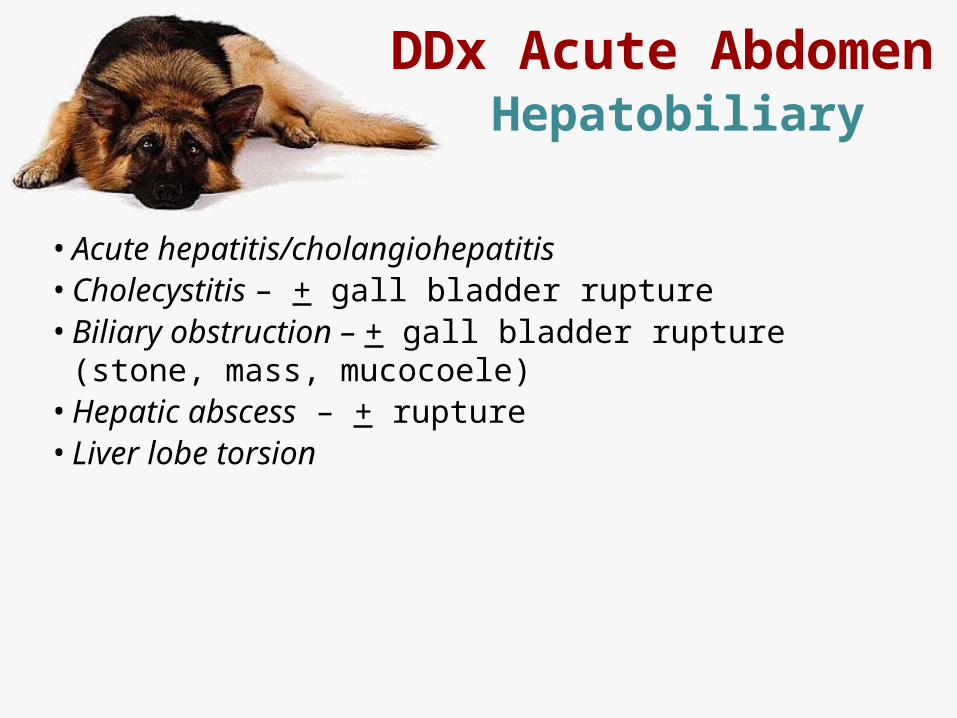
• Acute hepatitis/cholangiohepatitis• Cholecystitis – + gall bladder rupture• Biliary obstruction – + gall bladder rupture (stone, mass,
mucocoele)• Hepatic abscess – + rupture• Liver lobe torsion
Hepatobiliary
DDx Acute Abdomen
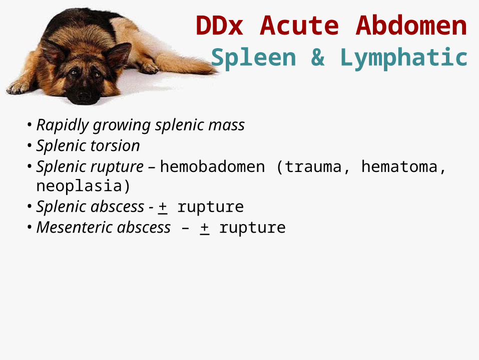
• Rapidly growing splenic mass• Splenic torsion• Splenic rupture – hemobadomen (trauma, hematoma,
neoplasia)• Splenic abscess - + rupture• Mesenteric abscess – + rupture
Spleen & Lymphatic
DDx Acute Abdomen
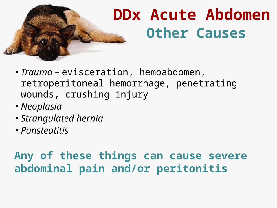
• Trauma – evisceration, hemoabdomen, retroperitoneal hemorrhage, penetrating wounds, crushing injury
• Neoplasia• Strangulated hernia• Pansteatitis
Any of these things can cause severe abdominal pain and/or peritonitis
Other Causes
DDx Acute Abdomen
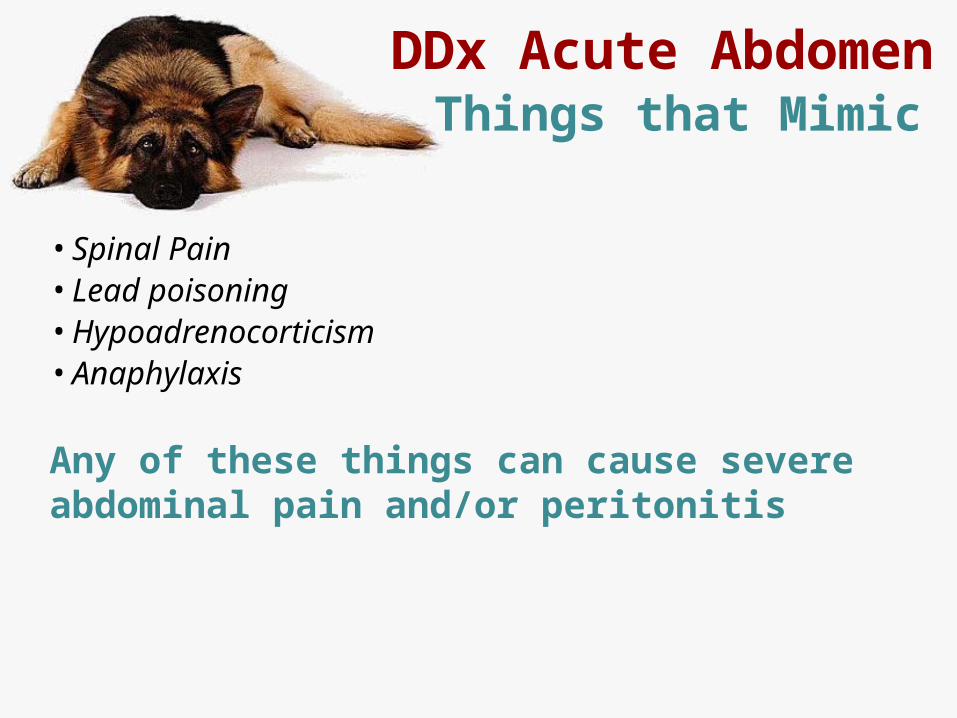
• Spinal Pain• Lead poisoning• Hypoadrenocorticism• Anaphylaxis
Any of these things can cause severe abdominal pain and/or peritonitis
Things that Mimic
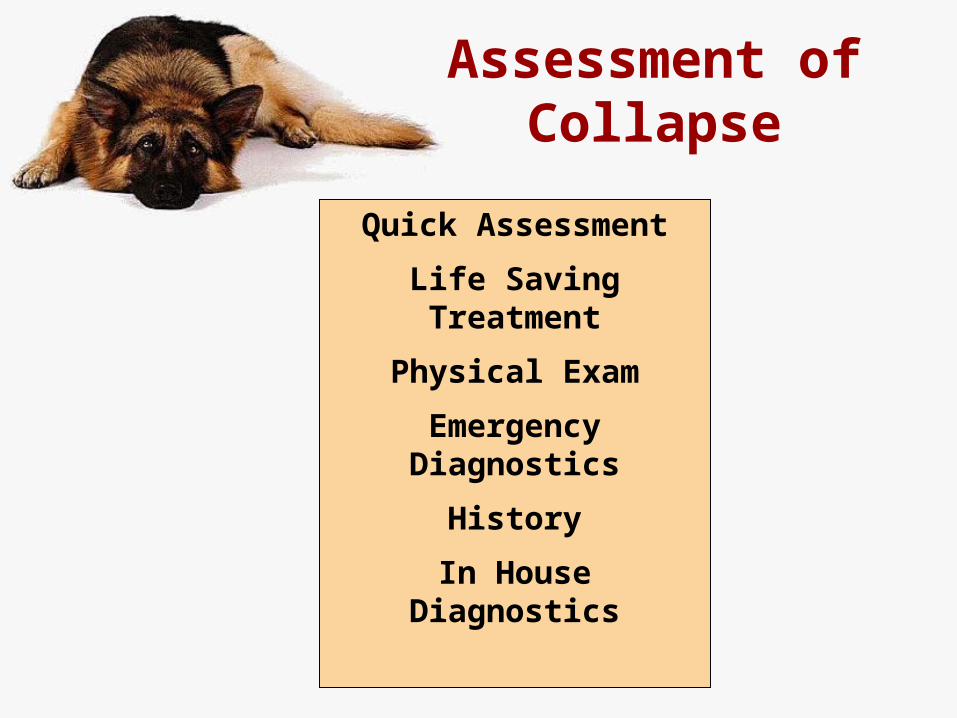
Assessment of Collapse
Quick Assessment
Life Saving Treatment
Physical Exam
Emergency Diagnostics
History
In House Diagnostics
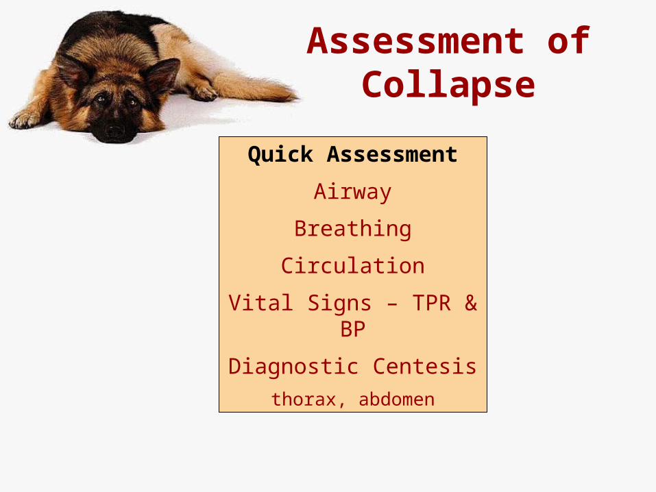
Assessment of Collapse
Quick Assessment
Airway
Breathing
Circulation
Vital Signs – TPR & BP
Diagnostic Centesis
thorax, abdomen
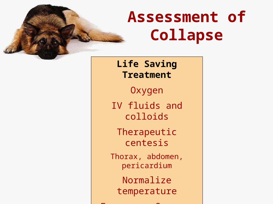
Assessment of Collapse
Life Saving Treatment
Oxygen
IV fluids and colloids
Therapeutic centesis
Thorax, abdomen, pericardium
Normalize temperature
Emergency Surgery
Assessment of Collapse
Physical Exam
General Exam
Cardiovascular Exam
Neurologic Exam
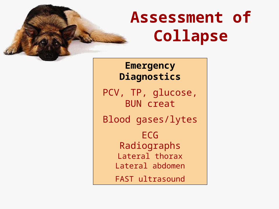
Assessment of Collapse
Emergency Diagnostics
PCV, TP, glucose, BUN creat
Blood gases/lytes
ECGRadiographsLateral thorax
Lateral abdomen
FAST ultrasound

Assessment of Collapse
Quick Assessment
Life Saving Treatment
Physical Exam
Emergency Diagnostics
History
In House Diagnostics
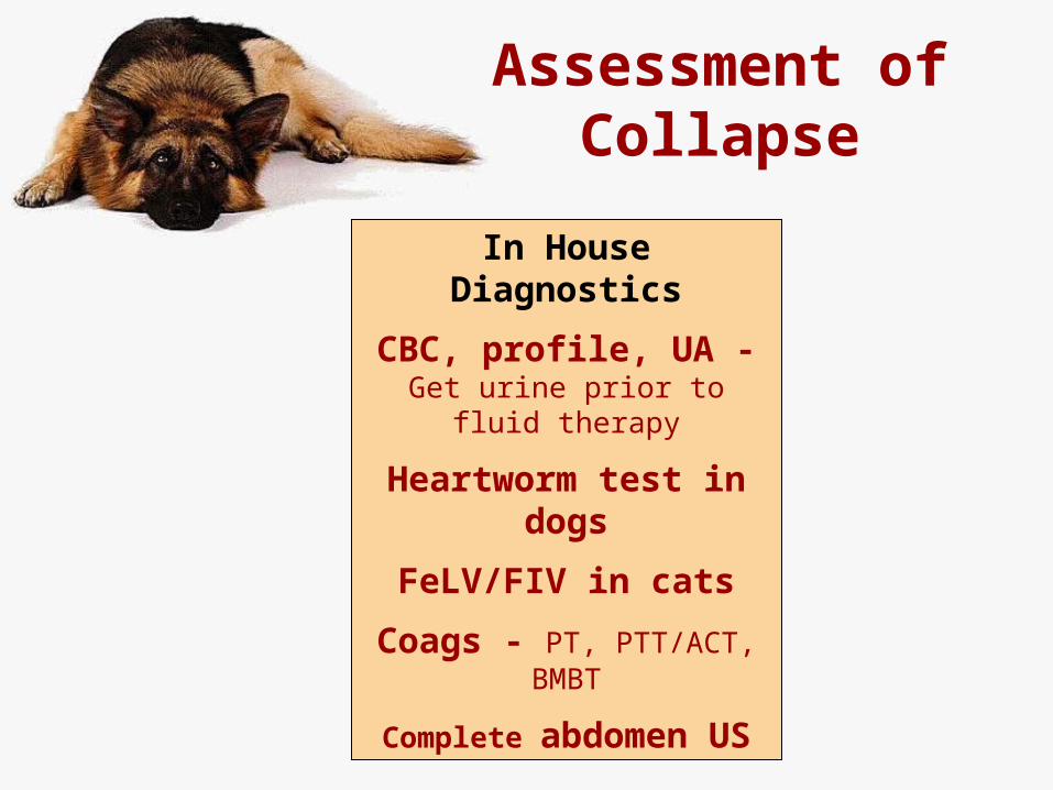
Assessment of Collapse
In House Diagnostics
CBC, profile, UA - Get urine prior to fluid therapy
Heartworm test in dogs
FeLV/FIV in cats
Coags - PT, PTT/ACT, BMBT
Complete abdomen US
Quick Assessment
Emergency Exam Form Cat
Emergency Dog Exam Form
Resuscitation Flow Sheet
Blood Pressure Handout
Quick Assessment
Get samples to run later• Blood (8-10cc)
• EDTA tube• Lithium heparin tube• Potassium citrate tube• Red top clot tube
• urine
Quick Assessment
Urinalysis• If you need a urinalysis later, you need a sample prior to
fluid therapy, before specific gravity is diluted• If fever, you may want urine for possible culture prior to
antibiotic therapy• Use a 5Fr infant feeding tube to catheterize male dog >
75 pounds• Use US guidance if needed for cystocentesis of small
bladder
Quick Assessment
Indications for Diagnostic Abdominocentesis• Palpable fluid wave• Owner reports abdominal bloating• Suspect abdominal hemorrhage
• Acute collapse, pale mucous membranes, weak pulses, low blood pressure, + anemia
• Suspect peritonitis – shock and abdominal pain• Fluid seen on FAST ultrasound
Quick Assessment
Diagnostic abdominal tap technique – 4 quadrants
• R cranial, L cranial, R caudal, L caudal• Syringe and 18-20g needle are fine• Put fluid in EDTA and red top tubes for analysis
• Spin down for cytology• Save red top tube for culture if needed• Run EDTA through CBC machine for cell counts• Be aware that you can fill the syringe with blood if
you hit a normal spleen – try RCrFluid Analysis Handout
Fluid Therapy
“Shock/Replacement Fluids”• Bolus of 10 ml/lb over 10-15 minutes, then reassess• NO shock fluids if there is anuria or CHF (Angel)
• Anuria - you can probably get way with one shock dose if the dog hasn’t had prior fluid therapy
• MONITOR URINE OUTPUT AFTER THE SHOCK DOSE
Fluid Therapy
“Shock/Replacement Fluids”• Bolus of 10 ml/lb over 10-15 minutes, then reassess• Two IV catheters may be necessary in a giant dog• CAREFUL if hypoalbuminemia
• If TP low, get albumin ASAPAggressive fluid therapy + hypoalbuminemia = pulmonary edema
• Replace colloids first – hetastarch• shock fluids may not be necessary, and could even lead
to volume overload
Fluid Therapy
Maintenance Fluids• 1-2 ml/lb/hr – fine tune later• To keep the IV line open while the patient is assessed• Most patients fall under this category
No Fluids – if CHF is possible• Heart murmur• Auscultable arrhythmia or pulse deficits• Undiagnosed thoracic effusion or ascites – modified transudate• Dyspneic animal who has not had chest x-rays yet
• Be especially careful with cats• Fluids, corticosteroids or x-rays can KILL a cat in CHF
Fluid Therapy
Colloids
• Hetastarch 15-20 ml/lb per 24 hours• Give over 15-20 minutes
• Plasma 5-10 ml/lb/day• Premedicate with diphenhydramine• Give over 1-2 hours
Ascites
Transudate or Modified Transudate• Remove enough fluid to alleviate dyspnea, and allow
comfortable chest x-rays & abdominal ultrasound• Bloodwork and abdominal ultrasound to determine the
cause, and treat accordingly• If cause is congestive heart failure, remove all fluid
Hemorrhage - usually a surgical problem, unless• Coagulopathy is identified and treated• Traumatic hemorrhage resolves spontaneously
Non-septic exudate, chyle – tap if dyspneic• Imaging determines whether the problem is surgical
Ascites
Septic exudate, uroabdomen, bile peritonitis – usually surgical
• Multiple species of bacteria suggest GI perforation• Plant material is very strong evidence• If no bacteria are seen, look for phagocytosed
bacteria in WBC, and for toxic changes in the neutrophils
Abdominal amylase and lipase elevated relative to serum with pancreatitis
Abdominal fluid glucose <50 mg/dl is often indicative of bacterial peritonitis

History
History
Acute collapse over minutes, then acute abdomen• Rapid hemorrhage – trauma or spontaneous
• Rupture of abdominal abscess
• Anaphylaxis
• after insect bite, snake bite, vaccination
• after heartworm prevention in untested dog (milbemycin)
• after going outside
History
Protracted dysuria, pollakuria• Urinary obstruction
Estrus about a month ago• pyometra
Eating carrion or garbage• HGE – hemorrhagic gastroenteritis• Foreign body obstruction• Botulism – flaccid paralysis• Roquefortine toxin – seizures and twitching
History
Pattern recognition for prostatitis• Male dog• Fever• Caudal abdominal pain• Hematuria (especially dripping)• Hindlimb stiffness or an abnormal gait• Signs of sepsis• Pollakuria or even urinary obstruction• dyschezia
History
Profuse and frequent vomiting• Foreign body obstruction• Pancreatitis
PU-PD• Sepsis, DKA, pyelonephritis, Addison’s
Melena• Gut necrosis• GI mass• Bleeding ulcer
Physical Exam
Heart Rate• Sinus Bradycardia – confirm with lead II ECG
• Impending death • Increased vagal tone –
• increased CSF pressure• abdominal disease• tracheal trauma• increased IOP• retching
• Give atropine or glycopyrrolate and recheck
Physical Exam
Heart Rate• Sinus Tachycardia – confirm with lead II ECG
• Pain or anxiety • give pain meds
• Hypovolemic shock• increase the fluid rate
• Heart failure• Pericardial tamponade
• Tap and give IV fluid bolus
Physical Exam
Mucous Membrane Color• Cyanosis
• Respiratory failure – airway obstruction, alveolar disease or pleural/pericardial disease (air/fluid/organs)
• Congestive heart failure• Pulmonary hypertension
• Differential cyanosis• Pink in front, blue in back (Reverse PDA or FATE)
• Brick red mucous membranes• Sepsis – do CBC, and albumin• HGE (hemorrhagic gastroenteritis in dogs)• SIRS
Physical Exam
Mucous Membrane Color• Icterus – yellow mucous membranes
• Check CBC first to rule out hemolysis• If anemic, check saline test for autoagglutination• If anemic, check blood smear cytology for Mycoplasma
haemofelis• If not anemic, you are left with hepatic disease (including
sepsis) or bile obstruction• No point doing bile acids if bilirubin is high (you know they are
high)• Abdominal US is more helpful

Physical Exam
Mucous Membrane Color
• Pallor• Pain• Cardiovascular shock• Anaphylactic shock• Anemia• Hypovolemia – hemorrhage, hypoproteinemia
CRT >2 sec means poor peripheral perfusion
Physical Exam
Respirations• Minimal chest excursions can indicate LMN
paralysis or severe shock• Exaggerated chest excursions
• Cardiovascular failure• Respiratory failure – lung disease, airway disease, third
space disease• Aspiration pneumonia due to vomiting-regurgitation
• Anemia or other oxygen carrying problem• Metabolic acidosis
Physical Exam
Pulses• Jugular pulses
• Hepatojugular reflux• apply pressure to the liver for 5-10 seconds• Filling of the jugular veins indicates right heart failure or
pericardial disease• Peripheral Pulses
• Weak pulses• CHF• Pericardial disease• Shock of any kind, especially hypovolemic• Hypertension
Physical Exam
Pulses• Peripheral Pulses
• Bounding pulses - Big difference in pressure between systole and diastole
• Fever/Sepsis (vasodilation makes diastolic pressure lower)• PDA (back flow during systole)• Aortic endocarditis (black flow during systole)• Extreme bradycardia (volume overload)• Anemia (low blood viscosity)
• Pulsus paradoxus – absent during peak inspiration• Pericardial effusion or hernia
• No pulses in only one area• Thromboembolic disease
Physical Exam
Skin• Hemorrhages might indicate coagulopathy – do coags
• Ecchymoses and petechiae• Peripheral edema
• Right heart failure• Vasculitis, venous or lymphatic obstruction• Hypoalbuminemia• Infiltrative tumor such as myxosarcoma can look like edema
Physical Exam
Abdominal Palpation• Distension
• Obesity, pendulous abdomen• Pregnancy, pyometra - ultrasound• Balotte fluid wave – tap• Palpate organomegaly – ultrasound
• Relieve urinary obstruction or express bladder• Abdominal mass – ultrasound
• If cystic masses, may not be safe to aspirate• Can aspirate solid masses later• Aspirate homogeneous enlarged spleen (MCT, Lymphoma)
Physical Exam
Abdominal Palpation• Distension
• Gut distended with gas – radiograph• Pass stomach tube if gastric• Pneumonperitoneum can cause some gas in the abdomen• Tympanic abdomen in a large dog is usually GDV
Physical Exam
Abdominal Palpation• Severe abdominal pain is often surgical, especially if
the pain is focal• Think peritonitis when you have shock and severe
abdominal pain• If a pyometra is painful, think peritonitis• Uroabdomen painful only if infected or acute
• How do you diagnose peritonitis and decide to go to surgery?• Get some abdominal fluid , spin down, look for bacteria• Diagnostic peritoneal lavage if necessary• Most but not all animals with peritonitis have ascites
Diagnostic Peritoneal lavage• Place a large bore IV catheter into the abdomen on
the ventral midline• Collect samples for fluid analysis, culture• Attach IV set and infuse 5-10 ml/lb sterile isotonic
fluids• Remove catheter, palpate abdomen• let sit for 30 minutes• Do single or 4 quadrant abdominocentesis• Drain out as much fluid as possible, saving some for
cytology, culture
Physical Exam
Differentiating abdominal pain from spinal pain• If neck pain, palpate the lower neck from ventral• Gently check range of motion of neck in all 4 directions• Palpate along the dorsal spinous processes without
touching the abdomen• Lift the tail• Check for neurologic deficits• Crying out when picked up is more often due to spinal
pain than abdominal pain
Physical Exam
Hemoabdomen• Pressure wrap on the cranial abdomen can slow
hemorrhage• Can collect the blood, run it through a blood
administration set and give it back to the IV compartment
Physical Exam
In House Diagnostics
Emergency Bloodwork• CBC with platelets• General health profile – include P, Ca++, albumin and
triglycerides• Electrolytes and blood gases• Urinalysis
Drug Therapy
Broad spectrum IV antibiotics• As soon as sepsis is suspected or identified• Ampicillin + cefixotin• Ampicillin + enrofloxacin• Ampicillin + amikacin (if not hypovolemic)• Can substitute metronidazole for ampicillin, but give
slowly, and it is less broad spectrum
Drug Therapy
Heparin • If DIC – 75 U/kg SC TID
Corticosteroids• Use for sepsis is controversial• Single moderate dose is best• Not with NSAIDs
Neupogen (Granulocyte Colony Stimulating Factor)• If neutrophilia is acute, this can improve survival
Imaging
Abdominal rads• Superior to US for finding intestinal obstruction or
foreign body• Better then US for evaluating gas in the abdomen• DDx Pneumonperitoneum:
• Rupture of hollow viscus• Gas forming bacteria• Therapeutic abdominocentesis or recent
surgery/laparoscopy – days to weeks• Penetrating injury
Imaging
Abdominal Ultrasound - FAST• Focused Assessment with Sonography in Trauma • early recognition of intraperitoneal blood • rapid, safe and sensitive• can be repeated if the patient’s status changes• help prioritize initial management in patients with multiple
penetrating injuries or an unknown missile trajectory• Sensitivity for determining need for surgery is 50% in people
Imaging
Acute Abdomen Surgery• Collect peritoneal fluid for analysis & culture/sensitivity• Identify and resolve sources of acute hemorrhage• Identify, debride, remove, lavage necrotic tissues• Identify and resolve GI/urinary obstruction
• Run the gut• Assess and treat for generalized or localized peritonitis
• Contain the evil spirits• Collect diagnostic samples
Imaging
Diagnostic Surgery – Canine Samples• Liver• Stomach, duodenum, jejunum, ileum• Mesenteric lymph node• Aspirate spleen for cytology• Evaluate and sample if abnormal:
• Spleen, kidneys, colon, bladder, prostate, ovaries/uterus• Other lymph nodes (hepatic, sublumbar)• Pancreas, gall bladder and adrenals only if absolutely
necessary
Imaging
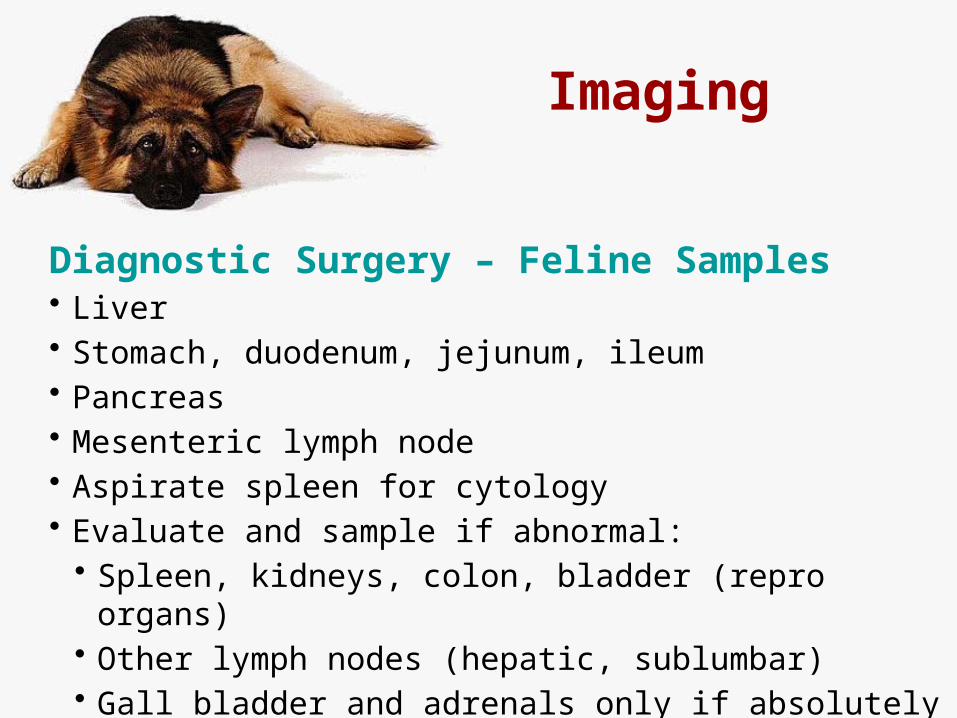
Diagnostic Surgery – Feline Samples• Liver• Stomach, duodenum, jejunum, ileum• Pancreas• Mesenteric lymph node• Aspirate spleen for cytology• Evaluate and sample if abnormal:
• Spleen, kidneys, colon, bladder (repro organs)• Other lymph nodes (hepatic, sublumbar)• Gall bladder and adrenals only if absolutely necessary
Handouts
• PowerPoint Presentation – after the gold tab• Clinic Handouts
• Coagulopathy Diagnostic Chart• Arrhythmia Treatment and Diagnostic Charts• CPR Flow Sheet• Emergency Exam Cat• Emergency Exam Dog• Measuring Blood Pressure in Dogs and Cats