Embed Size (px)
Citation preview

G-Biosciences ♦ 1-800-628-7730 ♦ 1-314-991-6034 ♦ [email protected]
A Geno Technology, Inc. (USA) brand name
think proteins! think G-Biosciences www.GBiosciences.com
PR064
AssaysforProteinQuantificationTeacher’sGuidebook
(Cat.#BE‐402)
Page 2 of 16
MATERIALS INCLUDED WITH THE KIT ................................................................................ 3
BIURET PROTEIN ASSAY ................................................................................................. 3
LOWRY PROTEIN ASSAY ................................................................................................. 3
CB PROTEIN ASSAY ........................................................................................................ 3
SPECIAL HANDLING INSTRUCTIONS ................................................................................... 3
ADDITIONAL EQUIPMENT REQUIRED ................................................................................ 3
TIME REQUIRED ................................................................................................................. 3
OBJECTIVES ........................................................................................................................ 4
BACKGROUND ................................................................................................................... 4
TEACHER’S PRE EXPERIMENT SET UP ................................................................................ 6
MATERIALS FOR EACH GROUP .......................................................................................... 6
BIURET PROTEIN ASSAY REAGENTS ............................................................................... 6
LOWRY PROTEIN ASSAY ................................................................................................. 6
CB PROTEIN ASSAY ........................................................................................................ 6
PROCEDURE ....................................................................................................................... 7
I. BIURET PROTEIN ASSAY .............................................................................................. 7
II. LOWRY PROTEIN ASSAY ............................................................................................. 8
III. BRADFORD PROTEIN ASSAY ...................................................................................... 9
IV. DETERMINATION OF THE CONCENTRATION OF THE UNKNOWN PROTEIN ............. 9
RESULTS, ANALYSIS & ASSESSMENT ................................................................................ 11
BIURET PROTEIN ASSAY (SECTION I) ............................................................................ 11
LOWRY PROTEIN ASSAY (SECTION II) ........................................................................... 11
BRADFORD PROTEIN ASSAY (SECTION III) .................................................................... 12
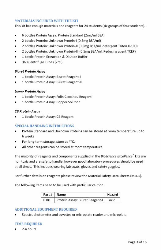
MATERIALSINCLUDEDWITHTHEKITThis kit has enough materials and reagents for 24 students (six groups of four students).
• 6 bottles Protein Assay: Protein Standard (2mg/ml BSA) • 2 bottles Protein: Unknown Protein‐I (0.5mg BSA/ml) • 2 bottles Protein: Unknown Protein‐II (0.5mg BSA/ml, detergent Triton X‐100) • 2 bottles Protein: Unknown Protein‐III (0.5mg BSA/ml, Reducing agent TCEP) • 1 bottle Protein Extraction & Dilution Buffer • 360 Centrifuge Tubes (2ml)
Biuret Protein Assay • 1 bottle Protein Assay: Biuret Reagent‐I • 1 bottle Protein Assay: Biuret Reagent‐II
Lowry Protein Assay • 1 bottle Protein Assay: Folin Ciocalteu Reagent • 1 bottle Protein Assay: Copper Solution
CB Protein Assay • 1 bottle Protein Assay: CB Reagent
SPECIALHANDLINGINSTRUCTIONS• Protein Standard and Unknown Proteins can be stored at room temperature up to
6 weeks • For long‐term storage, store at 4°C. • All other reagents can be stored at room temperature.
The majority of reagents and components supplied in the BioScience Excellence™ kits are non toxic and are safe to handle, however good laboratory procedures should be used at all times. This includes wearing lab coats, gloves and safety goggles.
For further details on reagents please review the Material Safety Data Sheets (MSDS).
The following items need to be used with particular caution.
Part # Name Hazard P381 Protein Assay: Biuret Reagent‐I Toxic
ADDITIONALEQUIPMENTREQUIRED• Spectrophotometer and cuvettes or microplate reader and microplate
TIMEREQUIRED• 2‐4 hours
Page 3 of 16
OBJECTIVES• Learn the principles of protein assays. • Explore the methods used for quantification of proteins. • Determine protein concentrations using three separate methods. • Study the effect of common laboratory reagents on the methods of for protein
quantification.
BACKGROUNDThe determination of protein concentration is an essential technique in all aspects of protein studies and proteomics. This lab activity is designed to teach students the principles behind protein estimation and three of the most widely used methods in protein estimation. The first two are the alkaline copper solution methods and the third is the dye binding protein assays, they are: Biuret Protein Assay, Lowry Protein Assay, and the CB Protein Assay.
The “Assays for Protein Quantification” kit provides all the reagents required to perform all three protein assays in a single lab activity. An often underestimated factor in quantifying protein is the presence of non‐protein interfering agents, such as salts and detergents. This kit teaches students about common laboratory agents that affect the protein assays, the reasoning behind their interferences and how to overcome the interference. Students also learn how to select a protein assay for different applications.
Although there are a wide variety of protein assays available none of the assays can be used without first considering their suitability for the application. Each method has its own advantages and limitations and often it is necessary to obtain more than one type of protein assay for research applications. Protein assays based on these methods are divided into two categories: dye binding protein assays and protein assays based on alkaline copper.
The dye binding protein assay (CB Protein Assay) is based on the binding of protein molecules to Coomassie dye under acidic conditions. The binding of protein to the dye results in a spectral shift, the color of Coomassie solution changes from brown (absorbance maximum 465nm) to blue (absorbance maximum 610nm). The change in color density is read at 595nm and is proportional to the protein concentration.
In the copper ion based protein assays, protein solutions are mixed with an alkaline solution of copper salt, cupric ions (Cu2+). The protein assay is based on the interaction of cupric ions with protein in an alkaline solution and is commonly referred to as the Biuret assay. The interaction of cupric ions (Cu2+) with protein results in a purple color that can be read at 545nm. The amount of color produced is proportional to protein concentration.
Page 4 of 16
Under alkaline conditions cupric ions (Cu2+) chelate with the peptide bonds resulting in reduction of cupric ions (Cu2+) to cuprous ions (Cu+). The Cuprous ions can also be detected with Folin Ciocalteu Reagent (phosphomolybdic/phosphotungstic acid); this method is commonly refereed to as the Lowry method. Cuprous ions (Cu+) reduction of Folin Ciocalteu Reagent produces a blue color that can be read at 650‐750nm. The amount of color produced is proportional to the amount of peptide bonds, i.e. size as well as the amount of protein/peptide.
In this lab activity students will study and analyze advantages and limitations of these widely used protein assay methods. This kit is provided with three protein assays and three different types of protein solutions as test samples, they are protein solution in sodium phosphate buffer, protein solution containing a detergent (Triton X‐100) that is widely used in protein research, and a protein solution containing a reducing agent TCEP (Tri [2‐carboxyethyl] phosphine).
Page 5 of 16
TEACHER’SPREEXPERIMENTSETUP1. Mix equal volume of Biuret Reagent‐I and Biuret Reagent‐II to make Working Biuret
Reagent just before the start of the experiment. Each student group needs 20ml Working Biuret Reagent.
2. Aliquot reagents for each student group according to the next section.
MATERIALSFOREACHGROUPSupply each group with the following components. Several components are shared by the whole class and should be kept on a communal table.
Biuret Protein Assay Reagents • Working Biuret Reagent (shared with whole class)
Lowry Protein Assay • Folin Ciocalteu Reagent (shared with whole class) • Copper Solution (shared with whole class)
CB Protein Assay • CB Reagent (shared with whole class) • 1 bottle Protein Standard (2mg/ml BSA) • 2 bottles Unknown Protein‐I (0.5mg BSA/ml) (shared with whole class) • 2 bottles Unknown Protein‐II (0.5mg BSA/ml, detergent Triton X‐100) (shared
with whole class) • 2 bottles Unknown Protein‐III (0.5mg BSA/ml, Reducing agent TECP) (shared
with whole class) • 4ml Protein Extraction & Dilution Buffer • 60 Centrifuge Tubes (2ml) • Marker Pen
Page 6 of 16
PROCEDURE
Wear gloves throughout the whole procedure.
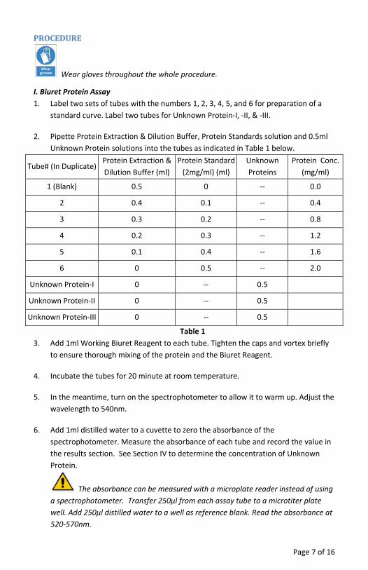
I. Biuret Protein Assay 1. Label two sets of tubes with the numbers 1, 2, 3, 4, 5, and 6 for preparation of a
standard curve. Label two tubes for Unknown Protein‐I, ‐II, & ‐III.
2. Pipette Protein Extraction & Dilution Buffer, Protein Standards solution and 0.5ml Unknown Protein solutions into the tubes as indicated in Table 1 below.
Tube# (In Duplicate) Protein Extraction & Dilution Buffer (ml)
Protein Standard (2mg/ml) (ml)
Unknown Proteins
Protein Conc. (mg/ml)
1 (Blank) 0.5 0 ‐‐ 0.0
2 0.4 0.1 ‐‐ 0.4
3 0.3 0.2 ‐‐ 0.8
4 0.2 0.3 ‐‐ 1.2
5 0.1 0.4 ‐‐ 1.6
6 0 0.5 ‐‐ 2.0
Unknown Protein‐I 0 ‐‐ 0.5
Unknown Protein‐II 0 ‐‐ 0.5
Unknown Protein‐III 0 ‐‐ 0.5
Table 1 3. Add 1ml Working Biuret Reagent to each tube. Tighten the caps and vortex briefly
to ensure thorough mixing of the protein and the Biuret Reagent.
4. Incubate the tubes for 20 minute at room temperature.
5. In the meantime, turn on the spectrophotometer to allow it to warm up. Adjust the wavelength to 540nm.
6. Add 1ml distilled water to a cuvette to zero the absorbance of the spectrophotometer. Measure the absorbance of each tube and record the value in the results section. See Section IV to determine the concentration of Unknown Protein.
The absorbance can be measured with a microplate reader instead of using a spectrophotometer. Transfer 250μl from each assay tube to a microtiter plate well. Add 250μl distilled water to a well as reference blank. Read the absorbance at 520‐570nm.
Page 7 of 16
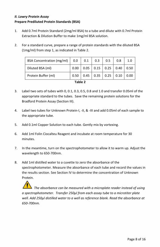
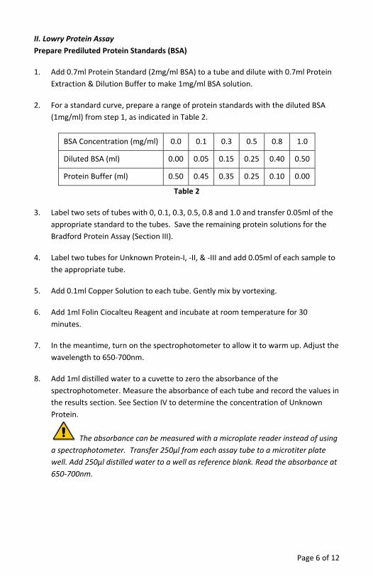
II. Lowry Protein Assay Prepare Prediluted Protein Standards (BSA)
1. Add 0.7ml Protein Standard (2mg/ml BSA) to a tube and dilute with 0.7ml Protein Extraction & Dilution Buffer to make 1mg/ml BSA solution.
2. For a standard curve, prepare a range of protein standards with the diluted BSA (1mg/ml) from step 1, as indicated in Table 2.
BSA Concentration (mg/ml) 0.0 0.1 0.3 0.5 0.8 1.0
Diluted BSA (ml) 0.00 0.05 0.15 0.25 0.40 0.50
Protein Buffer (ml) 0.50 0.45 0.35 0.25 0.10 0.00
Table 2
3. Label two sets of tubes with 0, 0.1, 0.3, 0.5, 0.8 and 1.0 and transfer 0.05ml of the appropriate standard to the tubes. Save the remaining protein solutions for the Bradford Protein Assay (Section III).
4. Label two tubes for Unknown Protein‐I, ‐II, & ‐III and add 0.05ml of each sample to the appropriate tube.
5. Add 0.1ml Copper Solution to each tube. Gently mix by vortexing.
6. Add 1ml Folin Ciocalteu Reagent and incubate at room temperature for 30 minutes.
7. In the meantime, turn on the spectrophotometer to allow it to warm up. Adjust the wavelength to 650‐700nm.
8. Add 1ml distilled water to a cuvette to zero the absorbance of the spectrophotometer. Measure the absorbance of each tube and record the values in the results section. See Section IV to determine the concentration of Unknown Protein.
The absorbance can be measured with a microplate reader instead of using a spectrophotometer. Transfer 250μl from each assay tube to a microtiter plate well. Add 250μl distilled water to a well as reference blank. Read the absorbance at 650‐700nm.
Page 8 of 16

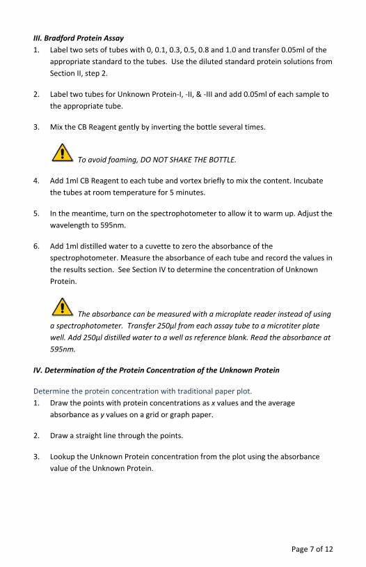
III. Bradford Protein Assay 1. Label two sets of tubes with 0, 0.1, 0.3, 0.5, 0.8 and 1.0 and transfer 0.05ml of the
appropriate standard to the tubes. Use the diluted standard protein solutions from Section II, step 2.
2. Label two tubes for Unknown Protein‐I, ‐II, & ‐III and add 0.05ml of each sample to the appropriate tube.
3. Mix the CB Reagent gently by inverting the bottle several times.
To avoid foaming, DO NOT SHAKE THE BOTTLE.
4. Add 1ml CB Reagent to each tube and vortex briefly to mix the content. Incubate the tubes at room temperature for 5 minutes.
5. In the meantime, turn on the spectrophotometer to allow it to warm up. Adjust the wavelength to 595nm.
6. Add 1ml distilled water to a cuvette to zero the absorbance of the spectrophotometer. Measure the absorbance of each tube and record the values in the results section. See Section IV to determine the concentration of Unknown Protein.
The absorbance can be measured with a microplate reader instead of using a spectrophotometer. Transfer 250μl from each assay tube to a microtiter plate well. Add 250μl distilled water to a well as reference blank. Read the absorbance at 595nm.
IV. Determination of the Protein Concentration of the Unknown Protein
Determine the protein concentration with traditional paper plot. 1. Draw the points with protein concentrations as x values and the average
absorbance as y values on a grid or graph paper.
2. Draw a straight line through the points.
3. Lookup the Unknown Protein concentration from the plot using the absorbance value of the Unknown Protein.
Page 9 of 16

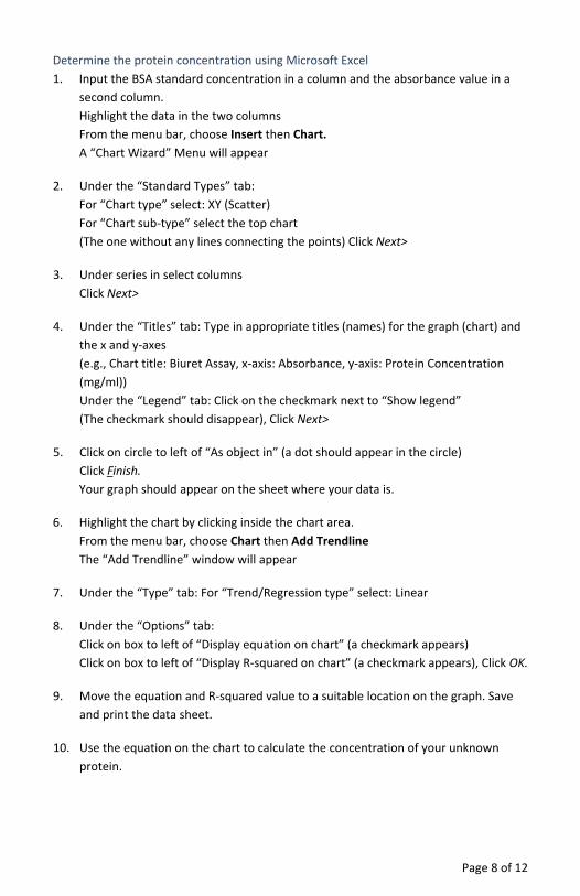
Determine the protein concentration using Microsoft Excel 1. Input the BSA standard concentration in a column and the absorbance value in a
second column. Highlight the data in the two columns From the menu bar, choose Insert then Chart. A “Chart Wizard” Menu will appear
2. Under the “Standard Types” tab: For “Chart type” select: XY (Scatter) For “Chart sub‐type” select the top chart (The one without any lines connecting the points) Click Next>
3. Under series in select columns Click Next>
4. Under the “Titles” tab: Type in appropriate titles (names) for the graph (chart) and the x and y‐axes (e.g., Chart title: Biuret Assay, x‐axis: Absorbance, y‐axis: Protein Concentration (mg/ml)) Under the “Legend” tab: Click on the checkmark next to “Show legend” (The checkmark should disappear), Click Next>
5. Click on circle to left of “As object in” (a dot should appear in the circle) Click Finish. Your graph should appear on the sheet where your data is.
6. Highlight the chart by clicking inside the chart area. From the menu bar, choose Chart then Add Trendline The “Add Trendline” window will appear
7. Under the “Type” tab: For “Trend/Regression type” select: Linear
8. Under the “Options” tab: Click on box to left of “Display equation on chart” (a checkmark appears) Click on box to left of “Display R‐squared on chart” (a checkmark appears), Click OK.
9. Move the equation and R‐squared value to a suitable location on the graph. Save and print the data sheet.
10. Use the equation on the chart to calculate the concentration of your unknown protein.
Page 10 of 16
RESULTS,ANALYSIS&ASSESSMENT
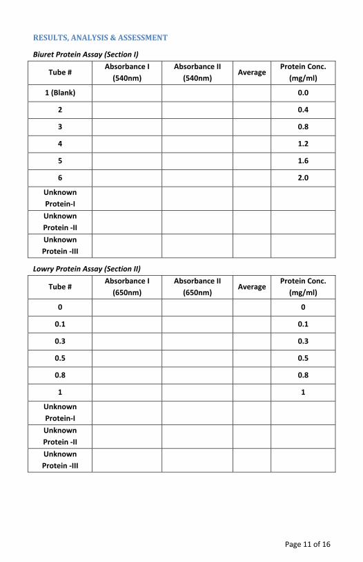
Biuret Protein Assay (Section I)
Tube # Absorbance I (540nm)
Absorbance II (540nm)
Average Protein Conc.
(mg/ml)
1 (Blank) 0.0
2 0.4
3 0.8
4 1.2
5 1.6
6 2.0
Unknown Protein‐I
Unknown Protein ‐II
Unknown Protein ‐III
Lowry Protein Assay (Section II)
Tube # Absorbance I (650nm)
Absorbance II (650nm)
Average Protein Conc.
(mg/ml)
0 0
0.1 0.1
0.3 0.3
0.5 0.5
0.8 0.8
1 1
Unknown Protein‐I
Unknown Protein ‐II
Unknown Protein ‐III
Page 11 of 16
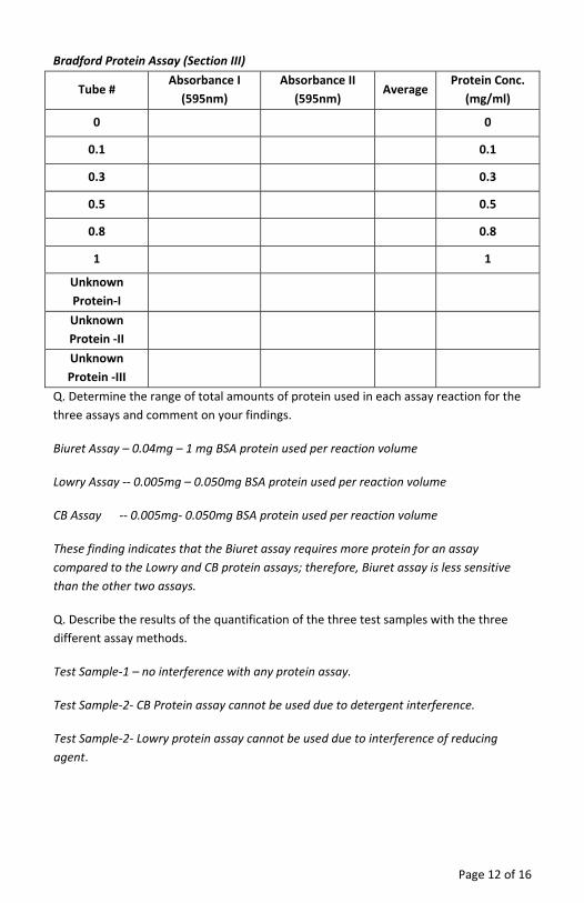
Bradford Protein Assay (Section III)
Tube # Absorbance I (595nm)
Absorbance II (595nm)
Average Protein Conc.
(mg/ml)
0 0
0.1 0.1
0.3 0.3
0.5 0.5
0.8 0.8
1 1
Unknown Protein‐I
Unknown Protein ‐II
Unknown Protein ‐III
Q. Determine the range of total amounts of protein used in each assay reaction for the three assays and comment on your findings.
Biuret Assay – 0.04mg – 1 mg BSA protein used per reaction volume
Lowry Assay ‐‐ 0.005mg – 0.050mg BSA protein used per reaction volume
CB Assay ‐‐ 0.005mg‐ 0.050mg BSA protein used per reaction volume
These finding indicates that the Biuret assay requires more protein for an assay compared to the Lowry and CB protein assays; therefore, Biuret assay is less sensitive than the other two assays.
Q. Describe the results of the quantification of the three test samples with the three different assay methods.
Test Sample‐1 – no interference with any protein assay.
Test Sample‐2‐ CB Protein assay cannot be used due to detergent interference.
Test Sample‐2‐ Lowry protein assay cannot be used due to interference of reducing agent.
Page 12 of 16
Page 13 of 16
Q. Briefly describe the principles behind the three protein assays and their weakness and strengths.
Biuret Protein Assay‐ based on binding of copper ions to peptide bonds under alkaline condition which produces purple color. Weakness – not very sensitive and requires large amounts of protein (0.04mg – 1 mg BSA protein per reaction volume).
Lowry Protein Assay‐ Under alkaline conditions cupric ions (Cu2+) chelate with the peptide bonds resulting in reduction of cupric (Cu2+) to cuprous ions (Cu+). The Cuprous ions are detected by the reduction of Folin Reagent (phosphomolybdic/phosphotungstic acid). Weakness – sensitive to reducing agents and other common lab reagents.
Dye Binding Protein Assay‐ based on the binding of protein molecules to Coomassie dye under acidic conditions. The binding of protein to the dye results in spectral shift, the color of Coomassie solution changes from brown to blue. Weakness‐ sensitive to detergents and other lab reagents.
Q. Select one protein assay at a time and explain how you will use that assay for determination of protein concentration.
Biuret Protein Assay – when large protein sample volume of high protein concentration (0.05mg protein per assay) is available and there is no risk of interference from non‐protein agents present in the protein solution.
Lowry Protein Assay – when protein sample is free from reducing agent and there is no risk of interference from non‐protein agents present in protein solution.
CB Protein Assay –when protein sample is free from detergents and there is no risk of interference from non‐protein agents present in the protein solution.
Q. For an unknown protein sample, which protein assay will you choose first?
For unknown protein sample the first choice is Biuret assay, unless there is not sufficient amount of protein available for assay. Followed by either the Lowey or CB protein assay, depending on whether or not the sample contains any interfering agents in the protein solution.
Q. How would you overcome interference in protein assay from common lab agents?
Interference from lab agents is removed by removing the interfering agents from protein solution, for detail on such techniques visits G‐Biosciences www.GBiosciences.com and visit protein assay pages.
Last saved: 9/13/2012 CMH

This page is intentionally left blank
Page 14 of 16

This page is intentionally left blank
Page 15 of 16

www.GBiosciences.com
Page 16 of 16

G-Biosciences ♦ 1-800-628-7730 ♦ 1-314-991-6034 ♦ [email protected]
A Geno Technology, Inc. (USA) brand name
think proteins! think G-Biosciences www.GBiosciences.com
PR065
AssaysforProteinQuantification
Student’sHandbook
(Cat.#BE‐402)
Page 2 of 12
OBJECTIVES ........................................................................................................................ 3
BACKGROUND ................................................................................................................... 3
MATERIALS FOR EACH GROUP .......................................................................................... 4
BIURET PROTEIN ASSAY REAGENTS ............................................................................... 4
LOWRY PROTEIN ASSAY ................................................................................................. 4
CB PROTEIN ASSAY ........................................................................................................ 4
PROCEDURE ....................................................................................................................... 5
I. BIURET PROTEIN ASSAY .............................................................................................. 5
II. LOWRY PROTEIN ASSAY ............................................................................................. 6
III. BRADFORD PROTEIN ASSAY ...................................................................................... 7
IV. DETERMINATION OF THE PROTEIN CONCENTRATION OF THE UNKNOWN PROTEIN ....................................................................................................................................... 7
RESULTS, ANALYSIS & ASSESSMENT .................................................................................. 9
BIURET PROTEIN ASSAY (SECTION I) .............................................................................. 9
LOWRY PROTEIN ASSAY (SECTION II) ............................................................................. 9
BRADFORD PROTEIN ASSAY (SECTION III) .................................................................... 10
OBJECTIVES• Learn the principles of protein assays. • Explore the methods used for quantification of proteins. • Determine protein concentrations using three separate methods. • Study the effect of common laboratory reagents on the methods of for protein
quantification.
BACKGROUNDThe determination of protein concentration is an essential technique in all aspects of protein studies and proteomics. This lab activity is designed to teach students the principles behind protein estimation and three of the most widely used methods in protein estimation. The first two are the alkaline copper solution methods and the third is the dye binding protein assays, they are: Biuret Protein Assay, Lowry Protein Assay, and the CB Protein Assay.
The “Assays for Protein Quantification” kit provides all the reagents required to perform all three protein assays in a single lab activity. An often underestimated factor in quantifying protein is the presence of non‐protein interfering agents, such as salts and detergents. This kit teaches students about common laboratory agents that affect the protein assays, the reasoning behind their interferences and how to overcome the interference. Students also learn how to select a protein assay for different applications.
Although there are a wide variety of protein assays available none of the assays can be used without first considering their suitability for the application. Each method has its own advantages and limitations and often it is necessary to obtain more than one type of protein assay for research applications. Protein assays based on these methods are divided into two categories: dye binding protein assays and protein assays based on alkaline copper.
The dye binding protein assay (CB Protein Assay) is based on the binding of protein molecules to Coomassie dye under acidic conditions. The binding of protein to the dye results in a spectral shift, the color of Coomassie solution changes from brown (absorbance maximum 465nm) to blue (absorbance maximum 610nm). The change in color density is read at 595nm and is proportional to the protein concentration.
In the copper ion based protein assays, protein solutions are mixed with an alkaline solution of copper salt, cupric ions (Cu2+). The protein assay is based on the interaction of cupric ions with protein in an alkaline solution and is commonly referred to as the Biuret assay. The interaction of cupric ions (Cu2+) with protein results in a purple color that can be read at 545nm. The amount of color produced is proportional to protein concentration.
Page 3 of 12
Under alkaline conditions cupric ions (Cu2+) chelate with the peptide bonds resulting in reduction of cupric ions (Cu2+) to cuprous ions (Cu+). The Cuprous ions can also be detected with Folin Ciocalteu Reagent (phosphomolybdic/phosphotungstic acid); this method is commonly refereed to as the Lowry method. Cuprous ions (Cu+) reduction of Folin Ciocalteu Reagent produces a blue color that can be read at 650‐750nm. The amount of color produced is proportional to the amount of peptide bonds, i.e. size as well as the amount of protein/peptide.
In this lab activity students will study and analyze advantages and limitations of these widely used protein assay methods. This kit is provided with three protein assays and three different types of protein solutions as test samples, they are protein solution in sodium phosphate buffer, protein solution containing a detergent (Triton X‐100) that is widely used in protein research, and a protein solution containing a reducing agent TCEP (Tri [2‐carboxyethyl] phosphine).
MATERIALSFOREACHGROUPSupply each group with the following components. Several components are shared by the whole class and should be kept on a communal table.
Biuret Protein Assay Reagents • Working Biuret Reagent (shared with whole class)
Lowry Protein Assay • Folin Ciocalteu Reagent (shared with whole class) • Copper Solution (shared with whole class)
CB Protein Assay • CB Reagent (shared with whole class) • 1 bottle Protein Standard (2mg/ml BSA) • 2 bottles Unknown Protein‐I (0.5mg BSA/ml) (shared with whole class) • 2 bottles Unknown Protein‐II (0.5mg BSA/ml, detergent Triton X‐100) (shared
with whole class) • 2 bottles Unknown Protein‐III (0.5mg BSA/ml, Reducing agent TECP) (shared
with whole class) • 4ml Protein Extraction & Dilution Buffer • 60 Centrifuge Tubes (2ml) • Marker Pen
Page 4 of 12
PROCEDURE
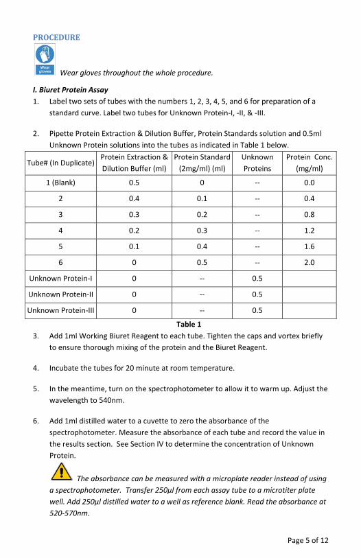
Wear gloves throughout the whole procedure.
I. Biuret Protein Assay 1. Label two sets of tubes with the numbers 1, 2, 3, 4, 5, and 6 for preparation of a
standard curve. Label two tubes for Unknown Protein‐I, ‐II, & ‐III.
2. Pipette Protein Extraction & Dilution Buffer, Protein Standards solution and 0.5ml Unknown Protein solutions into the tubes as indicated in Table 1 below.
Tube# (In Duplicate) Protein Extraction & Dilution Buffer (ml)
Protein Standard (2mg/ml) (ml)
Unknown Proteins
Protein Conc. (mg/ml)
1 (Blank) 0.5 0 ‐‐ 0.0
2 0.4 0.1 ‐‐ 0.4
3 0.3 0.2 ‐‐ 0.8
4 0.2 0.3 ‐‐ 1.2
5 0.1 0.4 ‐‐ 1.6
6 0 0.5 ‐‐ 2.0
Unknown Protein‐I 0 ‐‐ 0.5
Unknown Protein‐II 0 ‐‐ 0.5
Unknown Protein‐III 0 ‐‐ 0.5
Table 1 3. Add 1ml Working Biuret Reagent to each tube. Tighten the caps and vortex briefly
to ensure thorough mixing of the protein and the Biuret Reagent.
4. Incubate the tubes for 20 minute at room temperature.
5. In the meantime, turn on the spectrophotometer to allow it to warm up. Adjust the wavelength to 540nm.
6. Add 1ml distilled water to a cuvette to zero the absorbance of the spectrophotometer. Measure the absorbance of each tube and record the value in the results section. See Section IV to determine the concentration of Unknown Protein.
The absorbance can be measured with a microplate reader instead of using a spectrophotometer. Transfer 250μl from each assay tube to a microtiter plate well. Add 250μl distilled water to a well as reference blank. Read the absorbance at 520‐570nm.
Page 5 of 12
II. Lowry Protein Assay Prepare Prediluted Protein Standards (BSA)
1. Add 0.7ml Protein Standard (2mg/ml BSA) to a tube and dilute with 0.7ml Protein Extraction & Dilution Buffer to make 1mg/ml BSA solution.
2. For a standard curve, prepare a range of protein standards with the diluted BSA (1mg/ml) from step 1, as indicated in Table 2.
BSA Concentration (mg/ml) 0.0 0.1 0.3 0.5 0.8 1.0
Diluted BSA (ml) 0.00 0.05 0.15 0.25 0.40 0.50
Protein Buffer (ml) 0.50 0.45 0.35 0.25 0.10 0.00
Table 2
3. Label two sets of tubes with 0, 0.1, 0.3, 0.5, 0.8 and 1.0 and transfer 0.05ml of the appropriate standard to the tubes. Save the remaining protein solutions for the Bradford Protein Assay (Section III).
4. Label two tubes for Unknown Protein‐I, ‐II, & ‐III and add 0.05ml of each sample to the appropriate tube.
5. Add 0.1ml Copper Solution to each tube. Gently mix by vortexing.
6. Add 1ml Folin Ciocalteu Reagent and incubate at room temperature for 30 minutes.
7. In the meantime, turn on the spectrophotometer to allow it to warm up. Adjust the wavelength to 650‐700nm.
8. Add 1ml distilled water to a cuvette to zero the absorbance of the spectrophotometer. Measure the absorbance of each tube and record the values in the results section. See Section IV to determine the concentration of Unknown Protein.
The absorbance can be measured with a microplate reader instead of using a spectrophotometer. Transfer 250μl from each assay tube to a microtiter plate well. Add 250μl distilled water to a well as reference blank. Read the absorbance at 650‐700nm.
Page 6 of 12
III. Bradford Protein Assay 1. Label two sets of tubes with 0, 0.1, 0.3, 0.5, 0.8 and 1.0 and transfer 0.05ml of the
appropriate standard to the tubes. Use the diluted standard protein solutions from Section II, step 2.
2. Label two tubes for Unknown Protein‐I, ‐II, & ‐III and add 0.05ml of each sample to the appropriate tube.
3. Mix the CB Reagent gently by inverting the bottle several times.
To avoid foaming, DO NOT SHAKE THE BOTTLE.
4. Add 1ml CB Reagent to each tube and vortex briefly to mix the content. Incubate the tubes at room temperature for 5 minutes.
5. In the meantime, turn on the spectrophotometer to allow it to warm up. Adjust the wavelength to 595nm.
6. Add 1ml distilled water to a cuvette to zero the absorbance of the spectrophotometer. Measure the absorbance of each tube and record the values in the results section. See Section IV to determine the concentration of Unknown Protein.
The absorbance can be measured with a microplate reader instead of using a spectrophotometer. Transfer 250μl from each assay tube to a microtiter plate well. Add 250μl distilled water to a well as reference blank. Read the absorbance at 595nm.
IV. Determination of the Protein Concentration of the Unknown Protein
Determine the protein concentration with traditional paper plot. 1. Draw the points with protein concentrations as x values and the average
absorbance as y values on a grid or graph paper.
2. Draw a straight line through the points.
3. Lookup the Unknown Protein concentration from the plot using the absorbance value of the Unknown Protein.
Page 7 of 12
Determine the protein concentration using Microsoft Excel 1. Input the BSA standard concentration in a column and the absorbance value in a
second column. Highlight the data in the two columns From the menu bar, choose Insert then Chart. A “Chart Wizard” Menu will appear
2. Under the “Standard Types” tab: For “Chart type” select: XY (Scatter) For “Chart sub‐type” select the top chart (The one without any lines connecting the points) Click Next>
3. Under series in select columns Click Next>
4. Under the “Titles” tab: Type in appropriate titles (names) for the graph (chart) and the x and y‐axes (e.g., Chart title: Biuret Assay, x‐axis: Absorbance, y‐axis: Protein Concentration (mg/ml)) Under the “Legend” tab: Click on the checkmark next to “Show legend” (The checkmark should disappear), Click Next>
5. Click on circle to left of “As object in” (a dot should appear in the circle) Click Finish. Your graph should appear on the sheet where your data is.
6. Highlight the chart by clicking inside the chart area. From the menu bar, choose Chart then Add Trendline The “Add Trendline” window will appear
7. Under the “Type” tab: For “Trend/Regression type” select: Linear
8. Under the “Options” tab: Click on box to left of “Display equation on chart” (a checkmark appears) Click on box to left of “Display R‐squared on chart” (a checkmark appears), Click OK.
9. Move the equation and R‐squared value to a suitable location on the graph. Save and print the data sheet.
10. Use the equation on the chart to calculate the concentration of your unknown protein.
Page 8 of 12

RESULTS,ANALYSIS&ASSESSMENT
Biuret Protein Assay (Section I)
Tube # Absorbance I (540nm)
Absorbance II (540nm)
Average Protein Conc.
(mg/ml)
1 (Blank) 0.0
2 0.4
3 0.8
4 1.2
5 1.6
6 2.0
Unknown Protein‐I
Unknown Protein ‐II
Unknown Protein ‐III
Lowry Protein Assay (Section II)
Tube # Absorbance I (650nm)
Absorbance II (650nm)
Average Protein Conc.
(mg/ml)
0 0
0.1 0.1
0.3 0.3
0.5 0.5
0.8 0.8
1 1
Unknown Protein‐I
Unknown Protein ‐II
Unknown Protein ‐III
Page 9 of 12
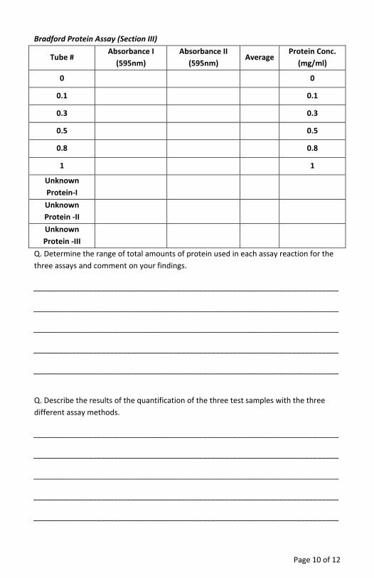
Bradford Protein Assay (Section III)
Tube # Absorbance I (595nm)
Absorbance II (595nm)
Average Protein Conc.
(mg/ml)
0 0
0.1 0.1
0.3 0.3
0.5 0.5
0.8 0.8
1 1
Unknown Protein‐I
Unknown Protein ‐II
Unknown Protein ‐III
Q. Determine the range of total amounts of protein used in each assay reaction for the three assays and comment on your findings.
________________________________________________________________________
________________________________________________________________________
________________________________________________________________________
________________________________________________________________________
________________________________________________________________________
Q. Describe the results of the quantification of the three test samples with the three different assay methods.
________________________________________________________________________
________________________________________________________________________
________________________________________________________________________
________________________________________________________________________
________________________________________________________________________
Page 10 of 12
Page 11 of 12
Q. Briefly describe the principles behind the three protein assays and their weakness and strengths.
________________________________________________________________________
________________________________________________________________________
________________________________________________________________________
________________________________________________________________________
________________________________________________________________________
Q. Select one protein assay at a time and explain how you will use that assay for determination of protein concentration.
________________________________________________________________________
________________________________________________________________________
________________________________________________________________________
________________________________________________________________________
________________________________________________________________________
Q. For an unknown protein sample, which protein assay will you choose first?
________________________________________________________________________
________________________________________________________________________
________________________________________________________________________
Q. How would you overcome interference in protein assay from common lab agents?
________________________________________________________________________
________________________________________________________________________
________________________________________________________________________
________________________________________________________________________
Last saved: 9/13/2012 CMH

www.GBiosciences.com
Page 12 of 12