Embed Size (px)
Citation preview
Aspherical magnetically modulated optical nanoprobes (MagMOONs)Jeffrey N. Anker, Caleb Behrend, and Raoul Kopelman Citation: Journal of Applied Physics 93, 6698 (2003); doi: 10.1063/1.1556926 View online: http://dx.doi.org/10.1063/1.1556926 View Table of Contents: http://scitation.aip.org/content/aip/journal/jap/93/10?ver=pdfcov Published by the AIP Publishing Articles you may be interested in Optical re-injection in cavity-enhanced absorption spectroscopy Rev. Sci. Instrum. 85, 093101 (2014); 10.1063/1.4893972 Planar lens integrated capillary action microfluidic immunoassay device for the optical detection of troponin I Biomicrofluidics 7, 064112 (2013); 10.1063/1.4837755 Magnetic property of polycrystalline magnetic garnet for voltage driven type magneto-optic spatial light phasemodulator J. Appl. Phys. 107, 09A948 (2010); 10.1063/1.3368114 Effective passivation of porous silicon optical devices by thermal carbonization J. Appl. Phys. 103, 083124 (2008); 10.1063/1.2910459 Magnetically modulated optical nanoprobes Appl. Phys. Lett. 82, 1102 (2003); 10.1063/1.1544435
[This article is copyrighted as indicated in the article. Reuse of AIP content is subject to the terms at: http://scitation.aip.org/termsconditions. Downloaded to ] IP:
130.63.180.147 On: Sat, 22 Nov 2014 06:24:02
Biomagnetism Robert Kraus and J. M. D. Coey, Co-Chairmen
Aspherical magnetically modulated optical nanoprobes „MagMOONs …Jeffrey N. Anker, Caleb Behrend, and Raoul Kopelmana)
Chemistry Department, The University of Michigan Ann Arbor, Michigan 48109-1055
~Presented on 12 November 2002!
Aspherical magnetic particles orient in a magnetic field due to magnetic shape anisotropy. They alsoemit different fluxes of light from their different geometric faces due to self-absorption and totalinternal reflection within the particles. The particles rotate in response to rotating magnetic fieldsand appear to blink as they rotate. We have made pancake and chain shaped particles andmagnetically modulated their fluorescent intensities. Demodulating the signal extracts the probefluorescence from electronic and optical backgrounds dramatically increasing signal to noise ratios.The probes have applications in sensitive and rapid immunoassays, improved intracellular sensors,and inexpensive single molecule analysis. ©2003 American Institute of Physics.@DOI: 10.1063/1.1556926#
I. INTRODUCTION
Fluorescence is the most sensitive method available formolecular detection and chemical imaging. It is even usedfor real time imaging of single molecules at ambient condi-tions with high spatial and spectral resolution.1,2 Fluorescentdyes are commonly used to: study intracellular chemicalconcentration changes,3,4 measure immunochemical concen-trations in fluids,5 tag molecules on cell surfaces or tissues,6
and research protein folding.7,8 They are critical to under-standing how cells function, for rapid drug discovery and fordetecting minute quantities of pathogens or DNA. Neverthe-less, background fluorescence from sample and instrumentoptics makes detecting low levels of fluorescence or smallchanges in fluorescence challenging. Rejecting backgroundfluorescence and increasing signal to noise ratios by ordersof magnitude would thus lead to important advances in mo-lecular and biomedical sciences.
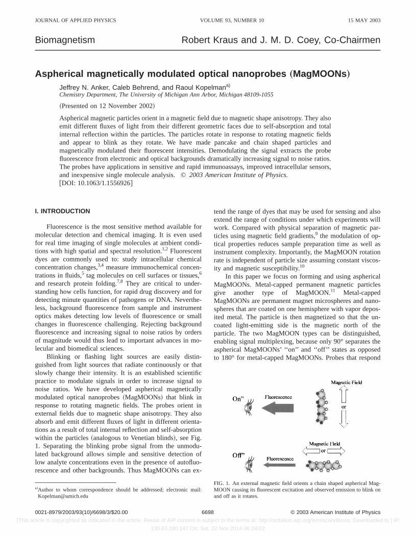
Blinking or flashing light sources are easily distin-guished from light sources that radiate continuously or thatslowly change their intensity. It is an established scientificpractice to modulate signals in order to increase signal tonoise ratios. We have developed aspherical magneticallymodulated optical nanoprobes~MagMOONs! that blink inresponse to rotating magnetic fields. The probes orient inexternal fields due to magnetic shape anisotropy. They alsoabsorb and emit different fluxes of light in different orienta-tions as a result of total internal reflection and self-absorptionwithin the particles~analogous to Venetian blinds!, see Fig.1. Separating the blinking probe signal from the unmodu-lated background allows simple and sensitive detection oflow analyte concentrations even in the presence of autofluo-rescence and other backgrounds. Thus MagMOONs can ex-
tend the range of dyes that may be used for sensing and alsoextend the range of conditions under which experiments willwork. Compared with physical separation of magnetic par-ticles using magnetic field gradients,9 the modulation of op-tical properties reduces sample preparation time as well asinstrument complexity. Importantly, the MagMOON rotationrate is independent of particle size assuming constant viscos-ity and magnetic susceptibility.10
In this paper we focus on forming and using asphericalMagMOONs. Metal-capped permanent magnetic particlesgive another type of MagMOON.11 Metal-cappedMagMOONs are permanent magnet microspheres and nano-spheres that are coated on one hemisphere with vapor depos-ited metal. The particle is then magnetized so that the un-coated light-emitting side is the magnetic north of theparticle. The two MagMOON types can be distinguished,enabling signal multiplexing, because only 90° separates theaspherical MagMOONs’ ‘‘on’’ and ‘‘off’’ states as opposedto 180° for metal-capped MagMOONs. Probes that respond
a!Author to whom correspondence should be addressed; electronic mail:[email protected]
FIG. 1. An external magnetic field orients a chain shaped aspherical Mag-MOON causing its fluorescent excitation and observed emission to blink onand off as it rotates.
JOURNAL OF APPLIED PHYSICS VOLUME 93, NUMBER 10 15 MAY 2003
66980021-8979/2003/93(10)/6698/3/$20.00 © 2003 American Institute of Physics
[This article is copyrighted as indicated in the article. Reuse of AIP content is subject to the terms at: http://scitation.aip.org/termsconditions. Downloaded to ] IP:
130.63.180.147 On: Sat, 22 Nov 2014 06:24:02
to modulated magnetic field gradients instead of field orien-tations give yet another distinguishable type of MagMOON.
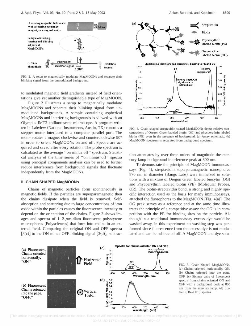
Figure 2 illustrates a setup to magnetically modulateMagMOONs and separate their blinking signal from un-modulated backgrounds. A sample containing asphericalMagMOONs and interfering backgrounds is viewed with anOlympus IMT2 epifluorescent microscope. A program writ-ten in Labview~National Instruments, Austin, TX! controls astepper motor interfaced to a computer parallel port. Themotor rotates a magnet clockwise and counterclockwise 90°in order to orient MagMOONs on and off. Spectra are ac-quired and saved after every rotation. The probe spectrum iscalculated as the average ‘‘on minus off’’ spectrum. Statisti-cal analysis of the time series of ‘‘on minus off’’ spectrausing principal components analysis can be used to furtherreduce interference from background signals that fluctuateindependently from the MagMOONs.
II. CHAIN SHAPED MagMOONs
Chains of magnetic particles form spontaneously inmagnetic fields. If the particles are superparamagnetic thenthe chains dissipate when the field is removed. Self-absorption and scattering due to large concentrations of ironoxide within the particles causes the fluorescence intensity todepend on the orientation of the chains. Figure 3 shows im-ages and spectra of 1–2-mm-diam fluorescent polystyrenemicrospheres~Polysciences! that form into chains in an ex-ternal field. Comparing the original ON and OFF spectra@3~c!# to the ON minus OFF blinking signal@3~d!#, subtrac-
tion attenuates by over three orders of magnitude the mer-cury lamp background interference peak at 800 nm.
To demonstrate the principle of MagMOON immunoas-says ~Fig. 4!, streptavidin superparamagnetic nanospheres870 nm in diameter~Bangs Labs! were immersed in solu-tions with a mixture of Oregon Green labeled biocytin~OG!and Phycoerythrin labeled biotin~PE! ~Molecular Probes,OR!. The biotin-streptavidin bond, a strong and highly spe-cific interaction used as the basis for many immunoassays,attached the fluorophores to the MagMOON@Fig. 4~a!#. TheOG peak serves as a reference and at the same time illus-trates the principle of a competitive assay: the OG is in com-petition with the PE for binding sites on the particle. Al-though in a traditional immunoassay excess dye would bewashed away, in this experiment no washing step was per-formed since fluorescence from the excess dye is not modu-lated and can be subtracted off. A MagMOON and dye solu-
FIG. 2. A setup to magnetically modulate MagMOONs and separate theirblinking signal from the unmodulated background.
FIG. 3. Chain shaped MagMOONs.~a! Chains oriented horizontally, ON.~b! Chains oriented into the page,OFF. ~c! Sixteen pairs of fluorescentspectra from chains oriented ON andOFF with a background peak at 800nm from the mercury lamp.~d! Six-teen~ON–OFF! spectra.
FIG. 4. Chain shaped streptavidin-coated MagMOONs detect relative con-centrations of Oregon Green labeled biotin~OG! and phycoerythrin labeledbiotin ~PE! even in the presence of background.~a! Assay schematic.~b!MagMOON spectrum is separated from background spectrum.
6699J. Appl. Phys., Vol. 93, No. 10, Parts 2 & 3, 15 May 2003 Anker, Behrend, and Kopelman
[This article is copyrighted as indicated in the article. Reuse of AIP content is subject to the terms at: http://scitation.aip.org/termsconditions. Downloaded to ] IP:
130.63.180.147 On: Sat, 22 Nov 2014 06:24:02
tion was added to one well in a 96 well plate, and thesolution was left overnight to let the biotin labeled dyes at-tach to the nanospheres. By orienting the MagMOON chainswith the computer-controlled magnet, MagMOON fluores-cence was separated from background fluorescence due toinstrument optics, dust, and free excess biotin-labeled dyes.Thirty-two pairs of ON and OFF spectra were collected, andthe average ON, OFF, and ON minus OFF spectra were plot-ted in Fig. 4~b!. The background mercury lamp peak at 800nm was attenuated by a factor of 2000.
Chains of magnetic particles can be linked together per-manently by heating the chains up above their glass transi-tion temperature~94 °C for polystyrene! in solution ~e.g.,within a 20 ml vial!. This process is simpler, and can yieldmore particles than previous methods such as: chemicallylinking magnetic chains,12 depositing microspheres in agroove shaped template to form chains and then meltingthem together in the groove,13 or passing microspheresthrough a microfluidic device where they are brieflyheated.14
III. PANCAKE AND ROLL SHAPED MagMOONs
We developed roll and pancake shaped microparticlesand breaded these with magnetic or fluorescent nano-crumbs
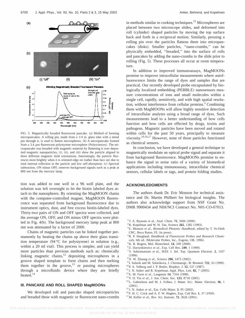
in methods similar to cooking techniques.15 Microspheres areplaced between two microscope slides, and deformed intoroll ~cylinder! shaped particles by moving the top surfaceback and forth in a reciprocal motion. Similarly, pressing arolling pin over the particles flattens them into micropan-cakes ~disks!. Smaller particles, ‘‘nano-crumbs,’’ can bephysically embedded, ‘‘breaded,’’ into the surface of rollsand pancakes by adding the nano-crumbs to the slide prior torolling ~Fig. 5!. These processes all occur at room tempera-ture.
In addition to improved immunoassays, MagMOONspromise to improve intracellular measurements where autof-luorescence limits the range of dyes and samples that arepractical. Our recently developed probe encapsulated by bio-logically localized embedding~PEBBLE! nanosensors mea-sure concentrations of ions and small molecules within asingle cell, rapidly, sensitively, and with high spatial resolu-tion, without interference from cellular proteins.3 Combiningthem with MagMOONs will allow highly sensitive detectionof intracellular analytes using a broad range of dyes. Suchmeasurements lead to a better understanding of how cellsfunction and how cells are effected by drugs, toxins andpathogens. Magnetic particles have been moved and rotatedwithin cells for the past 50 years, principally to measureviscosity.10,16,17However, none of those particles functionedas chemical sensors.
In conclusion, we have developed a general technique tomagnetically modulate an optical probe signal and separate itfrom background fluorescence. MagMOONs promise to en-hance the signal to noise ratio of a variety of biomedicalapplications including immunoassay, intracellular chemicalsensors, cellular labels or tags, and protein folding studies.
ACKNOWLEDGMENTS
The authors thank Dr. Eric Monson for technical assis-tance and Dr. Martin Philbert for biological insights. Theauthors also acknowledge support from NSF Grant No.DMR 9900434 and NIH/NCI Contract No. N01-CO-07013.
1T. A. Byasseeet al., Anal. Chem.72, 5606~2000!.2R. Kopelman and W. H. Tan, Science262, 1382~1993!.3E. Monsonet al., Biomedical Photonic Handbook, edited by T. Vo-DinhCRC, Boca Raton, FL~in press!.
4R. P. Haugland,Handbook of Fluorescent Probes and Research Chemi-cals, 6th ed.~Molecular Probes, Inc., Eugene, OR, 1996!.
5K. R. Rogers, Mol. Biotechnol.14, 109 ~2000!.6Z. Darzynkiewiczet al., Exp. Cell Res.249, 1 ~1999!.7V. Subramaniamet al., IEEE J. Sel. Top. Quantum Electron.2, 1107~1996!.
8X. W. Zhuanget al., Science296, 1473~2002!.9I. Safarik and M. Safarikova, J. Chromatogr., B: Biomed.722, 33 ~1999!.
10P. A. Valberg and J. P. Butler, Biophys. J.52, 537 ~1987!.11J. N. Anker and R. Kopelman, Appl. Phys. Lett.82, 7 ~2003!.12E. M. Furstet al., Langmuir14, 7334~1998!.13Y. D. Yin et al., J. Am. Chem. Soc.123, 8718~2001!.14L. Gabrielson and M. J. Folkes, J. Mater. Sci.: Mater. Electron.36, 1
~2001!.15J. N. Ankeret al., Eur. Cells Mater.3, 95 ~2002!.16F. H. C. Crick and A. F. W. Hughes, Exp. Cell Res.1, 37 ~1950!.17M. Keller et al., Rev. Sci. Instrum.72, 3626~2001!.
FIG. 5. Magnetically breaded fluorescent pancake.~a! Method of formingmicropancakes. A rolling pin, made from a 1/4 in. glass tube with a metalpin through it, is used to flatten microspheres.~b! A micropancake formedfrom a 3.4mm fluorescent polystyrene microsphere~Polysciences!. The mi-cropancake was breaded with magnetic material by flattening it over depos-ited magnetic nanoparticles;~i!, ~ii !, and ~iii ! show the particle aligned inthree different magnetic field orientations. Interestingly, the particle fluo-resces most brightly when it is oriented edge on~rather than face on! due tototal internal reflection in the particle and low self-absorption.~c! Spectralsubtraction, ON minus OFF, removes background signals such as a peak at800 nm from the mercury lamp.
6700 J. Appl. Phys., Vol. 93, No. 10, Parts 2 & 3, 15 May 2003 Anker, Behrend, and Kopelman
[This article is copyrighted as indicated in the article. Reuse of AIP content is subject to the terms at: http://scitation.aip.org/termsconditions. Downloaded to ] IP:
130.63.180.147 On: Sat, 22 Nov 2014 06:24:02