Embed Size (px)
Citation preview

patient and the third in the literature, following two cases
reported in children due to A. viridinutans [11].
Case report
A 56-year-old Caucasian male with diabetes mellitus type
II and rheumatoid arthritis, had been treated since 2003 with:
prednisolone, 17.5 mg/day; etarnecept, 50 mg/day; metho-
trexate, 2.5 mg 3 � /week; salazopirine, 1,500 mg/day; and
metformine, 1,700 mg/day. Due to the observation in March
2007 of a single pulmonary nodule in a routine chest X-ray,
a computed tomography (CT) was ordered which revealed
a nodular, 26 mm in diameter, hypodense, irregular, spicu-
lated lesion, suggestive of a neoplastic process, located in
the posterior segment of the upper lobe of the right lung.
The remaining upper lobe exhibited a micro-reticulo-nodular
pattern. Trans-thoracic needle aspiration biopsy and cyto-
logical examination of the aspirate revealed septate hyphae
suggestive of Aspergillus but the search for malignant cells
was negative. At the microbiology laboratory, septate
hyphae were found on direct exam (Fig. 1) but cultures of
the material, although incubated for 1 month, were nega-
tive. In the following weeks, the patient was afebrile, had
right-sided chest pain of moderate intensity, without inter-
ference with daily-life activities, and did not have other
associated complaints.
In April 2007 he started outpatient treatment with itra-
conazole PO 100 mg/day. One month later the lesion was
Received 11 September 2010 ; Received in fi nal revised from 8 January
2011 ; Accepted 18 January 2011
Correspondence: Maria Dolores Pinheiro, Laboratory of Microbiology,
Service of Clinical Pathology, Hospital de S. Jo ã o EPE, Alameda Prof.
Hern â ni Monteiro, 4200 319 Porto, Portugal. Tel: � 351 22 55 12 116;
telefax: � 351 22 55 12 261; E-mail: [email protected]
Case Reports
Aspergillus viridinutans: an agent of adult chronic
invasive aspergillosis
DANINA COELHO * , SUSANA SILVA * , LU Í S VALE-SILVA † , HELENA GOMES * , EUG É NIA PINTO † ,
ANT Ó NIO SARMENTO * & MARIA DOLORES PINHEIRO ‡
* Service of Infectious Diseases, Hospital de S. Jo ã o EPE, Porto, † Microbiology Service/CEQUIMED, Faculty of Pharmacy,
Universidade do Porto, and ‡ Laboratory of Microbiology, Service of Clinical Pathology, Hospital de S. Jo ã o EPE, Porto, Portugal
In contrast with the common hematogenous dissemination of invasive aspergillosis (IA), we present case with a protracted course through anatomical planes in an immunocom-promised adult male. The unusual clinical features and laboratory fi ndings led to fungal genotyping and identifi cation of the mold as Aspergillus viridinutans . It appears to be the fi rst described case of IA caused by this agent in an adult patient.
Keywords Aspergillus viridinutans , adult patient , invasive aspergillosis , fungal genotyping , antifungal susceptibility
Introduction
The incidence of invasive aspergillosis (IA) has been
increasing steadily for the past few decades [1,2].
The most common etiological agent of IA is Aspergillus fumigatus , followed by Aspergillus niger , Aspergillus fl avus , Aspergillus terreus [3,4] and other members of
Aspergillus section Fumigati [5].
Aspergillus viridinutans, a mold of Aspergillus section
Fumigati , was originally isolated from rabbit dung in
Australia and from soil samples collected from around the
world [6,7]. It has subsequently been identifi ed during ret-
rospective analyses of Aspergillus species in cultures [8 – 10]
but its clinical importance has not yet been defi ned [11].
Here we describe the clinical case of a 56-year-old
immunocompromised man who died from IA which
exhibited distinct clinical manifestations compared to infec-
tions caused by A. fumigatus . We also present the morpho-
logical characteristics, genotype-based identifi cation and the
antifungal susceptibility of the isolate. To the best of our
knowledge, the case described here is the fi rst in an adult
© 2011 ISHAM DOI: 10.3109/13693786.2011.556672
Medical Mycology October 2011, 49, 755–759
Med
Myc
ol D
ownl
oade
d fr
om in
form
ahea
lthca
re.c
om b
y C
olum
bia
Uni
vers
ity o
n 03
/18/
13Fo
r pe
rson
al u
se o
nly.
© 2011 ISHAM, Medical Mycology, 49, 755–759
756 Coelho et al .
found to be identical to that seen on the initial X-ray and
he again underwent trans - thoracic needle aspiration biopsy.
Septate hyphae were once more observed in direct exam,
but on this occasion a fi lamentous fungus, morphologically
identifi ed as Aspergillus spp., probably A . fumigatus was
recovered in culture.
At this point the patient was started on posaconazole
therapy 400 mg/day, continuously for 5 months, with
positive clinical and radiological response.
In October 2007, following unrelated abdominal sur-
gery, posaconazole was discontinued for a month. Due to
worsening of pulmonary imaging, a bronchoscopy was
performed but no endobronchial lesions were identifi ed.
Direct examination of bronchial and bronchoalveolar
lavage revealed hyphae and cultures were again positive
for Aspergillus spp. Upon discharge from hospital in
November, the patient was maintained on voriconazole PO
200 mg/day and continued with his usual medication,
including methotrexate and prednisolone, which he took
regularly during the course of the disease.
In early July 2008, a right cervical, deeply adherent
infl ammatory swelling, with a maximum diameter of 10 cm,
was observed. Aspiration needle biopsy and cytological
exam of the aspirate revealed fungal structures suggestive
of Aspergillus . Treatment with intravenous voriconazole
resulted in clinical improvement and decrease in mass size.
The patient was discharged with oral voriconazole. How-
ever, at the end of the second month of treatment, there
was a resurgence of the right cervical swelling and the
appearance of a second one with similar characteristics in
the thoracic region at the right anterior axillary line, 6th and
7th rib. This time there was a clear clinical worsening, includ-
ing fever of 38 – 40 ° C, deterioration of general condition,
swelling enlargement and chest X-ray changes. In late
August 2008, he was admitted and observed for the fi rst
time in the Infectious Diseases Service.
Medical treatment was optimized with the combination
of posaconazole, 200 mg four times a day and caspofungin,
50 mg daily and empirical therapy with amoxicillin and
clavulanic acid. While blood cultures were negative for
bacteria, mycobacteria and fungi, an Aspergillus spp. iso-
late was recovered from bronchial secretions.
The CT scan revealed multiple lymph adenopathies and
a right cervical conglomerate beginning in the supra-
clavicular region down to the 5th rib , where a 3.5 cm nod-
ule with central necrosis was seen. A right pleural effusion
with pleural thickening and a nodular lesion with spicu-
lated margins of 5 cm, located in the posterior aspect of
the right upper lobe, in contact with the pulmonary hilum,
were also found (Fig. 2).
The fever and lesions persisted despite spontaneous and
surgical drainage of both swellings. Cancer and infection
by other fungi, mycobacteria, or bacteria were excluded.
The clinical condition of the patient progressively wors-
ened with respiratory failure and oliguric irreversible renal
failure, and he died after 39 days of hospitalization. No
post mortem study was performed.
All isolates sent to the microbiology laboratory between
May 2007 and September 2008 presented the same mor-
phological and microscopic characteristics and were
identifi ed as Aspergillus spp., probably A. fumigatus . They
Fig. 1 Direct examination of the sample from the aspirative biopsy of
the pulmonary nodule. Notice the existence of septate hyphae.
Fig. 2 CT depicting the thorax with the nodular spiculated lesion on the
upper lobe of the right lung (black arrow).
Med
Myc
ol D
ownl
oade
d fr
om in
form
ahea
lthca
re.c
om b
y C
olum
bia
Uni
vers
ity o
n 03
/18/
13Fo
r pe
rson
al u
se o
nly.
© 2011 ISHAM, Medical Mycology, 49, 755–759
Invasive Aspergillus viridinutans in an adult patient 757
results were similar for the fi rst and last isolate tested.
Serum levels of administered antifungal drugs were not
measured.
Discussion
The case reported here shows a form of aspergillosis with
a clinical course different from that usually observed in
neutropenic and transplanted patients. Invasive aspergil-
losis typically presents as an acute, rapidly progressive
disease with predilection for angioinvasion and hematog-
enous dissemination. In contrast, this case and the two
others previously reported in the literature [11] showed
chronic evolution and progressive spread across anatom-
ical planes.
This third case strengthens the previous assertion that
A. viridinutans is able to cause a distinct form of aspergil-
losis [11]. In fact, the patients were immunocompromised
in some manner and the disease started in the lungs, likely
after inhalation of spores (the most frequent portal of entry
in IA), before disseminating to adjacent areas. Moreover,
the infection developed over several months in all cases (3
months in the 14-year-old boy, 7 months in the 8-year-old
boy and almost 18 months in this 56-year-old man), and
did not respond to medical and surgical therapy, emphasiz-
ing the indolent, albeit progressive, features of this form
of aspergillosis. An interesting observation is that all three
cases involved male patients, although the limited number
does not allow for conclusions on gender susceptibility (there
is no such predilection in usual forms of aspergillosis).
This case further evidences the need for genotypically-
based identifi cation of isolates of members of Aspergillus ,
section Fumigati [8 – 10]. Indeed, the mold isolated in the
present case was found to belong to that group, according
to morphological and microscopic characteristics, but its
features did not fully correspond to those of A. fumigatus
(for example, differences in growth patterns), which
prompted the use of molecular biology tools to obtain
defi nite species identifi cation.
The earlier therapeutic decisions deserve attention.
When the patient presented with a solitary nodule, he was
treated with 100 mg of itraconazole each day. Due to the
lack of a positive response, posaconazole PO 400 mg/day
was prescribed and, later, after surgery for unrelated rea-
sons, he was medicated with voriconazole PO 200 mg/day.
The under dose schedule employed for the three antifungal
drugs did not meet fully the Infectious Diseases Society of
America recommendations [14]. Although they included
the oral route for administration, it may not have been
adequate in this setting.
Finally, in vitro susceptibility tests of A. viridinutans
revealed peculiar fi ndings when compared to the antifun-
gal susceptibility of other Aspergillus spp. The MIC of
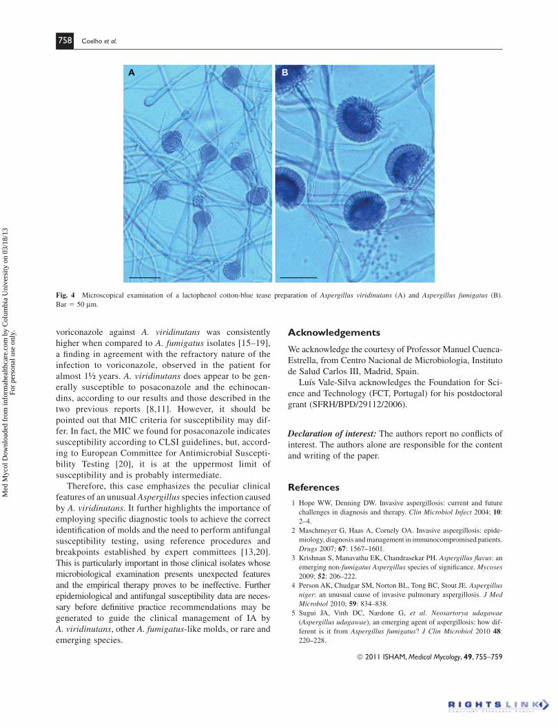
showed the usual features of members of the genus Asper-gillus including: ‘ foot cells ’ ; upright, usually nonseptate,
conidiophores, with swollen vesicles at the tips; vesicles
covered entirely or in part by the phialides [12]. However,
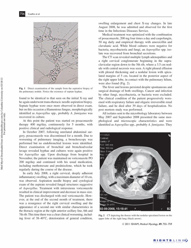
in comparison to A. fumigatus , colonies of the isolates
grew slower in Sabouraud-gentamicin-chloramphenicol
culture medium at 25 ° C, were initially white (Fig. 3) and
required additional incubation time to develop their green
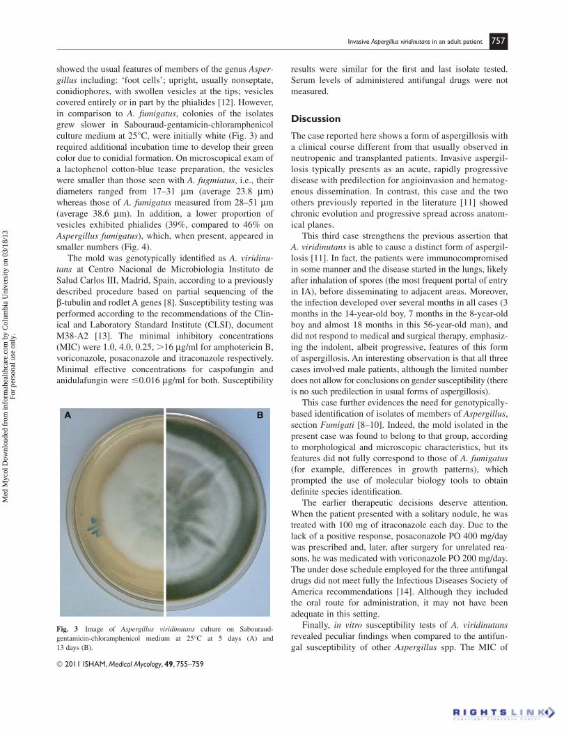
color due to conidial formation. On microscopical exam of
a lactophenol cotton-blue tease preparation, the vesicles
were smaller than those seen with A. fugmiatus , i.e., their
diameters ranged from 17 – 31 μ m (average 23.8 μ m)
whereas those of A. fumigatus measured from 28 – 51 μ m
(average 38.6 μ m). In addition, a lower proportion of
vesicles exhibited phialides (39%, compared to 46% on
Aspergillus fumigatus ), which, when present, appeared in
smaller numbers (Fig. 4).
The mold was genotypically identifi ed as A. viridinu-tans at Centro Nacional de Microbiologia Instituto de
Salud Carlos III, Madrid, Spain, according to a previously
described procedure based on partial sequencing of the
β -tubulin and rodlet A genes [8]. Susceptibility testing was
performed according to the recommendations of the Clin-
ical and Laboratory Standard Institute (CLSI), document
M38-A2 [13]. The minimal inhibitory concentrations
(MIC) were 1.0, 4.0, 0.25, � 16 μ g/ml for amphotericin B,
voriconazole, posaconazole and itraconazole respectively.
Minimal effective concentrations for caspofungin and
anidulafungin were � 0.016 μ g/ml for both. Susceptibility
Fig. 3 Image of Aspergillus viridinutans culture on Sabouraud-
gentamicin-chloramphenicol medium at 25 ° C at 5 days (A) and
13 days (B).
Med
Myc
ol D
ownl
oade
d fr
om in
form
ahea
lthca
re.c
om b
y C
olum
bia
Uni
vers
ity o
n 03
/18/
13Fo
r pe
rson
al u
se o
nly.
© 2011 ISHAM, Medical Mycology, 49, 755–759
758 Coelho et al .
voriconazole against A. viridinutans was consistently
higher when compared to A. fumigatus isolates [15 – 19],
a fi nding in agreement with the refractory nature of the
infection to voriconazole, observed in the patient for
almost 1 ½ years. A. viridinutans does appear to be gen-
erally susceptible to posaconazole and the echinocan-
dins, according to our results and those described in the
two previous reports [8,11]. However, it should be
pointed out that MIC criteria for susceptibility may dif-
fer. In fact, the MIC we found for posaconazole indicates
susceptibility according to CLSI guidelines, but, accord-
ing to European Committee for Antimicrobial Suscepti-
bility Testing [20], it is at the uppermost limit of
susceptibility and is probably intermediate.
Therefore, this case emphasizes the peculiar clinical
features of an unusual Aspergillus species infection caused
by A. viridinutans . It further highlights the importance of
employing specifi c diagnostic tools to achieve the correct
identifi cation of molds and the need to perform antifungal
susceptibility testing, using reference procedures and
breakpoints established by expert committees [13,20].
This is particularly important in those clinical isolates whose
microbiological examination presents unexpected features
and the empirical therapy proves to be ineffective. Further
epidemiological and antifungal susceptibility data are neces-
sary before defi nitive practice recommendations may be
generated to guide the clinical management of IA by
A. viridinutans , other A. fumigatus -like molds, or rare and
emerging species.
Acknowledgements
We acknowledge the courtesy of Professor Manuel Cuenca-
Estrella, from Centro Nacional de Microbiologia, Instituto
de Salud Carlos III, Madrid, Spain.
Lu í s Vale-Silva acknowledges the Foundation for Sci-
ence and Technology (FCT, Portugal) for his postdoctoral
grant (SFRH/BPD/29112/2006).
Declaration of interest: The authors report no confl icts of
interest. The authors alone are responsible for the content
and writing of the paper.
References
Hope WW, Denning DW. Invasive aspergillosis: current and future 1
challenges in diagnosis and therapy. Clin Microbiol Infect 2004; 10 :
2 – 4.
Maschmeyer G, Haas A, Cornely OA. Invasive aspergillosis: epide-2
miology, diagnosis and management in immunocompromised patients.
Drugs 2007; 67 : 1567 – 1601.
Krishnan S, Manavathu EK, Chandrasekar PH. 3 Aspergillus fl avus : an
emerging non- fumigatus Aspergillus species of signifi cance. Mycoses
2009; 52 : 206 – 222.
Person AK, Chudgar SM, Norton BL, Tong BC, Stout JE. 4 Aspergillus niger : an unusual cause of invasive pulmonary aspergillosis. J Med Microbiol 2010; 59 : 834 – 838.
Sugui JA, Vinh DC, Nardone G, 5 et al . Neosartorya udagawae
( Aspergillus udagawae ), an emerging agent of aspergillosis: how dif-
ferent is it from Aspergillus fumigatus ? J Clin Microbiol 2010 48 :
220 – 228.
Fig. 4 Microscopical examination of a lactophenol cotton-blue tease preparation of Aspergillus viridinutans (A) and Aspergillus fumigatus (B).
Bar � 50 μ m.
Med
Myc
ol D
ownl
oade
d fr
om in
form
ahea
lthca
re.c
om b
y C
olum
bia
Uni
vers
ity o
n 03
/18/
13Fo
r pe
rson
al u
se o
nly.
© 2011 ISHAM, Medical Mycology, 49, 755–759
Invasive Aspergillus viridinutans in an adult patient 759
McLennan EI, Tucker SC, Thrower LB. New soil fungi from Austral-6
ian heathland: Aspergillus , Penicillium , Spegazzinia . Austral J Bot 1954; 2 : 355 – 364.
Varga J, T ó th B, Rig ó K, Debets F, Kozakiewicz Z. Genetic variabil-7
ity within the Aspergillus viridinutans species. Folia Microbiol (Praha)
2000; 45 : 423 – 428.
Alcazar-Fuoli L, Mellado E, Alastruey-Izquierdo A, Cuenca-Estrella 8
M, Rodriguez-Tudela JL. Aspergillus section Fumigati : antifungal
susceptibility patterns and sequence-based identifi cation. Antimicrob Agents Chemother 2008; 52 : 1244 – 1251.
Katz ME, Dougall AM, Weeks K, Cheetham BF. Multiple genetically 9
distinct groups revealed among clinical isolates identifi ed as atypical
Aspergillus fumigatus . J Clin Microbiol 2005; 43 : 551 – 555.
Yaguchi T, Horie Y, Tanaka R, 10 et al . Molecular phylogenetics of mul-
tiple genes on Aspergillus section Fumigati isolated from clinical
specimens in Japan. Nip Ishinkin Gakkai Zasshi 2007; 48 : 37 – 46.
Vinh DC, Shea YR, Jones PA, 11 et al . Chronic invasive aspergillosis caused
by Aspergillus viridinutans . Emerg Infect Dis 2009; 15 : 1292 – 1294.
Sigler L, Kennedy MJ. 12 Aspergillus , Fusarium , and other opportunis-
tic moniliaceous fungi. In: Murray PR, Baron EJ, Pfaller MA, Ten-
over FC,Yolken RH (eds.) Manual of Clinical Microbiology , 7th edn.
Washington, DC: ASM Press, 1999: 1212 – 1241.
Clinical and Laboratory Standard Institute. 13 Reference Method for Broth Dilution Antifungal Susceptibility Testing of Filamentous Fung i; approved standard. Document M38-A2, 2nd edn, Wayne, PA:
CLSI, 2008.
Walsh TJ, Anaissie EJ, Denning DW, 14 et al . Treatment of aspergillo-
sis: clinical practice guidelines of the Infectious Diseases Society of
America. Clin Inf Dis 2008; 46 : 327 – 360.
Pfaller MA, Messer SA, Boyken L, 15 et al . In vitro survey of triazole
cross-resistance among more than 700 clinical isolates of Aspergillus
species. J Clin Microbiol 2008; 46 : 2568 – 2572.
Espinel-Ingroff A, Johnson E, Hockey H, Troke P. Activities of vori-16
conazole, itraconazole and amphotericin B in vitro against 590 moulds
from 323 patients in the voriconazole phase III clinical studies. J An-timicrob Chemother 2008; 61 : 616 – 620.
Cuenca-Estrella M, Gomez-Lopez A, Mellado E, 17 et al . Head-to-head
comparison of the activities of currently available antifungal agents
against 3,378 Spanish clinical isolates of yeasts and fi lamentous fungi.
Antimicrob Agents Chemother 2006; 50 : 917 – 921.
Sabatelli F, Patel R, Mann PA, 18 et al . In vitro activities of posaconazole,
fl uconazole, itraconazole, voriconazole, and amphotericin B against a
large collection of clinically important molds and yeasts. Antimicrob Agents Chemother 2006; 50 : 2009 – 2015.
Lass-Florl C, Mayr A, Perkhofer S, 19 et al . Activities of antifungal agents
against yeasts and fi lamentous fungi: assessment according to the meth-
odology of the European Committee on Antimicrobial Susceptibility
Testing. Antimicrob Agents Chemother 2008; 52 : 3637 – 3641.
European Committee for Antimicrobial Susceptibility Testing. Meth-20
od for the determination of broth dilution MICs of antifungal agents
for conidia forming moulds. Defi nitive document E.DEF9.1, 2008.
www.eucast.org
This paper was fi rst published online on Early Online on 11 February 2011.
Med
Myc
ol D
ownl
oade
d fr
om in
form
ahea
lthca
re.c
om b
y C
olum
bia
Uni
vers
ity o
n 03
/18/
13Fo
r pe
rson
al u
se o
nly.