Embed Size (px)
Citation preview
Plant 2017; 5(5-1): 1-12
http://www.sciencepublishinggroup.com/j/plant
doi: 10.11648/j.plant.s.2017050501.11
ISSN: 2331-0669 (Print); ISSN: 2331-0677 (Online)
Research Article
Asparagus racemosus Linn. Potentiates the Hypolipidemic and Hepatoprotective Activity of Fenofibrate in Alloxan-Induced Diabetic Rats
Abdullah Al Mamun1, *
, Mahbubul Hossain1, Ariful Islam
2, Sonia Zaman
1, Md. Sahab Uddin
1
1Department of Pharmacy, Southeast University, Dhaka, Bangladesh 2Department of Pharmacy, State University of Bangladesh, Dhaka, Bangladesh
Email address:
[email protected] (A. A. Mamun) *Corresponding author
To cite this article: Abdullah Al Mamun, Mahbubul Hossain, Ariful Islam, Sonia Zaman, Md. Sahab Uddin. Asparagus racemosus Linn. Potentiates the
Hypolipidemic and Hepatoprotective Activity of Fenofibrate in Alloxan-Induced Diabetic Rats. Plant. Special Issue: Phytotherapy.
Vol. 5, No. 5-1, 2017, pp. 1-12. doi: 10.11648/j.plant.s.2017050501.11
Received: July 12, 2016; Accepted: September 18, 2016; Published: October 18, 2016
Abstract: Diabetes mellitus is strongly connected with changes in lipid profile and also can cause damage of several organs
like liver over a long period of time. The purpose of this study was designed to evaluate the hypolipidemic and hepatoprotective
effects of ethanolic root extracts of Asparagus racemosus (EEAR) Linn. alone and in combination with a lipid lowering agent
(fenofibrate) in alloxan-induced diabetic rats. Diabetes was induced in male Wister albino rats by the administration of single
intra-peritoneal injection of alloxan monohydrate (120 mg/kg b.w.). Two different doses of EEAR (200 and 400 mg/kg b.w.)
alone, fenofibrate (30 mg/kg b.w.) and a combination of EEAR (200 mg/kg b.w.) with fenofibrate (30 mg/kg b.w.) were
administered orally for the period of 14 days. After the treatment period, hypolipidemic and hepatoprotective effects were
determined by examining serum biochemical markers including total cholesterol (TC), triglyceride (TG), low density lipoprotein
(LDL), very low density lipoprotein (VLDL), high density lipoprotein (HDL), serum glutamate oxaloacetate transaminases
(SGOT), serum glutamate pyruvate transaminases (SGPT) and total protein (TP) with the aid of commercially available kits. The
survival rate, body weight and organ weight were also measured. The ingestion of EEAR considerably (p < 0.05, p < 0.01, p <
0.001; p < 0.05, p < 0.01) modified the activity of TC, TG, LDL, VLDL and HDL cholesterol levels when compared to the
disease control and fenofibrate treated rats. The administration of combination therapy significantly (p < 0.001; p < 0.001)
improved the activity of TC, TG, LDL, VLDL and HDL levels when compared to that of disease control and fenofibrate treated
rats. The rats treated with EEAR markedly (p < 0.05, p < 0.01, p < 0.001; p < 0.05) reduced the level of SGOT, SGPT and TP as
compared to the disease control and fenofibrate treated rats. The suggested combination therapy significantly (p < 0.001; p <
0.001) decreased the level of SGOT, SGPT and TP when compared to that of disease control and fenofibrate treated rats indicated
amelioration in liver dysfunctions. The maximum survival rate was 100% found in combination therapy. During treatment period,
it was observed that the considerable (p < 0.01, p < 0.001; p < 0.05, p < 0.01, p < 0.001) changes in the body weight were found
in the EEAR treated rats and combination therapy on 10th
and 14th
day as compared to that of disease control and fenofibrate
treated rats. In case of organs weight, the weight of the liver and weight of the pancreas were significantly (p < 0.001; p < 0.05, p
< 0.01, p < 0.001) decreased in the rats treated with highest dose of EEAR (Alx+ EEAR 400) and combination therapy when
compared to the disease control and fenofibrate treated rats. The current study demonstrates that combination therapy of EEAR
and fenofibrate was more effective than that of monotherapy in controlling diabetes mellitus associated with cardiovascular
diseases and hepatic dysfunction in alloxan-induced diabetic rats.
Keywords: Diabetes Mellitus, Asparagus racemosus, Hypolipidemic Activity, Hepatoprotective Activity, Fenofibrate,
Combination Therapy
2 Abdullah Al Mamun et al.: Asparagus racemosus Linn. Potentiates the Hypolipidemic and
Hepatoprotective Activity of Fenofibrate in Alloxan-Induced Diabetic Rats
1. Introduction
Abnormal heightened levels of any or all lipids and or
lipoproteins in the blood is defined as hyperlipidemia which is
engendered by either genetic (primary hyperlipidemia) or a
poor diet and other particular factors (secondary
hyperlipidemia) arises because of other underlying reasons
such as diabetes [1]. In addition to hyperglycemia, there are
other two factors, hypercholesterolemia and
hypertriglyceridemia are also common complications of
diabetes mellitus [2]. Hyperlipidemia’s frequency in diabetes
is highly depending on the type of diabetes and its degree of
control. Diabetes mellitus (DM) is the most common
demonstrated metabolic disorder that is occurred in human
due to the high consumption of carbohydrates and lipids,
which has been affecting millions of people globally [3]. DM
is a metabolic disorder characterized by hyperglycemia as
well as hyperlipidemia [4]. Hyperlipidemia basically an
elevated level of total cholesterol (TC), triglycerides (TG) and
low-density lipoprotein (LDL) cholesterol, very low-density
lipoprotein (VLDL) cholesterol along with a lower level of
high density lipoprotein (HDL) cholesterol, is the forecaster of
coronary artery disease, fatty liver disease and carcinogenesis,
which correlates with the formation of reactive oxygen species
(ROS) [5, 6]. Over 70% of patients with type 2 diabetes
mellitus had one or more types of dyslipidemia [7].
Hyperlipidemia is a primal risk factor for cardiovascular
diseases (CVD) besides, accountable for the initiation and
progression of atherosclerotic impasse [8]. Hyperglycemia,
obesity, dyslipidemia, insulin resistance, hypertension,
inflammation, autonomic dysfunction and diminished
vascular responsiveness are contributors of CVD risk in DM.
High blood glucose level continuously generating ROS and
superoxide anions, that further aggravates the diabetic
complication by impairing the protein, deoxyribonucleic acid
and carbohydrate, which leading to increasing the oxidative
stress [9]. The concentration of the free radical production
could be favorably reduced through appropriate dietary intake
and drug therapy and thus less chance of diabetes and diabetic
associated CVS disorder [10]. Hence, it is dominant to control
not only blood glucose levels, but also blood lipid levels.
The liver is a large organ and its main function is managing
and controlling carbohydrates, lipids and protein metabolism.
To maintain normal blood glucose levels by taking and storing
glucose in the form of glycogen (glycogenesis), cleavage of
glycogen into glucose (glycogenolysis), and forming glucose
from non-carbohydrate sources for example amino acids
(gluconeogenesis) are some other functions of liver [11, 12].
Various studies have shown that alloxan has deleterious effects
on liver and kidney [12, 13]. Disruption in liver function that is
demonstrated with increasing in the alanine and aspartate
aminotransferases (ALT and AST) have been reported after one
week of alloxan injection [14, 15]. The enzymes,
gamma-glutamil transferases (γGT) and bilirubin are measured
for investigating liver function [15]. Aminotransferases are the
markers of the healthy hepatocyte [16]. Liver has a major role in
maintaining postprandial normal glucose concentration and it is
the main site of insulin clearance [17].
Alloxan is commonly used to instigate diabetes mellitus in
experimental animals as it causes severe necrosis of pancreatic
β-cells with the consequent lower level of insulin secretion
[18-20]. Oxidative stress, which is highly induced by alloxan is
also another possible mechanism of its diabetogenic action [21,
22]. Lipids play an essential role in maintaining the probity of
biomembrane structure and functions [23]. Modification in
cholesterol phospholipid molar ratio results in greater red cell
membrane permeability, fragility and reduced fluidity. Modified
lipids and lipoprotein metabolism in chronic diabetes mellitus are
associated with the pathogenesis of atherosclerosis and other
cardiovascular diseases [24]. Aberration in lipoprotein and
plasma lipid patterns due to insulin insufficiency has been well
documented, in both type I and type II DM [25]. Moreover, the
increment of the activities of AST, ALT and TP in plasma may be
primarily because of the leakage of these enzymes from the liver
cytosol into the blood stream which provides an indication of the
hepatotoxic effect of the alloxan [26].
Medicinal plants have an important role in the discovery of
new counteractive agents and received much more
consciousness as the source of biologically active substances
including, antioxidant, antihyperglycemic and
antihyperlipidemic agents [27]. Drugs from natural source are
less toxic and are considered to have fewer side effects than
synthetic drugs [28]. Medicinal plants exhibit natural remedies
that are considered to be effective and safe as alternative
treatments for hyperglycemia with hyperlipidemia and liver
toxicity [29]. The world health organization (WHO) estimates
that 80% of the population in some countries use medicines that
are from natural sources, for a number of specific aspects of
primary health care [30, 31]. In recent years, investigation of
herbal medicines has become deliberately influential in the
search for new, effective and safe therapeutic agent to treat
diabetes associated with hyperlipigemia and liver dysfunction.
The plant Asparagus racemosus (AR) Linn. is commonly
known in Bengali as Satamuli, Satavari, Satawar belongs to the
family Liliaceae found at low altitudes throughout Bangladesh,
India, Asia, Australia and Africa [32]. It grows one to two
meters tall and prefers to take root in gravelly, rocky soils high
up in piedmont plains, at 1,300–1,400 meters elevation [33]. It
is widely used for the treatment of diarrhea, dysentery,
rheumatism, nervous breakdown, and is thought to be an
aphrodisiac [34]. Some investigation reports indicate that the
pharmacological activities of AR root extract include antiulcer,
antioxidant, and antidiarrheal, antidiabetic and
immunomodulatory activities [35]. Root of AR has different
properties including emollient, cooling, nervine tonic,
constipating, galactogogue, and aphrodisiac, diuretic,
rejuvenating, carminative, stomachic, antiseptic and as tonic
[36]. Beneficial effects of the root of AR are suggested in
nervous disorders, dyspepsia, diarrhea, dysentery, tumors,
inflammations, hyper dipsia, neuropathy, hepatopathy, cough,
bronchitis, hyperacidity and certain infectious diseases [35, 37].
It has also been claimed that the root of this plant was used by

Plant 2017; 5(5-1): 1-12 3
traditional healers for various disease state [38]. AR possesses
the chemical constituents such as flavonoids, oligosaccharides,
amino acids, sulphur-containing acids and steroidal saponins
[39]. Various reports suggest that polysaccharides derived from
the plant exhibit antioxidant as well as radioprotective
properties [40, 41]. The polysaccharide kreskin also has been
shown to have inhibitory effects on the oxidation of low density
lipoprotein LDL [42, 43].
A preliminary study was shown that ethanolic extracts of
AR has antidiabetic and antihyperlipidemic activity in alloxan
induced diabetic animal’s model [32, 44]. The combined
effect of lipid lowering drug (fenofibrate), with AR had
determined for the first time, in this study. The current study
was designed to investigate the effect of ethanolic extract of
AR (EEAR) either potentiates the hypolipidemic and
hepatoprotective activity of the fenofibrate or not in
alloxan-induced diabetic rats.
2. Methodology
2.1. Chemicals and Drugs
Alloxan monohydrate was brought from Explicit
Chemicals, Pvt. Ltd, Pune, India. Ez Smart 168 (Tyson
Bioresearch, Inc. Chu-Nan, Taiwan) glucose test meter was
used for investigating the blood glucose level. Total
cholesterol (TC), triglyceride (TG), low density lipoprotein
(LDL), very low density lipoprotein (VLDL), high density
lipoprotein (HDL) kits were obtained from Human
Gesellschaft fur Biochemical mbH-Wiesbaden, Germany.
Serum glutamate oxaloacetate transaminases (SGOT), serum
glutamate pyruvate transaminases (SGPT) and total protein
(TP) kits were acquired from Linear chemicals-Barcelona,
Spain. The drug, fenofibrate was the generous gift sample
from Beximco pharmaceuticals limited, Dhaka, Bangladesh.
All other chemicals were purchased from local sources and
were of analytical grade.
2.2. Collection, Identification, Drying and Grinding of Plant
Material
In this experiment roots of AR were collected from the
neighboring area of Kurigram, Bangladesh, in February, 2014.
After collection roots were thoroughly washed with water. The
plant was recognized by specialist of Bangladesh National
Herbarium, Mirpur, Dhaka, Bangladesh. Accession number:
DACB-39527 for AR. The collected roots were washed and sun
dried under the shadow for two weeks. By using a suitable
grinder the dried roots were ground into a coarse powder.
2.3. Preparation of Plant Extract
The powdered roots (500 g) of the AR were taken in an
amber colored glass bottle and soaked in 2.5 liter of 98%
ethanol at room temperature. The bottle was stored at room
temperature and permitted to stand for 15 days with
occasional shaking and stirring. Afterward, the extracts were
filtered through cotton filter and then through Whatman filter
paper (No. 1). Then the liquid filtrates were concentrated and
evaporated to dry at temperature 40°C by using a rotary
evaporator under reduced pressure to get the crude extract
12.31 g, ultimately the dried crude extracts were kept in a
refrigerator at 4°C until further experiment.
2.4. Acute Oral Toxicity Study
An acute oral toxicity study was carried out for the EEAR
as per Organization for Economic Co-operation and
Development- 423 guidelines (acute toxic class method) [45].
Healthy male Wister albino rats were arbitrarily divided into
six groups with 5 animals in each group were used for acute
oral toxicity study. The rats were kept fasting overnight with
supplementation of water before oral dosing, then the EEAR
was administered orally with increasing doses [100, 200, 500,
1000, 1500, and 2000 mg/kg of body weight (b.w.)] by using
intragastric tube. The rats were carefully observed constantly
for 24 hrs for behavioral and any other adverse change and
consequently for any lethality.
2.5. Preparation of Dosage of Fenofibrate and Plant
Extract
The solution of fenofibrate was prepared by dissolving with
dimethyl sulfoxide (DMSO) and administered at the dose of
30 mg/kg b.w. The dosage of fenofibrate was selected based
on literature review [46]. A suspension of EEAR was prepared
by normal saline (pH 7.4) and administered orally to rats at
200 and 400 mg/kg b.w.
2.6. Animals
Weight of about 140 to 170g, healthy, adult male Wister
albino rats were purchased from the animal house of
Jahangirnagar University, Dhaka, Bangladesh. The rats used in
the studies were kept in hygienic individual polyethylene cages
in a well-ventilated room and they were maintained under
standard condition (12 hrs light and 12 hrs night cycle with a
temperature between 22–25°C and humidity 60–70%). The
animals were fed with standard pellet diet, provided by the same
institution. The use and care of rats were done according to the
guidelines for laboratory animals of the National Institutes of
Health (NIH) [47]. The experimental protocol used in this study
was approved by institutional animal ethical committee of the
Department of Pharmacy, Southeast University.
2.7. Experimental Design
In the experiment, a total of 40 rats were used. The rats
were divided into six groups and each group contains five
rats as follows:
Group 1: Control: Only food and water were administered
to rats (Con)
Group 2: Disease Control: Alloxan monohydrate 120
mg/kg b.w. was administered intraperitoneally to rats (Alx)
Group 3: Alloxan 120 mg/kg b.w.; i.p. + Plant extract 200
mg/kg b.w.; p.o. (Alx+ EEAR 200)
Group 4: Alloxan 120 mg/kg b.w.; i.p. + Plant extract 400
mg/kg b.w.; p.o. (Alx+ EEAR 400)
Group 5: Alloxan 120 mg/kg b.w.; i.p. + Fenofibrate 30
4 Abdullah Al Mamun et al.: Asparagus racemosus Linn. Potentiates the Hypolipidemic and
Hepatoprotective Activity of Fenofibrate in Alloxan-Induced Diabetic Rats
mg/kg b.w.; p.o. (Fen)
Group 6: Alloxan 120 mg/kg b.w.; i.p. + Plant extract 200
mg/kg b.w.; p.o. + Fenofibrate 30 mg/kg b.w.; p.o. (Alx+
EEAR 200 + Fen)
2.8. Induction of Diabetes
Alloxan monohydrate was dissolved in 0.9% saline and
administered to rats (120 mg/kg b.w., i.p.) to induce diabetes
in groups 2–7 by a single intra-peritoneal injection (i.p.) after
fasting 16 hrs [48]. After 1 week, with noticeable
hyperglycemia rats (blood sugar level higher than 11.5–13.5
mmol/L) were selected and used for this experiment.
Measurements of plasma glucose levels were examined by
glucometer using a blood sample from tail-vein of rats.
2.9. Lipid Profile and Hepatic Function Tests
After 14 days of the treatment period on 15th day, the rats from
all the experimental groups were sacrificed by using anesthesia
(diethyl ether) to open their chest to obtain blood sample. By
using heparinized syringes blood sample were withdrawn
directly from aorta of heart and kept in test tube containing
anticoagulant (EDTA). Ultra-centrifuge machine (Centurion,
UK) was used to centrifuge blood samples at an rpm of 4000 for
20 min to separate serum. Then the serum was preserved at
−20°C for investigating TC, TG, LDL-cholesterol,
VLDL-cholesterol, HDL-cholesterol, SGOT, SGPT and TP
concentrations by using UV spectrophotometric method
(Shimadzu UV-1200, Tokyo, Japan) with the help of wet reagent
diagnostic kits according to manufacturer’s protocol.
2.10. Measurement of Body Weight and Organ Weight
The body weight of rats of the entire experimental group
had been determined on 0th
, 5th
, 10th
and 14th
day of the
treatment period. After the sacrifice of the whole rat groups
the liver, kidney, pancreases, heart, and lung were being
detached and cleaning of the adjacent tissues had been
brought about. Ultimately, measurement of organ weight was
instantly being done. The ratio of organ weights to body
weight ratio (O/B) were estimated and kept in 10% formalin,
refrigerated at −20°C.
2.11. Statistical Analysis
Data were expressed as mean ± SEM. Statistical
comparison was performed by one-way (ANOVA) followed
by Bonferroni's multiple comparison test and the values were
considered as statistically significant when p values were less
than 0.05 (p < 0.05). Graph Pad Prism, Version-7 (GraphPad,
Software, Inc. 7825 Fay Avenue, Suite 230La Jolla, CA 92037
USA) and Microsoft Excel 2010 (Roselle, IL, USA) were used
for the statistical and graphical evaluations. The results were
considered as statistical significance at p < 0.05 compared to
disease control and fenofibrate treated groups.
3. Result
3.1. Determination of Acute Oral Toxicity
EEAR up to the dose level of 2000 mg/kg b.w. had no
adverse effect on behavioral, motor and neuronal responses of
the rats during 14 days of observation. Different doses of
EEAR displayed that there were no signs of alterations in the
skin, eyes, fur and body weight thus the extracts were
deliberated safe.
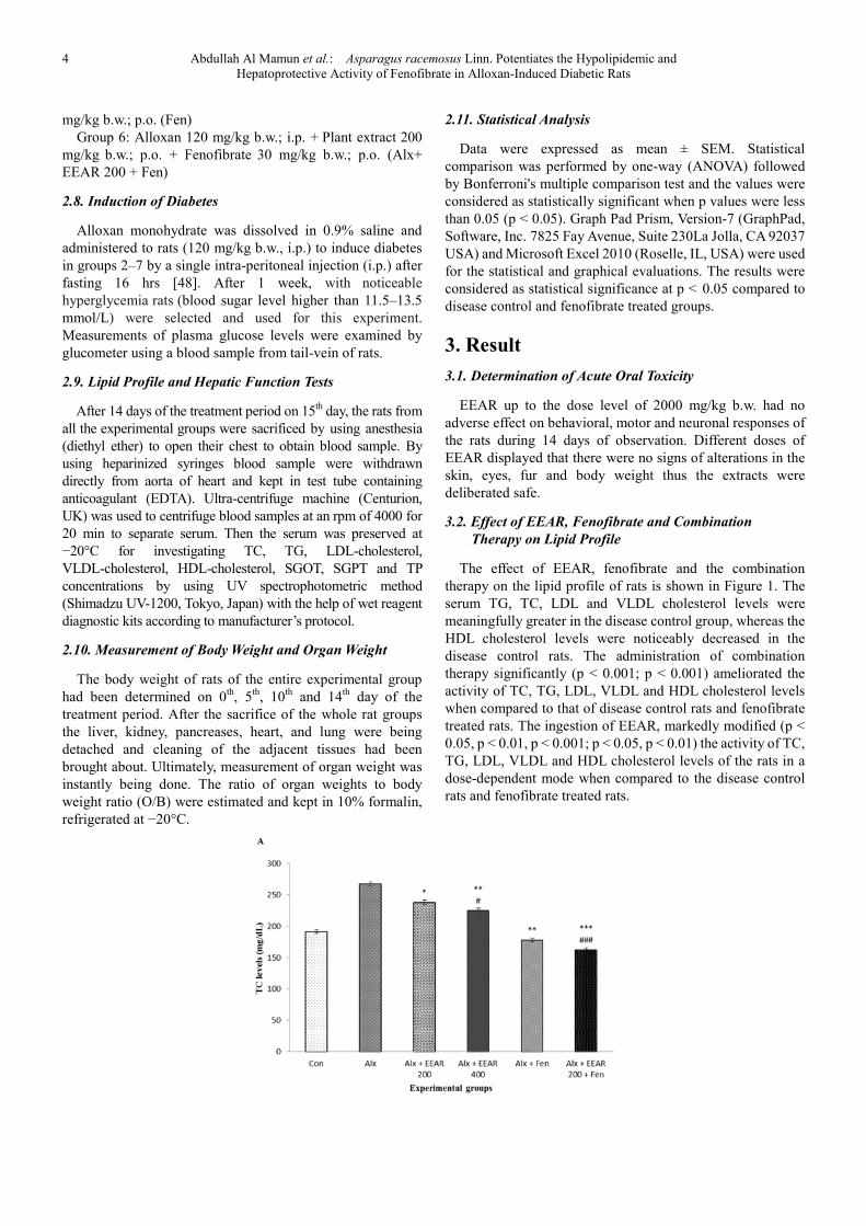
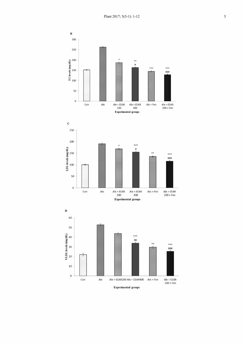
3.2. Effect of EEAR, Fenofibrate and Combination
Therapy on Lipid Profile
The effect of EEAR, fenofibrate and the combination
therapy on the lipid profile of rats is shown in Figure 1. The
serum TG, TC, LDL and VLDL cholesterol levels were
meaningfully greater in the disease control group, whereas the
HDL cholesterol levels were noticeably decreased in the
disease control rats. The administration of combination
therapy significantly (p < 0.001; p < 0.001) ameliorated the
activity of TC, TG, LDL, VLDL and HDL cholesterol levels
when compared to that of disease control rats and fenofibrate
treated rats. The ingestion of EEAR, markedly modified (p <
0.05, p < 0.01, p < 0.001; p < 0.05, p < 0.01) the activity of TC,
TG, LDL, VLDL and HDL cholesterol levels of the rats in a
dose-dependent mode when compared to the disease control
rats and fenofibrate treated rats.
Plant 2017; 5(5-1): 1-12 5

6 Abdullah Al Mamun et al.: Asparagus racemosus Linn. Potentiates the Hypolipidemic and
Hepatoprotective Activity of Fenofibrate in Alloxan-Induced Diabetic Rats
Figure 1. Hypolidemic effect of EEAR, fenofibrate and combination therapy on lipid profile in diabetic rats. Values were expressed as mean ± SEM (n = 5/group).
A. Total cholesterol; B. Triglycerides; C. Low density lipoprotein; D. Very low density lipoprotein; E. High density lipoprotein. *p < 0.05, **p < 0.01, ***p < 0.001
significant difference from the disease control group. #p < 0.05, ##p < 0.01, ###p < 0.001 significant difference from the fenofibrate treated group.
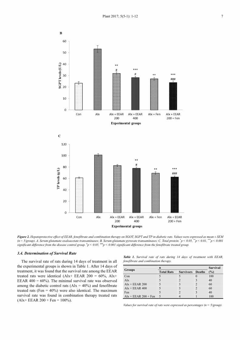
3.3. Effect of EEAR, Fenofibrate and Combination Therapy on Hepatic Functions
The effect of EEAR, fenofibrate treatment and the combination therapy on the hepatic functions of rats is shown in Figure 2.
The hepatic marker enzymes, including SGOT, SGPT and TP levels were noticeably higher in the disease control rats. The
administration of EEAR pointedly (p < 0.05, p < 0.01, p < 0.001; p < 0.05) reduced the liver enzymes level, such as SGOT, SGPT
and TP in a dose-dependent manner as compared to that of disease control rats and fenofibrate treated rats. The effect of
combination therapy significantly (p < 0.001; p < 0.001) decreased the SGOT, SGPT and TP hepatic marker enzyme levels when
compared to the disease control rats and fenofibrate treated rats indicated amelioration in liver dysfunctions.
Plant 2017; 5(5-1): 1-12 7
Figure 2. Hepatoprotective effect of EEAR, fenofibrate and combination therapy on SGOT, SGPT and TP in diabetic rats. Values were expressed as mean ± SEM
(n = 5/group). A. Serum glutamate oxaloacetate transaminases; B. Serum glutamate pyruvate transaminases; C. Total protein. *p < 0.05, **p < 0.01, ***p < 0.001
significant difference from the disease control group. #p < 0.05, ###p < 0.001 significant difference from the fenofibrate treated group.
3.4. Determination of Survival Rate
The survival rate of rats during 14 days of treatment in all
the experimental groups is shown in Table 1. After 14 days of
treatment, it was found that the survival rate among the EEAR
treated rats were identical (Alx+ EEAR 200 = 60%, Alx+
EEAR 400 = 60%). The minimal survival rate was observed
among the diabetic control rats (Alx = 40%) and fenofibrate
treated rats (Fen = 40%) were also identical. The maximum
survival rate was found in combination therapy treated rats
(Alx+ EEAR 200 + Fen = 100%).
Table 1. Survival rate of rats during 14 days of treatment with EEAR,
fenofibrate and combination therapy.
Groups n Survival
(%) Total Rats Survivors Deaths
Con 5 5 0 100
Alx 5 2 3 40
Alx + EEAR 200 5 3 2 60
Alx + EEAR 400 5 3 2 60
Fen 5 2 3 40
Alx + EEAR 200 + Fen 5 4 1 100
Values for survival rate of rats were expressed as percentages (n = 5/group).
8 Abdullah Al Mamun et al.: Asparagus racemosus Linn. Potentiates the Hypolipidemic and
Hepatoprotective Activity of Fenofibrate in Alloxan-Induced Diabetic Rats
3.5. Effect of EEAR and Combination Therapy on Body
Weight and Organ Weight Changes
Body weight changes in all the experimental groups of rats
are shown in Table 2. The body weight of disease control rats
showed significantly decreased during 14 days of the
treatment period. During treatment period, it was found that
the significant (p < 0.01, p < 0.001; p < 0.05, p < 0.01, p <
0.001) changes in the body weight were observed in the EEAR
and combination group on 10th
and 14th
day as compared to
that of disease control rats and fenofibrate treated rats. The
weight of heart, liver, lung, pancreas and kidney did not
change considerably after 14 days of treatment in all the
experimental groups of rats. Although the weight of liver and
weight of pancreas significantly decreased in disease control
group, after 14 days of treatment the values were normalized
(p < 0.001; p < 0.05, p < 0.01, p < 0.001) in maximum doses of
EEAR (Alx+ EEAR 400) and combination therapy treated rats
when compared to that of disease control rats and fenofibrate
treated rats.
Table 2. Effect of EEAR, fenofibrate and combination therapy on body weight changes in diabetic rats.
Groups Body Weight Changes (g)
0th Day 5th Day 10th Day 14th Day
Con 153.93 ± 2.257 156.67 ± 1.405 159.24 ± 1.514 160.33 ± 0.544
Alx 141.18± 1.828 136.92 ± 2.441 132.36 ± 2.518 126.26 ± 1.941
Alx + EEAR 200 153.10 ± 1.815 150.10 ± 3.837 151.37 ± 2.202**# 152.49 ± 1.110***#
Alx + EEAR 400 147.59 ± 1.872 152.32 ± 1.775 153.92 ± 0.887***# 157.69 ± 1.111***##
Fen 146.85 ± 4.632 147.84 ± 2.989 150.02 ± 3.224** 153.03 ± 3.104***
Alx + EEAR 200 + Fen 145.19 ± 3.729 149.23 ± 2.274 150.80 ± 1.511***### 154.58 ± 3.625***###
Values were expressed as mean ± SEM (n = 5/group). **p < 0.01, ***p < 0.001 significant difference from the disease control group. #p < 0.05, ##p < 0.01, ###p <
0.001 significant difference from the fenofibrate treated group.
Table 3. Effect of EEAR, fenofibrate treatment and combination therapy on organ weight changes in diabetic rats.
Groups Organ Weight Changes (g)
Heart Liver Lung Pancreas Kidney
Con 0.57 ± 0.0318 4.71 ± ± 0.2908 0.49 ± 0.0219 1.38 ± 0.0318 1.07 ± 0.0706
Alx 0.51 ± 0.0203 3.38 ± 0.0498 0.45 ± 0.0416 0.99 ± 0.0410 1.09 ± 0.0706
Alx + EEAR 200 0.55 ± 0.0463 4.33 ± 0.0899 0.51 ± 0.0115 1.12 ± 0.0291 0.97± 0.0088
Alx + EEAR 400 0.57 ± 0.0285 4.38 ± 0.0273***## 0.41 ± 0.0949 1.26 ± 0.0321***# 1.01 ± 0.0145
Fen 0.55 ± 0.0318 5.29 ± 0.0536 0.47 ± 0.0203 1.14 ± 0.0145 1.09 ± 0.0643
Alx + EEAR 200 + Fen 0.49 ± 0.0203 5.54 ± 0.0491***### 0.47 ± 0.0208 1.32 ± 0.0153***### 0.99 ± 0.0088
Values were expressed as mean ± SEM (n = 5/group). ***p < 0.001 significant difference from the disease control group. #p < 0.05, ##p < 0.01, ###p < 0.001
significant difference from the fenofibrate treated group.
4. Discussion
Dyslipidemia is one of the major risk factors for
cardiovascular disease in diabetes mellitus [48, 49]. The
relationship between diabetes mellitus and serum lipid profile
had been much discussed during the past decades [50-52].
Both lipid profile and diabetes have been shown to be the
important predictors for metabolic disturbances including
dyslipidemia, hypertension, cardiovascular diseases,
hyperinsulinemia, etc [53, 54].
In the present study, alloxan treatment showed a significant
elevation in glucose and lipid levels i.e., TC, TG, LDL and
VLDL and a reduction in HDL and also reported that elevation
of hepatic markers i.e., SGOT, SGPT and TP levels. The
possible mechanism might be that free radical generation by
the alloxan causes damage to β-cells of pancreas, leading to
insulin deficiency which results in hyperglycemia and also
associated with hyperlipidemia [55]. The mechanism of
alloxan induced hepatotoxicity could be attributed to decrease
in antioxidant enzymes, accompanied by a significant increase
in aldehyde products of lipid peroxidation, leading to hepatic
oxidative stress in rats [56].
Lipids play an important role in the pathogenesis of
diabetes mellitus [57]. The level of serum lipids is usually
raised in diabetes and such an elevation represents a risk factor
for coronary heart disease [58]. Lowering of serum lipids
levels through dietary or drugs therapy seems to be associated
with a decrease in the risk of vascular disease [59]. The
abnormal high concentration of serum lipids in diabetes is
mainly due to the increase in the mobilization of free fatty
acids from the peripheral depots, since insulin inhibits the
hormone sensitive lipase [60]. The elevated
hypertriglyceridemia was increased in the synthesis of
triglyceride rich lipoprotein particles, VLDL in liver
diminished catabolism in diabetic rats [61]. Since insulin has a
potent inhibitory effect on lipolysis in adipocytes, insulin
deficiency is associated with excess lipolysis and increased
influx of free fatty acids to the liver [62]. The increased levels
of LDL and VLDL in the diabetic animals might be due to
overproduction of LDL and VLDL by the liver due to the
stimulation of hepatic triglyceride synthesis as a result of free
fatty acid influx [63]. The HDL was significantly reduced in
the diabetic rats, which indicate a positive risk factor for
atheroscelerosis [64]. In the present study, alloxan induced
diabetic rats had an elevation in the serum lipids. The effect of
EEAR markedly modified the activity of TC, TG, LDL,
VLDL and HDL cholesterol levels when compared to the

Plant 2017; 5(5-1): 1-12 9
disease control rats and fenofibrate treated rats. The
administration of combination therapy meaningfully
improved the activity of TC, TG, LDL, VLDL and HDL
cholesterol levels as compared to that of disease control rats
and fenofibrate treated rats. In a previous study on lipid profile
by Dheeba et al., also reported analogous findings for the roots
extract of Asparagus racemosus in alloxan-induced diabetic
rats [65].
The liver is an important insulin-dependent tissue, which
plays a pivotal role in glucose and lipid homeostasis and is
severely affected by diabetes [66]. The liver is one of the
organs damaged by free radicals [67]. Several studies have
shown that oxidative free radicals generated by alloxan
administration being the most common etiology for the
destruction of vital organs of the body [68, 69]. It was evident
an increase in activities of the hepatic marker enzymes SGPT,
SGOT and TP indicated that diabetes might be induced due to
liver dysfunction [70]. Liver necrosis in alloxan-induced
diabetic rats augmented in the activities of SGPT, SGOT and
TP in plasma might be mainly due to the leakage of these
enzymes from the liver cytosol into the bloodstream [71, 72].
In our study, the levels of hepatic enzymes marker such as
SGOT, SGPT and TP levels were pointedly higher in the
disease control rats. After 14 days of treatment our study
observed that the rats treated with EEAR markedly reduced
the liver enzymes level, including SGOT, SGPT and TP when
compared to that of disease control and fenofibrate treated rats.
The administration of combination therapy expressively
decreased the SGOT, SGPT and TP hepatic marker enzyme
levels when compared to that of disease control rats and
fenofibrate treated rats disclosed amelioration in liver
dysfunctions. Rahimi et al., in the study on Carthamus
tinctorius oil in alloxan-induced diabetic rats claimed
noticeably improvement in the hepatic dysfunction [73].
The current investigation found that none of the rats died in
combination groups. The survival rate was expressively higher
in combination groups as compared to that of disease control
and fenofibrate treated rats. Rajendran et al., in the study on
nuts demonstrated similar outcomes when administered in
combinations with Emblica officinalis and honey [74].
In the present study alloxan induced diabetic rats had lower
body weight and organ weight (heart, liver, lung, pancreas and
kidney). The decrease in body weight could be due to an excess
breakdown of tissue proteins [75]. Increased breakdown of
glycogen and pronounced gluconeogenesis in diabetes might be
responsible for the reduction in liver weight of diabetic animals
[76]. Oral administration of combination therapy to diabetic rats
pointedly improved liver weight and pancreas weight when
compared to that of disease control rats and fenofibrate treated
rats. In the study on the effect of the bark extract of Ficus
racemosa in the body weight in alloxan-induced diabetic rats by
Sophia et al., reported similar results [77].
5. Conclusion
Present study exhibited that common lipid abnormalities
and liver dysfunction were observed in alloxan induced
diabetic rats. Outcomes suggest a greater dominance of
dyslipidemia, which might be playing a principal role in the
progression of CVD among diabetic rats. From our
experimental findings, it can be concluded that the effect of
EEAR potentiates the activity of fenofibrate by increasing
HDL level significantly, but reducing TC, TG, LDL, VLDL as
well as SGOT, SGPT and TP level. It causes rapid induction of
hypolipidemia as well as hepatoprotective effect in diabetic
rats. In this study, EEAR showed a natural key in
hypolipidemic and hepatoprotective activity. Further
identification and isolation of active phytochemical
constituents of AR and their fundamental mode of action
accountable for hypolipidemic and hepatoprotective activity
may be beneficial in developing a potent molecule for DM
associated with CVD and hepatic complications.
Abbreviations
DM: Diabetes mellitus; T1D: Type 1 diabetes; T2D: Type 2
diabetes; CVD: Cardiovascular diseases; AR: Asparagus
racemosus; EEAR: Ethanolic extract of Asparagus racemosus;
ALT: Alanine aminotransferase; AST: Aspartate
aminotransferase; WHO: World health organization; γGT:
gamma-glutamil transferases; b.w.: Body weight; p.o.: Per os
(by mouth); TC: Total cholesterol; TG: Triglycerides; LDL:
Low density lipoprotein; VLDL: Very low density lipoprotein;
HDL: High density lipoprotein. SGOT: Serum glutamate
oxaloacetate transaminases; SGPT: Serum glutamate pyruvate
transaminases; TP: Total protein; Con: Control; Alx: Alloxan;
Alx + EEAR 200: Alloxan 120 mg/kg b.w., i.p. + Plant extract
200 mg/kg b. w., p.o.; Alx+ EEAR 400: Alloxan 120 mg/kg
b.w., i.p. + Plant extract 400 mg/kg b.w., p.o.; Fen: Alloxan
120 mg/kg b.w., i.p. + Fenofibrate 30 mg/kg b.w., p.o.; Alx +
EEAR 200 + Fen: Alloxan 120 mg/kg b.w., i.p. + Plant extract
200 mg/kg b.w., p.o. + Fenofibrate 30 mg/kg b.w., p.o.
Ethical Approval
The protocol of the experiment was approved by the animal
ethics committee of the Department of Pharmacy, Southeast
University, Dhaka, Bangladesh. The animals care and health
were maintained according to the guidelines of NIH.
Author’s Contributions
AAM: Designed the study, wrote the protocol and managed
the analyses of the study and prepared the draft of the
manuscript. AAM and MH: Carried out the laboratory tests. AI:
Prepared the plant extracts and managed the literature searches.
AAM: Performed statistical and graphical evaluations. MSU
and SZ: Reviewed the scientific contents of the manuscript. All
the authors read and approved the final manuscript.
Conflict of Interests
The authors proclaim that there is no conflict of interests
exist about the content of this paper.

10 Abdullah Al Mamun et al.: Asparagus racemosus Linn. Potentiates the Hypolipidemic and
Hepatoprotective Activity of Fenofibrate in Alloxan-Induced Diabetic Rats
Acknowledgements
The authors wish to thank the Department of Pharmacy,
Southeast University, Dhaka, Bangladesh for providing
research facilities.
References
[1] Saha S, Mundle M, Ghosh S and Koley M. 2012. Effects of medical nutrition therapy on plasma lipoproteins of type II dyslipidemic patients: a short term pilot study. Asian J Pharm Clin Res, 5 (4): 207-211.
[2] Hossain MS, Ahmed M and Islam A. 2011. Hypolipidemic and hepatoprotective effects of different fractions of methanolic extract of Momordica charantia (linn.) in alloxan induced diabetic rats. Int J Pharma Sci Res, 2 (3): 601-607.
[3] Baynes HW. 2015. Classification, pathophysiology, diagnosis and management of diabetes mellitus. J Diabetes Metab, 6: 541.
[4] Deguchi Y, Miyazaki K. 2010. Anti-hyperglycemic and anti-hyperlipidemic effects of guava leaf extract. Nutri Meta, 7 (9): 1-10.
[5] Tian L, Long S, Fu M, Liu Y, Xu Y and Jia L. 2011. Characteristics of high-density lipoprotein subclasses distribution for subjects with desirable total cholesterol levels. Hea Dise, 10 (64): 1-9.
[6] Roberts CK, Barnard RJ, Sindhu RK, Jurczak M, Ehdaie A and Vaziri ND. 2006. Oxidative stress and dysregulation of NAD(P)H oxidase and antioxidant enzymes in dietinduced metabolic syndrome. Metabolism, 55: 928-934.
[7] Dixit AK, Dey R, Suresh A, Chaudhuri S, Panda AK, Mitra A, et al. 2014. The prevalence of dyslipidemia in patients with diabetes mellitus of ayurveda Hospital. J Dia Met Diss, 13 (58): 1-5.
[8] Deng R. 2009. Food and food supplements with hypocholesterolemic effects. Rec Pat Food, Nutri Agri, 1: 15-24.
[9] Tosato M, Zamboni V, Ferrini A and Cesari M. 2007. The aging process and potential interventions to extend life expectancy. Clin Interv Aging, 2 (3): 401-412.
[10] Kumar V, Anwar F, Ahmed D, Verma A, Ahmed A, Damanhouri ZA, et al. 2014. Paederia foetida Linn. leaf extract: An antihyperlipidemic, antihyperglycaemic and antioxidant activity. BMC Comp Alt Med, 14 (1): 76.
[11] Farokhi F, Farkhad NK, Togmechi A and band KS. 2012. Preventive effects of Prangos ferulacea (L.) Lindle on liver damage of diabetic rats induced by alloxan. Avicenna J Phytomed, 2 (2): 63-71.
[12] Giannini EG, Testa R and Savarino V. 2005. Liver enzyme alteration: a guide for clinicians. CMAJ, 172 (3): 367-379.
[13] Oršolić N, Sirovina D, Končić MZ, Lacković G and Gregorović G. 2012. Effect of Croatian propolis on diabetic nephropathy and liver toxicity in mice. BMC Comp Alt Med, 12 (117): 1-12.
[14] Yamatani K, Marubashi S, Wakasugi K, Saito K, Sato N, Takahashai K, et al. 1994. Catecholamine-induced cAMP
response in streptozotocin-induced diabetic rat liver. Tohoku J Exp Med, 173: 311-320.
[15] Zafar M, Naqvi S and Kaimkhani MA. 2009. Altered liver morphology and enzymes in streptozotocin induced diabetic rats. Int J Morphol, 27: 719-725.
[16] Pratt DS and Kaplan MM. 2009. Evaluation of abnormal liver enzyme results in asymptomatic patients. N Engl J Med, 342: 1266-1271.
[17] Ravikumar B, Gerrard J, Man CD, Firbank MJ, Lane A, English PT, et al. 2008. Pioglitazone decreases fasting and postprandial endogenous glucose production in proportion to decrease in hepatic triglyceride content. Diabetes, 57 (9): 2288-2295.
[18] Etuk, EU. 2010. Animals models for studying diabetes mellitus. Agric Biol J N Am, 1 (2): 130-134.
[19] Claudino M, Ceolin DS, Alberti S, Cestari TM, Spadella CT, Rubira-Bullen IRF, et al. 2007. Alloxan-induced diabetes triggers the development of periodontal disease in rats. PLoS ONE, 2 (12): e1320.
[20] Sunday RM, Ilesanmi OR and Obuotor, EM. 2016. Anti-Diabetic effect of Anthocleista vogelii ethanolic root extract in alloxan-induced diabetic rats. Res J Med Plants, 10: 79-88.
[21] Saravanan R and Pari L. 2005. Antihyperlipidemic and antiperoxidative effect of Diasulin, a polyherbal formulation in alloxan induced hyperglycemic rats. BMC Comp Alt Med, 5 (14): 1-8.
[22] Szkudelski T. 2001. The mechanism of alloxan and streptozotocin action in β-cells of the rat pancreas. Physiol res, 50: 537-546.
[23] Safarzade A, and Talebi-Garakani E. 2014. Short term resistance training enhanced plasma apoA-I and FABP4 levels in Streptozotocin-induced diabetic rats. J Dia Meta Dis, 13 (41): 1-8.
[24] Manninen V, Tenkanen L, Koskinien P, , Huttunen JK, Mänttäri M, Heinonen OP, et al. 1992. Joint effects of serum triglycerides and LDL cholesterol and HDL cholesterol concentrations on coronary heart disease risk in the Helsinki heart study: implications for treatment. Circulation, 85: 37-45.
[25] Sundaram R, Shanthi P, and Sachdanandam P. 2013. Effect of iridoidglucoside on plasma lipid profile, tissue fatty acid changes, inflammatory cytokines, and GLUT4 expression in skeletal muscle of streptozotocin-induced diabetic rats. Mol Cell Biochem, 380 (1–2): 43-55.
[26] Berredjem H, Reggami Y, Benlaifa M, Berredjem M, and Bouzerna N. 2015. antidiabetic and hypolipidemic potential of 3, 4-dihydroisoquinolin-2 (1H) - sulfonamide in alloxan induced diabetic rats. Int J Pharma, 11: 226-235.
[27] Uddin MS, Mamun AA, Hossain MS, Ashaduzzaman M, Noor MAA, Hossainet MS et al. 2016. Neuroprotective effect of Phyllanthus acidus L. on learning and memory impairment in scopolamine-induced animal model of dementia and oxidative stress: Natural wonder for regulating the development and progression of Alzheimer’s disease. Advances Alzheimer’s Dis, 5 (2), 53-72.
[28] Uddin MS, Mamun AA, Khanum S, Begum Y, and Alam MS. 2016. Analysis of in vitro antioxidant activity of Caryota urens L. leaves: A traditional natural remedy. J Coast Life Med, 4 (6): 483-489.

Plant 2017; 5(5-1): 1-12 11
[29] Sujatha S, and Shalin JJ. 2012. Complementary therapeutic potential: a focus on polyherbal products for hyperglycemia. Asian J Sci Res, 5: 1-13.
[30] Mamum AA, Uddin MS, Wahid F, Iqbal MA and Rahman M. 2016. Neurodefensive effect of Olea europaea L. in alloxan-induced cognitive dysfunction and brain tissue oxidative stress in mice: incredible natural nootropic. J Neurol Neurosci, 7: S3.
[31] Uddin MS, Mamun AA, Hossain MS, Akter F, Iqbal MA and Asaduzzaman M. 2016. Exploring the effect of Phyllanthus emblica L. on cognitive performance, brain antioxidant markers and acetylcholinesterase activity in rats: promising natural gift for the mitigation of Alzheimer’s disease. Ann Neurosci, 4: 218-229.
[32] Hannan JMA, Ali L, Khaleque J, Akhter M, Flatt PR, and Abdel-Wahab YHA. 2012, Antihyperglycaemic activity of Asparagus racemosus roots is partly mediated by inhibition of carbohydrate digestion and absorption, and enhancement of cellular insulin action. British J of Nutri, 107: 1316-1317.
[33] Anonymus. Asparagus racemosus. USA: Available: http://en.wikipedia.org/wiki/Asparagus_racemosus. Accessed: 12 December, 2015.
[34] Sharma PC, Yelne MB, and Dennis TJ. 2000. Database on medicinal plants used in Ayurveda. Central Cou Res Ayur Sidd, 1: 418-430.
[35] Sairam KS, Priyambada NC, Goel RK. 2003. Gastroduodenal ulcer protective activity of Asparagus racemosus: An experimental, biochemical and histological study. J Ethnopharmacol, 86 (1): 1-10.
[36] Alok S, Jain SK, Verma A, Kumar M, Mahor A, and Sabharwal M. 2013. Plant profile, phytochemistry and pharmacology of Asparagus racemosus (Shatavari): A review. Asian Pac J Trop Dis, 3 (3): 242-251.
[37] Uddin MS, Asaduzzaman M, Mamun AA, Iqbal MA, Wahid F and Rony RK. 2016. Neuroprotective activity of Asparagus racemosus Linn. against ethanol-induced cognitive impairment and oxidative stress in rats brain: auspicious for controlling the risk of Alzheimer’s disease. J Alzheimers Dis Parkinsonism, 6(4): 245.
[38] Hannan JMA, Marenah L and Ali L. 2007. Insulin secretory actions of extracts of Asparagus racemosus Root in Perfused Pancreas, Isolated Islets and Clonal Pancreatic β-Cells. J Endo, 192, 159-168.
[39] Shao Y, Chin CK, Ho CT, Ma W, Garrison SA and Huang MT. 1996. Antitumour activity of the crude saponins obtained from asparagus. Cancer Let, 104: 31-36.
[40] Gang ZZ, Li LZ, and Xian LX. 1997. Study on the isolation, purification and antioxidation properties of polysaccharides from Spirulina maxima. Acta Bota Sinica, 39: 77-81.
[41] Zeng, N., Meng, X., and Zhang, Y. 1997. Studies on the antioxidative effect of constituents of Herba epimedii (ESPS). Zhongguo Zhon Zazhi, 22: 46-48.
[42] Liu J, Yeo HC, Doniger SJ, and Ames BN. 1997. Assay of aldehydes from lipid peroxidation: gas chromatography-mass spectrometry compared to thioabarbituric acid. Analy Bioche, 245 161-166.
[43] Liu SX. Chen Y, Zhou M, and Wan J. 1997. Protective effect of
the polysaccharide kreskin on inhibition of lipo-polysaccharide-induced nitric oxide production in macrophages caused by oxidized low-density lipoprotein. Medi Sci Res, 25: 507-509.
[44] Mahammed NL, Jyothi G, Chary TN, Reddy CHV and Reddy GN. 2013. Antidiabetic and antihyperlipidamic activity of ethanolic extract of the leaf of Asperagus racemosus on streptozotocin induced diabetes rats. Int J Pharma Che Sci, 2 (2), 627-633.
[45] Organisation for Economic Cooperation and Development. 2002. OECD guidelines for the testing of chemicals: acute oral toxicity-acute toxic class method. Available: http://www.oecd-ilibrary.org/docserver/download/9742301e.pdf?ex. Accessed: 22 November 2015.
[46] Matsuura B, Kanno S, Minami H, Tsubouchi E, Iwai M, Matsui H, et al. 2004. Effects of antihyperlipidemic agents on hepatic insulin sensitivity in perfused Goto-Kakizaki rat liver. J Gastroenterol. 39 (4): 339-45.
[47] National Research Council. (2011) Guide for the care and use of laboratory animals. National Academies Press, Washington D. C. (USA).
[48] Shah NA and Khan MR. (2014) Antidiabetic Effect of Sida cordata in Alloxan Induced Diabetic Rats. BioMed Res Int, 2014, 1-5.
[49] Sowers JR, Epstein M and Frohlich ED. Diabetes, hypertension, and cardiovascular disease an update. Hypertens, 37: 1053-1059.
[50] Dixit AK, Dey R, Suresh A, Chaudhuri S, Panda AK, Mitra A, et al. 2014. The prevalence of dyslipidemia in patients with diabetes mellitus of ayurveda Hospital. J Diabetes Meta Dis, 13 (58): 1-8.
[51] Elinasri HA and Ahmed AM. 2008. Patterns of lipid changes among type 2 diabetes patients in Sudan. Eastern Mediter Health J, 14: 2.
[52] Mooradian AD. 2009. Dyslipidemia in type 2 diabetes mellitus. Nat Clin Pract Endocrin Metab, 5: 150-159.
[53] Ozder A. 2014. Lipid profile abnormalities seen in T2DM patients in primary healthcare in Turkey: a cross-sectional study. Lip Heal Dis, 13 (183) 1-6.
[54] Goldberg IJ. 2001. Diabetic dyslipidemia: Causes and consequences. J Clin Endo Metab, 8 (3): 965-971.
[55] Fernandes NPC, Lagishetty CV, Panda VS and Naik SR. 2007. An experimental evaluation of the antidiabetic and antilipidemic properties of a standardized Momordica charantia fruit extract. BMC Comple Alt Med, 7 (29): 1-8.
[56] Koyaguru N, Kumar VH, Jamadar MG, Huligol SV, Nayak N, Yendigeri SM, et al. 2013. Antidiabetic and hepatoprotective activities of Tamarindus indica fruit pulp in alloxan induced diabetic rats. Int J Pharma-col and Clin Sci, 2: 33-40.
[57] Averill MM and Bornfeldt KE. 2009. Lipids versus glucose in inflammation and the pathogenesis of macrovascular disease in diabetes. Curr Diab Rep, 9 (1): 18-25.
[58] Prince PSM, Menon VP and Gunasekaran G. 1998. Hypolipidaemic action of Tinospora cordifolia roots in alloxan diabetic rats. J Ethnopharmacol, 64 (1): 53-57.
12 Abdullah Al Mamun et al.: Asparagus racemosus Linn. Potentiates the Hypolipidemic and
Hepatoprotective Activity of Fenofibrate in Alloxan-Induced Diabetic Rats
[59] Valsa AK, Asha SK and Vijayalakshmi NR. 1998. Effect of catechin on intestinal lipid metabolism. Ind J Phy Pha, 42 (2): 286-90.
[60] Indradevi S, Ilavenil S, Kaleeswaran B, Srigopalram S and Ravikumar S. 2012. Ethanolic extract of Crinum asiaticum attenuates hyperglycemia-mediated oxidative stress and protects hepatocytes in alloxan induced experimental diabetic rats. J King Saud Uni-Sci, 24 (2): 171-177.
[61] Jr Brewer HB. 1999. Hypertriglyceridemia: changes in the plasma lipoproteins associated with an increased risk of cardiovascular disease. Am J Cardiol, 83 (9B): 3F-12F.
[62] Daradka HM, Abas MM, Mohammad MAM and Jaffar MM. 2014. Antidiabetic effect of Artemisia absinthium extracts on alloxan-induced diabetic rats. Compa Cli Pat, 23 (6): 1733-1742.
[63] Tacer KF and Rozman D. 2011. Nonalcoholic fatty liver disease: focus on lipoprotein and lipid deregulation. J Lipids, 2011: 1-14.
[64] Niacin VH. 2009. A re-emerging pharmaceutical for the treatment of dyslipidaemia. Bri J Pharmaco, 158 (2): 429-441.
[65] Dheeba B, Kumar PS, Kannan M and Saravana K. 2012. Antidiabetic and antihyperlipidemic activities of Asparagus racemosus in alloxan induced diabetic rats. J Pharm Res, 5 (5): 2469-2472.
[66] Kumar RN, Sundaram R, Shanthi P and Sachdanandam P. 2013. Protective role of 20-OH ecdysone on lipid profile and tissue fatty acid changes in streptozotocin induced diabetic rats. Eur J Pharmaco, 698: 489-498.
[67] Noeman SA, Hamooda HE and Baalash AA. 2011. Biochemical study of oxidative stress markers in the liver, kidney and heart of high fat diet induced obesity in rats. Diabe Met Syn, 3 (17): 1-8.
[68] de Andrade KQ, Moura FA, dos Santos JM, de Araújo ORP, de Farias Santos JC and Goulart MOF. 2015. Oxidative stress and inflammation in hepatic diseases: therapeutic possibilities of N-Acetylcysteine. Int J Mol Sci, 16 (12): 30269-30308.
[69] Sepici-Dincel A, Açıkgöz Ş, Çevik C, Sengelen M and Yeşilada E. 2007. Effects of in vivo antioxidant enzyme activities of myrtle oil in normoglycaemic and alloxan diabetic rabbits. J Ethnopharmaco, 110 (3): 498-503.
[70] Bairwa NK, Sethiya NK and Mishra SH. 2010. Protective effect of stem bark of Ceiba pentandra Linn. against paracetamol-induced hepatotoxicity in rats. Pharmacog Res, 2 (1): 1-6.
[71] Navarro CM, Montilla PM, Martin A, Jimenez J and Utrilla PM. 1993. Free radicals scavenger and antihepatotoxic activity of Rosmarinus. Plant Med, 59: 312-314.
[72] Nkosi CZ, Opoku AR and Terblanche SE. 2005. Effect of pumpkin seed (Cucurbita pepo) protein isolate on the activity levels of certain plasma enzymes in CCl4-induced liver injury in low protein fed rats. Phytother Res, 19: 341-5.
[73] Rahimi P, Asgary S and Kabiri N. 2014. Hepatoprotective and hypolipidemic effects of carthamus tinctorius oil in alloxan-induced type 1 diabetic rats. J HerbMed Pharmacol, 3 (2): 107-111.
[74] Mythilypriya R, Shanthi P and Sachdanandam P. Oral acute and subacute toxicity studies with Kalpaamrutha, a modified indigenous prepraration on rats. J Health Science, 2007; 53 (4): 351-8.
[75] Lecker SH, Solomon V, Mitch WE and Goldberg AL. 1999. Muscle protein breakdown and the critical role of the ubiquitin-proteasome pathway in normal and disease states. J Nutr, 129 (1), 227S-237S.
[76] Edgerton DS, Basu R, Ramnanan CJ, Farmer TD, Neal D, Scott M, et al. 2010. Effect of 11β-hydroxysteroid dehydrogenase-1 inhibition on hepatic glucose metabolism in the conscious dog. Ame J Phy-End Met, 298 (5): E1019-E1026.
[77] Sophia D and Manoharan S. 2007. Hypolipidemic activities of Ficus racemosa Linn. bark in alloxan induced diabetic rats. Afr J Trad CAM, 4 (3): 279-288.