Embed Size (px)
Citation preview
AS SIMPLE AS PRESSING the start button
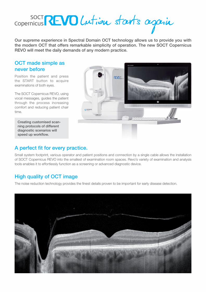
Our supreme experience in Spectral Domain OCT technology allows us to provide you with the modern OCT that offers remarkable simplicity of operation. The new SOCT Copernicus REVO will meet the daily demands of any modern practice.
OCT made simple as never beforePosition the patient and press
the START button to acquire
examinations of both eyes.
The SOCT Copernicus REVO, using
vocal messages, guides the patient
through the process increasing
comfort and reducing patient chair
time.
Creating customised scan-ning protocols of different diagnostic scenarios will speed up workflow.
A perfect fit for every practice.Small system footprint, various operator and patient positions and connection by a single cable allows the installation
of SOCT Copernicus REVO into the smallest of examination room spaces. Revo’s variety of examination and analysis
tools enables it to effortlessly function as a screening or advanced diagnostic device.
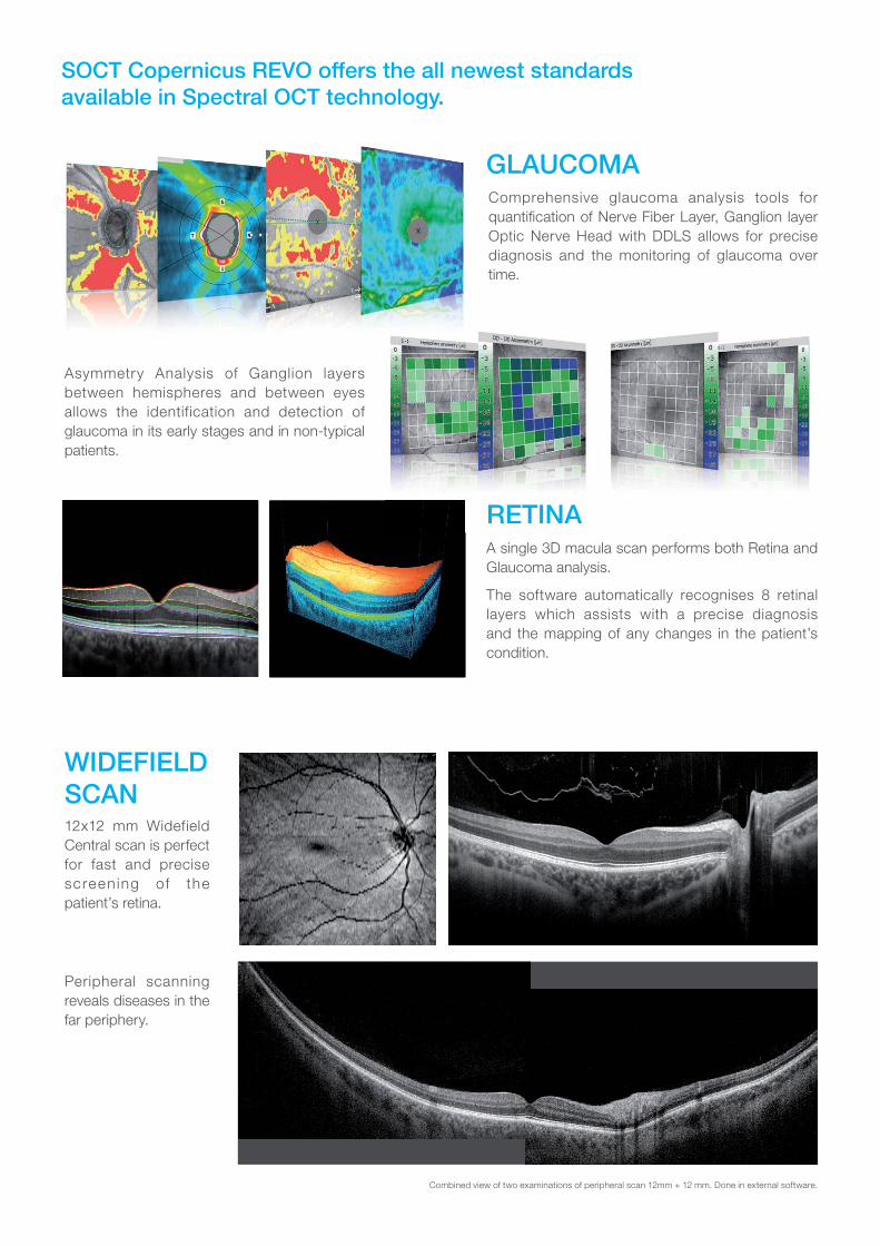
High quality of OCT imageThe noise reduction technology provides the finest details proven to be important for early disease detection.
lution starts again
WIDEFIELD SCAN12x12 mm Widefield
Central scan is perfect
for fast and precise
screen ing of the
patient’s retina.
Peripheral scanning
reveals diseases in the
far periphery.
Combined view of two examinations of peripheral scan 12mm + 12 mm. Done in external software.
RETINAA single 3D macula scan performs both Retina and
Glaucoma analysis.
The software automatically recognises 8 retinal
layers which assists with a precise diagnosis
and the mapping of any changes in the patient’s
condition.
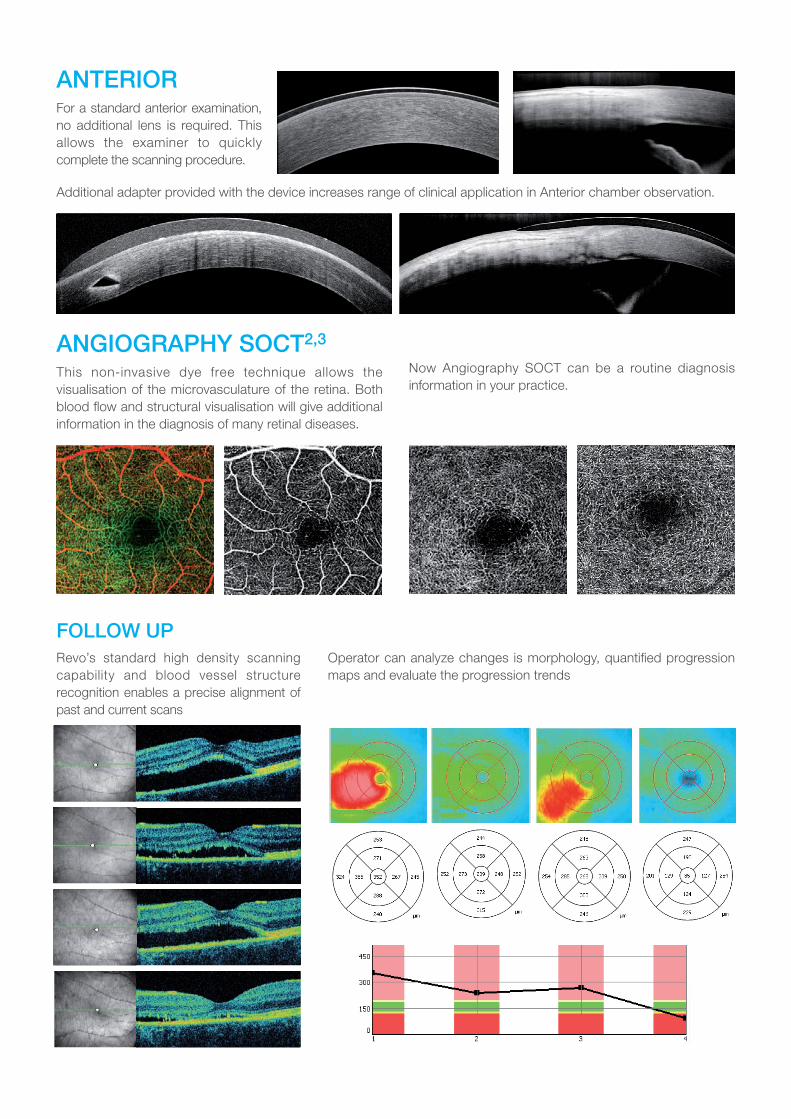
GLAUCOMAComprehensive glaucoma analysis tools for
quantification of Nerve Fiber Layer, Ganglion layer
Optic Nerve Head with DDLS allows for precise
diagnosis and the monitoring of glaucoma over
time.
Asymmetry Analysis of Ganglion layers
between hemispheres and between eyes
allows the identification and detection of
glaucoma in its early stages and in non-typical
patients.
SOCT Copernicus REVO offers the all newest standards available in Spectral OCT technology.
RETINA
ANGIOGRAPHY SOCTThis non-invasive dye free technique allows the
visualisation of the microvasculature of the retina. Both
blood flow and structural visualisation will give additional
information in the diagnosis of many retinal diseases.
Now Angiography SOCT can be a routine diagnosis
information in your practice.
FOLLOW UPRevo’s standard high density scanning
capability and blood vessel structure
recognition enables a precise alignment of
past and current scans
Operator can analyze changes is morphology, quantified progression
maps and evaluate the progression trends
ANTERIOR For a standard anterior examination,
no additional lens is required. This
allows the examiner to quickly
complete the scanning procedure.
Additional adapter provided with the device increases range of clinical application in Anterior chamber observation.
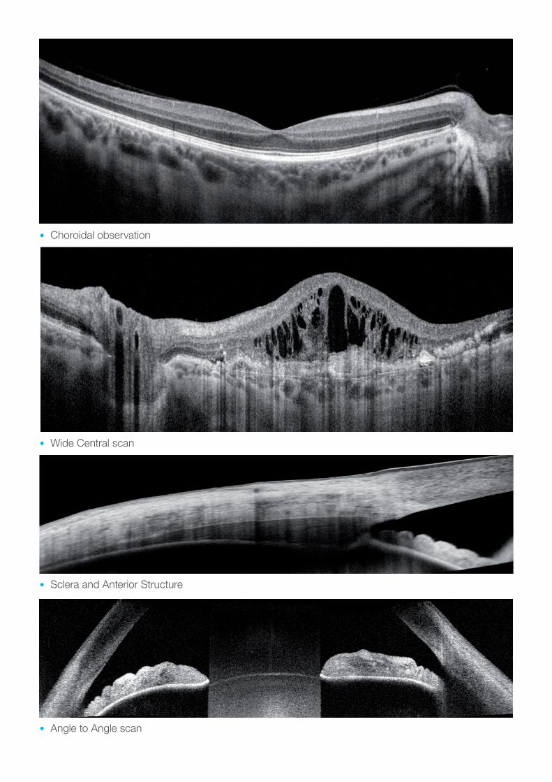
• Choroidal observation
• Wide Central scan
• Angle to Angle scan
• Sclera and Anterior Structure
Local Distributor:
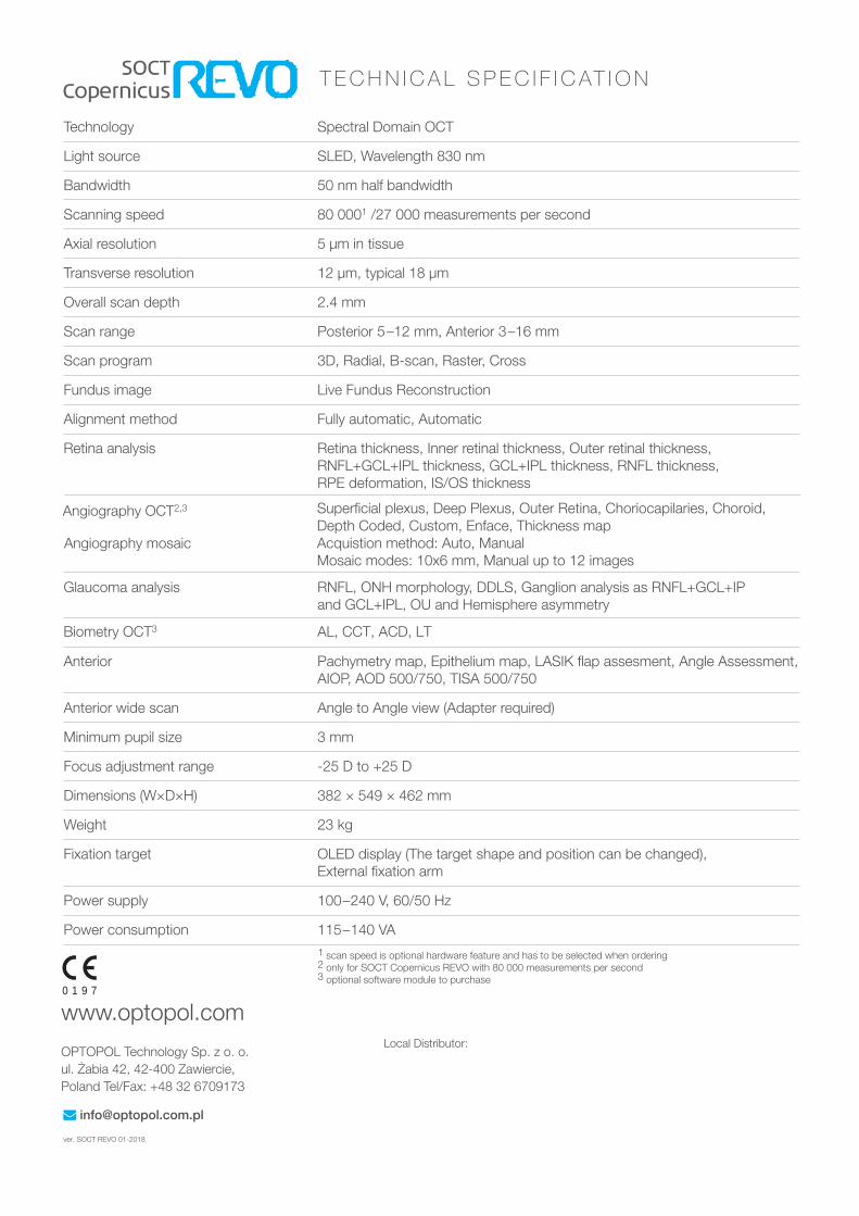
T E C H N I C A L S P E C I F I C AT I O N
ver. SOCT REVO 0 -201
Technology Spectral Domain OCT
Light source SLED, Wavelength 830 nm
Bandwidth 50 nm half bandwidth
Scanning speed 27 000 measurements per second
Axial resolution 5 μm in tissue
Transverse resolution 12 μm, typical 18 μm
Overall scan depth 2.4 mm
Scan range Posterior 5–12 mm, Anterior 3–16 mm
Scan program 3D, Radial, B-scan, Raster, Cross
Fundus image Live Fundus Reconstruction
Alignment method Fully automatic, Automatic
Retina analysis Retina thickness, Inner retinal thickness, Outer retinal thickness,
RNFL+GCL+IPL thickness, GCL+IPL thickness, RNFL thickness,
RPE deformation, IS/OS thickness
Glaucoma analysis RNFL, ONH morphology, DDLS, Ganglion analysis as RNFL+GCL+IP
and GCL+IPL, OU and Hemisphere asymmetry
Anterior Pachymetry map, Epithelium map, LASIK flap assesment, Angle Assessment,
AIOP, AOD 500/750, TISA 500/750
Anterior wide scan Angle to Angle view (Adapter required)
Minimum pupil size 3 mm
Focus adjustment range -25 D to +25 D
Dimensions (W×D×H) 382 × 549 × 462 mm
Weight 23 kg
Fixation target OLED display (The target shape and position can be changed),
External fixation arm
Power supply 100–240 V, 60/50 Hz
Power consumption 115–140 VA
www.optopol.com
OPTOPOL Technology Sp. z o. o.
ul. Żabia 42, 42-400 Zawiercie,
Poland Tel/Fax: +48 32 6709173
Angiography OCT2
0 1 9 7


![Simultaneous and absolute quantification of nucleoside ......9]UTP, 10 μM [15N 5, 13C 10]dATP, 10 μM[15N 5, 13C 10]dGTP, 10 μM [15N 3, 13C 9]dCTP, and 10 μM[15N 2, 13C 10]dTTP)](https://img.pdfslide.us/doc/110x75/6110c5cfc90cfe531510e3b4/simultaneous-and-absolute-quantification-of-nucleoside-9utp-10-m-15n.jpg)