Embed Size (px)
DESCRIPTION
Biology AS level practice exam questions on Transport in Animals - Sheet 4
Citation preview
Do notwrite inmarginQUESTIONSHEET 1
TOTAL /
TRANSPORT IN ANIMALSAS 4
7
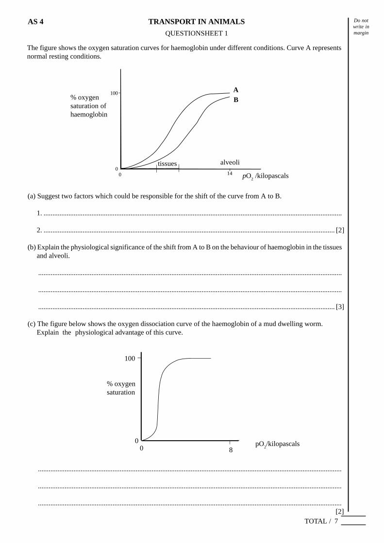
The figure shows the oxygen saturation curves for haemoglobin under different conditions. Curve A representsnormal resting conditions.
(a) Suggest two factors which could be responsible for the shift of the curve from A to B.
1. ........................................................................................................................................................................
2. .................................................................................................................................................................... [2]
(b) Explain the physiological significance of the shift from A to B on the behaviour of haemoglobin in the tissuesand alveoli.
...........................................................................................................................................................................
...........................................................................................................................................................................
....................................................................................................................................................................... [3]
(c) The figure below shows the oxygen dissociation curve of the haemoglobin of a mud dwelling worm.Explain the physiological advantage of this curve.
...........................................................................................................................................................................
...........................................................................................................................................................................
...........................................................................................................................................................................[2]
% oxygensaturation
pO2/kilopascals
% oxygensaturation ofhaemoglobin
pO2 /kilopascals
AB
1400
100
alveolitissues
100
800
Do notwrite inmarginQUESTIONSHEET 2
TOTAL /
TRANSPORT IN ANIMALSAS 4
7
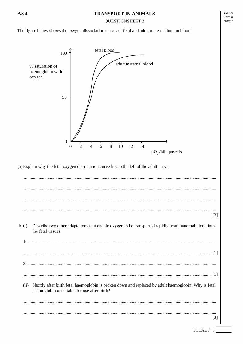
The figure below shows the oxygen dissociation curves of fetal and adult maternal human blood.
(a) Explain why the fetal oxygen dissociation curve lies to the left of the adult curve.
...........................................................................................................................................................................
...........................................................................................................................................................................
...........................................................................................................................................................................
...........................................................................................................................................................................[3]
(b) (i) Describe two other adaptations that enable oxygen to be transported rapidly from maternal blood intothe fetal tissues.
1: ........................................................................................................................................................................
....................................................................................................................................................................... [1]
2: ........................................................................................................................................................................
....................................................................................................................................................................... [1]
(ii) Shortly after birth fetal haemoglobin is broken down and replaced by adult haemoglobin. Why is fetalhaemoglobin unsuitable for use after birth?
...........................................................................................................................................................................
...........................................................................................................................................................................[2]
100
50
00 2 4 6 8 10 12 14
fetal blood
adult maternal blood% saturation ofhaemoglobin withoxygen
pO2 /kilo pascals
Do notwrite inmarginQUESTIONSHEET 3
TOTAL /
TRANSPORT IN ANIMALSAS 4
13
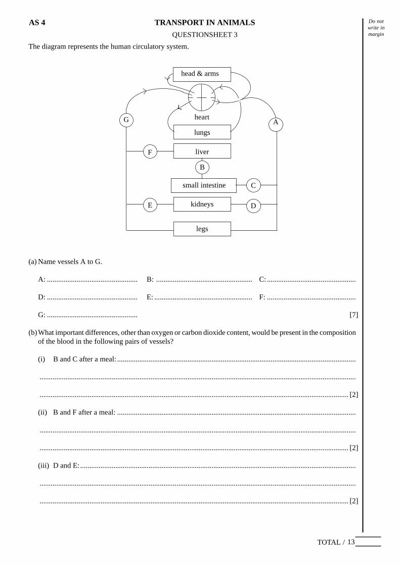
The diagram represents the human circulatory system.
(a) Name vessels A to G.
A: ................................................. B: .................................................... C: ................................................
D: ................................................. E: ..................................................... F: ................................................
G: ................................................. [7]
(b)What important differences, other than oxygen or carbon dioxide content, would be present in the compositionof the blood in the following pairs of vessels?
(i) B and C after a meal: .................................................................................................................................
...........................................................................................................................................................................
....................................................................................................................................................................... [2]
(ii) B and F after a meal: .................................................................................................................................
...........................................................................................................................................................................
....................................................................................................................................................................... [2]
(iii) D and E:.....................................................................................................................................................
...........................................................................................................................................................................
....................................................................................................................................................................... [2]
legs
small intestine
kidneys
liver
lungs
heart
head & arms
F
G
E D
C
A
B
Do notwrite inmarginQUESTIONSHEET 4
TOTAL /
TRANSPORT IN ANIMALSAS 4
14
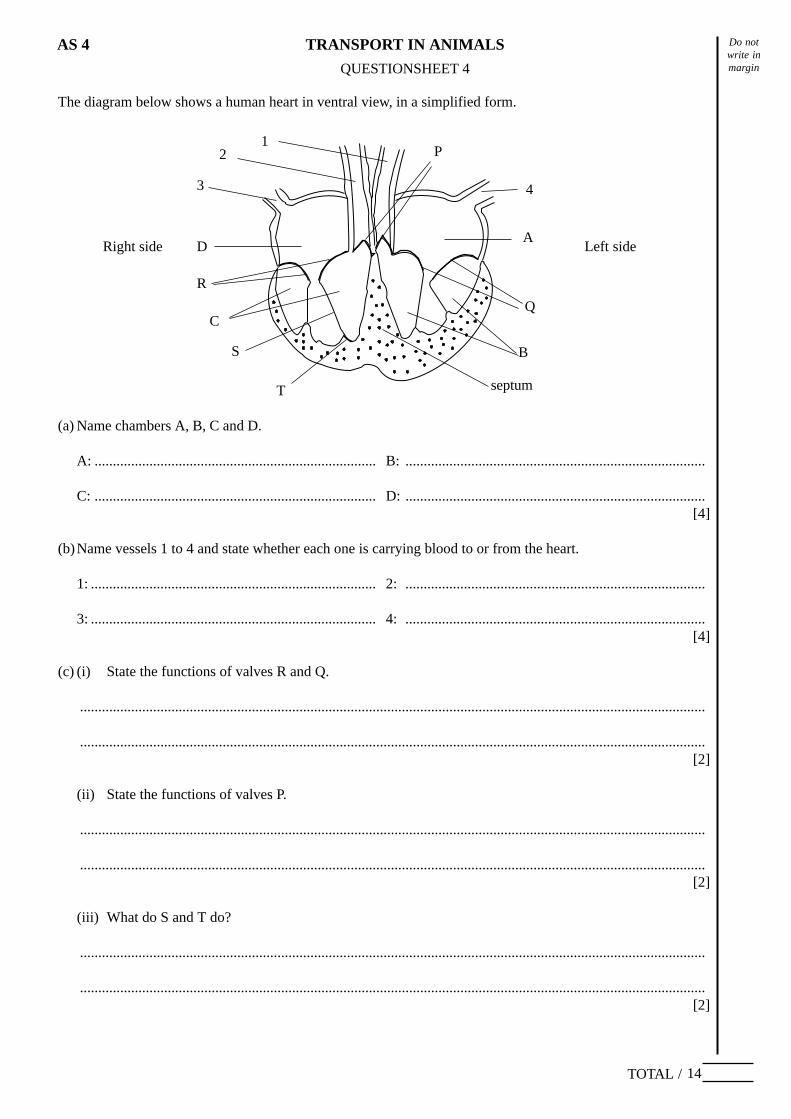
The diagram below shows a human heart in ventral view, in a simplified form.
(a) Name chambers A, B, C and D.
A: ............................................................................. B: ..................................................................................
C: ............................................................................. D: ..................................................................................[4]
(b)Name vessels 1 to 4 and state whether each one is carrying blood to or from the heart.
1: .............................................................................. 2: ..................................................................................
3: .............................................................................. 4: ..................................................................................[4]
(c) (i) State the functions of valves R and Q.
...........................................................................................................................................................................
...........................................................................................................................................................................[2]
(ii) State the functions of valves P.
...........................................................................................................................................................................
...........................................................................................................................................................................[2]
(iii) What do S and T do?
...........................................................................................................................................................................
...........................................................................................................................................................................[2]
S
P
A
Q
B
septumT
C
R
1
D
2
4
Left sideRight side
3
Do notwrite inmarginQUESTIONSHEET 5
TOTAL /
TRANSPORT IN ANIMALSAS 4
12
The diagram below shows the conducting system of the heart in a simplified form.
(a) (i) The heart muscle is said to be ‘myogenic’. What does this mean?
....................................................................................................................................................................... [1]
(ii) Name components A to D of the conducting system.
A: ............................................................................. B: ..................................................................................
C: ............................................................................. D: .............................................................................. [4]
(b)With reference to the parts of the conducting system of the heart, explain why:
(i) the right atrium contracts before the left atrium.
...........................................................................................................................................................................
....................................................................................................................................................................... [2]
(ii) the ventricles contract after the atria.
...........................................................................................................................................................................
....................................................................................................................................................................... [2]
(iii) the ventricles contract from the apex upwards.
....................................................................................................................................................................... [1]
(c) How is the frequency and force of the heartbeat modified to meet the body’s needs?
...........................................................................................................................................................................
...........................................................................................................................................................................
...........................................................................................................................................................................[2]
A
BC
D
right atrium
apex
Do notwrite inmarginQUESTIONSHEET 6
TOTAL /
TRANSPORT IN ANIMALSAS 4
16
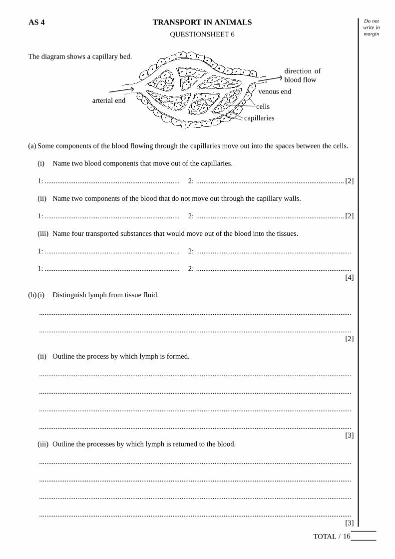
The diagram shows a capillary bed.
(a) Some components of the blood flowing through the capillaries move out into the spaces between the cells.
(i) Name two blood components that move out of the capillaries.
1: .......................................................................... 2: ................................................................................. [2]
(ii) Name two components of the blood that do not move out through the capillary walls.
1: .......................................................................... 2: ................................................................................. [2]
(iii) Name four transported substances that would move out of the blood into the tissues.
1: .......................................................................... 2: .....................................................................................
1: .......................................................................... 2: .....................................................................................[4]
(b) (i) Distinguish lymph from tissue fluid.
...........................................................................................................................................................................
...........................................................................................................................................................................[2]
(ii) Outline the process by which lymph is formed.
...........................................................................................................................................................................
...........................................................................................................................................................................
...........................................................................................................................................................................
...........................................................................................................................................................................[3]
(iii) Outline the processes by which lymph is returned to the blood.
...........................................................................................................................................................................
...........................................................................................................................................................................
...........................................................................................................................................................................
...........................................................................................................................................................................[3]
venous endarterial end
cells
capillaries
direction ofblood flow
Do notwrite inmarginQUESTIONSHEET 7
TOTAL /
TRANSPORT IN ANIMALSAS 4
10
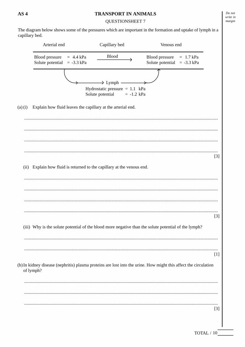
The diagram below shows some of the pressures which are important in the formation and uptake of lymph in acapillary bed.
(a) (i) Explain how fluid leaves the capillary at the arterial end.
...........................................................................................................................................................................
...........................................................................................................................................................................
...........................................................................................................................................................................
...........................................................................................................................................................................[3]
(ii) Explain how fluid is returned to the capillary at the venous end.
...........................................................................................................................................................................
...........................................................................................................................................................................
...........................................................................................................................................................................
...........................................................................................................................................................................[3]
(iii) Why is the solute potential of the blood more negative than the solute potential of the lymph?
...........................................................................................................................................................................
...........................................................................................................................................................................[1]
(b) In kidney disease (nephritis) plasma proteins are lost into the urine. How might this affect the circulationof lymph?
...........................................................................................................................................................................
...........................................................................................................................................................................
...........................................................................................................................................................................[3]
Arterial end Venous endCapillary bed
Blood pressure = 4.4 kPaSolute potential = -3.3 kPa
Blood pressure = 1.7 kPaSolute potential = -3.3 kPa
Lymph
Blood
Hydrostatic pressure = 1.1 kPaSolute potential = -1.2 kPa
Do notwrite inmarginQUESTIONSHEET 8
TOTAL /
TRANSPORT IN ANIMALSAS 4
% saturation ofhaemoglobin withoxygen
100
50
5 10 15 200
pO2 /kilopascals
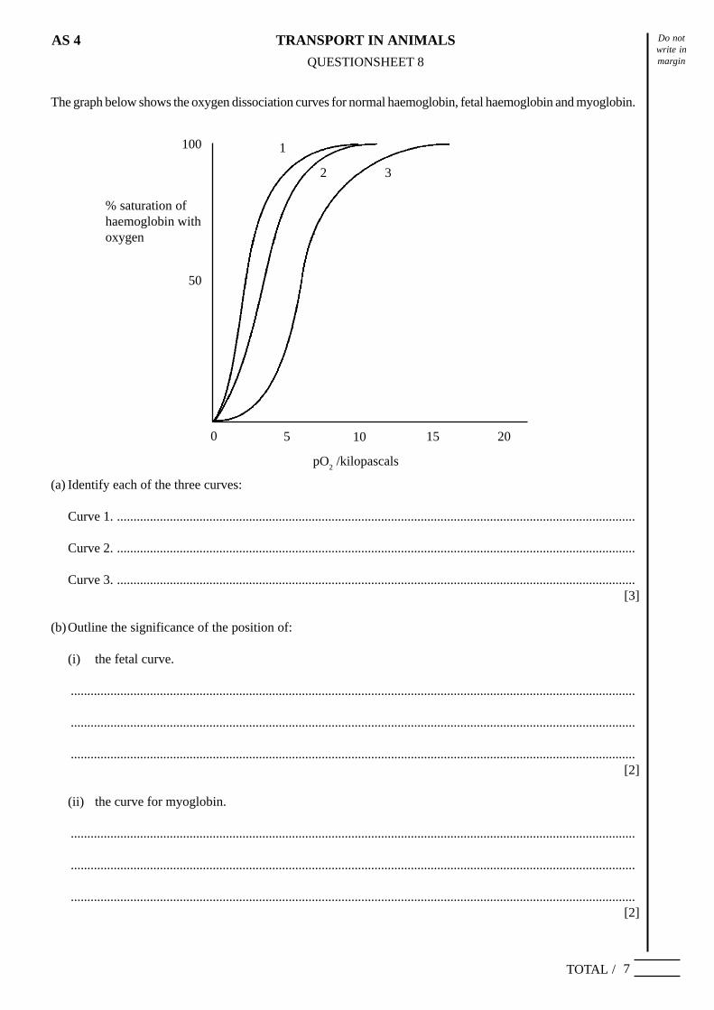
The graph below shows the oxygen dissociation curves for normal haemoglobin, fetal haemoglobin and myoglobin.
(a) Identify each of the three curves:
Curve 1. .............................................................................................................................................................
Curve 2. .............................................................................................................................................................
Curve 3. .............................................................................................................................................................[3]
(b)Outline the significance of the position of:
(i) the fetal curve.
...........................................................................................................................................................................
...........................................................................................................................................................................
...........................................................................................................................................................................[2]
(ii) the curve for myoglobin.
...........................................................................................................................................................................
...........................................................................................................................................................................
...........................................................................................................................................................................[2]
7
1
2 3
Do notwrite inmarginQUESTIONSHEET 9
TOTAL /
TRANSPORT IN ANIMALSAS 4
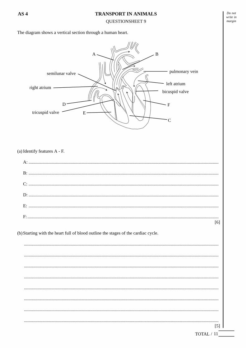
The diagram shows a vertical section through a human heart.
(a) Identify features A - F.
A: .......................................................................................................................................................................
B: .......................................................................................................................................................................
C: .......................................................................................................................................................................
D: .......................................................................................................................................................................
E: .......................................................................................................................................................................
F: ........................................................................................................................................................................[6]
(b)Starting with the heart full of blood outline the stages of the cardiac cycle.
...........................................................................................................................................................................
...........................................................................................................................................................................
...........................................................................................................................................................................
...........................................................................................................................................................................
...........................................................................................................................................................................
...........................................................................................................................................................................
...........................................................................................................................................................................
...........................................................................................................................................................................[5]
11
pulmonary vein
left atrium
F
E
D
C
A B
semilunar valve
tricuspid valve
right atriumbicuspid valve
Do notwrite inmarginQUESTIONSHEET 10
TOTAL /
TRANSPORT IN ANIMALSAS 4
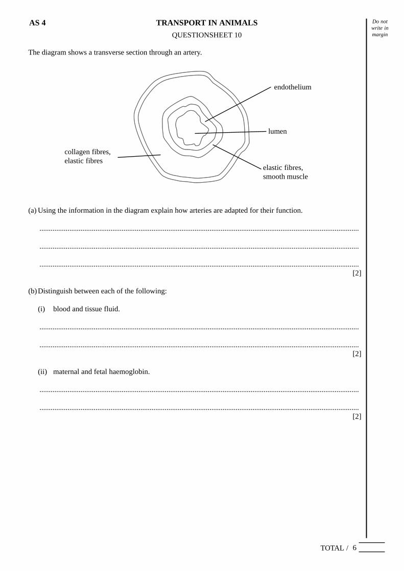
The diagram shows a transverse section through an artery.
6
(a) Using the information in the diagram explain how arteries are adapted for their function.
...........................................................................................................................................................................
...........................................................................................................................................................................
...........................................................................................................................................................................[2]
(b)Distinguish between each of the following:
(i) blood and tissue fluid.
...........................................................................................................................................................................
...........................................................................................................................................................................[2]
(ii) maternal and fetal haemoglobin.
...........................................................................................................................................................................
...........................................................................................................................................................................[2]
endothelium
elastic fibres,smooth muscle
lumen
collagen fibres,elastic fibres
Do notwrite inmarginQUESTIONSHEET 11
TOTAL /
TRANSPORT IN ANIMALSAS 4
15
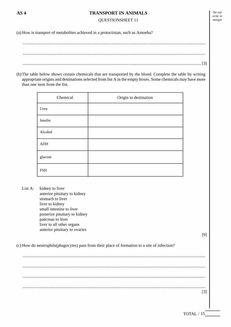
(b)The table below shows certain chemicals that are transported by the blood. Complete the table by writingappropriate origins and destinations selected from list A in the empty boxes. Some chemicals may have morethan one item from the list.
List A: kidney to liveranterior pituitary to kidneystomach to liverliver to kidneysmall intestine to liverposterior pituitary to kidneypancreas to liverliver to all other organsanterior pituitary to ovaries
[9]
(c) How do neutrophils(phagocytes) pass from their place of formation to a site of infection?
...........................................................................................................................................................................
...........................................................................................................................................................................
...........................................................................................................................................................................
...........................................................................................................................................................................[3]
Chemical Origin to destination
Urea
Insulin
Alcohol
ADH
glucose
FSH
(a) How is transport of metabolites achieved in a protoctistan, such as Amoeba?
...........................................................................................................................................................................
...........................................................................................................................................................................
....................................................................................................................................................................... [3]
Do notwrite inmarginQUESTIONSHEET 12
TOTAL /
TRANSPORT IN ANIMALSAS 4
16
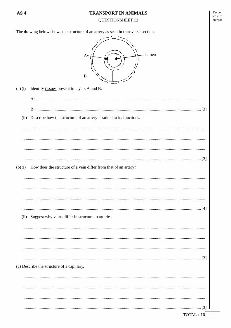
The drawing below shows the structure of an artery as seen in transverse section.
A
B
(a) (i) Identify tissues present in layers A and B.
A: ...............................................................................................................................................................
B: ........................................................................................................................................................... [3]
(ii) Describe how the structure of an artery is suited to its functions.
...........................................................................................................................................................................
...........................................................................................................................................................................
...........................................................................................................................................................................
....................................................................................................................................................................... [3]
(b) (i) How does the structure of a vein differ from that of an artery?
...........................................................................................................................................................................
...........................................................................................................................................................................
...........................................................................................................................................................................
....................................................................................................................................................................... [4]
(ii) Suggest why veins differ in structure to arteries.
...........................................................................................................................................................................
...........................................................................................................................................................................
...........................................................................................................................................................................
....................................................................................................................................................................... [3]
(c) Describe the structure of a capillary.
...........................................................................................................................................................................
...........................................................................................................................................................................
...........................................................................................................................................................................
....................................................................................................................................................................... [3]
lumen
Do notwrite inmarginQUESTIONSHEET 13
TOTAL /
TRANSPORT IN ANIMALSAS 4
11
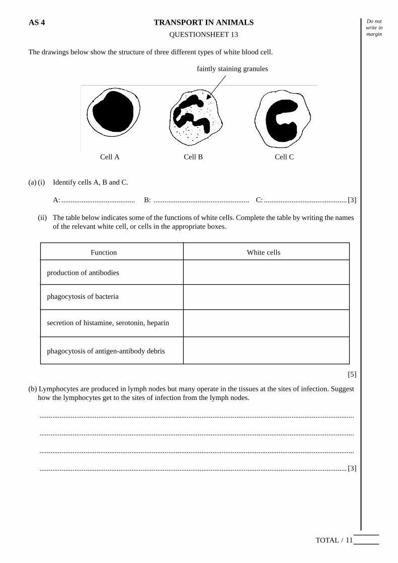
The drawings below show the structure of three different types of white blood cell.
faintly staining granules
Cell A Cell B Cell C
(a) (i) Identify cells A, B and C.
A: ........................................ B: .................................................... C: ............................................. [3]
(ii) The table below indicates some of the functions of white cells. Complete the table by writing the namesof the relevant white cell, or cells in the appropriate boxes.
[5]
(b) Lymphocytes are produced in lymph nodes but many operate in the tissues at the sites of infection. Suggesthow the lymphocytes get to the sites of infection from the lymph nodes.
...........................................................................................................................................................................
...........................................................................................................................................................................
...........................................................................................................................................................................
....................................................................................................................................................................... [3]
Function White cells
production of antibodies
phagocytosis of bacteria
secretion of histamine, serotonin, heparin
phagocytosis of antigen-antibody debris
Do notwrite inmarginQUESTIONSHEET 14
TOTAL /
TRANSPORT IN ANIMALSAS 4
9
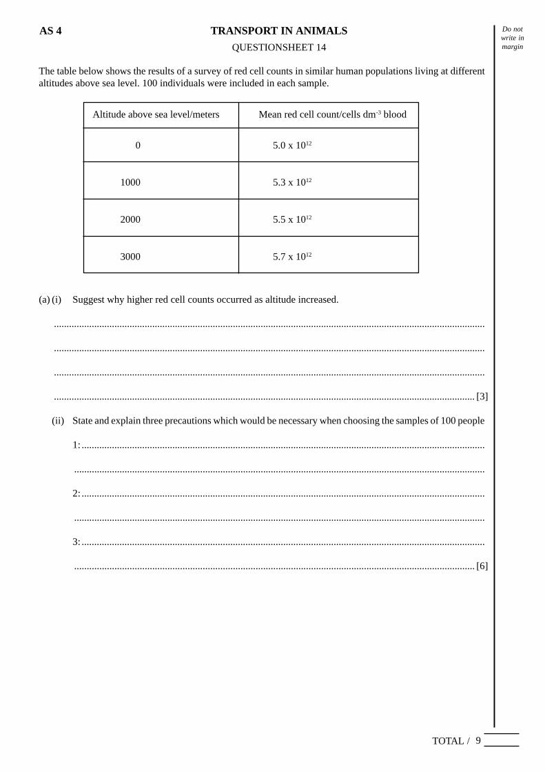
The table below shows the results of a survey of red cell counts in similar human populations living at differentaltitudes above sea level. 100 individuals were included in each sample.
Altitude above sea level/meters Mean red cell count/cells dm-3 blood
0 5.0 x 10¹²
1000 5.3 x 10¹²
2000 5.5 x 10¹²
3000 5.7 x 10¹²
(a) (i) Suggest why higher red cell counts occurred as altitude increased.
...........................................................................................................................................................................
...........................................................................................................................................................................
...........................................................................................................................................................................
....................................................................................................................................................................... [3]
(ii) State and explain three precautions which would be necessary when choosing the samples of 100 people
1: ................................................................................................................................................................
...................................................................................................................................................................
2: ................................................................................................................................................................
...................................................................................................................................................................
3: ................................................................................................................................................................
............................................................................................................................................................... [6]
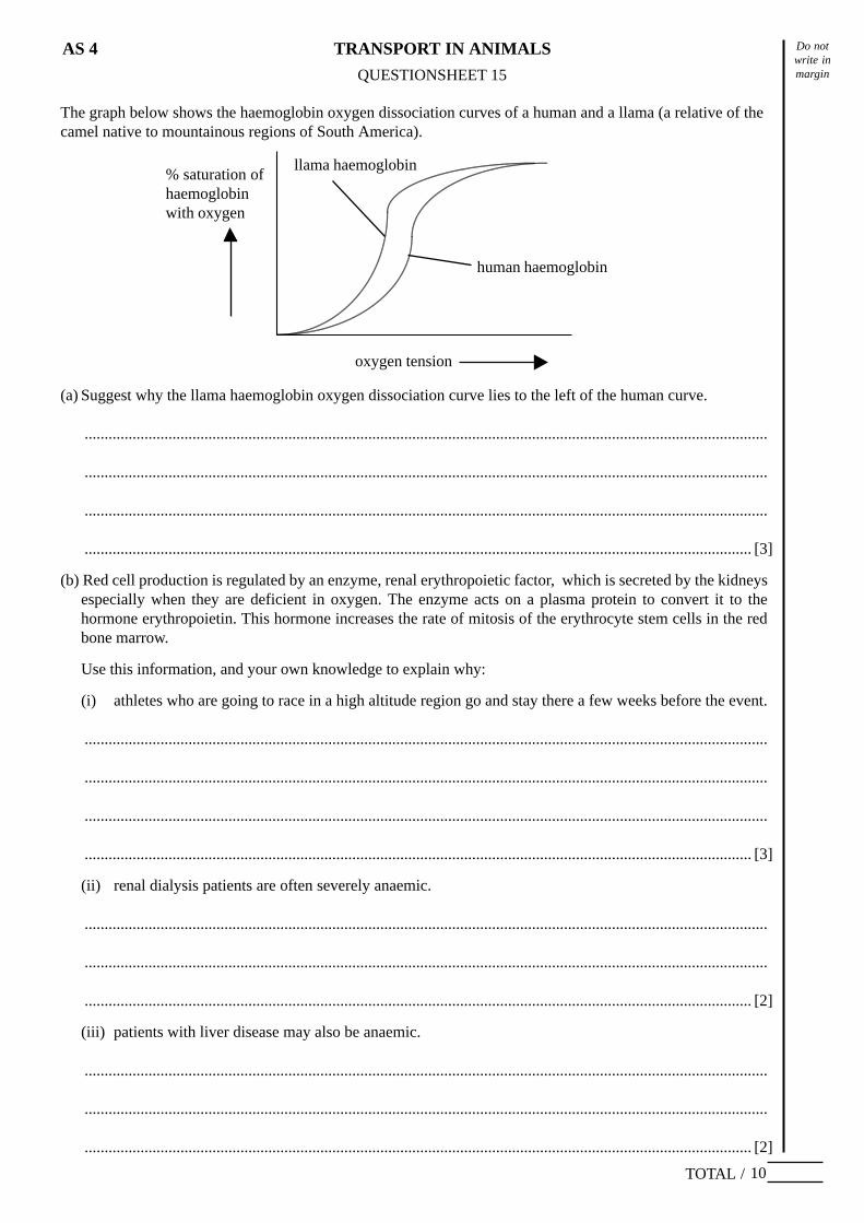
Do notwrite inmarginQUESTIONSHEET 15
TOTAL /
TRANSPORT IN ANIMALSAS 4
10
The graph below shows the haemoglobin oxygen dissociation curves of a human and a llama (a relative of thecamel native to mountainous regions of South America).
QUESTIONSHEET 15
% saturation ofhaemoglobinwith oxygen
oxygen tension
human haemoglobin
llama haemoglobin
(a) Suggest why the llama haemoglobin oxygen dissociation curve lies to the left of the human curve.
...........................................................................................................................................................................
...........................................................................................................................................................................
...........................................................................................................................................................................
....................................................................................................................................................................... [3]
(b) Red cell production is regulated by an enzyme, renal erythropoietic factor, which is secreted by the kidneysespecially when they are deficient in oxygen. The enzyme acts on a plasma protein to convert it to thehormone erythropoietin. This hormone increases the rate of mitosis of the erythrocyte stem cells in the redbone marrow.
Use this information, and your own knowledge to explain why:
(i) athletes who are going to race in a high altitude region go and stay there a few weeks before the event.
...........................................................................................................................................................................
...........................................................................................................................................................................
...........................................................................................................................................................................
....................................................................................................................................................................... [3]
(ii) renal dialysis patients are often severely anaemic.
...........................................................................................................................................................................
...........................................................................................................................................................................
....................................................................................................................................................................... [2]
(iii) patients with liver disease may also be anaemic.
...........................................................................................................................................................................
...........................................................................................................................................................................
....................................................................................................................................................................... [2]