Embed Size (px)
Citation preview

Arujuna, A. V., Housden, R. J., Ma, Y., Rajani, R., Gao, G., Nijhof,N., Cathier, P., Bullens, R., Gijsbers, G., Parish, V., Kapetanakis,S., Hancock, J., Rinaldi, C. A., Cooklin, M., Gill, J., Thomas, M.,O’Neill, M. D., Razavi, R. and Rhode, K. S. (2014) Novel systemfor real-time integration of 3-D echocardiography and fluoroscopyfor image-guided cardiac interventions: Preclinical validation andclinical feasibility evaluation. IEEE Journal of Translational Engi-
neering in Health and Medicine, 2. ISSN 2168-2372 Available from:http://eprints.uwe.ac.uk/25194
We recommend you cite the published version.The publisher’s URL is:http://dx.doi.org/10.1109/JTEHM.2014.2303799
Refereed: No
(c) 2014 IEEE. Personal use of this material is permitted. Permission fromIEEE must be obtained for all other users, including reprinting/ republishingthis material for advertising or promotional purposes, creating new collectiveworks for resale or redistribution to servers or lists, or reuse of any copyrightedcomponents of this work in other works.
Disclaimer
UWE has obtained warranties from all depositors as to their title in the materialdeposited and as to their right to deposit such material.
UWE makes no representation or warranties of commercial utility, title, or fit-ness for a particular purpose or any other warranty, express or implied in respectof any material deposited.
UWE makes no representation that the use of the materials will not infringeany patent, copyright, trademark or other property or proprietary rights.
UWE accepts no liability for any infringement of intellectual property rightsin any material deposited but will remove such material from public view pend-ing investigation in the event of an allegation of any such infringement.
PLEASE SCROLL DOWN FOR TEXT.

2
CARDIOVASCULAR DEVICES AND SYSTEMS
Received 9 October 2013; revised 20 November 2013; accepted 19 December 2013. Date of publication 30 January 2014;date of current version 14 February 2014.
Digital Object Identifier 10.1109/JTEHM.2014.2303799
Novel System for Real-Time Integration of 3-DEchocardiography and Fluoroscopy for
Image-Guided Cardiac Interventions: PreclinicalValidation and Clinical Feasibility Evaluation
ARUNA V. ARUJUNA1, R. JAMES HOUSDEN2, YINGLIANG MA2, RONAK RAJANI3, GANG GAO2,NIELS NIJHOF4, PASCAL CATHIER4, ROLAND BULLENS4, GEERT GIJSBERS4, VICTORIA PARISH3,
STAMATIS KAPETANAKIS3, JANE HANCOCK3, C. ALDO RINALDI1, MICHAEL COOKLIN3,JASWINDER GILL1, MARTYN THOMAS3, MARK D. O’NEILL1,
REZA RAZAVI1, AND KAWAL S. RHODE21Division of Imaging Sciences and Biomedical Engineering, King’s College London, London SE1 7EH, U.K., and the Department of Cardiology, Guy’s and St.
Thomas’ NHS Foundation Trust, London SE1 7EH, U.K.2Division of Imaging Sciences and Biomedical Engineering, King’s College London, London SE1 7EH, U.K.
3Department of Cardiology, Guy’s and St. Thomas’ NHS Foundation Trust, London SE1 7EH, U.K.4Philips Healthcare, Interventional X-Ray, Best 5680 DA, The Netherlands.
CORRESPONDING AUTHOR: R. J. HOUSDEN ([email protected])
This work was supported by a research grant from Philips Healthcare, Interventional X-Ray, Best, The Netherlands, and the NationalInstitute for Health Research Biomedical Research Centre at Guy’s and St. Thomas’ NHS Foundation Trust and King’s College London.
The work of A. Arujuna was supported by St. Jude Medical.
ABSTRACT Real-time imaging is required to guideminimally invasive catheter-based cardiac interventions.While transesophageal echocardiography allows for high-quality visualization of cardiac anatomy, X-rayfluoroscopy provides excellent visualization of devices. We have developed a novel image fusion system thatallows real-time integration of 3-D echocardiography and the X-ray fluoroscopy. The system was validatedin the following two stages: 1) preclinical to determine function and validate accuracy; and 2) in the clinicalsetting to assess clinical workflow feasibility and determine overall system accuracy. In the preclinical phase,the system was assessed using both phantom and porcine experimental studies. Median 2-D projection errorsof 4.5 and 3.3 mm were found for the phantom and porcine studies, respectively. The clinical phase focusedon extending the use of the system to interventions in patients undergoing either atrial fibrillation catheterablation (CA) or transcatheter aortic valve implantation (TAVI). Eleven patients were studied with nine inthe CA group and two in the TAVI group. Successful real-time view synchronization was achieved in allcases with a calculated median distance error of 2.2 mm in the CA group and 3.4 mm in the TAVI group.A standard clinical workflow was established using the image fusion system. These pilot data confirm thetechnical feasibility of accurate real-time echo-fluoroscopic image overlay in clinical practice, which may bea useful adjunct for real-time guidance during interventional cardiac procedures.
INDEX TERMS Cardiac interventional guidance, image fusion, X-ray fluoroscopy, 3D ultrasound, clinicalevaluation.
I. INTRODUCTIONCardiac anatomical heterogeneity amongst patients (e.g. leftatrial (LA) pulmonary vein (PV) anatomy, atrial septal defect(ASD) variations) can pose a challenge when performingminimally-invasive procedures [1]. The accuracy of navi-
gation during cardiac procedures is limited in part by thispatient-to-patient variability and in part by the operator’sability to manipulate the catheter/guide wire manually tothe intended intracardiac location. Precision, safety and asuccessful procedure hinge around better appreciation of
VOLUME 2, 20142168-2372 2014 IEEE. Translations and content mining are permitted for academic research only.
Personal use is also permitted, but republication/redistribution requires IEEE permission.See http://www.ieee.org/publications_standards/publications/rights/index.html for more information.
1900110
ARUJUNA et al.: Novel System for Real-Time Integration of 3-D Echocardiography and Fluoroscopy
structural anatomy and the relationship between catheters,guide wires and anatomical structures.
Most interventional devices are designed to be X-ray vis-ible and can be seen throughout the part of their length thatlies in the X-ray field of view (FOV). Two-dimensional (2D)X-ray imaging is the dominant imaging modality for guidingcardiac interventions. Typically, imaging can be performed athigh frame rates (up to 30 frames per second) and thereforethe cardiac motion and the motion of interventional devicesdo not cause significant motion artefacts in the acquiredimages.
In addition to X-ray, pre-procedural imaging of patientsundergoing cardiac interventions is becoming routine andplays an important part in patient management. Imagingmodalities such as computerised tomography (CT), magneticresonance imaging (MRI) and three-dimensional (3D) rota-tional X-ray angiography (RXA) or C-armCT have been inte-grated into the cardiac catheterisation laboratory in a varietyof ways [2]–[5], with successful clinical cases reported inthe literature. This is especially common for cardiac electro-physiology procedures where pre-procedural images can becombined with electroanatomical mapping systems such asEnsite (St. Jude Medical) and CARTO (Biosense Webster).These approaches have been shown to reduce procedure timeand radiation dose, and to improve outcome for some types ofprocedures [6], [7]. However, pre-procedural imaging repre-sents a static roadmap image of the heart and cannot monitorthe cardiac intervention in real-time. This can be importantsince tissue deformations do occur due to the presence ofinterventional devices and delivered treatments [8]. Further-more, elaborate techniques are required to compensate thestatic roadmaps for the contractile and respiratory motion ofthe heart [9].
In contrast to the relatively slow data acquisition of MRIand CT, 3D echocardiography is a real-time imagingmodalitywhich is increasingly performed during cardiac interven-tions. This technique visualises not only the relevant cardiacstructures but also the interventional devices and deliverysystems. Furthermore, 3D transesophageal echocardiogra-phy (TEE) confers improved spatial and temporal resolutionof the cardiac anatomy. 3D TEE is now routinely usedin a variety of cardiac interventional procedures includ-ing structural heart disease: with device closures, valvularheart disease: with mitral annulus resizing, mitral annu-lus repair, transcatheter aortic valve implantations (TAVI);and in electrophysiology with trans-septal puncture guid-ance and percutaneous left atrial appendage device closures[10]–[12]. More recently, intracardiac echocardiography(ICE) has been used to enable real time visualisation ofintracardiac chambers and adjacent structures and facilitateprecise guidance of catheter based procedures [13]. Howeverthis imaging modality confers a smaller FOV in comparisonto TEE, requires an additional femoral puncture and has costimplications with each ICE catheter being non-reuseable inEurope and being reusable only a few times in the UnitedStates.
Real-time 3D TEE [14] is presently used in combinationwith X-ray fluoroscopy and is particularly useful in defininglocation and morphology of the target cardiac structure andanatomical relation to neighbouring landmarks. Whilst 3DTEE allows for high quality visualisation of cardiac anatomy,there is limited visualisation of interventional devices dueto echo artefacts. X-ray has the potential to overcome thelimitations of 3D TEE by assisting in the optimum FOVselection for the 3D ultrasound and allowing a clearer defini-tion of the interventional device. Procedural guidance is thusdependent on visual information obtained from two separatescreens showing the 3D TEE and X-ray fluoroscopy. Thisapproach lacks the precise anatomical relationship betweenthe device and the cardiac structure of interest. In addition,continued X-ray fluoroscopy during device positioning anddeployment exposes patients, especially paediatric patientswith congenital defects, to ionising radiation which carriesa significant risk [15], [16]. A possible solution to this isintegration of the real-time 3D TEE and X-ray fluoroscopyto improve device to tissue spatial orientation.3D ultrasound and X-ray registration is a relatively new
research topic. Previous studies have used modified hardwareto track the probe. One approach focused on the registration oftransthoracic echocardiography (TTE) and X-ray [17] usinga mechanical tracking device. Based on the same principle,Jain et al. [18] reported a method to register TEE volumeswith X-ray by using an electromagnetic (EM) tracking sys-tem. Alternatively, a fiducial-based approach can be usedto track small, X-ray visible markers attached to the TEEdevice [19].We have previously developed an accurate and robust
image-based algorithm for registration of 3D TEE andX-ray fluoroscopy [20]. This image-based approach requiresnomodification of the existing hardware and so is more suitedto clinical translation. In the current paper, we develop thisfurther into a real-time clinical system to carry out the imagefusion. To our knowledge, this is the first real-time clinicalplatform developed using this concept. While we have pre-viously demonstrated successful registration of static clinicalimages in [20], we now perform a more extensive validationusing the real-time implementation with sequences of imagesover time. We perform pre-clinical experiments to validatethe platform and clinical feasibility experiments to establisha workflow for the system and to measure clinical accuracy.
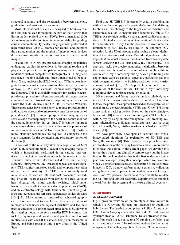
II. METHODSA. SYSTEM OVERVIEWFig. 1 gives an overview of the prototype clinical system inwhich live X-ray and 3D echo are integrated to obtain thehybrid view. The hardware comprises a Philips Allura XperFD10 C-arm X-ray system and a Philips iE33 3D ultrasoundsystemwith an X7-2t 3D TEE probe. Data is streamed in real-time from each image source to a PC running the fusion andvisualisation software. The software displays the live X-rayimages overlaid with a projection of the live 3D echo volume.
1900110 VOLUME 2, 2014
ARUJUNA et al.: Novel System for Real-Time Integration of 3-D Echocardiography and Fluoroscopy
FIGURE 1. Both the X-ray fluoroscopic image and echocardiographyimage are acquired separately and streamed into the visualisationplatform that allows for the real-time integration of the two modalities.
The two modalities are registered using our previouslydescribed method [20]. In brief, this uses a previouslyacquired nano-CT image of the TEE probe which is reg-istered to the projection image of the probe in the X-ray.The registration algorithm involves simulating the X-ray
projection process to generate projections of the 3D probeimage, called digitally reconstructed radiographs (DRRs).Using these images, the algorithm iteratively adjusts the posi-tion and orientation of the 3D probe image to obtain thebest match between its DRR and the X-ray projection. Fig. 2shows a typical X-ray view, the probemodel and a registrationof the two. The reader is referred to [20] for further details ofthe algorithm.In the clinical prototype, this iterative process is initialised
manually to approximately the correct position and ori-entation before running the automatic 3D-2D registration.Following the initial alignment, the automatic registrationis repeatedly rerun on the updating X-ray images to trackchanges in the probe position due to cardiac and respira-tory motion. This registration is GPU-accelerated (GraphicalProcessing Unit, NVIDIA GeForce 8800GTX) and updatesin this way at a rate of 1–2 Hz, sufficient for near-real-time tracking of the probe position. This is a significantimprovement on our previous implementation [20] in whichan equivalent setup required an average of 8 s per registration.
B. STUDY OVERVIEWThe protocol was divided into 2 phases. The pre-clinicalphase involved use of the system for data acquisition froma phantom model and during a porcine experimental study.The second, clinical phase involved use of the system fordata acquisition during catheter ablation (CA) in 9 patientsand TAVI in 2 patients following approval from the LocalResearch Ethics Committee.
C. PRE-CLINICAL STUDY (STAGE 1)1) PHANTOM MODELIn Stage 1, the prototype image-based TEE probe localisationalgorithm was validated using a phantom model. The phan-
FIGURE 2. Automatic overlay registration. (a) TEE probe model from a nano-CT scan of the probe head. (b) X-ray image with the projection of the TEE probeclearly visible. This example is from an AF ablation case. (c) Probe model registered to the X-ray by automatic 3D-2D registration.
VOLUME 2, 2014 1900110
ARUJUNA et al.: Novel System for Real-Time Integration of 3-D Echocardiography and Fluoroscopy
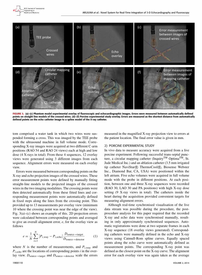
FIGURE 3. (a)–(c) Phantom model experimental overlay of fluoroscopic and echocardiographic images. Errors were measured between automatically definedpoints on straight line models of the crossed wires. (d)–(f) Porcine experimental study overlay. Errors are measured as the shortest distance from automaticallydefined points on the echo catheter image to a spline model of the X-ray catheter.
tom comprised a water tank in which two wires were sus-pended forming a cross. This was imaged by the TEE probewith the ultrasound machine in full volume mode. Corre-sponding X-ray images were acquired at two different C-armpositions (RAO 54 and RAO 24 views) each at high and lowdose (4 X-rays in total). From these 4 sequences, 12 overlayviews were generated using 3 different images from eachsequence. Alignment errors were measured on each overlayview.
Errors were measured between corresponding points on theX-ray and echo projection images of the crossed wires. Theseerror measurement points were defined by manually fittingstraight-line models to the projected images of the crossedwires in the two imagingmodalities. The crossing points werethen detected automatically from these fitted lines and cor-responding measurement points were automatically definedin fixed steps along the lines from the crossing point. Thisprovided up to 13 measurements per overlay view (minimum10 where the crossing point was near the edge of the view).Fig. 3(a)–(c) shows an example of this. 2D projection errorswere calculated between corresponding points and averagedto give an overall alignment error, e, for the overlay view asfollows
e =1N
N∑i=1
∣∣Pi,xray − Pi,echo∣∣ Dsource→target
Dsource→detector(1)
where N is the number of measurements, and Pi,xray andPi,echo are the locations of corresponding points i in the over-lay view. Dsource→target and Dsource→detector scale the errors
measured in the magnified X-ray projection view to errors atthe patient location. The final error value is given in mm.
2) PORCINE EXPERIMENTAL STUDYIn vivo data to measure accuracy were acquired from a liveporcine experiment. Following successful trans-septal punc-ture, a circular mapping catheter (InquiryTM OptimaTM, St.Jude Medical Inc.) and an ablation catheter (3.5 mm irrigatedtip catheter NaviStar R© ThermoCool R©, Biosense WebsterInc., Diamond Bar, CA, USA) were positioned within theleft atrium. Five echo volumes were acquired in full volumemode with the probe in different positions. At each posi-tion, between one and three X-ray sequences were recorded(RAO 30, LAO 30 and PA positions) with high X-ray dosesetting (9 X-ray views in total). The catheters inside theheart during the acquisition provided convenient targets formeasuring alignment errors.Although real-time synchronised visualisation of the live
data stream was possible during the procedure, the post-procedure analysis for this paper required that the recordedX-ray and echo data were synchronised manually, result-ing in only approximately synchronised sequences. Auto-matic registrations were done at two separate frames in eachX-ray sequence (18 overlay views generated). Correspond-ing catheters were manually defined in the echo and X-rayviews using Catmull-Rom spline curves. Equally spacedpoints along the echo curve were automatically defined asmeasurement points. The corresponding X-ray point wasdefined as the closest point on the X-ray curve. The alignmenterror for each overlay view was again taken as the average
1900110 VOLUME 2, 2014
ARUJUNA et al.: Novel System for Real-Time Integration of 3-D Echocardiography and Fluoroscopy
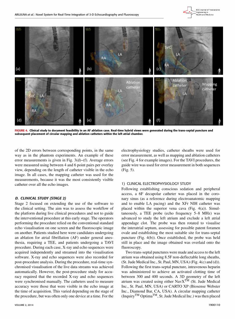
FIGURE 4. Clinical study to document feasibility in an AF ablation case. Real-time hybrid views were generated during the trans-septal puncture andsubsequent placement of circular mapping and ablation catheters within the left atrial chamber.
of the 2D errors between corresponding points, in the sameway as in the phantom experiments. An example of theseerror measurements is given in Fig. 3(d)–(f). Average errorswere measured using between 4 and 6 point pairs per overlayview, depending on the length of catheter visible in the echoimage. In all cases, the mapping catheter was used for themeasurements, because it was the most consistently visiblecatheter over all the echo images.
D. CLINICAL STUDY (STAGE 2)Stage 2 focused on extending the use of the software tothe clinical setting. The aim was to assess the workflow ofthe platform during live clinical procedures and not to guidethe interventional procedure at this early stage. The operatorsperforming the procedure relied on the conventional standardecho visualisation on one screen and the fluoroscopic imageon another. Patients studied here were candidates undergoingan ablation for atrial fibrillation (AF) under general anes-thesia, requiring a TEE, and patients undergoing a TAVIprocedure. During each case, X-ray and echo sequences wereacquired independently and streamed into the visualisationsoftware. X-ray and echo sequences were also recorded forpost-procedure analysis. During the procedure, real-time syn-chronised visualisation of the live data streams was achievedautomatically. However, the post-procedure study for accu-racy required that the recorded X-ray and echo sequenceswere synchronised manually. The catheters used to measureaccuracy were those that were visible in the echo image atthe time of acquisition. This varied depending on the stage ofthe procedure, but was often only one device at a time. For the
electrophysiology studies, catheter sheaths were used forerror measurement, as well as mapping and ablation catheters(see Fig. 4 for example images). For the TAVI procedures, theguide wire was used for error measurement in both sequences(Fig. 5).
1) CLINICAL ELECTROPHYSIOLOGY STUDYFollowing establishing conscious sedation and peripheralaccess, a 6F decapolar catheter was placed in the coro-nary sinus (as a reference during electroanatomic mappingand to enable LA pacing) and the XFr NIH catheter wasplaced within the superior vena cava (Fig. 4(a)). Simul-taneously, a TEE probe (echo frequency 5–8 MHz) wasadvanced to study the left atrium and exclude a left atrialappendage clot. The probe was then rotated to visualisethe interatrial septum, assessing for possible patent foramenovale and establishing the most suitable site for trans-septalpuncture (Fig. 4(b)). Once established, the probe was heldstill in place and the image obtained was overlaid onto thefluoroscopy.Two trans-septal punctures weremade and access to the left
atrium was obtained using 8.5F non-deflectable long sheaths,(St. JudeMedical Inc., St. Paul,MN, USA) (Fig. 4(c) and (d)).Following the first trans-septal puncture, intravenous heparinwas administered to achieve an activated clotting time ofbetween 300 and 400 seconds. A 3D geometry of the leftatrium was created using either NavXTM (St. Jude MedicalInc., St. Paul, MN, USA) or CARTO XP (Biosense WebsterInc., Diamond Bar, CA, USA). A circular mapping catheter(InquiryTM OptimaTM, St. JudeMedical Inc.) was then placed
VOLUME 2, 2014 1900110
ARUJUNA et al.: Novel System for Real-Time Integration of 3-D Echocardiography and Fluoroscopy
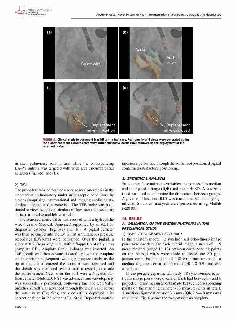
FIGURE 5. Clinical study to document feasibility in a TAVI case. Real-time hybrid views were generated duringthe placement of the Edwards core valve within the native aortic valve followed by the deployment of theprosthetic valve.
in each pulmonary vein in turn while the correspondingLA-PV antrum was targeted with wide area circumferentialablation (Fig. 4(e) and (f)).
2) TAVIThe procedure was performed under general anesthesia in thecatheterisation laboratory under strict aseptic conditions, bya team comprising interventional and imaging cardiologists,cardiac surgeons and anesthetists. The TEE probe was posi-tioned to view the left ventricular outflow tract and ascendingaorta, aortic valve and left ventricle.
The stenosed aortic valve was crossed with a hydrophilicwire (Terumo Medical, Somerset) supported by an AL1 5Fdiagnostic catheter (Fig. 5(a) and (b)). A pigtail catheterwas then advanced into the LV whilst simultaneous pressurerecordings (LV/aorta) were performed. Over the pigtail, asuper stiff 260-cm long wire, with a floppy tip of only 1 cm(Amplatz ST1, Amplatz Cook, Indiana) was inserted. An18F sheath was then advanced carefully over the Amplatzcatheter with a subsequent two-stage process: firstly, as thetip of the dilator entered the aorta, it was stabilised andthe sheath was advanced over it until it rested just insidethe aortic lumen. Next, over the stiff wire a Nucleus bal-loon catheter (NuMED, NY) was advanced and valvuloplastywas successfully performed. Following this, the CoreValveprosthesis itself was advanced through the sheath and acrossthe aortic valve (Fig. 5(c)) and successfully deployed in itscorrect position in the patient (Fig. 5(d)). Repeated contrast
injections performed through the aortic root positioned pigtailconfirmed satisfactory positioning.
E. STATISTICAL ANALYSISSummaries for continuous variables are expressed as medianand interquartile range (IQR) and mean ± SD. A student’st-test was used to determine the differences between groups.A p value of less than 0.05 was considered statistically sig-nificant. Statistical analyses were performed using Matlab(R2010b).
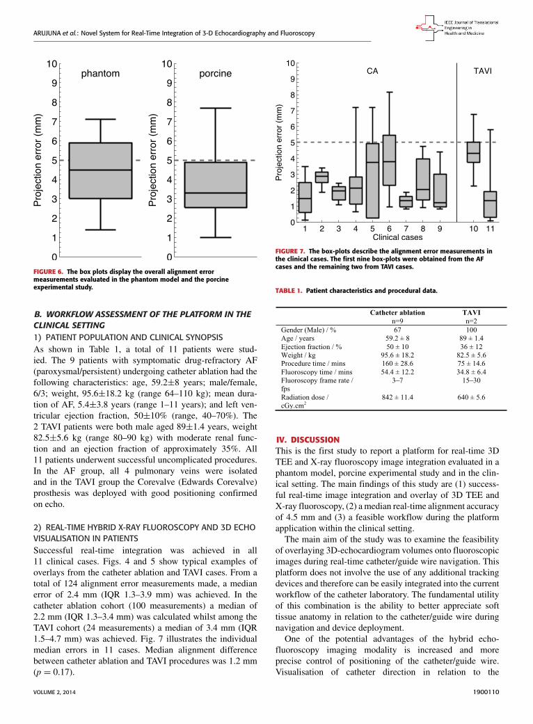
III. RESULTA. VALIDATION OF THE SYSTEM PLATFORM IN THEPRECLINICAL STUDY1) OVERLAY ALIGNMENT ACCURACYIn the phantom model, 12 synchronised echo-fluoro imagepairs were overlaid. On each hybrid image, a mean of 11.5measurements (range 10–13) between corresponding pointson the crossed wires were made to assess the 2D pro-jection error. From a total of 138 error measurements, amedian alignment error of 4.5 mm (IQR 3.0–5.9 mm) wascalculated.In the porcine experimental study, 18 synchronised echo-
fluoro image pairs were overlaid. Each had between 4 and 6projection error measurements made between correspondingpoints on the mapping catheter (83 measurements in total).A median alignment error of 3.3 mm (IQR 2.6–4.9 mm) wascalculated. Fig. 6 shows the two datasets as boxplots.
1900110 VOLUME 2, 2014
ARUJUNA et al.: Novel System for Real-Time Integration of 3-D Echocardiography and Fluoroscopy
FIGURE 6. The box plots display the overall alignment errormeasurements evaluated in the phantom model and the porcineexperimental study.
B. WORKFLOW ASSESSMENT OF THE PLATFORM IN THECLINICAL SETTING1) PATIENT POPULATION AND CLINICAL SYNOPSISAs shown in Table 1, a total of 11 patients were stud-ied. The 9 patients with symptomatic drug-refractory AF(paroxysmal/persistent) undergoing catheter ablation had thefollowing characteristics: age, 59.2±8 years; male/female,6/3; weight, 95.6±18.2 kg (range 64–110 kg); mean dura-tion of AF, 5.4±3.8 years (range 1–11 years); and left ven-tricular ejection fraction, 50±10% (range, 40–70%). The2 TAVI patients were both male aged 89±1.4 years, weight82.5±5.6 kg (range 80–90 kg) with moderate renal func-tion and an ejection fraction of approximately 35%. All11 patients underwent successful uncomplicated procedures.In the AF group, all 4 pulmonary veins were isolatedand in the TAVI group the Corevalve (Edwards Corevalve)prosthesis was deployed with good positioning confirmedon echo.
2) REAL-TIME HYBRID X-RAY FLUOROSCOPY AND 3D ECHOVISUALISATION IN PATIENTSSuccessful real-time integration was achieved in all11 clinical cases. Figs. 4 and 5 show typical examples ofoverlays from the catheter ablation and TAVI cases. From atotal of 124 alignment error measurements made, a medianerror of 2.4 mm (IQR 1.3–3.9 mm) was achieved. In thecatheter ablation cohort (100 measurements) a median of2.2 mm (IQR 1.3–3.4 mm) was calculated whilst among theTAVI cohort (24 measurements) a median of 3.4 mm (IQR1.5–4.7 mm) was achieved. Fig. 7 illustrates the individualmedian errors in 11 cases. Median alignment differencebetween catheter ablation and TAVI procedures was 1.2 mm(p = 0.17).
FIGURE 7. The box-plots describe the alignment error measurements inthe clinical cases. The first nine box-plots were obtained from the AFcases and the remaining two from TAVI cases.
TABLE 1. Patient characteristics and procedural data.
IV. DISCUSSIONThis is the first study to report a platform for real-time 3DTEE and X-ray fluoroscopy image integration evaluated in aphantom model, porcine experimental study and in the clin-ical setting. The main findings of this study are (1) success-ful real-time image integration and overlay of 3D TEE andX-ray fluoroscopy, (2) a median real-time alignment accuracyof 4.5 mm and (3) a feasible workflow during the platformapplication within the clinical setting.The main aim of the study was to examine the feasibility
of overlaying 3D-echocardiogram volumes onto fluoroscopicimages during real-time catheter/guide wire navigation. Thisplatform does not involve the use of any additional trackingdevices and therefore can be easily integrated into the currentworkflow of the catheter laboratory. The fundamental utilityof this combination is the ability to better appreciate softtissue anatomy in relation to the catheter/guide wire duringnavigation and device deployment.One of the potential advantages of the hybrid echo-
fluoroscopy imaging modality is increased and moreprecise control of positioning of the catheter/guide wire.Visualisation of catheter direction in relation to the
VOLUME 2, 2014 1900110
ARUJUNA et al.: Novel System for Real-Time Integration of 3-D Echocardiography and Fluoroscopy
anatomical structures facilitates faster and more accuratecatheter movement to the intended anatomical site. Figs. 4and 5 show typical examples of overlays from clinical cases.An echo volume on its own can be difficult to interpretbecause of its limited FOV and lack of context for theecho coordinate system relative to the patient. Catheters anddevices tend to produce artefacts in the ultrasound data reduc-ing the clarity of the images. In the overlay view, the echovolume is displayed in a coordinate system that can be moreeasily related to the patient. Also, the highly visible cathetersin the X-ray image help with identifying the catheters in theecho and so can be related to the cardiac anatomy via the echoimage. During an ablation procedure, the precise location ofthe catheter tip and electrodes on the mapping catheter inrelation to atrial tissue permits better targeted lesion deliverythan just visualising the catheter as a whole on fluoroscopy.Similarly during a TAVI procedure, being able to visualiseboth the native valve and the prosthesis on a single image willfacilitate delivery and deployment of the prosthetic valve.
The accuracy requirement for a clinically useful imageguidance system depends on many factors including thepatient and the type of procedure being performed [21], [22].An accuracy of less than 5 mm is often deemed as a suitabletarget for many applications and this threshold is highlightedon the plots in Figs. 6 and 7wheremost of the errors are belowthe 5 mm threshold. Based on these results, the system issufficiently accurate to guide catheter/guide wire navigation.It should be noted that the location of the catheter in the echovolume will affect the accuracy, as objects further from theprobe will have a greater misalignment due to errors in theprobe’s orientation.
The standard deviations noted within the figures are, inpart, likely to arise from some data streaming delay in thevisualisation software and, in part, time lag in X-ray regis-tration. The largest standard deviations are due to respira-tory motion acting on the probe, which is not immediatelycompensated by the 1–2 Hz registration updates. While allsequences encounter respiratory motion, variations in theprobe position and X-ray direction produce different scalesof potential error in each sequence. The median alignmenterror difference between the TAVI and ablation procedures(3.4 mm vs 2.2 mm) may be attributed to the rapid movementof the guide wire/device that is located near the aortic valvein contrast to the lasso and ablating catheter located withinthe left atrium. In comparison to the phantom data, both theporcine and clinical study had lower alignment errors. Thismay be due to the error measurement techniques: full 2Derrors were measured in the phantom experiment whereas inthe porcine and clinical experiments it was only possible tomeasure to the nearest point on the corresponding catheter.
Other studies on echo-fluoroscopy overlay have also foundclinically-useful accuracies. In [18], the mean error wasapproximately 2.0 mm when imaging a heart phantom andtracking the TEE probe with an electromagnetic trackingsystem. More recently, Lang et al. [19] found a mean error ofapproximately 1.2 mm in vivo, by tracking fiducial markers
attached to the TEE probe. These are both better than our sys-tem’s average accuracy, but require devices to be attached tothe probe. The results therefore highlight a trade-off betweenaccuracy and ease of clinical integration.In this study, we have presented a platform for real-time
hybrid X-ray fluoroscopy and 3D echo visualisation and havesuccessfully demonstrated a feasible workflow within theclinical setting. We anticipate that by gaining familiarity andconfidence within this hybrid viewing system, there will bea lesser need for repeated X-ray fluoroscopy use to estab-lish catheter position. On-going research is actively seekingto reduce the use of, or even replace, X-ray fluoroscopyin cardiac interventional procedures, especially for paedi-atrics [23]–[25]. With increased usage over time, we pre-dict a greater reliance on echo views to assist catheter/guidewire navigation. In the long run, this will reduce overallfluoroscopy time and patient exposure to ionising radiation.Ultimately, randomised comparisons of real-time hybrid
X-ray fluoroscopy and 3D echo image-guided therapy withstandard visualisation approaches are necessary to defini-tively establish its clinical utility and translate it from anexperimental method to a clinical routine.
A. PRESENT LIMITATIONSIt is important to recognise that one of the main objectives ofthis study was to assess the clinical feasibility of the proposedmethod. Although all the porcine experimental and clinicaldata were acquired specifically for this study, the data analysiswas performed off-line. The porcine experiment in particularmay suffer from artificially large errors due to the echo andfluoroscopy images not being acquired simultaneously. Ourfuture work will focus on evaluating the accuracy of the livesystem and further improving the co-registration.In addition, our approach of measuring to the closest point
accounts for 2D in plane errors and does not necessarilycapture the complete error as there can also be misalignmenttangentially to the catheters. However, many of the catheterswere curved, which reduces the severity of this limitation.The clinical case numbers in this study at the present time
are small. While we have established the clinical feasibility ofthis system, further studies involving larger patient groups arerequired to demonstrate that this novel technique translatesinto reduced fluoroscopy time, reduced complications andbetter overall patient outcome.
V. CONCLUSIONIn this study, we have developed a clinically-integrated sys-tem for real-time hybrid X-ray fluoroscopy and 3D echo visu-alisation. The practicability of accurate real-time hybrid visu-alisation was demonstrated in both a phantom model and aporcine experimental study. The clinical series demonstrateda feasible clinical workflow, with successful image fusion inall cases. Further clinical validation and a randomised com-parison between real-time hybrid echo-fluoro image-guidedtherapy versus a standard visualisation approach is neededto translate this tool into routine clinical practice, where it
1900110 VOLUME 2, 2014
ARUJUNA et al.: Novel System for Real-Time Integration of 3-D Echocardiography and Fluoroscopy
may prove useful for real-time guidance of catheterisationinterventions.
REFERENCES[1] V. Y. Reddy, P. Neuzil, Z. J. Malchano, R. Vijaykumar, R. Cury,
S. Abbara, et al., ‘‘View-synchronized robotic image-guided therapy foratrial fibrillation ablation: Experimental validation and clinical feasibility,’’Circulation, vol. 115, no. 21, pp. 2705–2714, May 2007.
[2] K. S. Rhode, D. L. G. Hill, P. J. Edwards, J. Hipwell, D. Rueckert,G. Sanchez-Ortiz, et al., ‘‘Registration and tracking to integrate X-ray andMR images in an XMR facility,’’ IEEE Trans. Med. Imag., vol. 22, no. 11,pp. 1369–1378, Nov. 2003.
[3] H. Yu, R. Fahrig, and N. J. Pelc, ‘‘Co-registration of X-ray and MR fieldsof view in a hybrid XMR system,’’ J. Magn. Reson. Imag., vol. 22, no. 2,pp. 291–301, Aug. 2005.
[4] S. De Buck, F. Maes, J. Ector, J. Bogaert, S. Dymarkowski,H. Heidbuchel, et al., ‘‘An augmented reality system for patient-specific guidance of cardiac catheter ablation procedures,’’ IEEE Trans.Med. Imag., vol. 24, no. 11, pp. 1512–1524, Nov. 2005.
[5] J. Sra, G. Narayan, D. Krum, A. Malloy, R. Cooley, A. Bhatia, et al.,‘‘Computed tomography-fluoroscopy image integration-guided catheterablation of atrial fibrillation,’’ J. Cardiovascular Electrophysiol., vol. 18,no. 4, pp. 409–414, Apr. 2007.
[6] E. Bertaglia, G. Brandolino, F. Zoppo, F. Zerbo, and P. Pascotto, ‘‘Inte-gration of three-dimensional left atrial magnetic resonance images intoa real-time electroanatomic mapping system: Validation of a registra-tion method,’’ Pacing Clin. Electrophysiol., vol. 31, no. 3, pp. 273–282,Mar. 2008.
[7] M. J. Earley, R. Showkathali, M. Alzetani, P. M. Kistler, D. Gupta,D. J. Abrams, et al., ‘‘Radiofrequency ablation of arrhythmias guided bynon-fluoroscopic catheter location: A prospective randomized trial,’’ Eur.Heart J., vol. 27, no. 10, pp. 1223–1229, May 2006.
[8] D. J. Hawkes, D. Barratt, J. M. Blackall, C. Chan, P. J. Edwards,K. Rhode, et al., ‘‘Tissue deformation and shape models in image-guidedinterventions: A discussion paper,’’ Med. Image Anal., vol. 9, no. 2,pp. 163–175, Apr. 2005.
[9] A. P. King, R. Boubertakh, K. S. Rhode, Y. L. Ma, P. Chinchapatnam,G. Gao, et al., ‘‘A subject-specific technique for respiratory motion cor-rection in image-guided cardiac catheterisation procedures,’’ Med. ImageAnal., vol. 13, no. 3, pp. 419–431, Jun. 2009.
[10] G. H. Baker, G. Shirali, J. M. Ringewald, T. Y. Hsia, and V. Bandisode,‘‘Usefulness of live three-dimensional transesophageal echocardiographyin a congenital heart disease center,’’ Amer. J. Cardiol., vol. 103, no. 7,pp. 1025–1028, Apr. 2009.
[11] G. B. Mackensen, D. Hegland, D. Rivera, D. B. Adams, andT. D. Bahnson, ‘‘Real-time 3-dimensional transesophagealechocardiography during left atrial radiofrequency catheter ablationfor atrial fibrillation,’’ Circulat., Cardiovascular Imag., vol. 1, no. 1,pp. 85–86, Jul. 2008.
[12] F. E. Silvestry, R. E. Kerber, M. M. Brook, J. D. Carroll, K. M. Eberman,S. A. Goldstein, et al., ‘‘Echocardiography-guided interventions,’’ J. Amer.Soc. Echocardiography, vol. 22, no. 3, pp. 213–231, Mar. 2009.
[13] Z. M. Hijazi, K. Shivkumar, and D. J. Sahn, ‘‘Intracardiac echocardiog-raphy during interventional and electrophysiological cardiac catheteriza-tion,’’ Circulation, vol. 119, no. 4, pp. 587–596, Feb. 2009.
[14] N. A. Marsan, L. F. Tops, P. Nihoyannopoulos, E. R. Holman, and J. J. Bax,‘‘Real-time three dimensional echocardiography: Current and future clini-cal applications,’’ Heart, vol. 95, no. 22, pp. 1881–1890, Nov. 2009.
[15] P. Kovoor, M. Ricciardello, L. Collins, J. B. Uther, and D. L. Ross,‘‘Risk to patients from radiation associated with radiofrequency abla-tion for supraventricular tachycardia,’’ Circulation, vol. 98, no. 15,pp. 1534–1540, Oct. 1998.
[16] B. Modan, L. Keinan, T. Blumstein, and S. Sadetzki, ‘‘Cancer followingcardiac catheterization in childhood,’’ Int. J. Epidemiol., vol. 29, no. 3,pp. 424–428, Jun. 2000.
[17] Y. Ma, G. P. Penney, D. Bos, P. Frissen, C. A. Rinaldi, R. Razavi, et al.,‘‘Hybrid echo and X-ray image guidance for cardiac catheterization proce-dures by using a robotic arm: A feasibility study,’’Phys. Med. Biol., vol. 55,no. 13, pp. N371–N382, Jul. 2010.
[18] A. Jain, L. Gutierrez, and D. Stanton, 3D TEE Registration withX-Ray Fluoroscopy for Interventional Cardiac Applications (LectureNotes in Computer Science), vol. 5528, N. Ayache, H. Delingette,and M. Sermesant, Eds. Berlin, Germany: Springer-Verlag, 2009,pp. 321–329.
[19] P. Lang, P. Seslija, M. W. A. Chu, D. Bainbridge, G. M. Guiraudon,D. L. Jones, et al., ‘‘US-fluoroscopy registration for transcatheter aor-tic valve implantation,’’ IEEE Trans. Biomed. Eng., vol. 59, no. 5,pp. 1444–1453, May 2012.
[20] G. Gao, G. Penney, Y. Ma, N. Gogin, P. Cathier, A. Arujuna, et al., ‘‘Reg-istration of 3D trans-esophageal echocardiography to X-ray fluoroscopyusing image-based probe tracking,’’ Med. Image Anal., vol. 16, no. 1,pp. 38–49, Jan. 2012.
[21] C. A. Linte, J. Moore, and T. M. Peters, ‘‘How accurate is accurateenough? A brief overview on accuracy considerations in image-guidedcardiac interventions,’’ inProc. Annu. Int. Conf. IEEE Eng.Med. Biol. Soc.,Aug./Sep. 2010, pp. 2313–2316.
[22] C. A. Linte, P. Lang,M. E. Rettmann, D. S. Cho, D. R. Holmes, R. A. Robb,et al., ‘‘Accuracy considerations in image-guided cardiac interventions:Experience and lessons learned,’’ Int. J. Comput. Assist. Radiol. Surgery,vol. 7, no. 1, pp. 13–25, Jan. 2012.
[23] R. Razavi, D. L. G. Hill, S. F. Keevil, M. E. Miquel, V. Muthurangu,S. Hegde, et al., ‘‘Cardiac catheterisation guided by MRI in childrenand adults with congenital heart disease,’’ Lancet, vol. 362, no. 9399,pp. 1877–1882, Dec. 2003.
[24] A. Tzifa, G. A. Krombach, N. Krämer, S. Krüger, A. Schütte,M. Von Walter, et al., ‘‘Magnetic resonance-guided cardiac interventionsusing magnetic resonance-compatible devices: A preclinical study andfirst-in-man congenital interventions,’’ Circulat., Cardiovascular Inter-vent., vol. 3, no. 6, pp. 585–592, Dec. 2010.
[25] A. Tzifa, T. Schaeffter, andR. Razavi, ‘‘MR imaging-guided cardiovascularinterventions in young children,’’ Magn. Reson. Imag. Clin. North Amer.,vol. 20, no. 1, pp. 117–128, Feb. 2012.
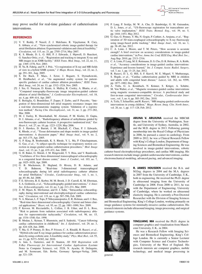
ARUNA V. ARUJUNA received the MBChBdegree from the University of Washington, Seat-tle, and University of Sydney, Australia, in 2002,and the M.D. degree in 2013. Following gainingmembership into the Royal College of Physiciansin 2006, he pursued a career in cardiology. From2009 to 2012, he was a Clinical Research Fellowwith King’s College London, Department of Imag-ing Sciences and Biomedical Engineering. He wasinvolved in image-guided interventions, robotic
catheter-based electrophysiology procedures, and cardiac MR imaging. Hisresearch interests include image-guided cardiovascular interventions, cardiacelectromechanical modeling, advanced pacing, and advanced imaging.
R. JAMES HOUSDEN received the B.A. andM.Eng. degrees in 2004 and the M.A. degreesin 2007 from the University of Cambridge, U.K.,both in engineering. He received the Ph.D. degreein ultrasound imaging from the University ofCambridge in 2008. From 2008 to 2011, he waswith the Department of Engineering, Universityof Cambridge, where he worked on ultrasoundelasticity imaging. He is currently a Post-DoctoralResearcher with the Division of Imaging Sciences
and Biomedical Engineering, King’s College London, working primarily onimage guidance systems for minimally-invasive cardiac catheterization. Hisresearch interests include ultrasound imaging, image processing, and surgicalguidance systems.
YINGLIANG MA received the Ph.D. degree incomputer graphics and visualization from Manch-ester University, U.K., in 2004.
He was a Research Fellow with Imaging Sci-ences and Biomedical Engineering, King’s Col-lege London. He is currently a Senior Lecturerwith Computer Science and Creative Technolo-gies, University of the West of England. Hisresearch interests are computer graphics, mobiletechnology, and medical image processing in
general.
VOLUME 2, 2014 1900110
ARUJUNA et al.: Novel System for Real-Time Integration of 3-D Echocardiography and Fluoroscopy
RONAK RAJANI, photograph and biography not available at the time ofpublication.
GANG GAO, photograph and biography not available at the time of publi-cation.
NIELS NIJHOF, photograph and biography not available at the time ofpublication.
PASCAL CATHIER, photograph and biography not available at the time ofpublication.
ROLAND BULLENS, photograph and biography not available at the time ofpublication.
GEERT GIJSBERS, photograph and biography not available at the time ofpublication.
VICTORIA PARISH, photograph and biography not available at the time ofpublication.
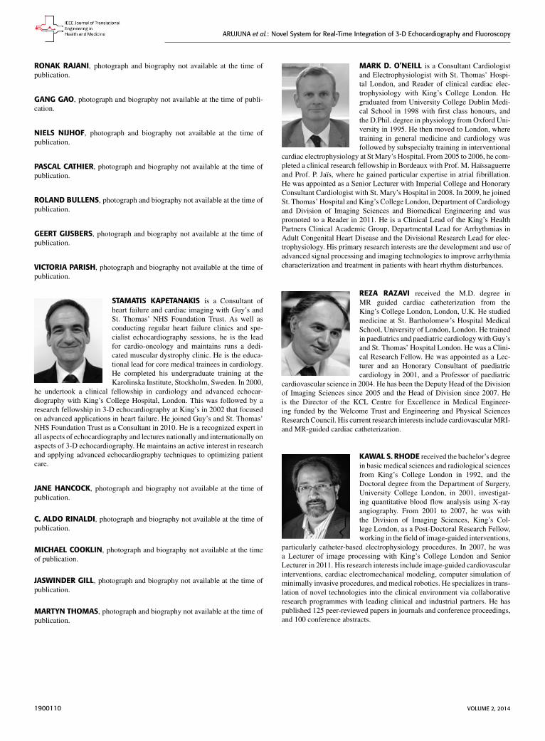
STAMATIS KAPETANAKIS is a Consultant ofheart failure and cardiac imaging with Guy’s andSt. Thomas’ NHS Foundation Trust. As well asconducting regular heart failure clinics and spe-cialist echocardiography sessions, he is the leadfor cardio-oncology and maintains runs a dedi-cated muscular dystrophy clinic. He is the educa-tional lead for core medical trainees in cardiology.He completed his undergraduate training at theKarolinska Institute, Stockholm, Sweden. In 2000,
he undertook a clinical fellowship in cardiology and advanced echocar-diography with King’s College Hospital, London. This was followed by aresearch fellowship in 3-D echocardiography at King’s in 2002 that focusedon advanced applications in heart failure. He joined Guy’s and St. Thomas’NHS Foundation Trust as a Consultant in 2010. He is a recognized expert inall aspects of echocardiography and lectures nationally and internationally onaspects of 3-D echocardiography. He maintains an active interest in researchand applying advanced echocardiography techniques to optimizing patientcare.
JANE HANCOCK, photograph and biography not available at the time ofpublication.
C. ALDO RINALDI, photograph and biography not available at the time ofpublication.
MICHAEL COOKLIN, photograph and biography not available at the timeof publication.
JASWINDER GILL, photograph and biography not available at the time ofpublication.
MARTYN THOMAS, photograph and biography not available at the time ofpublication.
MARK D. O’NEILL is a Consultant Cardiologistand Electrophysiologist with St. Thomas’ Hospi-tal London, and Reader of clinical cardiac elec-trophysiology with King’s College London. Hegraduated from University College Dublin Medi-cal School in 1998 with first class honours, andthe D.Phil. degree in physiology from Oxford Uni-versity in 1995. He then moved to London, wheretraining in general medicine and cardiology wasfollowed by subspecialty training in interventional
cardiac electrophysiology at StMary’s Hospital. From 2005 to 2006, he com-pleted a clinical research fellowship in Bordeaux with Prof. M. Haïssaguerreand Prof. P. Jaïs, where he gained particular expertise in atrial fibrillation.He was appointed as a Senior Lecturer with Imperial College and HonoraryConsultant Cardiologist with St. Mary’s Hospital in 2008. In 2009, he joinedSt. Thomas’ Hospital and King’s College London, Department of Cardiologyand Division of Imaging Sciences and Biomedical Engineering and waspromoted to a Reader in 2011. He is a Clinical Lead of the King’s HealthPartners Clinical Academic Group, Departmental Lead for Arrhythmias inAdult Congenital Heart Disease and the Divisional Research Lead for elec-trophysiology. His primary research interests are the development and use ofadvanced signal processing and imaging technologies to improve arrhythmiacharacterization and treatment in patients with heart rhythm disturbances.
REZA RAZAVI received the M.D. degree inMR guided cardiac catheterization from theKing’s College London, London, U.K. He studiedmedicine at St. Bartholomew’s Hospital MedicalSchool, University of London, London. He trainedin paediatrics and paediatric cardiology with Guy’sand St. Thomas’ Hospital London. He was a Clini-cal Research Fellow. He was appointed as a Lec-turer and an Honorary Consultant of paediatriccardiology in 2001, and a Professor of paediatric
cardiovascular science in 2004. He has been the Deputy Head of the Divisionof Imaging Sciences since 2005 and the Head of Division since 2007. Heis the Director of the KCL Centre for Excellence in Medical Engineer-ing funded by the Welcome Trust and Engineering and Physical SciencesResearch Council. His current research interests include cardiovascularMRI-and MR-guided cardiac catheterization.
KAWAL S. RHODE received the bachelor’s degreein basic medical sciences and radiological sciencesfrom King’s College London in 1992, and theDoctoral degree from the Department of Surgery,University College London, in 2001, investigat-ing quantitative blood flow analysis using X-rayangiography. From 2001 to 2007, he was withthe Division of Imaging Sciences, King’s Col-lege London, as a Post-Doctoral Research Fellow,working in the field of image-guided interventions,
particularly catheter-based electrophysiology procedures. In 2007, he wasa Lecturer of image processing with King’s College London and SeniorLecturer in 2011. His research interests include image-guided cardiovascularinterventions, cardiac electromechanical modeling, computer simulation ofminimally invasive procedures, and medical robotics. He specializes in trans-lation of novel technologies into the clinical environment via collaborativeresearch programmes with leading clinical and industrial partners. He haspublished 125 peer-reviewed papers in journals and conference proceedings,and 100 conference abstracts.
1900110 VOLUME 2, 2014


![Qcl 14-v3 [college canteen kitchen]-[banasthali university]_[rajani ranjan]](https://img.pdfslide.us/doc/110x75/55a2025b1a28ab47268b46df/qcl-14-v3-college-canteen-kitchen-banasthali-universityrajani-ranjan.jpg)