Embed Size (px)
Citation preview
In the format provided by the authors and unedited.
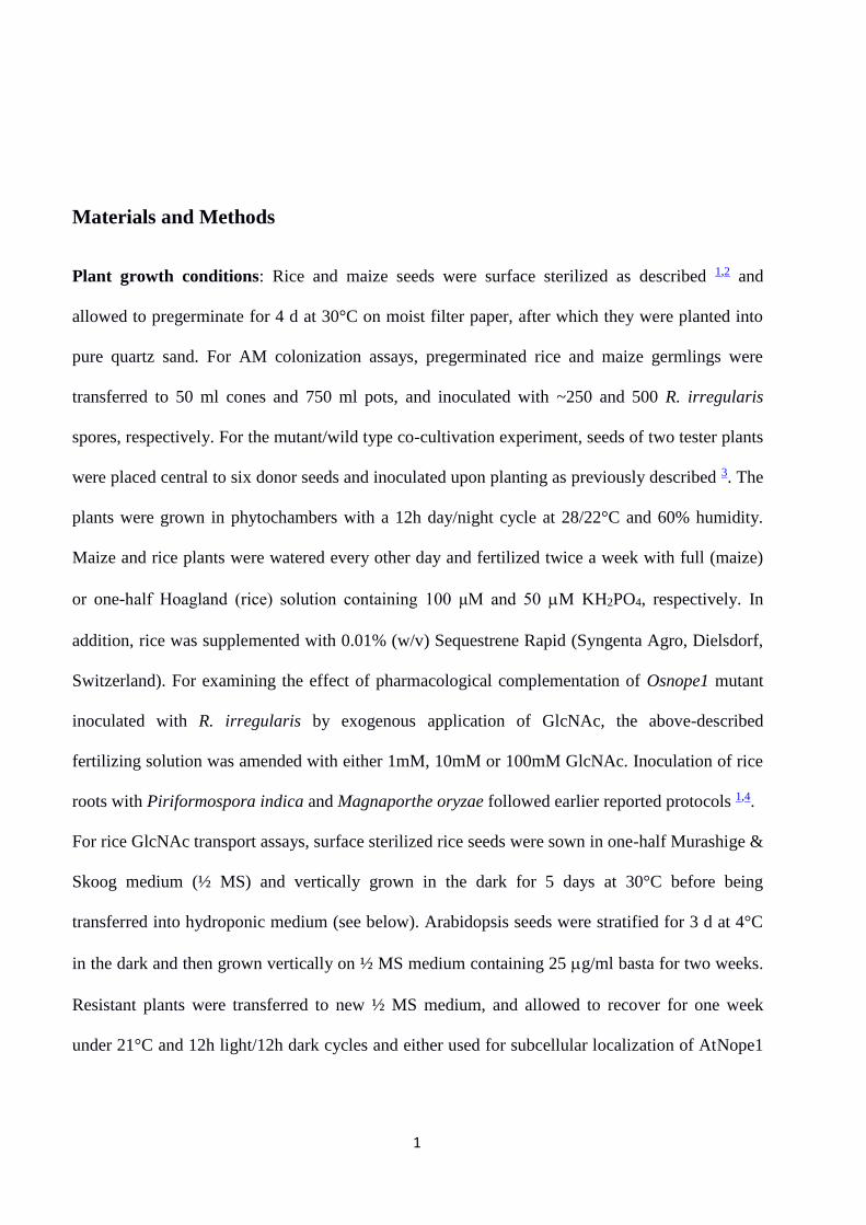
An N-acetylglucosamine transporter requiredfor arbuscular mycorrhizal symbiosesin rice and maizeMarina Nadal1,2‡, Ruairidh Sawers2†‡, Shamoon Naseem3, Barbara Bassin4, Corinna Kulicke1†,Abigail Sharman1, Gynheung An5, Kyungsook An5, Kevin R. Ahern6, Amanda Romag6,Thomas P. Brutnell6†, Caroline Gutjahr2†, Niko Geldner2, Christophe Roux7,Enrico Martinoia4, James B. Konopka3 and Uta Paszkowski1,2*‡
Most terrestrial plants, including crops, engage in beneficial interactions with arbuscular mycorrhizal fungi. Vital to theassociation is mutual recognition involving the release of diffusible signals into the rhizosphere. Previously, we identifiedthe maize no perception 1 (nope1) mutant to be defective in early signalling. Here, we report cloning of ZmNope1 on thebasis of synteny with rice. NOPE1 encodes a functional homolog of the Candida albicans N-acetylglucosamine (GlcNAc)transporter NGT1, and represents the first plasma membrane GlcNAc transporter identified from plants. In C. albicans,exposure to GlcNAc activates cell signalling and virulence. Similarly, in Rhizophagus irregularis treatment with ricewild-type but not nope1 root exudates induced transcriptome changes associated with signalling function, suggesting arequirement of NOPE1 function for presymbiotic fungal reprogramming.
Arbuscular mycorrhizal (AM) symbiosis is a mutually beneficialrelationship between plants and fungi in which plant rootsexchange photoassimilates for fungus-delivered soil minerals.
The resulting interaction may profoundly influence plant perform-ance, in both wild and cultivated systems. For the symbiosis tobegin, plant roots and AM fungi (AMF) exchange signals via secretionof diffusible compounds1, including fungal chitin-based molecules(reviewed in refs 2 and 3) detected by Lysine Motif (LysM) containingplasma membrane receptor-like kinases4,5. Central to the perceptionof AMF in rice is the α/β-hydrolase DWARF14 LIKE (D14L) andthe F-box protein DWARF3 (D3)6, although, in this instance, thesignal molecules remain uncharacterized.
A number of plant-derived factors are known to stimulate mor-phological changes in AMF, promoting fungal–host encounters1,including flavonoids that enhance hyphal tip elongation7, 2-hydro-xid fatty acids (2-OH-FA) that trigger hyphal branching8 and strigo-lactones (SLs) that induce changes in fungal metabolism, coupledwith profuse hyphal ramifications9–11. Despite their presymbioticeffect on AMF growth, plant SL and flavonoid biosyntheticmutants are still partially or fully colonized12,13. Once the fungushas reached the plant’s surface, cutin monomers induce hyphopo-dium differentiation, the anchoring structure for entry of AMFinto the root epidermal cell layer14. As the fungal genome lacksgenes for the de novo biosynthesis of certain fatty acids15, cutinmay have an additional nutritional role.
To better understand signalling during symbiotic establishment,we analysed the maize no perception1 (nope1) mutant, which doesnot form AM symbioses16. We identified NOPE1 to encode anN-acetylglucosamine (GlcNAc) transporter, a function not describedpreviously in plants, but characterized in fungi17. Notably, GlcNAchas been shown to stimulate the fungal pathogen Candida albicansto undergo morphological changes and increase expression of viru-lence genes that promote pathogenic interactions with the host18.Our analyses provide the first evidence that a previously unknownplant GlcNAc transporter plays a role in the initiation of rootcolonization by AMF.
ResultsCloning of maize ZmNope1 on the basis of synteny with rice. Themaize Zmnope1 mutant is unable to establish AM symbioses16.Limited physical interaction of Zmnope1 with AMF suggested afailure in presymbiotic signal exchange. As genetic mapping hadlinked the Nope1 locus to the marker UMC1336 on chromosome10, we searched for rice candidate genes exhibiting atranscriptional response to mycorrhizal root colonization locatedin the region syntenic to maize nope1 (refs 19 and 20), andidentified the gene LOC_Os04g01520 (ref. 19) (SupplementaryFig. 1A). To investigate a potential role in AM symbiosis,rice plants segregating for a transfer-DNA insertion inLOC_Os04g01520 (4A-01057; Supplementary Fig. 1B,C) were
1Department of Plant Sciences, University of Cambridge, Cambridge, CB2 3EA, UK. 2Department of Plant Molecular Biology, University of Lausanne, 1015Lausanne, Switzerland. 3Department of Molecular Genetics and Microbiology, Stony Brook University, Stony Brook, New York 11794-5222, USA. 4Instituteof Plant Biology, University of Zurich, 8008 Zurich, Switzerland. 5Crop Biotech Institute and Graduate School of Biotechnology, Kyung Hee University,Yongin 17104, Korea. 6Boyce Thompson Institute for Plant Research, Ithaca, New York 14853, USA. 7UPS, UMR5546, Laboratoire de Recherche en SciencesVégétales, Université de Toulouse, BP 42617, F-31326 Castanet-Tolosan CEDEX, France. †Present addresses: Laboratorio Nacional de Genómica para laBiodiversidad, Centro de Investigación y de Estudios Avanzados, Campus Guanajuato, PO Box 629, Irapuato Guanajuato, 36821 Mexico (R.S.); DonaldDanforth Plant Science Center, 975 North Warson Road St. Louis, Missouri 63132, USA (T.P.B.); Faculty of Biology, Genetics, University of Munich, 82152Martinsried, Germany (C.G.); Sir William Dunn School of Pathology, University of Oxford, South Parks Road, Oxford OX1 3RE, UK (C.K). ‡These authorscontributed equally to this work. *e-mail: [email protected]
ARTICLESPUBLISHED: XX XX 2017 | VOLUME: 3 | ARTICLE NUMBER: 17073
NATURE PLANTS 3, 17073 (2017) | DOI: 10.1038/nplants.2017.73 | www.nature.com/natureplants 1
© 2017 Macmillan Publishers Limited, part of Springer Nature. All rights reserved.
SUPPLEMENTARY INFORMATIONVOLUME: 3 | ARTICLE NUMBER: 17073
NATURE PLANTS | DOI: 10.1038/nplants.2017.73 | www.nature.com/natureplants
1
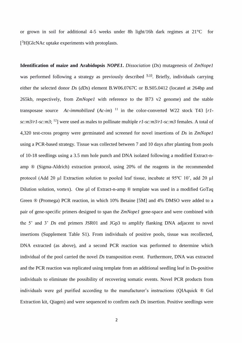
Materials and Methods
Plant growth conditions: Rice and maize seeds were surface sterilized as described 1,2 and
allowed to pregerminate for 4 d at 30°C on moist filter paper, after which they were planted into
pure quartz sand. For AM colonization assays, pregerminated rice and maize germlings were
transferred to 50 ml cones and 750 ml pots, and inoculated with ~250 and 500 R. irregularis
spores, respectively. For the mutant/wild type co-cultivation experiment, seeds of two tester plants
were placed central to six donor seeds and inoculated upon planting as previously described 3. The
plants were grown in phytochambers with a 12h day/night cycle at 28/22°C and 60% humidity.
Maize and rice plants were watered every other day and fertilized twice a week with full (maize)
or one-half Hoagland (rice) solution containing 100 μM and 50 M KH2PO4, respectively. In
addition, rice was supplemented with 0.01% (w/v) Sequestrene Rapid (Syngenta Agro, Dielsdorf,
Switzerland). For examining the effect of pharmacological complementation of Osnope1 mutant
inoculated with R. irregularis by exogenous application of GlcNAc, the above-described
fertilizing solution was amended with either 1mM, 10mM or 100mM GlcNAc. Inoculation of rice
roots with Piriformospora indica and Magnaporthe oryzae followed earlier reported protocols 1,4.
For rice GlcNAc transport assays, surface sterilized rice seeds were sown in one-half Murashige &
Skoog medium (½ MS) and vertically grown in the dark for 5 days at 30°C before being
transferred into hydroponic medium (see below). Arabidopsis seeds were stratified for 3 d at 4°C
in the dark and then grown vertically on ½ MS medium containing 25 g/ml basta for two weeks.
Resistant plants were transferred to new ½ MS medium, and allowed to recover for one week
under 21°C and 12h light/12h dark cycles and either used for subcellular localization of AtNope1
2
or grown in soil for additional 4-5 weeks under 8h light/16h dark regimes at 21°C for
[3H]GlcNAc uptake experiments with protoplasts.
Identification of maize and Arabidopsis NOPE1. Dissociation (Ds) mutagenesis of ZmNope1
was performed following a strategy as previously described 9,10. Briefly, individuals carrying
either the selected donor Ds (dDs) element B.W06.0767C or B.S05.0412 (located at 264bp and
265kb, respectively, from ZmNope1 with reference to the B73 v2 genome) and the stable
transposase source Ac-immobilized (Ac-im) 11 in the color-converted W22 stock T43 [r1-
sc:m3/r1-sc:m3; 12] were used as males to pollinate multiple r1-sc:m3/r1-sc:m3 females. A total of
4,320 test-cross progeny were germinated and screened for novel insertions of Ds in ZmNope1
using a PCR-based strategy. Tissue was collected between 7 and 10 days after planting from pools
of 10-18 seedlings using a 3.5 mm hole punch and DNA isolated following a modified Extract-n-
amp ® (Sigma-Aldrich) extraction protocol, using 20% of the reagents in the recommended
protocol (Add 20 µl Extraction solution to pooled leaf tissue, incubate at 95ºC 10’, add 20 µl
Dilution solution, vortex). One µl of Extract-n-amp ® template was used in a modified GoTaq
Green ® (Promega) PCR reaction, in which 10% Betaine [5M] and 4% DMSO were added to a
pair of gene-specific primers designed to span the ZmNope1 gene-space and were combined with
the 5’ and 3’ Ds end primers JSR01 and JGp3 to amplify flanking DNA adjacent to novel
insertions (Supplement Table S1). From individuals of positive pools, tissue was recollected,
DNA extracted (as above), and a second PCR reaction was performed to determine which
individual of the pool carried the novel Ds transposition event. Furthermore, DNA was extracted
and the PCR reaction was replicated using template from an additional seedling leaf in Ds-positive
individuals to eliminate the possibility of recovering somatic events. Novel PCR products from
individuals were gel purified according to the manufacturer’s instructions (QIAquick ® Gel
Extraction kit, Qiagen) and were sequenced to confirm each Ds insertion. Positive seedlings were
3
grown to maturity and self-pollinated to create a segregating homozygous nope1::Ds insertion
stock. The A. thaliana NOPE1 homologs were identified based on sequence similarities. The rice
OsNope1 cDNA and OsNope1 protein sequences were used as query for blastn and blastp
searches in the Arabidopsis database (www.arabidopsis.org). Two A. thaliana genes, A1g18000
and At1g18010, are identical in sequence and showed the highest similarities. Using the reciprocal
blast strategy (RBS) confirmed rice OsNope1 (LOC_Os04g01520) as the most similar rice
sequence to A1g18000 and At1g18010.
Phylogenetic analysis
Sequences for the viridiplantae-specific orthogroup corresponding to OsNope1 was downloaded
from Phytozome V.11.1 (https://phytozome.jgi.doe.gov/pz/portal.html) as CDS sequences.
Alignments were undertaken in the translation align function generated in Geneious version 7.1.8,
and trees generated by FastTree, under default settings but under a GTR+I+gamma model of
nucleotide substitution.
Mycorrhizal quantification: Performed by staining roots with Trypan Blue as described earlier 1.
For detailed inspection of fungal morphology, roots were stained with WGA-AlexaFluor488
conjugate (Invitrogen, Paisley, UK), counterstained with propidium iodide and imaged with a
Leica SP5 confocal microscope (Leica Microsystems, Milton Keyes, UK).
Laser Scanning Confocal Microscopy: For the assessment of subcellular localization of
AtNope1, roots of 7d old A. thaliana line 4731-y were stained with propidium iodine and imaged
with a Zeiss LSM 700 confocal microscope (Zeiss, Feldbach, Switzerland). Excitation/Emission
windows used were: 488/500-550 nm for WGA; 488/590-660 nm for PI and 514/525-550 for
YFP.
4
Gigaspora rosea germ tube branching bioassay: Four G. rosea spores per plate were germinated
and incubated at 2% [CO2] and 30°C in the dark in minimal medium supplemented with 10 µM
quercetin and gelled with 0.6% W/V Phytagel (Sigma-Aldrich, Saint-Quentin Fallavier, FR).
Seven days after inoculation, each spore produced a single germ tube growing upwards. Two
small wells were produced into the gel on each side of the germ tube tip with a Pasteur pipette tip
and 5 µl of the test solution (10-7 M GR24) in 0.1% acetonitrile (positive control), or 0.1%
acetonitrile (negative control), or plant exudates (complemented with 0.1% final acetonitrile) were
injected into each well. After 24 h, germ tube branching was recorded by counting newly formed
hyphal tips. Three to five plates (9–15 spores) were used for each treatment. The mean numbers of
branches for all conditions tested were compared by the Kruskal–Wallis test and, when
significant, pair comparison was made by the student test. The experiment was three times
independently repeated.
Nucleic acid extraction and manipulation. Rice genomic DNA was extracted by the urea
extraction method 13, digested with EcoRI enzyme (Promega, Dübendorf, Switzerland) and treated
with RNAse A (Sigma-Aldrich, Buchs, Switzerland). The digested DNA was separated by gel
electrophoresis and transferred overnight to a nylon membrane (GE Healthcare, Glattbrugg,
Switzerland). An 870 bp fragment of the phosphinothricin-acetyl-transferase resistance gene (bar)
was amplified with primers MN047 and MN048 (Supplemental Table S4) from plasmid pTF101.1
14 and used as probe for hybridization. For probe labelling and detection the non-radioactive DIG-
system (Roche, Basel, Switzerland) was employed according to the manufacturer’s guidance.
Southern blot was performed according to standard protocols 15.
Plant RNA extraction, cDNA synthesis and quantitative RT-PCR were performed as described
earlier 1. Briefly, RNA was extracted from ~100mg of root (rice and maize) or cauline leaf tissue
5
(Arabidopsis) with Trizol prepared in the lab 16 and treated with DNaseI (ThermoFisher Scientific,
Ecublens, Switzerland and Thermo Electron Basingstoke, UK) to remove contaminating gDNA.
Absence of residual gDNA was confirmed by control PCR on DNAseI treated RNA before
reverse transcription. First strand cDNA was synthesized using Superscript II reverse transcriptase
(ThermoFisher Scientific, Ecublens, Switzerland and Thermo Electron Basingstoke, UK)
combined with oligo(dT) primers (Promega, Dübendorf, Switzerland and Southhampton, UK).
For detecting NOPE1 transcripts from rice, maize or Arabidopsis lines, cDNA was synthesized
using Superscript III reverse transcriptase (ThermoFisher Scientific, Ecublens, Switzerland and
Thermo Electron Basingstoke, UK).
Fungal RNA was extracted from fungal mycelium produced from spores using RNeasy Plant Mini
Kit (Qiagen, Hombrechtikon, Switzerland and Manchester, UK). Reverse transcription applied the
same protocols as for plants described above. Gene specific primers for plant AM marker genes 1
and AtNope1, and fungal genes are described in Supplemental Table S2 and Supplemental Table
S3, respectively. Real-time RT-PCR was performed as previously described 1. The following
constitutively expressed genes were used to normalize target gene transcript levels in each
organisms: rice, CYCLOPHILIN2 (CP2), LOC_Os02g02890; maize, GLYCERALDEHYDE-3-
PHOSPHATE DEHYDROGENASE (GAPDH), GRMZM2G046804; Arabidopsis, ISOPENTENYL-
DIPHOSPHATE-DELTA-ISOMERASE II (IPP2), A13g02780; R. irregularis, ELONGATION
FACTOR 1-α (RiEF), fgenesh1_kg.21690_#_7.
To determine the full-length cDNA of rice NOPE1 Gene-Race kit (ThermoFisher Scientific,
Ecublens, Switzerland) was used to amplify 5’ and 3’ ends of rice NOPE1 cDNA following
manufacturer recommendations.
Generation of constructs and genetic transformation of A. thaliana and C. albicans: A.
thaliana constructs were generated based on Gateway Cloning Technology (ThermoFisher
6
Scientific, Ecublens, Switzerland). Briefly, the silencing construct pAtNope1 was generated by
amplifying ~200 bp fragment complementary to the 5’end of the At1g18000 coding region using
primers MN042 and MN043, followed by cloning into pDONR207 (ThermoFisher Scientific,
Ecublens, Switzerland) and transfer into the destination vector pB7GWIWG2-II 18. To generate
Ubqprom::YPF::AtNope1 for subcellular localization, AtNope1 ORF was transferred from clone
U14731 (http://signal.salk.edu/) into destination vector pNIGEL07 19. A. thaliana transgenic lines
were generated by floral dipping as previously described 20. Primers used to generate constructs
for plant transformation are described in Supplemental Table S4.
The construct for genetic complementation of C. albicans, pOsNope1, was generated by
synthesizing a codon optimized version of rice NOPE1 cDNA for expression in C. albicans
(http://www.idtdna.com/site). This cDNA version was then modified by amplification with
primers carrying 50 bp of homology to the regions flanking the CaNGT1 gene to replace the
CaNGT1 gene in plasmid pDDB57 by homologous recombination in S. cerevisiae 7. The resulting
pOsNope1 plasmid and the corresponding parental CaNGT1 plasmids were digested in the
promoter region with Bsa BI and transformed into C. albicans ngt1 strain YJA2 7 using a URA3
gene for selection. Control ngt1 cells transformed at the same time received only the URA3 gene.
Resulting colonies were PCR-confirmed with primers shown in Supplemental Table S1 to contain
the appropriate gene.
Library preparation and RNAseq sequencing. RNA extracted from treated R. irregularis
spores was quantified using a Nanodrop fluorospectrometer (ThermoFisher Scientific Inc.,
Villebon sur Yvette, France) and quality was determined by electrophoresis on an Agilent
Bioanalyzer (Agilent Technologies, Les Ulis Cedex, France). Samples with RNA Integrity
Number (RIN) higher than eight were selected. RNAseq libraries were constructed as previously
described 21. The libraries were prepared according to Illumina’s protocols using the Illumina
7
TruSeq RNA Sample Prep Kits v2 (Illumina Inc., San Diego, USA). Polyadenylated RNA was
purified from at least 1µg of total RNA using beads containing oligo-dT. Subsequently, each
mRNA sample was fragmented to generate double stranded cDNA for sequencing. Double-
stranded cDNA was synthesized using SuperScript II Reverse Transcriptase (Life Technologies,
Saint Aubin, France) and random primers for first strand cDNA synthesis followed by second
strand synthesis using DNA Polymerase I and RNaseH (ThermoFisher Scientific Inc., Villebon
sur Yvette, France) for removal of mRNA. Double-stranded cDNA was purified using Agencourt
AMPure XP beads (Beckman Couter Inc., Villepinte, France) as recommended in the TruSeq
RNA Sample Prep Guide. cDNAs were end-repaired by T4 DNA polymerase and Klenow DNA
Polymerase, and phosphorylated by T4 polynucleotide kinase. The blunt ended cDNA was
purified using Agencourt AMPure XP beads (Beckman Couter Inc., Villepinte, France). The
cDNA products were incubated with Klenow DNA Polymerase (ThermoFisher Scientific Inc.,
Villebon sur Yvette, France) to add an ‘A’ base (Adenine) to the 3’ end of the blunt
phosphorylated DNA fragments and then purified using Agencourt AMPure XP beads. DNA
fragments were ligated to Illumina adapters with a single ‘T’ base (Thymine) overhang at their
3’ends. Adapter-ligated products were purified using Agencourt AMPure XP beads and amplified
in a Linker Mediated PCR reaction (LM-PCR) for 12 cycles using PhusionTM DNA Polymerase
and Illumina's PE genomic DNA primer set followed by purification using Agencourt AMPure
XP beads. Size selection (ranging from 200-300 bp) was performed on E-gel (ThermoFisher
Scientific, Villebon-sur-Yvette, France). Quality of finished libraries was assessed using an
Agilent Bioanalyzer (Agilent Technologies, Les Ulis Cedex, France). Libraries were quantified by
qPCR using the KAPA Library Quantification Kit (PN11 KK4824- KAPA Biosciences, Nanterre,
France) to obtain an accurate quantification.
GlcNAc transport assays in C. albicans and A. thaliana: C. albicans GlcNAc uptake and
8
competition assay were performed as previously described 7. Transport experiments using
protoplast were performed by a slight modification of a previously described method 25.
Specifically, the abaxial epidermis of leaves from 6- to 8-week-old plants were abraded with P500
sandpaper, and immediately floated on medium A, containing 500 mM sorbitol, 1 mM CaCl2, and
10mM MES-KOH, pH 5.6, supplemented with 1 mg ml-1 BSA in petri dishes. Subsequently,
leaves were incubated for 2 h at 30°C with their abaxial side on medium A containing 10 mg ml-1
cellulase R10 and 5 mg ml-1 macerozyme R10 (Serva Electrophoresis, Heidelberg, Germany). The
suspensions with released protoplasts were collected into 50 ml Falcon tubes, each of which was
underlied with 2 ml of Percoll solution, pH 6: 500 mM sorbitol, 1mM CaCl2, and 20 mM MES in
100% Percoll (GE Healthcare, Glattbrugg, Switzerland). After centrifugation at 1500 g for 8 min
at 4°C, the supernatant was aspired and the concentrated protoplasts were resuspended in the
remaining solution. This solution was adjusted with medium A or Percoll solution to give a final
Percoll concentration of about 35%. The protoplasts were overlaid with medium A containing
25% Percoll and medium A containing 2% Percoll. After centrifugation for 8 min at 1,200 g, the
protoplasts were recovered from the upper interphase. The purified protoplasts were mixed with 2
parts of medium B: 500 mM betaine, 1 mM CaCl2, and 10 mM MES-KOH, pH 5.6. For transport
experiments, 50µl of medium A containing 50% Percoll was placed at the bottom of 400 µL
polyethylene tubes and overlaid with 200µl of silicon oil AR200. Transport experiments were
initiated by adding the substrate to protoplasts and terminated by overlaying 100 µL of the
protoplast suspension on the preformed silicon oil gradients and a subsequent 20 s centrifugation
at 10,000 g at the times indicated . The polyethylene tubes were frozen overnight. The bottoms of
the frozen tubes were cut with a razor blade and the bottom of the tube was transferred to the
scintillation liquid and counted directly. GlcNAc uptake values was estimated based on five
biological replicates per genotype.
9
Statistical analysis: Biological replicates corresponded to individual plants of independently
grown experiments. In experiments to evaluate the level of fungal, differences among genotypes
were assessed per structure using the non-parametric Kruskal-Wallis test and post hoc Dunn test
to assign means groups on the basis of pair wise comparisons (R statistics dunn.test::dunn.test;
Dinno, 2016). To control for multiple testing, p-values were adjusted using the method of
Benjamini and Hochberg (Benjamini and Hochberg, 1995) and significant differences called at p
< 0.05. In GlcNAc and transcript accumulation experiments, differences among genotypes were
assessed using ANOVA (R statistics; R Core Team, 2016) and post hoc means groups assigned
using Tukey HSD (R statistics agricolae::HSD.test; de Mendiburu, 2016; p < 0.05).
Alexis Dinno (2016). dunn.test: Dunn's Test of Multiple Comparisons Using Rank Sums. R
package, version 1.3.2. https://CRAN.R-project.org/package=dunn.test; accessed June 2016.
de Mendiburu F. 2016. agricolae: Statistical Procedures for Agricultural Research. R package
version 1.2-4. https://CRAN.R-project.org/package=agricolae; accessed June 2016.
R Core Team. 2016. R: A language and environment for statistical computing. R Foundation for
Statistical Computing, Vienna, Austria. URL https://www.R-project.org/; accessed June 2016.
References
1 Gutjahr, C. et al. Arbuscular mycorrhiza-specific signaling in rice transcends the common symbiosis signaling pathway. Plant Cell 20, 2989-3005, doi:10.1105/tpc.108.062414 (2008).
2 Paszkowski, U., Jakovleva, L. & Boller, T. Maize mutants affected at distinct stages of the arbuscular mycorrhizal symbiosis. Plant J. 47, 165-173 (2006).
3 Gutjahr, C. et al. The half-size ABC transporters STR1 and STR2 are indispensable for mycorrhizal arbuscule formation in rice. Plant J 69, 906-920, doi:10.1111/j.1365-313X.2011.04842.x (2012).
4 Marcel, S., Sawers, R., Oakeley, E., Angliker, H. & Paszkowski, U. Tissue-adapted invasion strategies of the rice blast fungus Magnaporthe oryzae. Plant Cell 22, 3177-3187, doi:10.1105/tpc.110.078048 (2010).
5 Bécard, G. & Fortin, J. A. Early events of vesicular–arbuscular mycorrhiza formation on Ri T-DNA transformed roots. New Phytologist 108, 211–218 (1988).
6 Buee, M., Rossignol, M., Jauneau, A., Ranjeva, R. & Bécard, G. The presymbiotic growth of arbuscular mycorrhizal fungi is induced by a branching factor partially purified from plant root exudates. Molecular Plant Microbe Interaction 13, 693-698 (2000).
10
7 Alvarez, F. J. & Konopka, J. B. Identification of an N-acetylglucosamine transporter that mediates hyphal induction in Candida albicans. Molecular biology of the cell 18, 965-975, doi:10.1091/mbc.E06-10-0931 (2007).
8 Güimil, S. et al. Comparative transcriptomics of rice reveals an ancient pattern of response to microbial colonization. Proc Natl Acad Sci 102, 8066-8070 (2005).
9 Ahern, K. R. et al. Regional mutagenesis using Dissociation in maize. Methods 49, 248-254, doi:10.1016/j.ymeth.2009.04.009 (2009).
10 Vollbrecht, E. et al. Genome-wide distribution of transposed Dissociation elements in maize. Plant Cell 22, 1667-1685, doi:10.1105/tpc.109.073452 (2010).
11 Conrad, L. J. & Brutnell, T. P. Ac-immobilized, a stable source of Activator transposase that mediates sporophytic and gametophytic excision of Dissociation elements in maize. Genetics 171, 1999-2012, doi:10.1534/genetics.105.046623 (2005).
12 Alleman, M. & Kermicle, J. L. Somatic variegation and germinal mutability reflect the position of transposable element Dissociation within the maize R gene. Genetics 135, 189-203 (1993).
13 Chen, J. & Dellaporta, S. Urea-based plant miniprep. In Freeling, M. & V. Walbot (eds), The maize handbook. Spriner, New York, 526-527 (1994).
14 Paz, M. M. et al. Assessment of conditions affecting Agrobacteriummediated soybean transformation using the cotyledonary node explant. Euphytica 136, 167-179 (2004).
15 Sambrook, J. & David, W. R. Molecular Cloning: A Laboratory Manual, Third Edition Vol. 2 (Cold Spring Harbor Laboratory Press, 2001).
16 Chomczynski, P. & Sacchi, N. Single-step method of RNA isolation by acid guanidinium thiocyanate-phenol-chloroform extraction. Analytical biochemistry 162, 156-159, doi:10.1006/abio.1987.9999 (1987).
17 Yang, S. Y. et al. Nonredundant regulation of rice arbuscular mycorrhizal symbiosis by two members of the phosphate transporter1 gene family. Plant Cell 24, 4236-4251, doi:10.1105/tpc.112.104901 (2012).
18 Karimi, M., Inze, D. & Depicker, A. GATEWAY vectors for Agrobacterium-mediated plant transformation. Trends Plant Sci 7, 193-195 (2002).
19 Geldner, N. et al. Rapid, combinatorial analysis of membrane compartments in intact plants with a multicolor marker set. Plant J 59, 169-178, doi:10.1111/j.1365-313X.2009.03851.x (2009).
20 Clough, S. J. & Bent, A. F. Floral dip: a simplified method for Agrobacterium-mediated transformation of Arabidopsis thaliana. The Plant journal : for cell and molecular biology 16, 735-743 (1998).
21 Tisserant, E. et al. The transcriptome of the arbuscular mycorrhizal fungus Glomus intraradices (DAOM 197198) reveals functional tradeoffs in an obligate symbiont. New Phytol 193, 755-769, doi:10.1111/j.1469-8137.2011.03948.x (2012).
22 Baggerly, K. A., Deng, L., Morris, J. S. & Aldaz, C. M. Differential expression in SAGE: accounting for normal between-library variation. Bioinformatics 19, 1477-1483 (2003).
23 Benjamini, Y. & Hochberg, Y. Controlling the false discovery rate: a practical and powerful approach to multiple testing. Journal of the Royal Statistical Society Series B, 289-300 (1995).
24 Falcon, S. & Gentleman, R. Using GOstats to test gene lists for GO term association. Bioinformatics 23, 257-258, doi:10.1093/bioinformatics/btl567 (2007).
25 Tohge, T. et al. Toward the storage metabolome: profiling the barley vacuole. Plant physiology 157, 1469-1482, doi:10.1104/pp.111.185710 (2011).
11
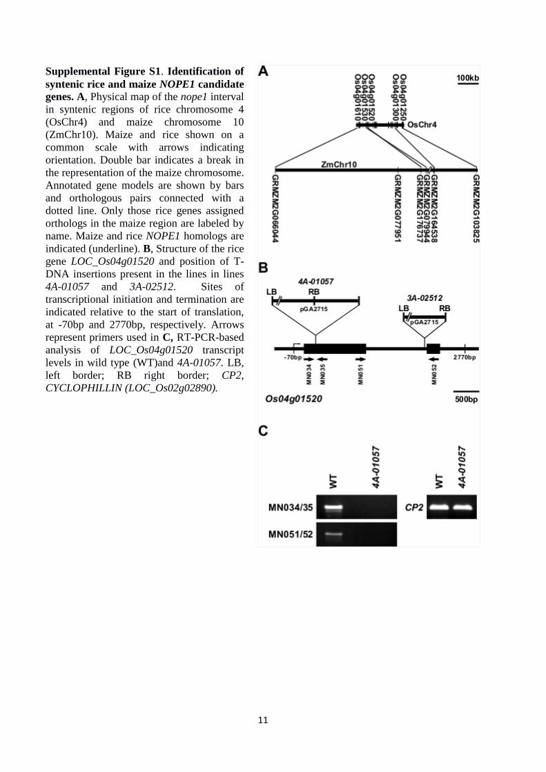
Supplemental Figure S1. Identification of
syntenic rice and maize NOPE1 candidate
genes. A, Physical map of the nope1 interval
in syntenic regions of rice chromosome 4
(OsChr4) and maize chromosome 10
(ZmChr10). Maize and rice shown on a
common scale with arrows indicating
orientation. Double bar indicates a break in
the representation of the maize chromosome.
Annotated gene models are shown by bars
and orthologous pairs connected with a
dotted line. Only those rice genes assigned
orthologs in the maize region are labeled by
name. Maize and rice NOPE1 homologs are
indicated (underline). B, Structure of the rice
gene LOC_Os04g01520 and position of T-
DNA insertions present in the lines in lines
4A-01057 and 3A-02512. Sites of
transcriptional initiation and termination are
indicated relative to the start of translation,
at -70bp and 2770bp, respectively. Arrows
represent primers used in C, RT-PCR-based
analysis of LOC_Os04g01520 transcript
levels in wild type (WT)and 4A-01057. LB,
left border; RB right border; CP2,
CYCLOPHILLIN (LOC_Os02g02890).
12
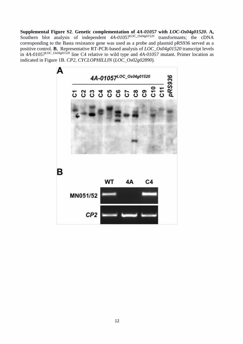
Supplemental Figure S2. Genetic complementation of 4A-01057 with LOC-Os04g01520. A,
Southern blot analysis of independent 4A-01057LOC_Os04g01520 transformants; the cDNA
corresponding to the Basta resistance gene was used as a probe and plasmid pRS936 served as a
positive control. B, Representative RT-PCR-based analysis of LOC_Os04g01520 transcript levels
in 4A-01057LOC_Os04g01520 line C4 relative to wild type and 4A-01057 mutant. Primer location as
indicated in Figure 1B. CP2, CYCLOPHILLIN (LOC_Os02g02890).
13
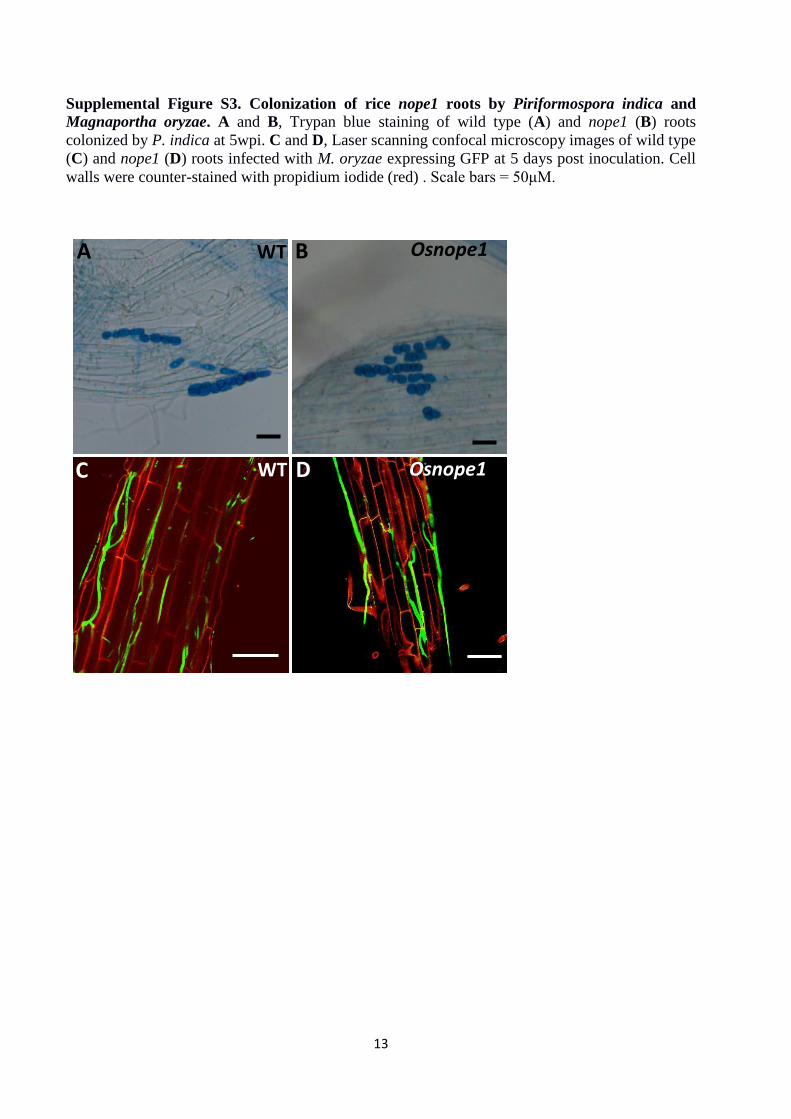
Supplemental Figure S3. Colonization of rice nope1 roots by Piriformospora indica and
Magnaportha oryzae. A and B, Trypan blue staining of wild type (A) and nope1 (B) roots
colonized by P. indica at 5wpi. C and D, Laser scanning confocal microscopy images of wild type
(C) and nope1 (D) roots infected with M. oryzae expressing GFP at 5 days post inoculation. Cell
walls were counter-stained with propidium iodide (red) . Scale bars = 50μM.
B Osnope1 A WT
C WT D Osnope1
14
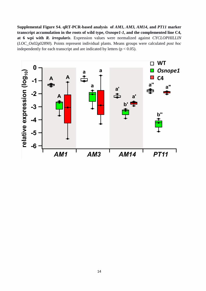
Supplemental Figure S4. qRT-PCR-based analysis of AM1, AM3, AM14, and PT11 marker
transcript accumulation in the roots of wild type, Osnope1-1, and the complemented line C4,
at 6 wpi with R. irregularis. Expression values were normalized against CYCLOPHILLIN
(LOC_Os02g02890). Points represent individual plants. Means groups were calculated post hoc
independently for each transcript and are indicated by letters (p < 0.05).
WT Osnope1 C4
15
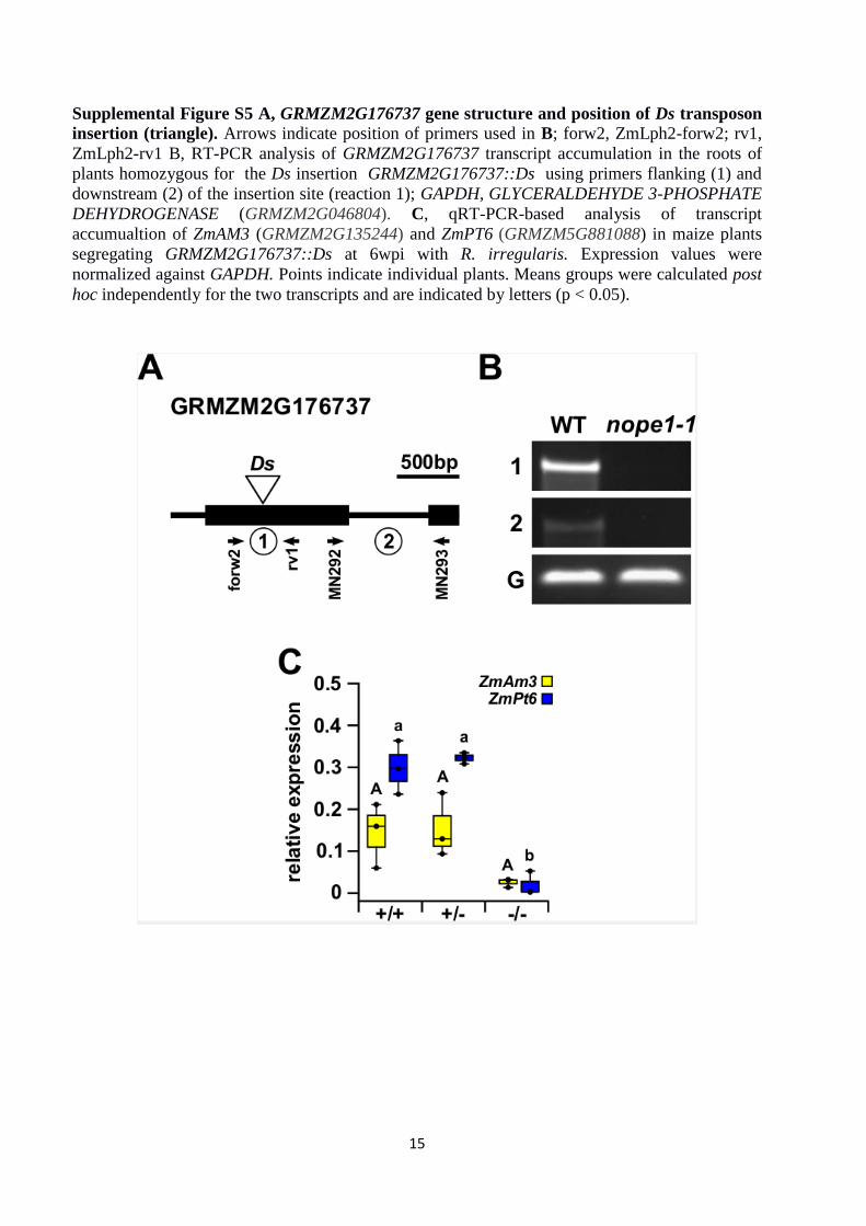
Supplemental Figure S5 A, GRMZM2G176737 gene structure and position of Ds transposon
insertion (triangle). Arrows indicate position of primers used in B; forw2, ZmLph2-forw2; rv1,
ZmLph2-rv1 B, RT-PCR analysis of GRMZM2G176737 transcript accumulation in the roots of
plants homozygous for the Ds insertion GRMZM2G176737::Ds using primers flanking (1) and
downstream (2) of the insertion site (reaction 1); GAPDH, GLYCERALDEHYDE 3-PHOSPHATE
DEHYDROGENASE (GRMZM2G046804). C, qRT-PCR-based analysis of transcript
accumualtion of ZmAM3 (GRMZM2G135244) and ZmPT6 (GRMZM5G881088) in maize plants
segregating GRMZM2G176737::Ds at 6wpi with R. irregularis. Expression values were
normalized against GAPDH. Points indicate individual plants. Means groups were calculated post
hoc independently for the two transcripts and are indicated by letters (p < 0.05).
16
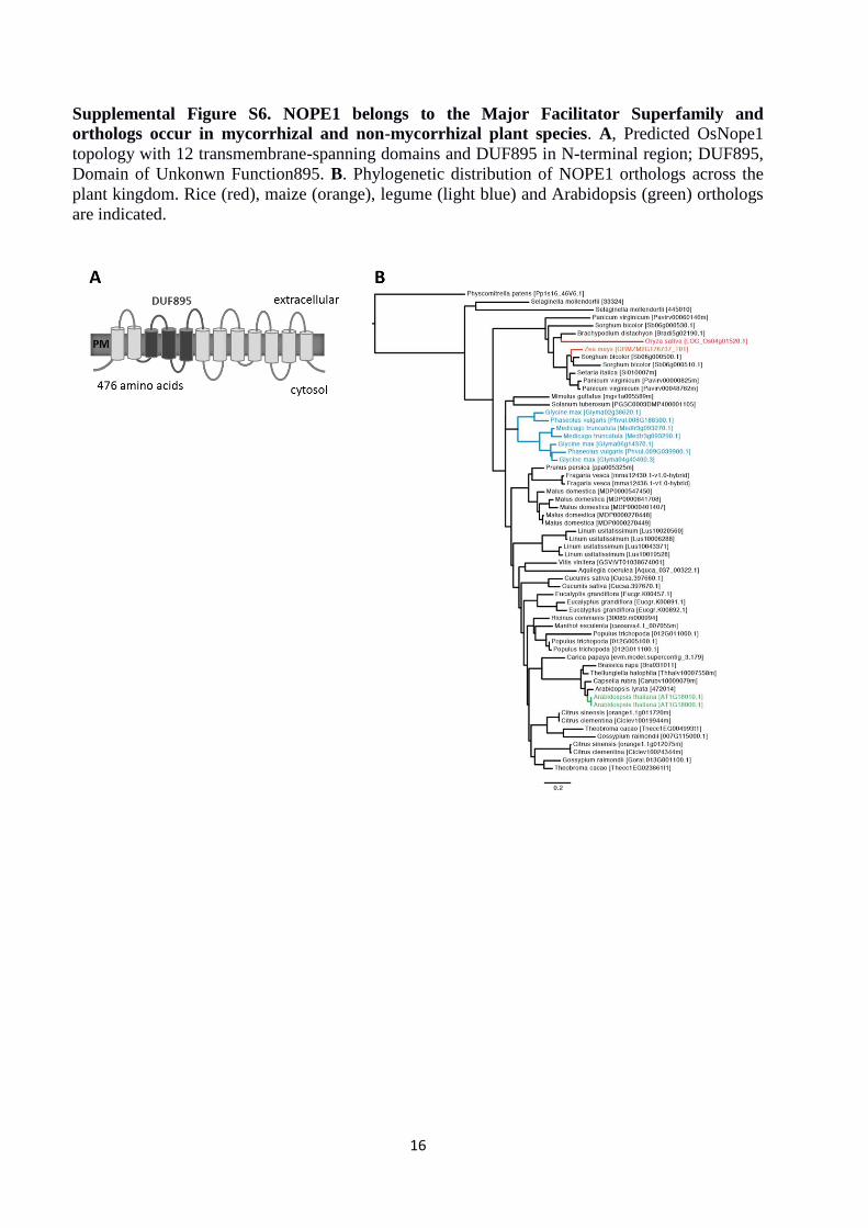
Supplemental Figure S6. NOPE1 belongs to the Major Facilitator Superfamily and
orthologs occur in mycorrhizal and non-mycorrhizal plant species. A, Predicted OsNope1
topology with 12 transmembrane-spanning domains and DUF895 in N-terminal region; DUF895,
Domain of Unkonwn Function895. B. Phylogenetic distribution of NOPE1 orthologs across the
plant kingdom. Rice (red), maize (orange), legume (light blue) and Arabidopsis (green) orthologs
are indicated.
17
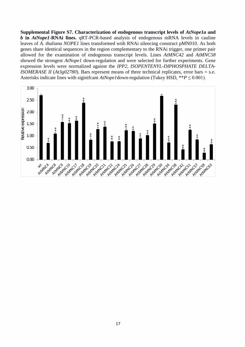
Supplemental Figure S7. Characterization of endogenous transcript levels of AtNope1a and
b in AtNope1-RNAi lines. qRT-PCR-based analysis of endogenous mRNA levels in cauline
leaves of A. thaliana NOPE1 lines transformed with RNAi silencing construct pMN010. As both
genes share identical sequences in the region complementary to the RNAi trigger, one primer pair
allowed for the examination of endogenous transcript levels. Lines AtMNC42 and AtMNC58
showed the strongest AtNope1 down-regulation and were selected for further experiments. Gene
expression levels were normalized against the IPP2, ISOPENTENYL-DIPHOSPHATE DELTA-
ISOMERASE II (At3g02780). Bars represent means of three technical replicates, error bars = s.e.
Asterisks indicate lines with siginifcant AtNope1down-regulation (Tukey HSD, **P ≤ 0.001).
18
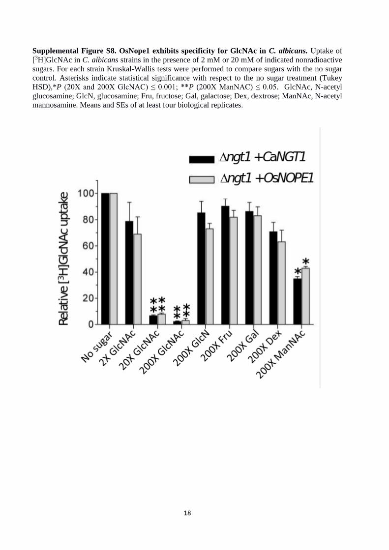
Supplemental Figure S8. OsNope1 exhibits specificity for GlcNAc in C. albicans. Uptake of
[3H]GlcNAc in C. albicans strains in the presence of 2 mM or 20 mM of indicated nonradioactive
sugars. For each strain Kruskal-Wallis tests were performed to compare sugars with the no sugar
control. Asterisks indicate statistical significance with respect to the no sugar treatment (Tukey
HSD),*P (20X and 200X GlcNAC) ≤ 0.001; **P (200X ManNAC) ≤ 0.05. GlcNAc, N-acetyl
glucosamine; GlcN, glucosamine; Fru, fructose; Gal, galactose; Dex, dextrose; ManNAc, N-acetyl
mannosamine. Means and SEs of at least four biological replicates.
19
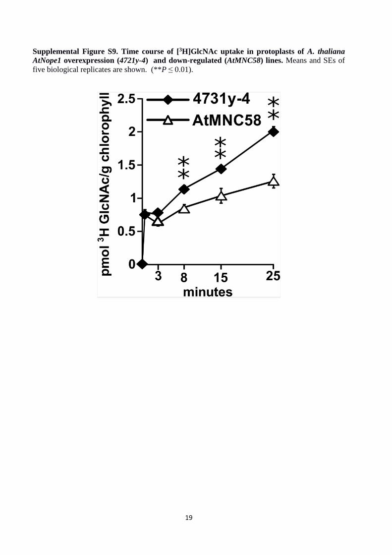
Supplemental Figure S9. Time course of [3H]GlcNAc uptake in protoplasts of A. thaliana
AtNope1 overexpression (4721y-4) and down-regulated (AtMNC58) lines. Means and SEs of
five biological replicates are shown. (**P ≤ 0.01).
20
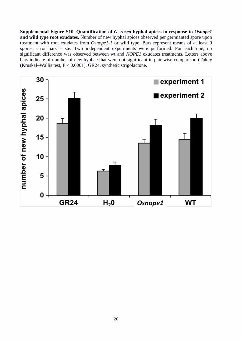
Supplemental Figure S10. Quantification of G. rosea hyphal apices in response to Osnope1
and wild type root exudates. Number of new hyphal apices observed per germianted spore upon
treatment with root exudates from Osnope1-1 or wild type. Bars represent means of at least 9
spores, error bars = s.e. Two independent experiments were performed. For each one, no
significant difference was observed between wt and NOPE1 exudates treatments. Letters above
bars indicate of number of new hyphae that were not significant in pair-wise comparison (Tukey
(Kruskal–Wallis test, P < 0.0001). GR24, synthetic strigolactone.
Osnope1
21
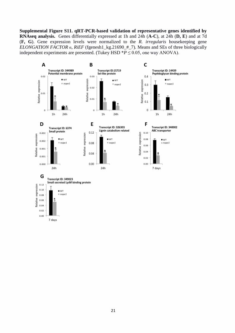
Supplemental Figure S11. qRT-PCR-based validation of representative genes identified by
RNAseq analysis. Genes differentially expressed at 1h and 24h (A-C), at 24h (D, E) and at 7d
(F, G). Gene expression levels were normalized to the R. irregularis housekeeping gene
ELONGATION FACTOR α, RiEF (fgenesh1_kg.21690_#_7). Means and SEs of three biologically
independent experiments are presented. (Tukey HSD *P ≤ 0.05, one way ANOVA).
22
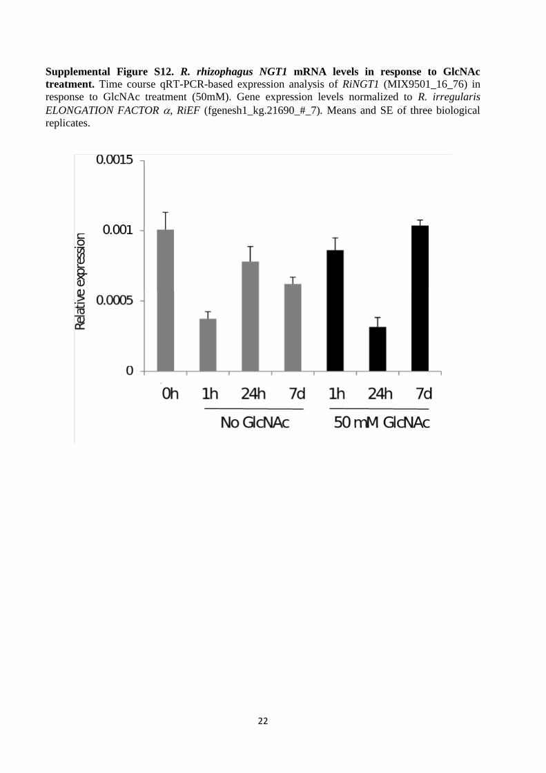
Supplemental Figure S12. R. rhizophagus NGT1 mRNA levels in response to GlcNAc
treatment. Time course qRT-PCR-based expression analysis of RiNGT1 (MIX9501_16_76) in
response to GlcNAc treatment (50mM). Gene expression levels normalized to R. irregularis
ELONGATION FACTOR , RiEF (fgenesh1_kg.21690_#_7). Means and SE of three biological
replicates.
23
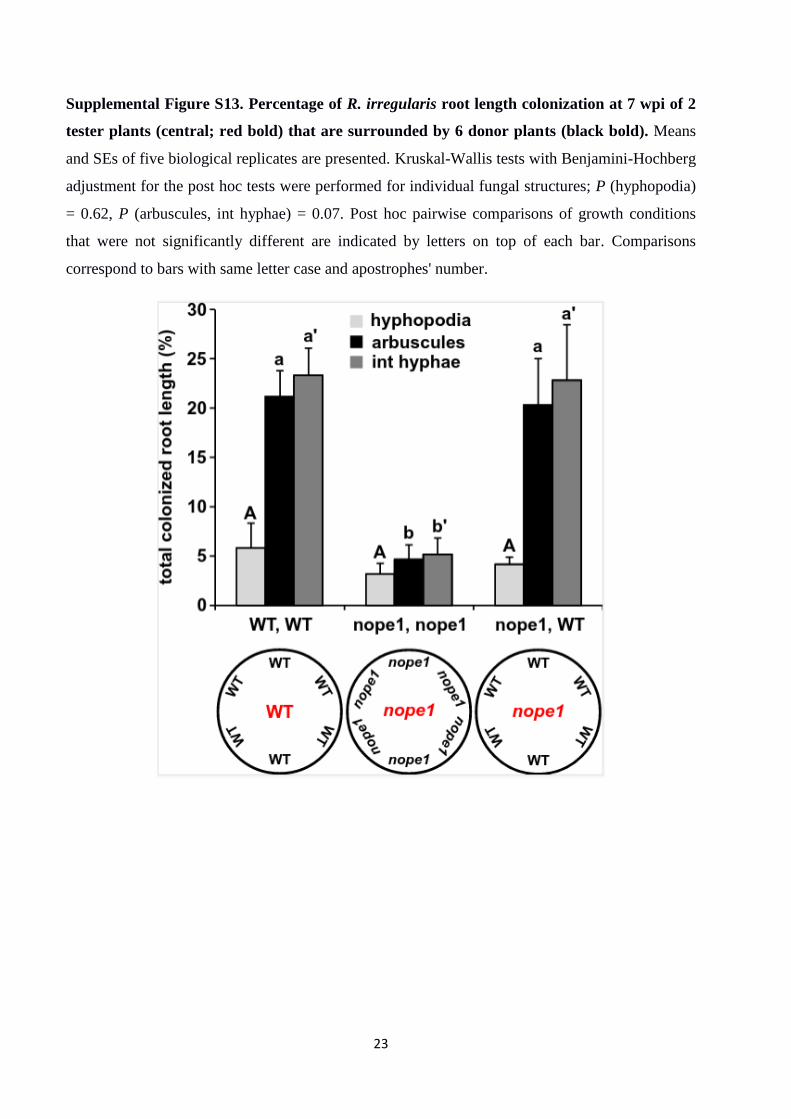
Supplemental Figure S13. Percentage of R. irregularis root length colonization at 7 wpi of 2
tester plants (central; red bold) that are surrounded by 6 donor plants (black bold). Means
and SEs of five biological replicates are presented. Kruskal-Wallis tests with Benjamini-Hochberg
adjustment for the post hoc tests were performed for individual fungal structures; P (hyphopodia)
= 0.62, P (arbuscules, int hyphae) = 0.07. Post hoc pairwise comparisons of growth conditions
that were not significantly different are indicated by letters on top of each bar. Comparisons
correspond to bars with same letter case and apostrophes' number.
24
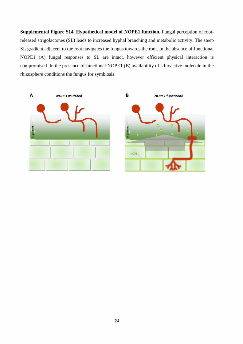
Supplemental Figure S14. Hypothetical model of NOPE1 function. Fungal perception of root-
released strigolactones (SL) leads to increased hyphal branching and metabolic activity. The steep
SL gradient adjacent to the root navigates the fungus towards the root. In the absence of functional
NOPE1 (A) fungal responses to SL are intact, however efficient physical interaction is
compromised. In the presence of functional NOPE1 (B) availability of a bioactive molecule in the
rhizosphere conditions the fungus for symbiosis.
25
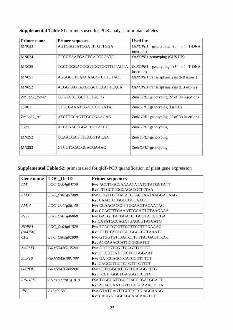
Supplemental Table S1: primers used for PCR analysis of mutant alleles
Primer name Primer sequence Used for
MN033 AGTCGGTATCGATTTGTTGGA OsNOPE1 genotyping (5’ of T-DNA
insertion)
MN034 GCCGTAATGAGTGACCGCATC OsNOPE1 genotyping (GUS RB)
MN035 TGGCGGGAGGGGTGGTGGTTGTAGTA OsNOPE1 genotyping (3’ of T-DNA
insertion)
MN051 AGGGCCTCAACAACGTCTTCTACT OsNOPE1 transcript analysis (RB exon1)
MN052 ACGGTAGTAAGCGCCCAATTCACA OsNOPE1 transcript analysis (LB exon2)
ZmLph2_forw2 CCTCATCTGCTTCTGCTG ZmNOPE1 genotyping (5’ of Ds insertion)
JSR01 GTTCGAAATCGATCGGGATA ZmNOPE1 genotyping (Ds RB)
ZmLph2_rv1 ATCTTCCAGTTGGCGAAGAG ZmNOPE1 genotyping (3’ of Ds insertion)
JGp3 ACCCGACCGGATCGTATCGG ZmNOPE1 genotyping
MN292 CCAACCAGCTCAGCTACAA
ZmNOPE1 genotyping
MN293 GTCCTCCACCGACGAAAC
ZmNOPE1 genotyping
Supplemental Table S2: primers used for qRT-PCR quantification of plant gene expression
Gene name LOC_Os ID Primer sequences
AM1 LOC_Os04g04750 Fw: ACCTCGCCAAAATATATGTATGCTATT
Rv: TTTGCTTGCCACACGTTTTAA
AM3 LOC_Os01g57400 Fw: CTGTTGTTACATCTACGAATAAGGAGAAG
Rv: CAACTCTGGCCGGCAAGT
AM14 LOC_Os11g26140 Fw: CCAACACCGTTGCAAGTACAATAC
Rv: GCACTTTGAAATTGGACTGTAAGAAA
PT11 LOC_Os01g46860 Fw: GATGTTACGGATCTGGGTATATCGA
Rv:CATATCCCAGATGAGCGTATCATG
NOPE1
(AM156)
LOC_Os04g01520 Fw: TCAGTGTGTTCCTTCCTTTGAAAG
Rv: TTTCTATACCATGGCCCCTAAATC
CP2 LOC_Os02g02890 Fw: GTGGTGTTAGTCTTTTTATGAGTTCGT
Rv: ACCAAACCATGGGCGATCT
ZmAM3 GRMZM2G135244 Fw: ATCTGTCGTTGCGTTCCTCT
Rv: GCATCTATC ACTGCGGGAAT
ZmPT6 GRMZM5G881088 Fw: GATCCAGCTCATCGGTTTCT
Rv: GAGCGTGGTGTGTTTGTTCT
GAPDH GRMZM2G046804 Fw: CTTCGGCATTGTTGAGGGTTTG
Rv: TCCTTGGCTGAGGGTCCGTC
AtNOPE1 At1g1800/At1g1810 Fw: TCGCCATTGGTTACGTGATGGACT
Rv: ACACGAATGGTCCCGCAAACTCTA
IPP2 A13g02780 Fw: GTATGAGTTGCTTCTCCAGCAAAG
Rv: GAGGATGGCTGCAACAAGTGT
26
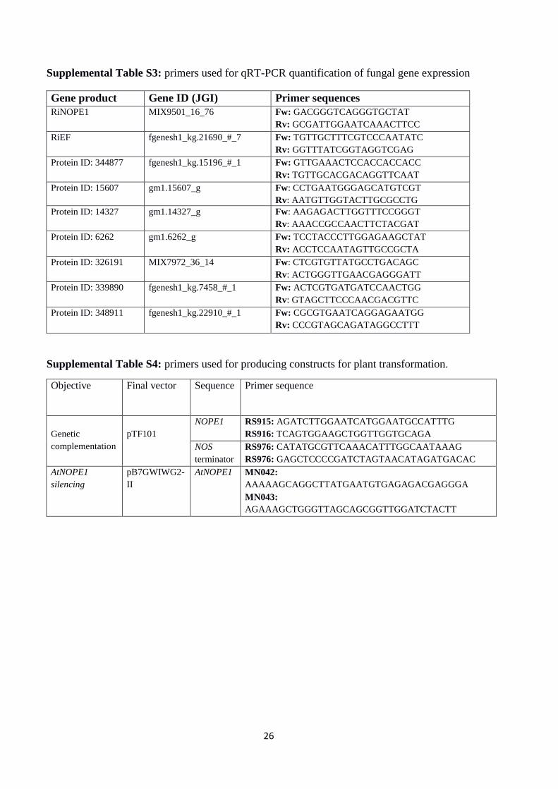
Supplemental Table S3: primers used for qRT-PCR quantification of fungal gene expression
Gene product Gene ID (JGI) Primer sequences
RiNOPE1 MIX9501_16_76 Fw: GACGGGTCAGGGTGCTAT
Rv: GCGATTGGAATCAAACTTCC
RiEF fgenesh1_kg.21690_#_7 Fw: TGTTGCTTTCGTCCCAATATC
Rv: GGTTTATCGGTAGGTCGAG
Protein ID: 344877 fgenesh1_kg.15196_#_1 Fw: GTTGAAACTCCACCACCACC
Rv: TGTTGCACGACAGGTTCAAT
Protein ID: 15607 gm1.15607_g Fw: CCTGAATGGGAGCATGTCGT
Rv: AATGTTGGTACTTGCGCCTG
Protein ID: 14327 gm1.14327_g Fw: AAGAGACTTGGTTTCCGGGT
Rv: AAACCGCCAACTTCTACGAT
Protein ID: 6262 gm1.6262_g Fw: TCCTACCCTTGGAGAAGCTAT
Rv: ACCTCCAATAGTTGCCGCTA
Protein ID: 326191 MIX7972_36_14 Fw: CTCGTGTTATGCCTGACAGC
Rv: ACTGGGTTGAACGAGGGATT
Protein ID: 339890 fgenesh1_kg.7458_#_1 Fw: ACTCGTGATGATCCAACTGG
Rv: GTAGCTTCCCAACGACGTTC
Protein ID: 348911 fgenesh1_kg.22910_#_1 Fw: CGCGTGAATCAGGAGAATGG
Rv: CCCGTAGCAGATAGGCCTTT
Supplemental Table S4: primers used for producing constructs for plant transformation.
Objective Final vector Sequence Primer sequence
Genetic
complementation
pTF101
NOPE1 RS915: AGATCTTGGAATCATGGAATGCCATTTG
RS916: TCAGTGGAAGCTGGTTGGTGCAGA
NOS
terminator
RS976: CATATGCGTTCAAACATTTGGCAATAAAG
RS976: GAGCTCCCCGATCTAGTAACATAGATGACAC
AtNOPE1
silencing
pB7GWIWG2-
II
AtNOPE1 MN042:
AAAAAGCAGGCTTATGAATGTGAGAGACGAGGGA
MN043:
AGAAAGCTGGGTTAGCAGCGGTTGGATCTACTT
27
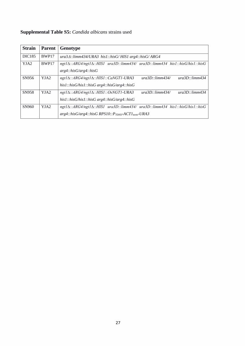
Supplemental Table S5: Candida albicans strains used
Strain Parent Genotype
DIC185 BWP17 ura3::limm434/URA3 his1::hisG/ HIS1 arg4::hisG/ ARG4
YJA2 BWP17 ngt1∆::ARG4/ngt1∆::HIS1 ura3D::limm434/ ura3D::limm434 his1::hisG/his1::hisG
arg4::hisG/arg4::hisG
SN956 YJA2 ngt1∆::ARG4/ngt1∆::HIS1::CaNGT1-URA3 ura3D::limm434/ ura3D::limm434
his1::hisG/his1::hisG arg4::hisG/arg4::hisG
SN958 YJA2 ngt1∆::ARG4/ngt1∆::HIS1::OsNGT1-URA3 ura3D::limm434/ ura3D::limm434
his1::hisG/his1::hisG arg4::hisG/arg4::hisG
SN960 YJA2 ngt1∆::ARG4/ngt1∆::HIS1 ura3D::limm434/ ura3D::limm434 his1::hisG/his1::hisG
arg4::hisG/arg4::hisG RPS10::PTDH3-ACT1term-URA3