Embed Size (px)
Citation preview
A R T I C L E S
A M E L T I N G P O I N T FOR T H E
B I R E F R I N G E N T C O M P O N E N T OF M U S C L E
J O H N F . A R O N S O N
From the Department of Biological Structure, University of Washington, Seattle
A B S T R A C T
The A filament of the striated muscle sarcomere is an ordered aggregate of one or a few species of proteins. Ordering of these filaments into a parallel array is the basis of birefringence in the A region, and loss of birefringencc is therefore a measure of decreased order. Heat ing caused a large decrease in the birefringence of glycerinated rabbit psoas muscle fibers over a narrow temperature range ( ~ 3°C) and a large decrease in both the birefringence and op- tical density of the A region of Drosophila melanogaster fibrils. These changes were interpreted as a loss of A filament structure and were used to define a transition temperature (Ttr) as a measure of the stability of the A region. Since the transition temperature was sensitive to pH, ionic strength, and urea, solvent conditions which often affect protein structure, it is an ex- perimentally useful indicator for factors affecting the structure of the A filament. Fibers from glycerinated frog muscle were less stable over a wide pH range than fibers from glycerinated rabbit muscle, a fact which demonstrates a species difference in structure. Glycerinated rabbit fibrils heated to 70°C shortened to about 40% of their initial length. The extent of shortening was not correlated with the loss of blrefringence, and phase-contrast microscopy showed that this shortening occurred in the I region as well as in the A region. This response may be useful for studying the I filament and actin in much the same way that the decrease in birefringence was used for studying the A filament and myosin. The observations pre- sented show that some properties of muscle proteins can be studied essentially in situ without the necessity of first dispersing the structure in solutions of high or low ionic strength.
I N T R O D U C T I O N
I intend to show in this paper that heat can be used in studying the structure of the A region of striated muscle. This region is characterized by filaments composed largely of the protein myosin which, because of their birefringence, can be optically isolated in a s t a t c approaching that found in vivo.
Changes in the state of aggregation and orien-
tation of many proteins can be induced by heat, since most are stabilized by relatively weak non- covalent bonds. These changes may be inde- pendent of, correlated with, or in addition to, changes in the conformation of individual mole- cules or parts of molecules. Chemical modifica- tion, solvent properties, and competitive bonding agents, each studied as a function of the rate of
453
on April 3, 2019jcb.rupress.org Downloaded from http://doi.org/10.1083/jcb.30.3.453Published Online: 1 September, 1966 | Supp Info:
h e a t d en a t u r a t i o n , o r less des i rab ly of a de- n a t u r a t i o n t e m p e r a t u r e , can modi fy in te rac t ions a n d give some ind ica t ion of the types and i mp o r - t ance of " b o n d s " w h i c h d e t e r m i n e a given m a c r o - molecu la r s t ruc ture (1).
Bi re f r ingence and X - r a y d i f f rac t ion are two proper t ies of the A region of s t r ia ted muscle w h i c h have been used to de t ec t h e a t - i n d u c e d c b a n g es in aggrega te s t ructure . Vles (2) ob- served a decrease in the b i re f r ingence of freshly isolated muscle f rom the tho rax of the beetle , Dytiscus, f rom the a b d o m e n of the crayfish, a n d f rom uniden t i f i ed frog and mussel muscles w h e n these were hea t ed to a b o u t 50°C. Shor t en ing of the muscle compl i ca t ed the in t e rp re t a t ion of the b i re f r ingence changes . A s t b u r y (3) and also R u d a l l (4) have similar ly no ted changes in the wide angle X - r a y d i f f rac t ion p a t t e rn of frog stri- a t ed muscle h ea t ed at a b o u t 60°C and of acto- myos in films w h e n these were hea t ed to above 40°C.
T h e fol lowing work shows tha t changes de- tec table wi th the l ight microscope a c c o m p a n y the hea t ing of s t r ia ted muscle a n d uses these changes to def ine p a r a m e t e r s wh ich affect the stabil i ty o f the A f i laments.
M A T E R I A L S A N D M E T H O D S
Flight muscle fibrils were obtained from an Oregon R strain of Drosophila melanogast~r. Preparations of fibrils were made by teasing pieces of thoracic muscle in a drop of a solution containing 0. l M KC1, 5 uaM MgCl2, 2.5 mM EDTA1,5 mM potassium phosphate , and 2.5 m~ sodium A T P 2 at a final p H of 7.0. A cover glass with silicone grease 3 on two opposite edges was placed on the drop and pressed to make the fibrils stick to the cover glass. Free fibrils, sarcosomes, and other debris were then washed away before the isolating med ium was exchanged for a solution containing 0.1 M KCI and 5 mM potassium phosphate at p H 7.0 and then for the desired experimental medium. Finally, the prepara t ion was sealed with silicone grease. Phase- contrast observations were made with an NA 1.25 phase objective in conjunct ion with a long working distance condenser. Polarized light observations were made with a pr ism-equipped polarizing microscope by utilizing an NA 0.65 objective, an NA 0.5 objec- tive used as a condenser, and a mica compensator. I observed preparat ions of fibrils being heated by using a slide with a t ransparent resistance film (Arthur H. Thomas, Co., Philadelphia, Pennsylvania).
* EDTA, ethylene diamine tetraacetate. 2 ATP, adenosine tr iphosphate. a Silicone grease, Dow-Corning High Vacuum, Mid- land, Michigan.
Rabbi t psoas muscle was separated into bundles of fibers which were tied to sticks and placed in ice cold 50% glycerol containing 5 mM potassium phosphate, 20 mM potassium chloride, and 2 m u EDTA at a final p H of 7.0. The fibers were placed in fresh glycerol solution 24 hr later and stored at -- 10°C for 6 wk to 1 yr. Before use, small fiber bundles ( ~ 0.2 m m in diameter) were equilibrated in 0.1 M KC1 con- taining 5 mM potassium phosphate at p H 7.0, for at least 1 hr. Single fiber lengths about 5 m m long were teased from these bundles and mounted without flattening between a slide and cover glass. The prep- aration was then sealed with a silicone grease.
Glycerinated frog muscle was prepared from the lower hind leg of a frog (Rana pipiens). Skin and the gastrocnemius muscle were removed, after which the leg was tied to a straight stick. The glycerination pro- cedure was the same one used for rabbi t psoas muscle, except that the frog muscles were not separated into smaller bundles and were left at tached to their inser- tions. Preparations of frog fibers were usually made from the peripheral part of the soleus muscle in the same manner as rabbi t psoas fiber preparations.
Glycerinated frog and rabbi t muscle fibers were observed with a polarizing microscope at a magnifica- tion of 200, a particular region of the fiber where the birefringence was uniform was noted, and the re tarda- tion and fiber width at this point were measured. After these initial measurements, the slide was heated for 2 rain, and then the re tardat ion and width were measured again.
Slides were heated by immersion in a beaker of water which was slowly stirred. The tempera ture decrease of the bath over a 2 min period at 50°C was about half a degree, and the nominal temperature was considered to be the initial temperature. Experi- ments with heating times longer than 2 rain were per- formed in a bath regulated to 4-0.1 °C.
I measured birefringence with a quartz wedge using a 10 X NA 0.25 objective as a condenser in conjunct ion with a 20 X NA 0.5 objective and a 10 X friar micrometer. A water-cooled GE AH-6 mercury arc used with a Kodak 77 A filter served as the source.
Reagent grade inorganic chemicals and distilled water were used throughout. Mersalyl 4 was obtained from Winthrop Laboratories, New York, New York, A T P from Pabst Research Laboratories, Milwaukee, Wisconsin, N-acetyl t ryp tophan and N-acetyl histi- dine from California Corporat ion for Biochemical Research, Los Angeles, California; sodium deoxycho- late from Difco Laboratories, Inc. Detroit, Michigan, and sodium dodecylsulfate was a commercial grade preparat ion from E. H. Sargent & Co., Chicago, Illinois. The p H of all solutions was determined at room tempera ture and this value was used as the
4 Mersalyl (Salyrgan), salicyl-(T-hydroxymercuri-/3- methoxypropyl) -amide- O-acetic acid.
454 THE JOIrRNAL OF CELL BIoIxmY • VOL~YME 30, 1966
nominal pH at other temperatures. The effect of temperature on the phosphate buffer generally used was not studied, but the work of Bates and Acree (5) indicates that the change in pH between 24 ° and 60°C will be small at pH 7.
R E S U L T S
Drosophila
CHANGES OBSERVED BY L I G H T MICROSCOPY
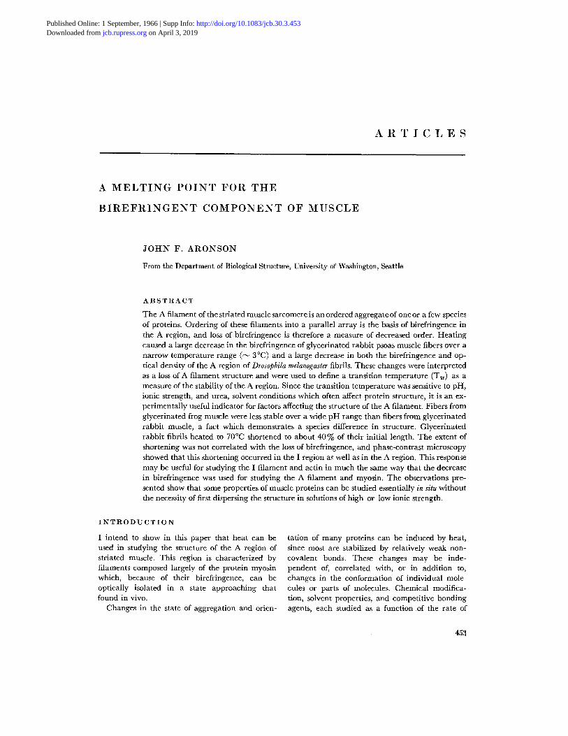
P H A S E - C O N T R A S T M I C R O S C O P Y : Th e phase- contras t appearance of a freshly isolated Drosophila fibril is shown in the first pho tograph of Fig. 1. H and I zones are clear, wi th the I zone being the wider of the two. A Z line may be visible bu t is rarely obvious, while each half A band has a suggestion of three or four faint striae which may be optical artifacts.
W h e n a Drosophila fibril was heated, a charac- teristic sequence of changes was visible by phase- contras t microscopy. Fig. 1 shows the pa t te rn of changes as the fibril was hea ted to progressively higher tempera tures in 0.1 M KCI at p H 7. After an init ial decrease in diameter , the first sign of change was in the H zone which became wider. As this zone cont inued to widen, an M line be- came visible and gradually denser, giving the appearance of a C m band. The denser regions remain ing at the edges of the A region in adja- cent sarcomeres gave the appearance of doublets centered on the Z line. Wi th fur ther heating, these doublets often merged into a single band. A similar pa t t e rn of changes was seen in 0.1 M KC1 at p H 6 and at p H 4 and in 0.1 M KC1 conta in ing 2.5 mM A T P + 5 mM MgCl~ + 2.5 mM E D T A at pH 7.0. W h e n fibrils in 0.1 M KC1 at pH 8.5 were heated, the changes which occurred were similar bu t not so clear and may have been com- plicated by extraction. The use of letters to refer to the striae of heated muscle is a convenience in describing the relat ive position and density of a region and does not imply tha t the striae of heated muscle are necessarily structurally comparable to the striae of unhea ted muscle.
There was little decrease in sarcomere length (less t han 2 % in Fig. l) when fibrils were heated in 0.1 M KC1 at p H 7 to tempera tures at which there were marked cytological changes, bu t short- tening to abou t 90% of the init ial length was often seen when fibrils were heated in 0.1 M KC1 at p H 8.5.
P O L A R I Z E D L I G H T M I C R O S C O P Y : T h e image of relaxed Drosophila fibrils in polarized l ight is complex, as shown in Fig. 2. T h e struc-
tura l significance of this image is uncer ta in and fibrils which have been dehydra ted in acetone and immersed in n i t robenzene may provide a more direct image of the intrinsic structure of the myofibril. Acetone caused a decrease in fibril d iameter of roughly 6 0 % to a d iameter of about 1 #, and the n i t robenzene gives an approximate refractive index match. 5 Such fibrils do not show hyperstr iae in the A region, a tr iplet Z structure, or a birefr ingent M line. Instead, the A region is more evenly birefringent, a l though a slightly less birefr ingent central region can often be de- tected.
In order to compare the phase-contrast and the polarized l ight appearances of fibrils after heat ing, prepara t ions of isolated Drosophila fibrils in 0.1 M KC1 were hea ted to a given t empera tu re for 2 min. They were then observed by phase-contrast microscopy to de te rmine the range of sarcomere pa t te rns characterist ic of the prepara t ion . T h e same slides were then dehydra ted in acetone, immersed in ni t robenzene, and examined with polarized light. Var iabi l i ty among fibrils within a prepara t ion occurred and appeared to be a proper ty of the fibrils r a the r t han of large regions within the preparat ions.
A comparison of polarized l ight and phase- contrast images for heated fibrils is shown in Fig. 3. The first change in pa t te rn seen by phase contras t was widening of the H zone at abou t 48°C (0.1 M KCI, pH 7, 2 min heat ing) . This zone was not obviously present in polarized l ight at 48°C, and a large, easily noticed decrease in birefrin- gence was not seen unti l abou t 52°C. At this t empera tu re the center of the sarcomere an d the ends of the A region appeared more "s tab le" t han the in tervening regions. Wi th fur ther heat- ing, the birefringence decreased everywhere, bu t the same pa t te rn was retained.
IN VIvo COMPARISON
T h e relat ion of the s tructure of isolated fibrils to fibril s tructure within the an imal was studied by heat ing in tac t flies wi th a 2 min immersion. No cytological signs of strong shortening in terms of Cz and C m bands were seen in fibrils f rom
5 The refractive index of nitrobenzene is intermediate to the refractive indices of acetone-dehydrated Dro- sophila muscle fibrils. When one observes a fibril with green polarized light at 25°C by phase-contrast mi- croscopy, it is possible to see images which are matched, overmatched, or undermatched, depending on the plane of polarization.
J. F. ARONSON Melting of Muscle Birefringence 455
FiGm~n 1 Changes seen when a fibril from Drosophila indirect flight muscle was slowly heated from ~4°C to about 70°C in 0.1 M KC1 at pH 7. The first photograph in the series was taken at room tempera- ture. Phase-contrast. Scale divisions are 10 #. X ~600.
456
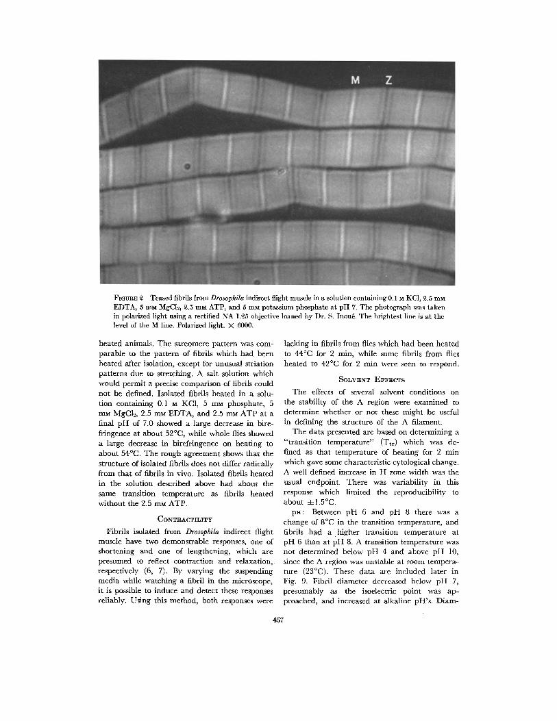
FIOVRE 3 Teased fibrils from Drosophila indirect flight muscle in a solution containing 0.1 M KCI, 3.5 mM EDTA, 5 mM MgCI2, 3.5 m ~ ATP, and 5 mM potassium phosphate at pH 7. The photograph was taken in polarized light using a rectified NA 1.35 objective loaned by Dr. S. Inou~. The brightest line is at the level of the M line, Polarized light. X 6000,
hea ted animals. The sarcomere pa t te rn was com- parable to the pa t te rn of fibrils which had been hea ted after isolation, except for unusual striation pa t te rns due to stretching. A salt solution which would permi t a precise comparison of fibrils could not be defined. Isolated fibrils hea ted in a solu- t ion conta in ing 0.1 M KC1, 5 rnM phosphate, 5 mM MgC12, 2.5 mM EDTA, and 2.5 mM A T P at a final p H of 7.0 showed a large decrease in bire- fr ingence at about 52°C, while whole flies showed a large decrease in birefr ingence on hea t ing to about 54°C. The rough agreement shows tha t the s tructure of isolated fibrils does not differ radically f rom tha t of fibrils in vivo. Isolated fibrils hea ted in the solution described above had about the same transi t ion tempera ture as fibrils heated wi thout the 2.5 m u ATP.
CONTRACTILITY
Fibrils isolated from Drosophila indirect flight muscle have two demonst rab le responses, one of shortening and one of lengthening, which are presumed to reflect contrac t ion and relaxation, respectively (6, 7). By varying the suspending media while watching a fibril in the microscope, i t is possible to induce and detect these responses reliably. Using this method, bo th responses were
lacking in fibrils from flies which had been hea ted to 44°C for 2 min, while some fibrils f rom flies hea ted to 42°C for 2 min were seen to respond.
SOLVENT EFFECTS
The effects of several solvent condit ions on the stability of the A region were examined to de te rmine whe ther or no t these might be useful in defining the structure of the A filament.
T h e da ta presented are based on de te rmin ing a " t rans i t ion t empera tu re" (Ttr) which was de- fined as tha t t empera ture of hcat ing for 2 rain which gave some characterist ic cytological change. A well defined increase in H zone width was the usual cndpoint . There was variabi l i ty in this response which l imited the reproducibi l i ty to about -4-1.5°C.
pH: Between p H 6 and p H 8 there was a change of 8°C in the t ransi t ion tempera ture , and fibrils h a d a h i g h e r t ransi t ion tempera ture at p H 6 t han at pH 8. A transi t ion t empera tu re was not de termined below p H 4 and above pH 10, since the A region was unstable at room tempera- turc (23°C). These da ta are included later in Fig. 9. Fibri l d iameter decreased below p H 7, presumably as the isoclectric point was ap- proached, and increased at alkaline pH's. D iem-
457
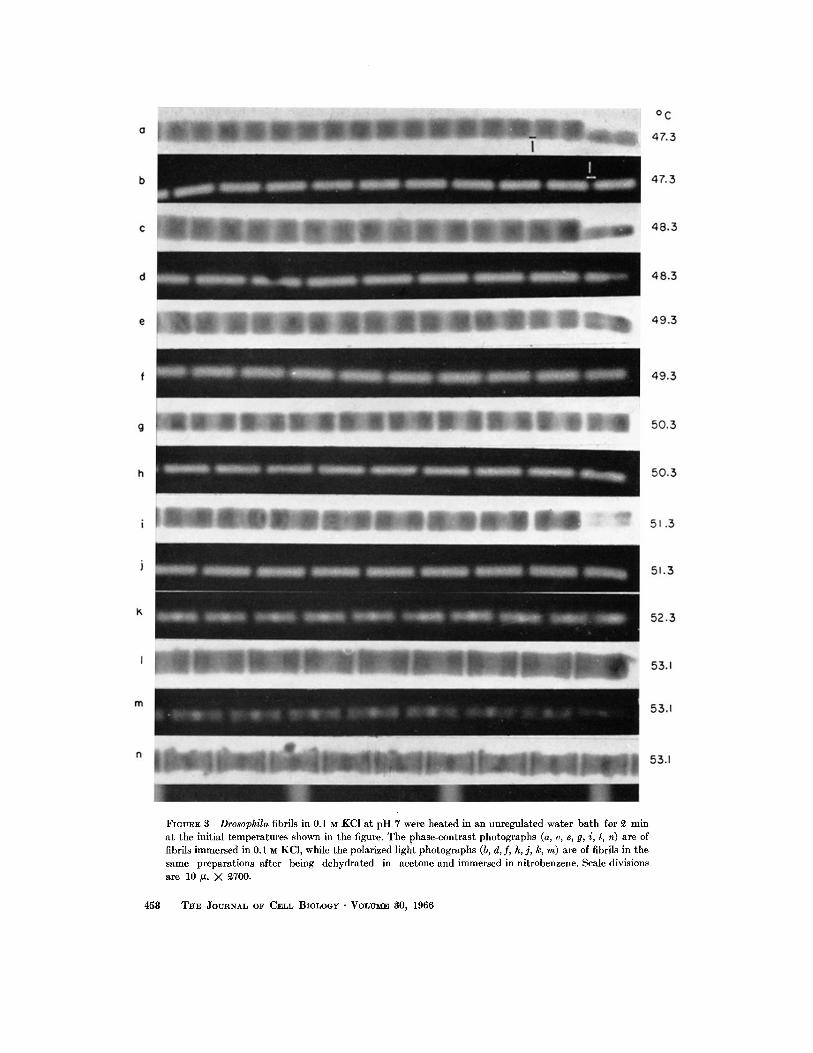
FIGURE 3 Drosophila fibrils in 0.1 M KCI a t pH 7 were heated in an unregulated water ba th for ~ min a t the initial temperatures shown in the figure. The phase-contrast photographs (a, c, e, g, i, l, n) are of fibrils immersed in 0.1 M KC1, while the polarized light photographs (b, d, fi h, j, k, m) are of fibrils in the same preparations after being dehydrated in acetone and immersed in nitrobenzene. Scale divisions are 10/z. )< 2700.
458 THE JOURNAL OF CELL BIOLOGr • VOLV~E 30, 1966
eter changes between p H 5 and p H 9 appeared reversible despite some extract ion at pH 9.
C A T I O N S : Sodium, potassium, and l i th ium chlorides at p H 7 reduced the t ransi t ion tempera- ture in a similar way at concentra t ions of 0.2 and 0.4 molar. Calc ium and, less markedly, magne- sium chlorides at equivalent ionic strengths de ~ creased the thermal stability even more.
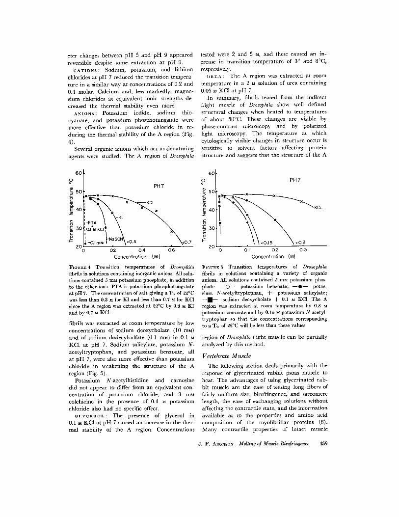
A N I O N S : Potassium iodide, sodium thio- cyanate, and potassium phosphotungsta te were more effective than potassium chloride in re- ducing the the rmal stability of the A region (Fig.
4). Several organic anions which act as dena tu r ing
agents were studied. The A region of Drosophila
6O O o
5O :3
E 4 0
g :~ so C O
2O
tested were 2 and 5 M, and these caused an in- crease in t ransi t ion t empera tu re of 3 ° and 8°C,
respectively. UREA: The A region was extracted at room
tempera ture in a 2 M solution of urea conta in ing
0.05 M KC1 at p H 7. In summary, fibrils teased from the indirect
flight muscle of Drosophila show well defined structural changes when heated to tempera tures of abou t 50°C. These changes are visible by phase-contrast microscopy and by polarized light microscopy. The tempera ture at which cytologically visible changes in structure occur is sensitive to solvent factors affecting protein structure and suggests tha t the structure of the A
6O (D
PH7 o 50
KCI ~.
-PTA g
, - o . , , . , , , , • 2C
o O2 0.4 0.6 Concentration (M)
FIGURE ~ Transition temperatures of Drosophila fibrils in solutions containing inorganic anions. All solu- tions contained 5 mM potassium phosphate, in addition to the other ions. PTA is potassium phosphotungstate at pH 7. The concentration of salt giving a Ttr of ~2°C was less than 0.S M for KI and less than 0.7 ~ for KCI since the A region was extracted at ~£°C by 0.3 M KI and by 0.7 M KC1.
fibrils was extracted at room tempera tu re by low concentra t ions of sodium deoxycholate (10 raM) and of sodium dodccylsulfate (0.1 re_M) in 0.1 M KC1 at p H 7. Sodium salicylate, potassium N- ace ty l twptophan , and potassium benzoate, all a t p H 7, were also more effective than potassium chloride in weakening the structure of the A region (Fig. 5).
Potassium N-acetylhist idine and carnosine did not appear to differ from an equivalent con- cent ra t ion of potassium chloride, and 3 mM colchicine in the presence of 0.1 M potassium chloride also had no specific effect.
OLYCF.ROL: The presence of glycerol in 0.1 M KC1 at p H 7 caused an increase in the ther- mal stability of the A region. Concent ra t ions
PH7
T \\ \ 0 0.1 0.2 0.5
Concentration (M)
FIGURE 5 Transition temperatures of Drosophila fibrils in solutions containing a variety of organic anions. All solutions contained 5 mM potassium phos- phate. - - O - - potassium benzoate; - - @ - - potas- sium N-acetyltryptophan, + potassium salicylate; - - . - - sodium deoxycholate + 0.1 M KC1. The A region was extracted at room temperature by 0.3 M potassium benzoate and by 0.15 M potassium N-acetyl- tryptophan so that the concentrations corresponding to a Tt, of ~ ° C will be less than these values.
region of Drosophila f i g h t muscle can be part ial ly analyzed by this method.
Vertebrate Muscle
The following section deals pr imari ly wi th the response of glycerinated r abb i t psoas muscle to heat. The advantages of using glycerinated rab- bi t muscle are the ease of teasing long fibers of fairly uniform size, birefringcnce, and sarcomere length, the case of exchanging solutions wi thout affecting the contracti le state, and the informat ion avai lable as to the propert ies and amino acid composit ion of the myofibri l lar proteins (8). M a n y contracti le properties of in tac t muscle
J. F. ARONSON Melting of Muscle Birefringence 459
I00
75
0 0
x 50
o.__o 25
X K IcU
xWidth (u) x N
• • Retardat ion ( • ) $
• . t I
pH ZO • 0,I M KCI
X
50 55 60 65 Temperature °C
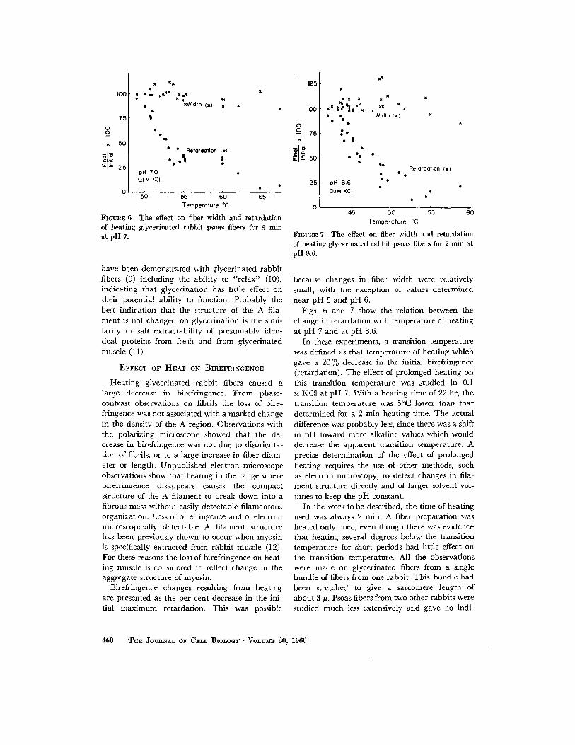
FIGURE 6 The effect on fiber width and retardation of heating glycerinated rabbit psoas fibers for ~ rain at pH 7.
have been demonstrated with glycerinated rabbit fibers (9) including the ability to "re lax" (10), indicating that glycerination has little effect on their potential ability to function. Probably the best indication that the structure of the A fila- ment is not changed on glycerination is the simi- larity in salt extractability of presumably iden- tical proteins from fresh and from glycerinated muscle (11).
E F F E C T OF H E A T ON B I R E F R I N G E N C E
Heat ing glycerinated rabbit fibers caused a large decrease in birefringence. From phase- contrast observations on fibrils the loss of bire- fringence was not associated with a marked change in the density of the A region. Observations with the polarizing microscope showed that the de- crease in birefringence was not due to disorienta- tion of fibrils, or to a large increase in fiber diam- eter or length. Unpublished electron microscope observations show that heating in the range where birefringence disappears causes the compact structure of the A filament to break down into a fibrous mass without easily detectable filamentous organization. Loss of birefringence and of electron microscopically detectable A filament structure has been previously shown to occur when myosin is specifically extracted from rabbit muscle (12). For these reasons the loss of birefringence on heat- ing muscle is considered to reflect change in the aggregate structure of myosin.
Birefringence changes resulting from heating are presented as the per cent decrease in the ini- tial maximum retardation. This was possible
125
100
0 _o 75 x
25
x
x x x x x x
x x x x t • Width (x) x
• x
g , • !
• e : •
$e Retardat ion ( • )
~ e ¢ • oH 8 .6
O . I M K C I
45 5O 55 6O Temperature °C
FIGURE 7 The effect OR fiber width and retardation of heating glycerinated rabbit psoas fibers for £ min at pH 8.6.
because changes in fiber width were relatively small, with the exception of values determined near pH 5 and pH 6.
Figs. 6 and 7 show the relation between the change in retardation with temperature of heating at pH 7 and at pH 8.6.
In these experiments, a transition temperature was defined as that temperature of heating which gave a 20% decrease in the initial birefringence (retardation). The effect of prolonged heating on this transition temperature was studied in 0.I M KC1 at pH 7. With a heating time of 22 hr, the transition temperature was 5°C lower than that determined for a 2 min heating time. The actual difference was probably less, since there was a shift in pH toward more alkaline values which would decrease the apparent transition temperature. A precise determination of the effect of prolonged heating requires the use of other methods, such as electron microscopy, to detect changes in fila- ment structure directly and of larger solvent vol- umes to keep the p H constant.
In the work to be described, the time of heating used was always 2 rain. A fiber preparation was heated only once, even though there was evidence that heating several degrees below the transition temperature for short periods had little effect on the transition temperature. All the observations were made on glycerinated fibers from a single bundle of fibers from one rabbit. This bundle had been stretched to give a sarcomere length of about 3 #. Psoas fibers from two other rabbits were studied much less extensively and gave no indi-
460 THE JOURNAL OF CELL BIOLOGy " VOLUME 80, 1966
::L v
£ 2 (D
( D
E I O o
09
X x
x
oH7
0.I N KCI
X
0 20 6'0 8'0 ,oo
Temperature °C
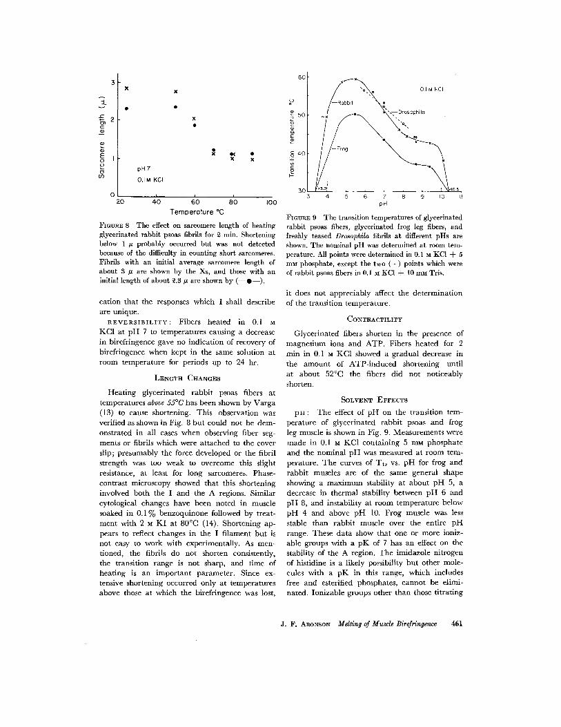
FIGURE 8 The effect on sareomere length of heating glycerinated rabbit psoas fibrils for ~ rain. Shortening below 1 IZ probably occurred but was not detected because of the difficulty in counting short sarcomeres. Fibrils with an initial average sarcomere length of about 3 # are shown by the Xs, and those with an initial length of about ~.3/~ are shown by ( - - 0 - - ) .
cation tha t the responses which I shall describe are unique.
REVERSIBILITY: Fibers heated in 0.1 M KCI at p H 7 to tempera tures causing a decrease in birefringenee gave no indicat ion of recovery of birefringence when kept in the same solution at room tempera ture for periods up to 24 hr.
LENGTH CHANGES
Hea t ing glycerinated rabb i t psoas fibers at tempera tures above 55°C has been shown by Varga (13) to cause shortening. This observation was verified as shown in Fig. 8 bu t could not be dem- onstrated in all cases when observing fiber seg- ments or fibrils which were a t tached to the cover slip; presumably the force developed or the fibril s t rength was too weak to overcome this slight resistance, at least for long sarcomeres. Phase- contras t microscopy showed tha t this shortening involved bo th the I and the A regions. Similar cytological changes have been noted in muscle soaked in 0 .1% benzoquinone followed by treat- mcn t with 2 M K I at 80°C (14). Shor tening ap- pears to reflect changes in the I f i lament bu t is not easy to work with experimentally. As men- tioned, the fibrils do not shorten consistently, the t ransi t ion range is not sharp, and t ime of heat ing is an impor t an t parameter . Since ex- tensive shortening occurred only at tempera tures above those at which the birefringence was lost,
6 0
/ ~ ' - x " o ~ . ~ 0.1 KCI M
/-R0bb. \ ; [ , ~ ' . -- Drosophilo
e / " . . ~.. .~,,
J i
30 ,3.3a <I05 4 ; ; ~ ; ; ,0 ,
pH
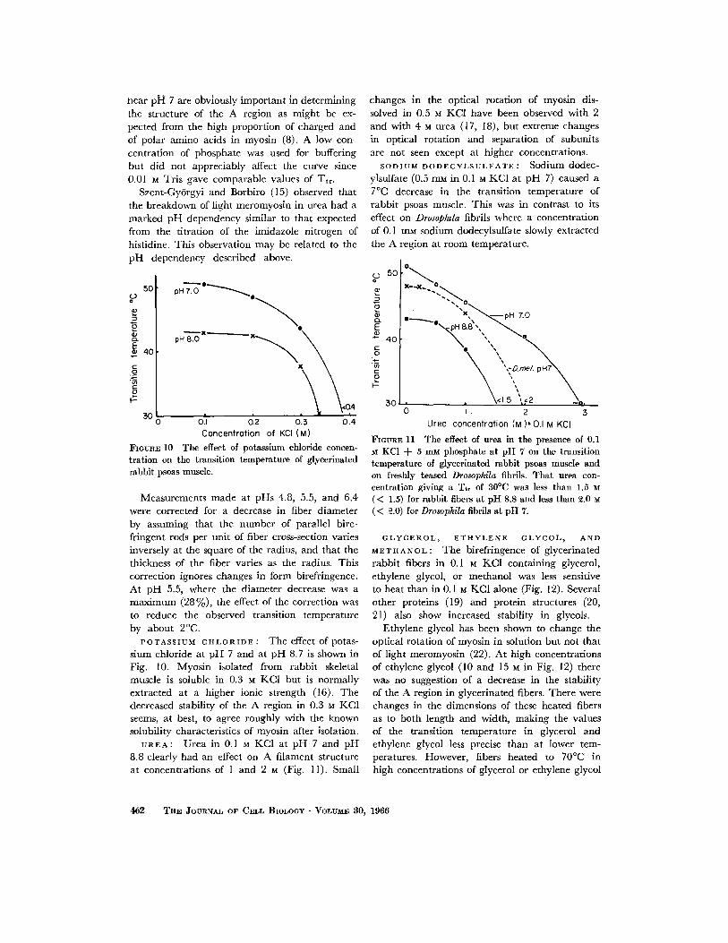
F m ~ E 9 The transition temperatures of glyeerinated rabbit psoas fibers, glyeerinated frog leg fibers, and freshly teased Drosophila fibrils at different pHs are shown. The nominal pH was determined at room tem- perature. All points were determined in 0.1 ~ KC1 + 5 mM phosphate, except the two (+) points which were of rabbit psoas fibers in 0.1 M KCI + 10 mM Tris.
it does not appreciably affect the de te rmina t ion of the t ransi t ion temperature .
CONTRACTILITY
Glycer inated fibers shorten in the presence of magnes ium ions and ATP. Fibers hea ted for 2 rain in 0.1 M KC1 showed a gradual decrease in the a m o u n t of ATP- induced shortening unt i l a t abou t 52°C the fibers did not not iceably shorten.
SOLVENT EFFECTS
pH : The effect of pH on the transition tem-
perature of glycerinated rabbit psoas and frog
leg muscle is shown in Fig. 9. Measurements were made in 0.1 u KC1 conta in ing 5 mM phosphate and the nomina l p H was measured at room tem- perature. The curves of Tt r vs. pH for frog and r abb i t muscles are of the same general shape showing a m a x i m u m stability at about p H 5, a decrease in the rmal stability between p H 6 and p H 8, and instabil i ty at room tempera tu re below p H 4 and above p H 10. Frog muscle was less stable t han rabb i t muscle over the entire pH range. These da ta show tha t one or more ioniz- able groups wi th a p K of 7 has an effect on the stability of the A region. The imidazole n i t rogen of histidine is a likely possibility bu t other mole- cules with a p K in this range, which includes free and esterified phosphates, canno t be elimi- nated. Ionizable groups other than those t i t ra t ing
J. F. ARONSON Melting of Muscle Birefringence 4{i1
near plaI 7 are obviously important in determining the structure of the A region as might be ex- pected from the high proportion of charged and of polar amino acids in myosin (8). A low con- centration of phosphate was used for buffering but did not appreciably affect the curve since 0.01 M Tris gave comparable values of Ttr.
Szent-Gy6rgyi and Borbiro (15) observed that the breakdown of light meromyosin in urea had a marked pH dependency similar to that expected from the titration of the imidazole nitrogen of histidine. This observation may be related to the pH dependency described above.
50 u
r~ E 40
.8 -g g
3O 0
pH 7.---'~ ° ~ e
.4 0.1 0,2 0 .3 0 . 4
Concentrcflion of KCI (M)
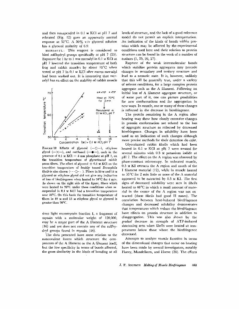
FIGURE 10 The effect of potassium chloride concen- tration on the transition temperature of glycerinated rabbit psoas muscle.
Measurements made at pHs 4.8, 5.5, and 6.4 were corrected for a decrease in fiber diameter by assuming that the number of parallel bire- fringent rods per unit of fiber cross-section varies inversely at the square of the radius, and that the thickness of the fiber varies as the radius. This correction ignores changes in form birefringence. At pH 5.5, where the diameter decrease was a maximum (28 %), the effect of the correction was to reduce the observed transition temperature by about 2°C.
POTASSIUM CHLORIDE : The effect of potas- sium chloride at pH 7 and at pH 8.7 is shown in Fig. 10. Myosin i~olated from rabbit skeletal muscle is soluble in 0.3 M KC1 but is normally extracted at a higher ionic strength (16). The decreased stability of the A region in 0.3 M KC1 seems, at best, to agree roughly with the known solubility characteristics of myosin after isolation.
UREA: Urea in 0.1 M KCI at pH 7 and pH 8.8 clearly had an effect on A filament structure at concentrations of 1 and 2 M (Fig. 11). Small
changes in the optical rotation of myosin dis- solved in 0.5 M KC1 have been observed with 2 and with 4 u urea (17, 18), but extreme changes in optical rotation and separation of subunits are not seen except at higher concentrations.
SODIUM DODECYLSULFATE : Sodium dodec- ylsulfate (0.5 mM in 0.1 M KC1 at pH 7) caused a 7°C decrease in the transition temperature of rabbit psoas muscle. This was in contrast to its effect on Drosop&la fibrils where a concentration of 0.1 mM sodium dodecylsulfate slowly extracted the A region at room temperature.
50°~
e ~ , pH Z 0
A ",, ""x. 40 ~ ~ e g
s o . , \ . , s o I 2 3
Ureo concentrofion (M)*0.1M KCI
FIGURE 11 The effeet of urea in the presence of 0.1 M KC1 + 5 mM phosphate at pH 7 on the transition temperature of glycerinated rabbit psoas muscle and on freshly teased Drosophila fibrils. That urea con- centration giving a Tt, of S0°C was less than 1.5 M (< 1.5) for rabbit fibers at pH 8.8 and less than 2.0 M (< ~.0) for Drosophila fibrils at pH 7.
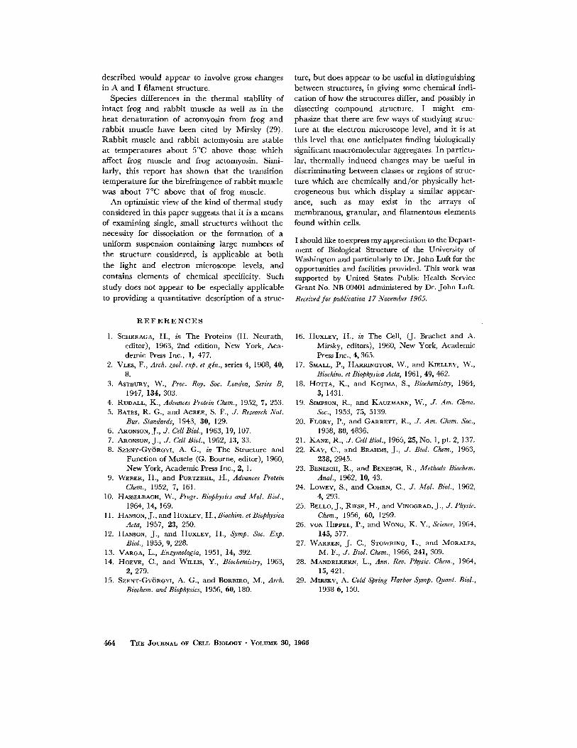
GLYCEROL, ETHYLENE GLYCOL~ AND METHANOL: The birefringence of glycerinated rabbit fibers in 0.1 M KCI containing glycerol, ethylene glycol, or methanol was less sensitive to heat than in 0.1 u KC1 alone (Fig. 12). Several other proteins (19) and protein structures (20, 21) also show increased stability in glycols.
Ethylene glycol has been shown to change the optical rotation of myosin in solution but not that of light meromyosin (22). At high concentrations of ethylene glycol (10 and 15 M in Fig. 12) there was no suggestion of a decrease in the stability of the A region in glycerinated fibers. There were changes in the dimensions of these heated fibers as to both length and width, making the values of the transition temperature in glycerol and ethylene glycol less precise than at lower tem- peratures. However, fibers heated to 70°C in high concentrations of glycerol or ethylene glycol
462 THE JOURNAL OF CELL BIOLOOY • VOLUMIg S0, 1966
and then resuspended in 0. I M KC1 at p H 7 and reheated (Fig. 12) gave an apparent ly normal response at 52°C. A 5 0 % v / v glycerol solution has a glycerol molari ty of 6.9.
M E R S A L Y L : This reagent is considered to b ind sulfhydryl groups specifically at pH 7 (23). Exposure for 1 hr to 1 rn~ mersalyl in 0.1 M KC1 at p H 7 lowered the t ransi t ion t empera tu re of bo th frog and rabb i t muscles by about 10°C when tested at pH 7 in 0.1 u KC1 after excess mersalyl had been washed out. I t is interesting tha t mer- salyl has an effect on the stability of r abb i t muscle
7O o x >70 ° x >70 °
oO He(It ol 70% for 2 rain
! / / ~ / / . / / Resuspend in o. I " 0.1M KCI, pH 7 E 60
o o x t--
. . . . '0 '5 50 0 2 4 6 I I Concentrot ion (M)+ 0.1 M KCI, pH 7
FIOUR~I~ Effects of glycerol ( - -O- - ) , ethylene glycol ( - - X - - ) , and methanol ( - -O- - ) , each in the presence of 0.1 M KCl + 5 mM phosphate at pH 7, on the transition temperature of glycerinated rabbit psoas fibers. The effect of glycerol -t- 0.1 M KCl on the transition temperature of freshly teased Drosophda fibrils is also shown (- - -O- - -). Fibers in 10 M and 15 M glycerol or ethylene glycol did not give any indication of loss of birefringence when heated to 70°C for ~ min. As shown on the right side of the figure, fibers which were heated to 70°C under these conditions when re- suspended in 0.1 M KC1 had a transition temperature near 5£°C. On this basis the transition temperature of fibers in 10 M and 15 M ethylene glycol or glycerol is greater than 70°C.
since l ight meromyosin fraction I, a f ragment of myosin with a molecular weight of 120,000, may be a major pa r t of the A f i lament structure (16) and yet does not contain any of the sulfhy- dryl groups found in myosin (24).
The data presented have some relat ion to the noncovalent forces which structure the com- ponents of the A f i lament or the A fi lament itself, bu t the low specificity in terms of bonds affected, the gross similarity in the kinds of bonding at all
levels of structure, and the lack of a good reference model do not permi t an explicit in terpreta t ion. An indicat ion of the kinds of bonds within pro- teins which may be affected by the exper imenta l condit ions used here and their re la t ion to protein structure can be found in the work of a n u m b e r of authors (I , 25, 26, 27).
Rup tu r e of the weak in termolecular bonds which stabilize protein aggregates may precede changes in secondary and tert iary structure and lead to a nemat ic state. I t is, however, unlikely tha t this will be generally true, under a var iety of solvent conditions, for a large complex protein aggregate such as the A fi lament. Following an initial loss of A f i lament aggregate structure, or of some pa r t of it, one can picture possibilities for new conformations and for aggregat ion in new ways. In muscle, one or m a n y of these changes is reflected in the decrease in birefringence.
The protein remain ing in the A region after hea t ing may show how closely extensive changes in protein conformat ion are related to the loss of aggregate s tructure as reflected by decreased birefringence. Changes in solubility have been used as an indicat ion of such changes a l though more precise methods for their detect ion do exist.
Glycer inated rabb i t fibrils which had been heated in 0.1 M KC1 at p H 7 were t reated for several minutes with 0.5 M potassium iodide at p H 7. The effect on the A region was observed by phase-contrast microscopy. In unhea ted muscle, 0.5 M K I extracts the A region and m u c h of the I f i lament mater ia l (12), while in muscle heated to 55°C for 2 min little or none of the A mater ia l appeared to be extracted by 0.5 M KI . The first signs of decreased solubility were seen in fibrils heated to 48°C in which a small a m o u n t of mate- rial in the center of the A region was not ex- t racted (these fibrils had good H zones). The correlat ion between hea t - induced birefr ingence changes and decreased solubility demonst ra tes tha t tempera tures which reduce the birefr ingence have effects on protein structure in addi t ion to disaggregation. This was also shown by the gradual decrease in s t rength of ATP- induced shortening seen when fibrils were heated at tem- peratures below those where the birefr lngence decreased.
At tempts to analyze muscle funct ion in terms
of the dimensional changes tha t occur on hea t ing
have been made by several investigators, no tab ly Florey, Mandelkern , and Hoeve (28). The effects
J. F. ARONSON Mdting of Muscle Birefringence 453
described would appear to involve gross changes in A and I filament structure.
Species differences in the thermal stability of intact frog and rabbit muscle as well as in the heat denaturation of actomyosin from frog and rabbit muscle have been cited by Mirsky (29). Rabbi t muscle and rabbit actomyosin are stable at temperatures about 5°C above those which affect frog muscle and frog actomyosin. Simi- larly, this report has shown that the transition temperature for the birefringence of rabbit muscle was about 7°C above that of frog muscle.
An optimistic view of the kind of thermal study considered in this paper suggests that it is a means of examining single, small structures without the necessity for dissociation or the formation of a uniform suspension containing large numbers of the structure considered, is applicable at both
the light and electron microscope levels, and contains elements of chemical specificity. Such study does not appear to be especially applicable to providing a quantitative description of a struc-
R E F E R E N C E S
1. SCHERAGA, H., in The Proteins (H. Neurath, editor), 1963, 2nd edition, New York, Aca- demic Press Inc., l , 477.
2. VLES, F., Arch. zool. exp. et gin., series 4, 1908, 40, 8.
3. ASTBURY, W., Proc. Roy. Soc. London, Series B, 1947, 134, 303.
4. RUDALL, K., Advances Protein Chem., 1952, 7, 253. 5. BATES, R. G., and ACREE, S. F., J. Research Nat.
Bur. Standards, 1943, 30, 129. 6. ARONSON, J., or. CellBiol., 1963, 19, 107. 7. ARONSON, J., J. Cell Biol., 1962, 13, 33. 8. SZENT-GYORGYI, A. G., in The Structure and
Function of Muscle (G. Bourne, editor), 1960, New York, Academic Press Inc., 2, 1.
9. WEBER, H., and PORTZEHL, H., Advances Protein Chem., 1952, 7, 161.
10. HASSELBACH, W., Progr. Biophysics and Mol. Biol., 1964, 14, 169.
11. HANSON, J., and HUXLEy, H., Biochim. et Biophysica Acta, 1957, 23, 250.
12. HANSON, J. , and HUXLEY, H., Symp. Soc. Exp. Biol., 1955, 9, 228.
13. VAROA, L., Enzymologia, 1951, 14, 392. 14. HOEVE, C., and WILLIS, Y., Biochemistry, 1963,
2, 279. 15. SZENT-GYOROYI, A. G., and BORBIRO, M., Arch.
Biochem. and Biophysics, 1956, 60, 180.
ture, but does appear to be useful in distinguishing between structures, in giving some chemical indi- cation of how the structures differ, and possibly in dissecting compound structure. I might em- phasize that there are few ways of studying struc- ture at the electron microscope level, and it is at this level that one anticipates finding biologically significant macromolecular aggregates. In particu- lar, thermally induced changes may be useful in discriminating between classes or regions of struc- ture which are chemically and /o r physically het- erogeneous but which display a similar appear- ance, such as may exist in the arrays of membranous, granular, and filamentous elements found within cells.
I should like toexpress my appreciation to the Depart- ment of Biological Structure of the University of Washington and particularly to Dr. John Luft for the opportunities and facilities provided. This work was supported by United States Public Health Service Grant No. NB 00401 administered by Dr. John Luft.
Received for publication 17 November 1965.
16. HUXLEY, H., in The Cell, (J. Brachet and A. Mirsky, editors), 1960, New York, Academic Press Inc., 4, 365.
17. SMALL, P., HARmNGTON, W., and KmLLEY, W., Biochim. et Biophysica Acta, 1961, 49, 462.
18. HOTTA, K., and KojIr~A, S., Biochemistry, 1964, 3, 1431.
19. SIMPSON, R., and KAUZMANN, W., J. Am. Chem. Soc., 1953, 75, 5139.
20. FLORY, P., and GARm~TT, R., J. Am. Chem. Soc., 1958, 80, 4836.
21. KANE, R., J. CellBiol., 1965, 25, No. 1, pt. 2, 137. 22. KAY, C., and BRAHMS, J., J. Biol. Chem., 1963,
238, 2945. 23. BENESCH, R., and BENESCH, R., Methods Biochem.
Anal., 1962, 10, 43. 24. LOWEY, S., and COHEN, C., J. Mol. Biol., 1962,
4, 293. 25. BELLO, J., RIESE, H., and VINOORAD, J., J. Physic.
Chem., 1956, 60, 1299. 26. YON HIm~EL, P., and WONO, K. Y., Science, 1964,
145, 577. 27. WARREN, J. C., STOWRINC, L., and MORALES,
M. F., J. Biol. Chem., 1966, 241, 309. 28. MANDELKERN, L., Ann. Rev. Physic. Chem., 1964,
15, 421. 29. MIRSXV, A. Cold Spring Harbor Symp. Quant. Biol.,
1938 6, 150.
464 TOE JOURNAL OF CELL BIOLOGY • VOLUME 30, 1966