Embed Size (px)
Citation preview
Cardiovascular Pathology xx (2009) xxx–xxx
ARTICLE IN PRESS
Case Report
Second harmonic generation microscopy to investigate collagenconfiguration: a pericarditis case study
Jonathan Bélislea, Tiffany Zigrasb,c, Santiago Costantinoa, Raymond Cartierc,d, Jagdish Butanyf,Paul W. Wisemana,e, Richard L. Leaskb,c,⁎
aDepartment of Physics, McGill University, Montreal, CanadabDepartment of Chemical Engineering, McGill University, Montreal, Canada
cResearch Center, Montreal Heart Institute, Montreal, CanadadDepartment of Cardiac Surgery, Montreal Heart Institute, Montreal, Canada
eDepartment of Chemistry, McGill University, Montreal, CanadafToronto General Hospital, Department of Pathology, Toronto, Canada
Received 24 November 2008; received in revised form 3 June 2009; accepted 8 June 2009
Abstract
We have used second-harmonic-generation (SHG) to image collagen fibers in pericardial tissue removed from a patient with constrictivepericarditis and compared this to healthy pericardium. SHG imaging allowed for the visualization of collagen fibers without the need forstaining or pretreatment. Images were compared to stained histology slides. Collagen fibers in SHG and histology images displayed thesame structure and morphology. The mature collagen of the parietal pericardium was easily distinguishable from the new collagenaccumulation due to the pericarditis. SHG imaging can provide a convenient and valuable architectural profile of collagen organization.Crown Copyright © 2009 Published by Elsevier Inc. All rights reserved.
Keywords: Pericarditis; Collagen; Diagnostic imaging; Connective tissue
1. Clinical history
The patient initially presented with an umbilical herniaand abdominal distention. An abdominal echo showedascites and a hepatic condition. The 26-year-old male patientwas referred to our hospital diagnosed with constrictivepericarditis and fibrosis-causing restrictive cardiac syn-drome. The patient was neither a smoker nor diabetic, withknown allergies to penicillin and clarithromycin. The patientdid not respond to the prescribed steroids and thus underwenta pericardectomy via stenotomy at our institution. After thesurgery, the patient rapidly lost 30 lb and progressed withoutany complications. A follow-up echocardiogram revealedthe patient's heart was functioning and he was dischargedhome after 8 postsurgical days.
⁎ Corresponding author. Department of Chemical Engineering/Montreal Heart Institute, Montreal, Quebec, Canada.
E-mail address: [email protected] (R.L. Leask).
1054-8807/09/$ – see front matter. Crown Copyright © 2009 Published by Elsevdoi:10.1016/j.carpath.2009.06.001
2. Methods and results
Second-harmonic-generation (SHG) microscopy usesnonlinear photon scattering caused by the noncentrosym-metic assembly of collagen to create an image. It canprovide fibril resolution without extrinsic dyes [1,2]. SHGalso allows for optical sectioning and the ability to image atvarious depths in excised tissue, due to the deep penetrationof the infrared (IR) excitation laser wavelengths and thenonlinear intensity dependence of the SHG emission.
Small pieces of diseased and healthy pericardial tissuemeasuring 5×3 cm, from the anterior wall of the pericardialsac, were collected for SHG imaging and for histology.The healthy pericardial tissue was removed duringcoronary artery bypass graft surgery from a 66-year-oldmale. The samples were first imaged by SHG microscopy(homebuilt) and then sent to histology. SHG images werecollected in the x-y plane and imaged through the tissuedepth (z-plane) creating z-stacks (available online). Both
ier Inc. All rights reserved.
2 J. Bélisle et al. / Cardiovascular Pathology xx (2009) xxx–xxx
ARTICLE IN PRESS
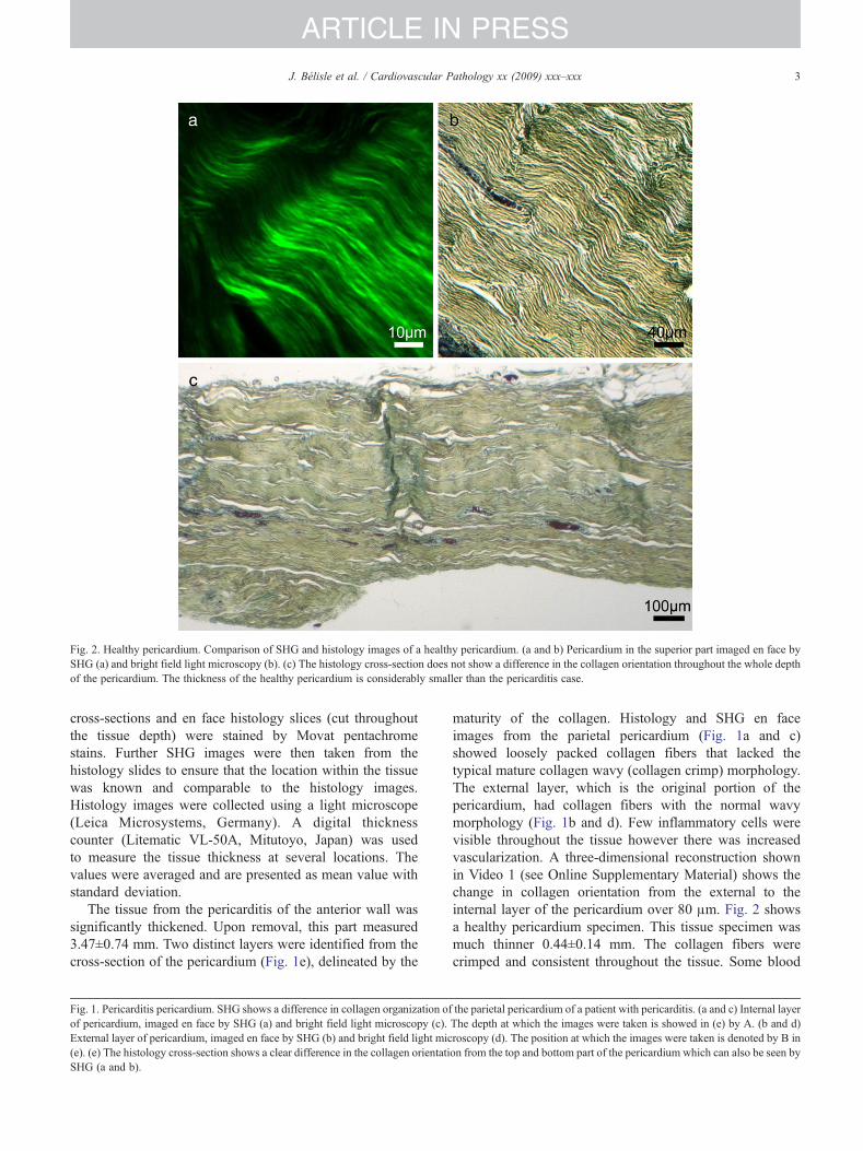
Fig. 2. Healthy pericardium. Comparison of SHG and histology images of a healthy pericardium. (a and b) Pericardium in the superior part imaged en face bySHG (a) and bright field light microscopy (b). (c) The histology cross-section does not show a difference in the collagen orientation throughout the whole depthof the pericardium. The thickness of the healthy pericardium is considerably smaller than the pericarditis case.
3J. Bélisle et al. / Cardiovascular Pathology xx (2009) xxx–xxx
ARTICLE IN PRESS
cross-sections and en face histology slices (cut throughoutthe tissue depth) were stained by Movat pentachromestains. Further SHG images were then taken from thehistology slides to ensure that the location within the tissuewas known and comparable to the histology images.Histology images were collected using a light microscope(Leica Microsystems, Germany). A digital thicknesscounter (Litematic VL-50A, Mitutoyo, Japan) was usedto measure the tissue thickness at several locations. Thevalues were averaged and are presented as mean value withstandard deviation.
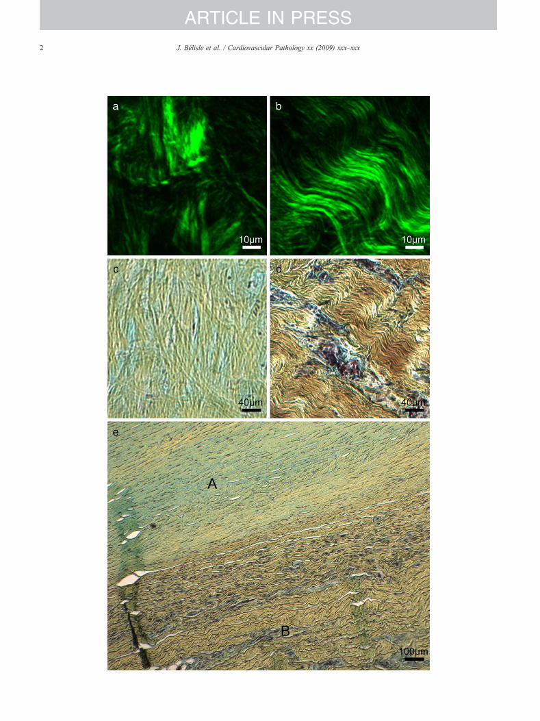
The tissue from the pericarditis of the anterior wall wassignificantly thickened. Upon removal, this part measured3.47±0.74 mm. Two distinct layers were identified from thecross-section of the pericardium (Fig. 1e), delineated by the
Fig. 1. Pericarditis pericardium. SHG shows a difference in collagen organization oof pericardium, imaged en face by SHG (a) and bright field light microscopy (c).External layer of pericardium, imaged en face by SHG (b) and bright field light mic(e). (e) The histology cross-section shows a clear difference in the collagen orientatiSHG (a and b).
maturity of the collagen. Histology and SHG en faceimages from the parietal pericardium (Fig. 1a and c)showed loosely packed collagen fibers that lacked thetypical mature collagen wavy (collagen crimp) morphology.The external layer, which is the original portion of thepericardium, had collagen fibers with the normal wavymorphology (Fig. 1b and d). Few inflammatory cells werevisible throughout the tissue however there was increasedvascularization. A three-dimensional reconstruction shownin Video 1 (see Online Supplementary Material) shows thechange in collagen orientation from the external to theinternal layer of the pericardium over 80 μm. Fig. 2 showsa healthy pericardium specimen. This tissue specimen wasmuch thinner 0.44±0.14 mm. The collagen fibers werecrimped and consistent throughout the tissue. Some blood
f the parietal pericardium of a patient with pericarditis. (a and c) Internal layerThe depth at which the images were taken is showed in (e) by A. (b and d)roscopy (d). The position at which the images were taken is denoted by B inon from the top and bottom part of the pericardium which can also be seen by
4 J. Bélisle et al. / Cardiovascular Pathology xx (2009) xxx–xxx
ARTICLE IN PRESS
components were visible, but there was no indication ofinflammation or neovascularization.
3. Discussion
SHG microscopy is a quick imaging tool that is non-destructive and allows for clear visualization of collagenfibers throughout connective tissue without any staining orpretreatment. When compared to histology images there isgood agreement between the collagen morphology andorientation. A three-dimensional reconstruction of collagenfibers is possible with this technique which allows for thedepth of the tissue to be examined and easily revealsdetails and spatial understanding which would requiremultiple histology slices. SHG microscopy is powerful toolfor studying tissue structure, and with continued researchand improvements in resolution, a variety of applicationswill benefit. It can be used to measure collagen fibrilsdown to the submicron scale which is useful for under-standing the progression of diseases related to theextracellular matrix [3]. Tissue obtained from biopsiescould be quickly evaluated. Eventually, an SHG endoscopecould be used to visualize collagen structure in vivo, a
prototype for which has already been developed and testedin animals [4].
Acknowledgments
Thank you to Dr. Leung from the Montreal Heart Institutefor his expertise in pathology on cardiovascular tissue. PWWacknowledges grant support from the Natural Sciences andEngineering Research Council of Canada (NSERC), theCanada Foundation for Innovation (CFI), and the CanadianInstitute for Photonic Innovations. R.L.L., R.C., and J.B.grant support from NSERC and the Canadian Institute ofHealth Research.
References
[1] Fine S, Hansen WP. Optical second harmonic generation in biologicalsystems. Appl Opt 1971;10(10):2350–3.
[2] Freund I, Deutsch M. Second-harmonic microscopy of biological tissue.Opt Lett 1986;11(2):94–6.
[3] Chu S-W, et al. Thickness dependence of optical second harmonicgeneration in collagen fibrils. Opt Express 2007;15(19):12005–10.
[4] Fu L, et al. Nonlinear optical endoscopy based on a double-cladphotonic crystal fiber and a MEMS mirror. Opt Express 2006;14(3):1027–32.