Embed Size (px)
Citation preview

1
2
3
4
567
8
91011121314151617181920212223
42
43
44
45
46
47
48
49
50
51
52
53
54
55
56
57
58
59
Experimental Neurology xxx (2010) xxx–xxx
YEXNR-10443; No. of pages: 13; 4C:
Contents lists available at ScienceDirect
Experimental Neurology
j ourna l homepage: www.e lsev ie r.com/ locate /yexnr
ARTICLE IN PRESS
F
Mitochondrial-dependent apoptosis in Huntington's disease human cybrids
Ildete L. Ferreira a, Maria V. Nascimento a, Márcio Ribeiro a, Sandra Almeida a, Sandra M. Cardoso a,Manuela Grazina a,b, João Pratas a, Maria João Santos a, Cristina Januário c,Catarina R. Oliveira a,b, A. Cristina Rego a,b,⁎a Center for Neuroscience and Cell Biology, Faculty of Medicine, University of Coimbra, Portugalb Institute of Biochemistry, Faculty of Medicine, University of Coimbra, Portugalc Neurology Unit, Coimbra University Hospital, Coimbra, Portugal
⁎ Corresponding author. Center for Neuroscience andBiochemistry, Faculty of Medicine, Ruas Larga, UnivCoimbra, Portugal. Fax: +351 239 822776.
E-mail addresses: [email protected], acrego@
0014-4886/$ – see front matter © 2010 Published by Edoi:10.1016/j.expneurol.2010.01.002
Please cite this article as: Ferreira, I.L., et al.,doi:10.1016/j.expneurol.2010.01.002
O
a b s t r a c t
a r t i c l e i n f o24
25
26
27
28
29
30
31
32
33
34
Article history:Received 10 September 2009Revised 26 November 2009Accepted 5 January 2010Available online xxxx
Keywords:ApoptosisHuman cybridsHuntington's diseaseMitochondria3-Nitropropionic acid
35
36
37
38
TEDPROWe investigated the involvement of mitochondrial-dependent apoptosis in Huntington's disease (HD) vs.control (CTR) cybrids, obtained from the fusion of human platelets with mitochondrial DNA-depleted NT2cells, and further exposed to 3-nitropropionic acid (3-NP) or staurosporine (STS). Untreated HD cybrids didnot exhibit significant modifications in the activity of mitochondrial respiratory chain complexes I–IV or inmtDNA sequence variations suggestive of a primary role in mitochondrial susceptibility in the subpopulationof HD carriers studied. However, a slight decrease in mitochondrial membrane potential and increasedformation of intracellular hydroperoxides was observed in HD cybrids under basal conditions. Furthermore,apoptotic nuclei morphology and a moderate increase in caspase-3 activation, as well as increased levels ofsuperoxide ions and hydroperoxides were observed in HD cybrids upon 3-NP or STS treatment. 3-NP-evokedapoptosis in HD cybrids involved cytochrome c and AIF release from mitochondria, which was associatedwith mitochondrial Bax translocation. CTR cybrids subjected to 3-NP showed increased mitochondrial Baxand Bim levels and the release of AIF, but not cytochrome c, suggesting a different mode of cell death, linkedto the loss of membrane integrity. Additionally, increased mitochondrial Bim and Bak levels, and a slightrelease of cytochrome c in untreated HD cybrids may help to explain their moderate susceptibility tomitochondrial-dependent apoptosis.
39
Cell Biology, and Institute ofersity of Coimbra, 3004-504
cnc.cj.uc.pt (A.C. Rego).
lsevier Inc.
Mitochondrial-dependent apoptosis in Huntin
© 2010 Published by Elsevier Inc.
4041
C60
61
62
63
64
65
66
67
68
69
70
71
72
73
74
75
76
UNCORREIntroduction
Huntington's disease (HD) is an autosomal dominant disordercharacterized by uncontrolled body movements known as chorea,changes in personality and a loss of cognitive ability eventuallyleading to dementia. HD is caused by an expansion of the trinucleotideCAG repeat in the huntingtin gene, producing a proteinwith increasednumber of polyglutamines at the N-terminal (mutant huntingtin).Neuropathological changes are caused by the death of GABAergicprojection medium-spiny neurons of the neostriatum (caudate andputamen) and neurons in the cerebral cortex, the two most severelyaffected brain structures in HD (e.g., Gil and Rego, 2008). However,the mechanisms by which mutant huntingtin causes selectivedegeneration of striatal and cortical neurons in HD are largelyunknown.
Neuronal abnormalities involving aberrant protein–protein inter-actions caused bymutant huntingtinmay lead to deregulation in gene
77
78
79
80
81
expression in human HD striatum (Kuhn et al., 2007). Furthermore,excitotoxicity linked to decreased Ca2+ homeostasis, mitochondrialdysfunction, impairment in energy metabolism (Schapira, 1998; Beal,2005; Sas et al., 2007; Sorolla et al., 2008; Yang et al., 2008), caspaseactivation and apoptosis (Brouillet et al., 1998; Beal, 2005; Milakovicand Johnson, 2005; Rego and de Almeida, 2005; Fan and Raymond,2007) have been reported in HD-affected individuals. In addition,oxidative stress and damage to specific macromolecules alsoparticipate in HD progression (Sorolla et al., 2008).
Analysis of post-mortem striatal tissue from HD patients revealeda decrease in the activity of the respiratory chain complexes II/III andIV (Schapira 1998; Tabrizi et al., 1999). Mutant huntingtin may causemitochondrial dysfunction by either perturbing transcription ofnuclear-encoded mitochondrial proteins or by directly interactingwith the organelle, thus evoking defects in mitochondrial dynamics,organelle trafficking and fission and fusion, which, in turn, may resultin bioenergetic failure in HD (Bossy-Wetzel et al., 2008). Indeed, mildor gradual energy disturbances may lead to the release of pro-apoptotic factors from the mitochondria, such as cytochrome c,leading to apoptotic cell death. However, if the energy supply of thecell drops dramatically, cells die by necrosis (Vanlangenakker et al.,2008).
gton's disease human cybrids, Exp. Neurol. (2010),

C
82
83
84
85
86
87
88
89
90
91
92
93
94
95
96
97
98
99
100
101
102
103
104
105
106
107
108
109
110
111
112
113
114
115
116
117
118
119
120
121
122
123
124
125
126
127
128
129
130
131
132
133
134
135
136
137
138
139
140
141
142
143
144
145
146
147
148
149
150
151
152
153
154
155
156
157
158
159
160
161
162
163
164
165
166
167
168
169
170
171
172
173
174
175
176
177
178
179
180
181
182
183
184
185
186
187
188
189
190
191
192
193
194
195
196
197
198
199
200
201
202
203
2 I.L. Ferreira et al. / Experimental Neurology xxx (2010) xxx–xxx
ARTICLE IN PRESS
UNCO
RRE
Mutant huntingtin is widely expressed in the HD brain (Aroninet al., 1995; Trottier et al., 1995), but also in peripheral tissues.Thus, abnormalities outside the brain can also be expected. Accordingly,mutant huntingtin was reported to be associated with mitochondrialcomplex II/III dysfunction, mitochondrial depolarization, cytochrome crelease and increased caspases activity in skeletal muscle (Ciammola etal., 2006; Turner et al., 2007), and decreased catalase activity in skinfibroblast cultures from HD patients (del Hoyo et al., 2006). Lympho-blasts derived from HD patients also show increased stress-inducedapoptotic cell death associated with caspase-3 activation, abnormalcalcium homeostasis and mitochondrial dysfunction (Sawa et al., 1999;Panov et al., 2002; Bezprozvanny and Hayden, 2004). Recently, wedemonstrated that HD human peripheral blood cells, particularly Blymphocytes, are endowed with increased expression of Bax anddecreased mitochondrial membrane potential (Almeida et al., 2008),further suggesting that an adverse effect of mutant huntingtin is notlimited to neurons.
It is widely accepted that mitochondrial DNA (mtDNA) abnormal-ities play an important role in neurodegenerative diseases (Grazina etal., 2006; Onyango et al., 2006), even if they are not a primarytriggering factor (Mancuso et al., 2008). mtDNA mutations maymodify the age of onset, as a result of an impairment of mitochondrialrespiratory chain and/or translational mechanisms thus contributingto the neurodegenerative process (Grazina et al., 2006). Even thoughthe studies concerning HD and mtDNA mutations are rare andheterogeneous, mtDNAmutations have been suggested to occur in HDpathophysiology (Kasraie et al., 2008; Yang et al., 2008). It wasrecently demonstrated that mitochondrial DNA damage is an earlybiomarker for HD-associated neurodegeneration supporting thehypothesis that mtDNA lesions may contribute to the pathogenesisobserved in HD (Acevedo-Torres et al., 2009). Indeed, recent datashowed that HD patients' lymphocytes have higher frequencies ofmtDNA deletions and oxidative stress, suggesting that CAG repeatsinstability and mutant huntingtin are a causative factor in mtDNAdamage (Banoei et al., 2007). Nevertheless, previous studies in HDcybrids (a valuable cellular tool to isolate mitochondrial-encodedhuman defects) showed no changes in mitochondrial respiratorychain activity or oxidative stress (Swerdlow et al., 1999), evidencingno major changes in mitochondrial function, even if considering theoccurrence of point mtDNA mutations. Notwithstanding, mutanthuntingtin was previously shown to interact with neuronal mito-chondria of YAC72 transgenic mice suggesting that mitochondrialcalcium abnormalities associatedwith HD pathogenesismay be due toa direct effect of mutant huntingtin on the organelle (Panov et al.,2002). Moreover, mutant huntingtin fragments can directly inducethe opening of the mitochondrial permeability transition pore inisolated mouse liver mitochondria, with the consequent release ofcytochrome c (Choo et al., 2004), favoring the hypothesis that mutanthuntingtin interacting with mitochondria may well lead to mito-chondrial modifications independently of damage on mtDNA.
Thus, in the present study, we studied mitochondrial-dependentapoptotic events and oxidative stress in human cybrid lines, obtainedfrom the fusion of HD or control platelets with NT2 ρ0 cells, depletedof mitochondrial DNA. We report increased susceptibility of asubpopulation of HD cybrids, an ex vivo mitochondrial HD humanmodel, to undergo mitochondrial-dependent apoptosis when sub-jected to complex II inhibition with 3-nitropropionic acid (3-NP) or toapoptosis with the classic inducer staurosporine (STS).
Materials and methods
Materials
Optimem was purchased from GIBCO (Paisley, UK). Proteaseinhibitor cocktail (chymostatin, pepstatin, A, leupeptin and antipain),3-nitropropionic acid, penicillin/streptomycin, oligomycin, carbonyl-
Please cite this article as: Ferreira, I.L., et al., Mitochondrial-dependent apdoi:10.1016/j.expneurol.2010.01.002
TEDPR
OOF
cyanide-p-(trifluoromethoxyphenyl)hydrazone (FCCP), dichlorphe-nolindophenol (DCPIP), thenoyltrifluoroacetone (TTFA), 5,59-dithiobis (2-nitrobenzoic acid) (DTNB) and anti-α-tubulin werefrom Sigma Chemical Co. (St Louis, MO, USA). N-acetyl-Asp-Glu-Val-Asp-p-nitroanilide (Ac-DEVD-pNA) was obtained from Calbiochem(Darmstadt, Germany). Anti-cytochrome c was from BD Pharmingen(San Diego, CA, USA); anti-Bax from Cell Signaling (Beverly, MA,USA); anti-Bcl-2 and anti-AIF from Santa Cruz Biotechnology (SantaCruz, CA, USA); anti-Bim from Stressgen (Assay Designs, Inc.,Michigan, USA); and anti-Bak from Abcam Inc. (Cambridge, USA).Secondary antibodies conjugated to alkaline phosphatase (anti-mouse and anti-rabbit) were purchased from Amersham Biosciences(Buckinghamshire, UK). The fluorescence probes tetramethylrhoda-mine methyl ester (TMRM+), dihydroethidium (DHE), 2′,7′-dichlor-odihydrofluorescein diacetate (DCFH2-DA), Hoechst 33342 and anti-cytochrome c oxidase I (COX I) were obtained from Molecular Probes(Invitrogen, USA). All other reagents were of analytical grade.
Participants
Five to six genetically and clinically confirmed HD patients frompre-identified Portuguese families and three age-matched healthycontrols, without any neurological disease, were studied. The numberof CAG repeats present in HD gene for all the patients were between42 and 44, which gives rise to the most common adult-onset form ofthe disease. The patients were characterized according to the UnifiedHuntington's Disease Rating Scale (UHDRS) (Huntington StudyGroup, 1996) and neurological evaluation was performed by anexperienced neurologist. The studywas performed in accordancewiththe Ethical Committee of Coimbra University Hospital, and all thesubjects gave informed consent.
Cybrid production, culture and incubation with 3-NP and STS
Cybrids (cytoplasmic hybrid systems) were produced after fusionof mitochondrial DNA-depleted human teratocarcinoma cells (ρ° NT2cells), obtained from Dr. R. H. Swerdlow (University of Virginia,Charlottesville, VA, USA), with human platelets. Production andselection of the cybrids were performed as described previously(Cardoso et al., 2004). Cybrids were cultured in Optimem mediumsupplemented with 10% of fetal calf serum, penicillin (100 U/ml),streptomycin (100 μg/ml) and maintained at 37 °C in humidifiedincubator containing 95% air and 5% CO2. Since mitochondria dividemainly in response to the energy needs of the cell, i.e., independentlyof the cell cycle (Sas et al., 2007) and to account for the auto-selectionof the remaining functional mitochondria, experiments were per-formed with cybrids less than 2 months in culture, as previouslydescribed (e.g., Cardoso et al., 2004). Cybrids were plated on glasscoverslips, multiwell chambers or flasks at a density of 0.06×10⁶
cells/cm2 one day before the experiments in order to allow thedesired confluence. Cells were then incubated in culture medium inthe absence or presence of 3-NP (0.1, 1 or 10 mM) for 24 h or STS (0.1,1 or 10 nM) for 15 h, as described in figure legends.
Assay of enzymatic activities of mitochondrial electron transport chain
Cybrids were extracted in a sucrose buffer (250 mM sucrose;20 mM HEPES-KOH, pH 7.5; 100 mM KCl; 1.5 mMMgCl2; 1 mM EDTAand 1 mM EGTA), and centrifuged at 2300 rpm for 12 min at 4 °C. Thesupernatant was analyzed for mitochondrial complex activities on aUV/VIS spectrophotometer (model 2401; Shimadzu Scientific Instru-ments, Columbia, MD).
NADH–ubiquinone oxidoreductase assayComplex I activity was determined at 340 nm by following the
decrease in NADH absorbance that occurs when ubiquinone is
optosis in Huntington's disease human cybrids, Exp. Neurol. (2010),

204
205
206
207
208
209
210
211
212
213
214
215
216
217
218
219
220
221
222
223
224
225
226
227
228
229
230
231
232
233
234
235
236
237
238
239
240
241
242
243
244
245
246
247
248
249
250
251
252
253
254
255
256
257
258
259
260
261
262
263
264
265
266
267
268
269
270
271
272
273
274
275
276
277
278
279
280
281
282
283
284
285
286
287
288
289
290
291
292
293
294
295
296
297
298
299
300
301
302
303
304
305
306
307
308
309
310
311
312
313
314
315
316
317
318
319
320
3I.L. Ferreira et al. / Experimental Neurology xxx (2010) xxx–xxx
ARTICLE IN PRESS
UNCO
RREC
reduced to ubiquinol. The reaction was started by adding the sampleto the reaction mixture (in mM: 20 K2HPO4, pH 7.2, 10 MgCl2, 0.15NADH, 2.5 mg/ml BSA fatty-acid free, 1 KCN) containing 50 μMdecylubiquinone, at 30 °C. After 8 min, rotenone (10 μM) was addedand the reaction was registered for further 8 min. Complex I activitywas expressed in nanomoles per minute per milligram of protein andcorrespond to the rotenone sensitive rate. The enzyme activity wascorrected for citrate synthase activity.
Succinate–ubiquinone oxidoreductase assayComplex II activity was monitored at 600 nm by following the
reduction of 6,6-dichlorophenolindophenol (DCPIP) by the ubiquinolformed in the reaction. The assay was started by adding the sample tothe reaction mixture (in mM: 50 K2HPO4, pH 7.4, 20 succinate, 0.1EDTA, 1 KCN, 0.01 rotenone) containing 50 μM decylubiquinone, at30 °C. After 8 min, 1 mM 2-thenoyltrifluoroacetone (TTFA) was addedand the reaction registered for further 8 min. Complex II activity wasexpressed in nanomoles per minute per milligram of protein andcorrespond to the TTFA sensitive rate. The enzyme activity wascorrected for citrate synthase activity.
Ubiquinol–cytochrome c reductase assayComplex III activity was monitored at 550 nm by following the
reduction of cytochrome c by ubiquinol. The assay was started byadding the sample to the reactionmixture (inmM: 35 K2HPO4, pH 7.2,1 EDTA, 5MgCl2, 1 KCN, 5 μM rotenone) containing 15 μM cytochromec and 15 μM ubiquinol, at 30 °C. Complex III activity was expressed inrate constant (k) per minute per milligram of protein and correctedfor citrate synthase activity.
Cytochrome c oxidase assayComplex IV activity was determined at 550 nm by measuring the
oxidation of reduced cytochrome c by cytochrome c oxidase. Thereduced cytochrome c was prepared by mixing its oxidized form withascorbate and then dialysed for 24 h against a 0.01 M phosphatebuffer, pH 7.0, at 4 °C. The assay was started by adding the sample tothe reaction buffer (10 mM K2HPO4, pH 7) containing 50 μM reducedcytochrome c and 1mM ferricyanide, at 30 °C. Complex IV activity wasexpressed in rate constant (k) perminute permilligram of protein andcorrected for citrate synthase activity.
Citrate synthase assayCitrate synthase (CS) activity was performed at 412 nm following
the reduction of 0.2 mM 5,5′-dithio-bis(2-nitrobenzoic acid) in thepresence of 0.2 mM acetyl-CoA and 0.1 mM oxaloacetate in a mediumwith 100 mM Tris–HCl, pH 8.0 and 0.1% Triton X-100. CS activity wasexpressed in nanomoles per minute per milligram of protein.
Analysis of mitochondrial membrane potential
The mitochondria membrane potential was determined by usingthe cationic fluorescent probe tetramethyl rhodamine methyl ester(TMRM+), which accumulates predominantly in polarized mitochon-dria (Ward et al., 2000). Thus the variation of TMRM+ retention wasstudied in order to estimate changes in mitochondrial membranepotential. Following a washing step with Na+ medium containing (inmM): 135 NaCl, 5 KCl, 0.4 KH2PO4, 1.8 CaCl2, 1 MgSO4, 20 HEPES, and5.5 glucose, pH 7.4, cells were incubated in Na+ medium containing150 nM TMRM+ (quench mode) for 1 h at 37 °C. Basal fluorescence(540 nm excitation and 590 emission) was measured using aMicroplate Spectrofluorometer Gemini EM (Molecular Devices, USA)for 5 min, followed by the addition of 1 μM FCCP and 2 μg/mloligomycin, which produced maximal mitochondrial depolarization.Results were expressed as the difference between the increase ofTMRM+
fluorescence upon addition of FCCP plus oligomycin and basalfluorescence values.
Please cite this article as: Ferreira, I.L., et al., Mitochondrial-dependent apdoi:10.1016/j.expneurol.2010.01.002
TEDPR
OOF
Analysis of apoptotic nuclei
The nuclear morphology of HD and CTR cybrids exposed to 3-NPor STS was analyzed by fluorescence microscopy, by using a double-staining procedure with Hoechst 33342 and propidium iodide.Following a washing step with Na+ medium, the cells wereincubated with 7.5 μg/ml Hoechst 33342 and 4 μg/ml propidiumiodide, in the dark, for 3 min, at room temperature. Cells werewashed 3 times in Na+ medium in order to remove extracellulardyes and further examined and scored using the Axioscope 2 Plusupright microscope (Zeiss, Jena, Germany).
Lactate dehydrogenase (LDH) measurements
The integrity of the plasma membrane was determined bymonitoring the activity of the cytoplasmic enzyme LDH in theextracellular incubation medium, which represents a common proce-dure to determine membrane leakage and necrotic cell damage. Afterexposure to 3-NP or STS, the incubation medium was collected(extracellular) and the cells were lysed in 10 mM HEPES (pH 7.4) plus0.01% Triton X-100 (intracellular) and frozen at −80 °C. Cell debris inboth sampleswere removedby centrifugation at 14,000 rpm(EppendorfCentrifuge 5417R), for 10 min. LDH was determined spectrophotomet-rically, by following the rate of conversion of reduced nicotinamideadenine dinucleotide (NADH) to oxidized NAD+ at 340 nm (Bergmeyerand Bernt, 1974). LDH released into the extracellular medium wasexpressed as a percentage of the total LDH activity in the cells [% of LDHreleased=extracellular LDH/(extracellular LDH+ intracellular LDH)].
Analysis of intracellular superoxide ions
The cybridswere incubated for 60minat 37 °C in thepresenceof 5 μMDHE, in Na+medium. DHE is a cell-permeable fluorescent dye that, onceinternalized, is oxidized by superoxide to fluorescent ethidium bromide,which intercalates into DNA. DHE itself shows a blue fluorescence(355nmexcitation, 420nmemission) in cell cytoplasmuntil oxidation toform ethidium, which becomes red fluorescent (518 nm excitation,605 nm emission) upon DNA intercalation. Ethidium bromide fluores-cence intensity was measured continuously for 1 h at 37 °C, and therelative level of superoxide production quantified, using a MicroplateSpectrofluorometer Gemini EM (Molecular Devices, USA). At the end ofeach experiment, the cells were scrapped to quantify cell protein in eachwell, using the BioRad protein assay, and ethidium fluorescence wascorrected for variations in total protein between wells. The values werenormalized to the percentage of control (untreated cybrids).
Intracellular hydroperoxides analysis
Cybrids were incubated for 30 min in the presence of 20 μMDCFH2-DA, a stable non-fluorescent cell permeable compound, at37 °C in Na+ medium, pH 7.4. When internalized by the cell, DCFH2-DA is hydrolyzed to DCFH2 by intracellular esterases and rapidlyoxidized to the highly green fluorescent component 2,7-dichloro-fluorescein (DCF) by endogenous hydroperoxides. Intracellular levelsof peroxides were measured by following DCF fluorescence (480 nmexcitation, 550 nm emission) at 37 °C continuously for 1 h, using amicroplate reader Spectrofluorometer Gemini EM (Molecular Devices,USA). In order to correct the DCF fluorescence values for variations intotal protein content in the wells, cell protein in each well wasquantified by the BioRad protein assay. The values were normalized tothe percentage of the control (untreated cybrids).
Caspase-3 activity assay
After washing, cells were scrapped at 4 °C in lysis buffer containing25 mM HEPES, 2 mM MgCl2, 1 mM EDTA and 1 mM EGTA, pH 7.5,
optosis in Huntington's disease human cybrids, Exp. Neurol. (2010),

321
322
323
324
325
326
327
328
329
330
331
332
333
334
335
336
337
338
339
340
341
342
343
344
345
346
347
348
349
4 I.L. Ferreira et al. / Experimental Neurology xxx (2010) xxx–xxx
ARTICLE IN PRESS
supplemented with 2 mM DTT, 0.1 mM phenylmethylsulphonylfluoride (PMSF) and 1:1000 of protease inhibitor cocktail (chymos-tatin, pepstatin A, leupeptin and antipain). Cells were frozen twotimes in liquid N2 and centrifuged at 14,000 rpm for 10 min(Eppendorf Centrifuge 5417R). The resulting supernatants wereassayed for protein content by the BioRad protein assay. To measurecaspase-3 activity, 30 μg protein were added to a reaction buffer[25 mM HEPES, 10% (m/v) sucrose, 0.1% (m/v) 3-[(3-cholamidopro-pyl) dimethylammonio]-1-propane-sulfonate CHAPS), pH 7.5] con-taining the colorimetric substrate (100 μM) for caspase-3 (Ac-DEVD-pNA)-like activity. The reactionmixturewas incubated at 37 °C for 2 h,and the formation of pNAwas measured at 405 nm using amicroplatereader Spectra Max Plus 384 (Molecular Devices, USA). Caspase-likeactivity was calculated as the increase above control for equal amountof loaded protein.
UNCO
RREC
Fig. 1. Mitochondrial specific activities of complexes I-IV and mitochondrial membranenormalized for the activity of citrate synthase (E) and mitochondrial membrane potential (were observed in the respiratory complexes activities. Data are the mean±S.E.M. of 6–8 intwo-way ANOVA, followed by Bonferroni post test. ⁎pb0.05, ⁎⁎pb0.01 when compared to
Please cite this article as: Ferreira, I.L., et al., Mitochondrial-dependent apdoi:10.1016/j.expneurol.2010.01.002
Western blot analysis in mitochondrial and cytosolic subcellular fractions
After a washing step, cybrids were scrapped at 4 °C in sucrosebuffer containing 250 mM sucrose, 20 mM HEPES, 100 mM KCl,1.5 mM MgCl2, 1 mM EDTA and 1 mM EGTA, pH7.5/KOH,supplemented with 1 mM DTT, 1 mM PMSF and 1:1000 proteasecocktail inhibitor (chymostatin, pepstatin A, leupeptin and antipain).Cellular extracts were homogenized (20 strokes) and centrifuged at500×g for 12 min to pellet the nucleus and cell debris. Thesupernatant was further centrifuged at 12,000×g for 20 min andthe resulting pellet (mitochondrial fraction) was resuspended insupplemented TNC buffer containing 10 mM Tris acetate pH 8, 0.5%Nonidet P40, 5 mM CaCl2 supplemented with 1:1000 of proteasecocktail inhibitor. TCA 15% was added to the supernatant andcentrifuged at 15,000×g for 10 min. The resulting pellet (cytosolic
TEDPR
OOF
potential in HD and CTR cybrids. Mitochondrial complex enzymatic activities (A–D)F) were determined as described in Materials and methods. No significant differencesdependent experiments performed in duplicates. Statistical analysis was performed byCTR cybrids; ###pb0.001 when compared to control (untreated) conditions.
optosis in Huntington's disease human cybrids, Exp. Neurol. (2010),

TED
350
351
352
353
354
355
356
357
358
359
360
361
362
363
364
365
366
367
368
369
370
371Q1372
373
374
375
376
377
378
379
380
381
382
383
384
385
386
387
388
389
390
391
392
393
394
395
396
397
398
399
400
401
402
403
404
405
406
407
408
409
410
411
412
413
414
415
416
417
418
419
420
421
422
423
424
425
426
427
428
429
430
431
432
433
434
Table 1 t1:1
Summary of mtDNA investigation in HD and control cybrids.t1:2t1:3Sample mtDNA
sequencevariations
Status (according to MITOMAP) Gene
t1:4HD-1 Nonet1:5HD-2 Nonet1:6HD-3 3348ANG CRP MTND1t1:711719GNA CRP UnP in oral cancer MTND4t1:814766CNT CRP MTCYBt1:9HD-4 3618TNC⁎ CRP MTND1t1:10HD-5 3394TNC CRP; SM in acute leukaemia; PM “unclear”
in LHON/NIDDM/CPT deficiencyMTND1
t1:114216TNC CRP; haplogrup marker JT MTND1t1:1211719GNA CRP; UnP in oral cancer MTND4t1:1313708GNA CRP; haplogroup marker J MTND5t1:1414766CNT CRP MTCYBt1:1514798TNC CRP MTCYBt1:16CTR-1 7621TNC CRP MTCO2t1:178291ANG Novel MTNC7t1:18CTR-2 3348ANG CRP MTND1t1:194172TNA Novel MTND1t1:207566GNA⁎ Novel MTTD
(tRNAasp)
t1:2111719GNA CRP; UnP in oral cancer MTND4t1:2211938CNT CRP MTND4t1:2314766CNT CRP MTCYBt1:24CTR-3 3348ANG CRP MTND1t1:254172TNA Novel MTND1t1:267566GNA⁎ Novel MTTD
(tRNAasp)
t1:2711719GNA CRP; UnP in oral cancer MTND4t1:2811938CNT CRP MTND4t1:2914766CNT CRP MTCYB
Note: the nomenclature of genes is presented according to MITOMAP; CRP: codingregion polymorphism; ⁎heteroplasmy; SM: somatic mutation; PM: point mutation;UnP: unpublished polymorphism; LHON: Leber hereditary optic neuropathy; NIDDM:non-insulin dependent diabetes mellitus; CPT: carnitine palmitoyl transferase. t1:30
5I.L. Ferreira et al. / Experimental Neurology xxx (2010) xxx–xxx
ARTICLE IN PRESS
UNCO
RREC
fraction) was resuspended in supplemented sucrose buffer andadjusted to pH 7 with 2.5 M KOH. Protein content was determinedby BioRad method, and the samples were denaturated with 6 timesconcentrated denaturating buffer at 95 °C, for 5 min. Equivalentamounts of protein were separated on a 15% SDS-PAGE gelelectrophoresis and electroblotted onto polyvinylidene difluoride(PVDF) membranes. The membranes were further blocked with 5%fat-free milk and incubated with antibodies directed against thedenatured form of cytochrome c (Cyt c, 1:500), AIF (1.1000), Bax(1:1000), Bim (1:1000), Bak (1:5000), Bcl-2 (1:500), α-tubulin(1:20000) and mitochondrial DNA-encoded cytochrome c oxidasesubunit I (COX-I, 1:500). In somemembranes retainingmitochondrialsamples where labeling with COX-I was not possible, we used theantibody directed against α-tubulin (1:20000) to normalize theamount of protein per lane. Tubulin is an inherent component ofmitochondrial membranes (Carré et al., 2002) and its levels did notchange in any of the treatments used in this study. Immunoreactivebands were visualized by alkaline phosphatase activity after incuba-tion with ECF reagent on a BioRad Versa Doc 3000 Imaging System.
Mitochondrial DNA (mtDNA) screening
Total DNA was extracted from 5 HD and 3 CTR cybrids by usingstandard methods (Treco et al., 1992) and quantified by UVspectrophotometry (λ=260 nm). Automated sequencing analysiswere used, according to the manufacturer's instructions (3130 ABIPrism sequencing system), using BigDye® Terminator Ready ReactionMix v1.1 (Applied Biosystems), for investigation of 11 mtDNA regionscorresponding to nucleotides 1435-1917, 3150-3769, 4074-4703,4886-5021, 7241-7644, 8222-8461, 8915-9413, 11720-11819, 13515-13727, 14420-14855, 15023-15450, allowing the screening of 31confirmed pathogenic mutations, 105 reported mutations and 288polymorphisms, including 4 haplogroup markers, according toMITOMAP (www.mitomap.org). Evolutionary conservation analysisamong species for positions with novel variants identified wasachieved using ENSEMBL® tools.
Statistical analysis
No significant differences in biochemical studies were observedbetween the HD cybrid lines and the three CTR cybrids used in thiswork. Therefore data were expressed as the mean±S.E.M. of thenumber of experiments indicated in the figure legends. Comparisonsbetween multiple groups were performed with a two-way analysis ofvariance (ANOVA), followed by Bonferroni post-test for comparisonbetween experimental groups. Student's t test was also performed forcomparison between two Gaussian populations, as described in figurelegends. Significance was accepted at pb0.05.
Results
Mitochondrial electron transport chain activities and mitochondrialmembrane potential
We measured the activity of mitochondrial respiratory chaincomplexes and the mitochondrial membrane potential in HD and CTRcybrids. Our data show no significant changes in the activities ofrespiratory complexes I, II, III and IV (Figs. 1A–D) or citrate synthase(Fig. 1E) in HD vs. CTR cybrids. Although the putative basal leakcurrent and the coupling between oxygen consumption and ATPsynthesis may be underestimated by assaying the catalytic activitiesof mitochondrial complexes, the latter measurement is important toevaluate putative changes between our HD cybrids and the datagenerated by Swerdlow et al. (1999).
We determined TMRM+ mitochondrial accumulation after com-plete mitochondrial depolarization due to the addition of FCCP plus
Please cite this article as: Ferreira, I.L., et al., Mitochondrial-dependent apdoi:10.1016/j.expneurol.2010.01.002
PROO
F
oligomycin, which gave rise to a measurable “spike” of cellfluorescence as a result of TMRM+ dequenching. We observed aslight, but significant, decrease in TMRM+ release frommitochondria,suggestive of decreased mitochondrial membrane potential inuntreated HD cybrids (Fig. 1F). The 3-NP evoked decrease in TMRM+
release was similarly exacerbated (50% decrease, pb0.001) uponexposure of both HD and CTR cybrids to 10mM 3-NP, but not 1 mM 3-NP (Fig. 1F).
Mitochondrial DNA (mtDNA) screening
Despite the lack of differences in mitochondrial complexesactivities, both HD and CTR cybrids were subjected for mtDNAscreening. Results depicted in Table 1 summarize mtDNA findings incybrids derived from HD patients and CTR subjects. The results wereheterogeneous, revealing different patterns of mtDNA variations, bothin controls and HD patients and sequence variations were found in 3(60%) out of 5 patients. One pathogenic mutation, 3394A N G, withstatus “unclear,” according to MITOMAP (www.mitomap.org) wasfound in one (HD-5) of 5 patients (20%) with 38-year-olds and 25/44CAG repeats genotype, together with other polymorphic variants.Furthermore, we found 3 novel sequence variations in the controlsubjects, occurring in genetic regions that are phylogeneticallymoderate or highly conserved.
Effect of 3-NP and STS on cell viability
In order to evaluate the susceptibility of HD vs. CTR cybrids, westudied the effect of 3-NP and STS on nuclei morphology and LDHrelease. The cybrids were incubated with 1 and 10 mM 3-NP (Figs. 2A,
optosis in Huntington's disease human cybrids, Exp. Neurol. (2010),

UNCO
RREC
TEDPR
OOF
Fig. 2. Apoptotic and necrotic cell death induced by 3-NP (A, B, and E) or STS (C, D, and E) in HD andCTR cybrids. Cells cultured in glass coverslipswere incubatedwith 3-NP (1, 10mM)or STS (1, 10 nM) for 24 h or 15 h, respectively. At the end of the incubation the cells were incubated for 3 min with Hoechst 33342 plus propidium iodide and observed underfluorescencemicroscopy for nuclei morphology (200–300 cells per field and 3 fields per conditionwere counted). (E) shows a representative image of the cells. Data are themean±S.E.M. of 3 independent experiments performed in duplicates. Statistical analysis was performed by two-way ANOVA, followed by Bonferroni post test. ⁎pb0.05, ⁎⁎pb0.01 whencompared to CTR cybrids; #pb0.05, ##pb0.01 and ###pb0.001 when compared to control (untreated) conditions.
6 I.L. Ferreira et al. / Experimental Neurology xxx (2010) xxx–xxx
ARTICLE IN PRESS
Please cite this article as: Ferreira, I.L., et al., Mitochondrial-dependent apoptosis in Huntington's disease human cybrids, Exp. Neurol. (2010),doi:10.1016/j.expneurol.2010.01.002

435
436
437
438
439
440
441
442
443
444
445
446
447
448
449
450
451
452
453
454
455
456
457
458
459
460
461
462
463
464
465
466
467
468
469
470
471
472
473
474
475
476
477
478
479
480
481
482
483
484
7I.L. Ferreira et al. / Experimental Neurology xxx (2010) xxx–xxx
ARTICLE IN PRESS
B and E) or 1 and 10 nM STS (Figs. 2C–E) and compared with non-treated cells (control). A significant number of apoptotic cellsdisplaying condensed and/or fragmented chromatin was observedin HD cybrids incubated with 1 mM 3-NP (pb0.05) and 10 mM 3-NP(pb0.001) when compared to untreated HD cybrids (Fig. 2A). In theseconditions, CTR cybrids exhibited a significant increase in the numberof apoptotic cells only when incubated in the presence of 10 mM 3-NP(pb0.01) (Fig. 2A). For the higher 3-NP concentration tested asignificant difference between HD and CTR cybrids was observed(pb0.01), suggesting an increased susceptibility of HD cybrids toundergo an apoptotic mode of cell death upon exposure to 3-NP (Fig.2A). Incubation of HD and CTR cybrids with 1 mM 3-NP did notsignificantly affect the number of necrotic cells, compared tountreated conditions (control) (Fig. 2B). Conversely, CTR cybridsexhibited morphological characteristics of necrosis following expo-sure to 10 mM 3-NP, which were significantly more evident than inHD cybrids (pb0.01) (Fig. 2B). These results evidence a highersusceptibility of CTR cybrids to undergo a necrotic mode of cell deathmode in response to 3-NP exposure. Analysis of LDH releaseconfirmed these observations (Fig. 3A). A significant increase in LDHrelease was observed in both CTR and HD cybrids subjected to 10 mM3-NP; however, CTR cybrids showed a preferential mode of necroticcell death, as determined by a higher loss of plasma membraneintegrity (Fig. 3A).
We also tested the effect of STS on apoptotic and necrotic nucleimorphology on both HD and CTR cybrids (Figs. 2C–E). Our results
UNCO
RREC
485
486
487
488
489
490
491
492
493
494
495
496
497
498
499
500
501
502
503
504
505
506
507
508
509
510
511
512
513
514
515
516
517
518
519
520
521
522
Fig. 3. Effect of 3-NP (A) or STS (B) on LDH release in HD and CTR cybrids. Cells wereincubated in the absence or in the presence of 3-NP (1, 10 mM) or STS (1, 10 nM) for 24h or 15 h, respectively, and LDHwas determined spectrophotometrically as described inthe Material and Methods. Data are the mean±S.E.M. of 4–8 independent experimentsperformed in duplicates. Statistical analysis was performed by two-way ANOVA,followed by Bonferroni post test. ⁎⁎⁎pb0.001 when compared to HD cybrids and###pb0.001 when compared to control (untreated) conditions.
Please cite this article as: Ferreira, I.L., et al., Mitochondrial-dependent apdoi:10.1016/j.expneurol.2010.01.002
TEDPR
OOF
show that 1 nM STS produced a small, but significant, increase in thenumber of HD cybrids undergoing apoptosis, compared with CTRcybrids (pb0.05) (Fig. 2C). This effect was more pronounced in thepresence of 10 nM STS, since both CTR (pb0.01) and HD cybrids(pb0.001) showed a higher number of apoptotic nuclei compared tountreated cybrids (control) (Figs. 2C and E). Under these conditions,HD cybrids were more susceptible to apoptosis induced by 10 nM STScompared to CTR cybrids (pb0.01). In cells exposed to 10 nM STS, wealso observed a small increase in the number of necrotic cells, whencompared to untreated conditions (control) (pb0.05), but nodifferences were observed between HD and CTR cybrids (Fig. 2D).However, no significant differences caused by STS (1 or 10 nM) wereobserved on LDH release in both CTR and HD cybrids (Fig. 3B).
These results show that HD cybrids exhibit morphologicalcharacteristics of apoptosis following 3-NP or STS treatment, beingthe HD cybrids more susceptible to apoptosis compared with CTRcybrids. Conversely, upon 3-NP exposure, CTR cybrids appear topreferentially undergo a necrotic mode of cell death, whereasincubation with STS does not differentially affect necrotic cell deathin HD and CTR cybrids.
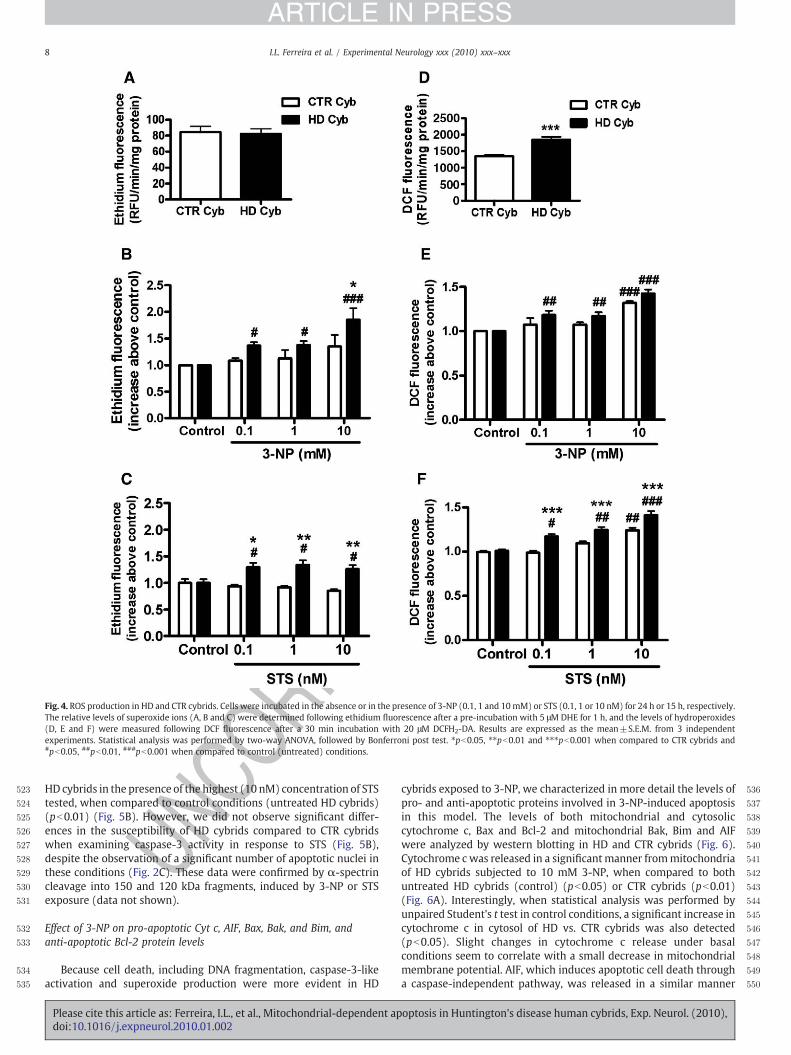
Effect of 3-NP and STS on reactive oxygen species production
To explain the higher susceptibility of HD cybrids when exposed totoxic stimuli, we examined the production of endogenous reactiveoxygen species (ROS). For this purpose, HD or CTR cybrids wereincubated in the absence or presence of 3-NP or STS and the levels ofsuperoxide ions and hydroperoxides were analyzed by measuringethidium or DCF fluorescence, respectively.
Our results show no differences on superoxide levels between HDand CTR cybrids under basal conditions (untreated HD vs. CTRcybrids) (Fig. 4A). However, superoxide production increased in HDcybrids upon exposure to 3-NP (0.1–10 mM), compared withuntreated conditions (control) (Fig. 4B). A significant difference insuperoxide production in HD compared to CTR cybrids was onlyobserved for the higher concentration of 3-NP tested (10 mM)(pb0.05). Incubation with STS (0.1–10 nM) caused a significantincrease in superoxide production in HD cybrids, as compared to CTRor untreated cybrids. Similarly to 3-NP, exposure to STS did not affectthe levels of superoxide in CTR cybrids (Fig. 4C).
By measuring DCF fluorescence we demonstrate that under basalconditions HD cybrids are endowed with a significant higher amountof hydroperoxides production, compared to CTR cybrids (pb0.01)(Fig. 4D); however, no differences between HD and CTR cybrids wereobserved when the cells were subjected to increasing concentrationsof 3-NP (Fig. 4E). Incubation with 10 mM 3-NP increased hydroper-oxides production, in HD and CTR cybrids, compared to untreatedconditions (control) (pb0.001) (Fig. 4E). Incubation with STSproduced a dose-dependent increase in hydroperoxides productionin HD cybrids compared to untreated conditions (control) and CTRcybrids (Fig. 4F), suggesting that HD cybrids are more susceptible toSTS-induced hydroperoxide production. A significant increase inhydroperoxide production in CTR cybrids was only observed in thepresence of the highest concentration of STS tested (10 nM) (pb0.01).
Effect of 3-NP and STS on caspase-3 activation
Since HD cybrids exhibited a higher percentage of cells displayingapoptotic morphology (as determined in Fig. 2), we also investigatedthe effect of 3-NP and STS on caspase-3 activation in both CTR and HDcybrids (Figs. 5A and B). Although no significant changes wereobserved in CTR cybrids subjected to mitochondrial inhibition,treatment with 10 mM 3-NP was effective in inducing caspase-3-like activity in HD cybrids, when compared to untreated cybrids(control) (pb0.001) or with CTR cybrids (pb0.01) (Fig. 5A). STSincubation also caused a significant increase in caspase-3 activity in
optosis in Huntington's disease human cybrids, Exp. Neurol. (2010),
RREC
TEDPR
OOF
523
524
525
526
527
528
529
530
531
532
533
534
535
536
537
538
539
540
541
542
543
544
545
546
547
548
549
550
Fig. 4. ROS production in HD and CTR cybrids. Cells were incubated in the absence or in the presence of 3-NP (0.1, 1 and 10mM) or STS (0.1, 1 or 10 nM) for 24 h or 15 h, respectively.The relative levels of superoxide ions (A, B and C) were determined following ethidium fluorescence after a pre-incubation with 5 μM DHE for 1 h, and the levels of hydroperoxides(D, E and F) were measured following DCF fluorescence after a 30 min incubation with 20 μM DCFH2-DA. Results are expressed as the mean±S.E.M. from 3 independentexperiments. Statistical analysis was performed by two-way ANOVA, followed by Bonferroni post test. ⁎pb0.05, ⁎⁎pb0.01 and ⁎⁎⁎pb0.001 when compared to CTR cybrids and#pb0.05, ##pb0.01, ###pb0.001 when compared to control (untreated) conditions.
8 I.L. Ferreira et al. / Experimental Neurology xxx (2010) xxx–xxx
ARTICLE IN PRESS
UNCOHD cybrids in the presence of the highest (10 nM) concentration of STS
tested, when compared to control conditions (untreated HD cybrids)(pb0.01) (Fig. 5B). However, we did not observe significant differ-ences in the susceptibility of HD cybrids compared to CTR cybridswhen examining caspase-3 activity in response to STS (Fig. 5B),despite the observation of a significant number of apoptotic nuclei inthese conditions (Fig. 2C). These data were confirmed by α-spectrincleavage into 150 and 120 kDa fragments, induced by 3-NP or STSexposure (data not shown).
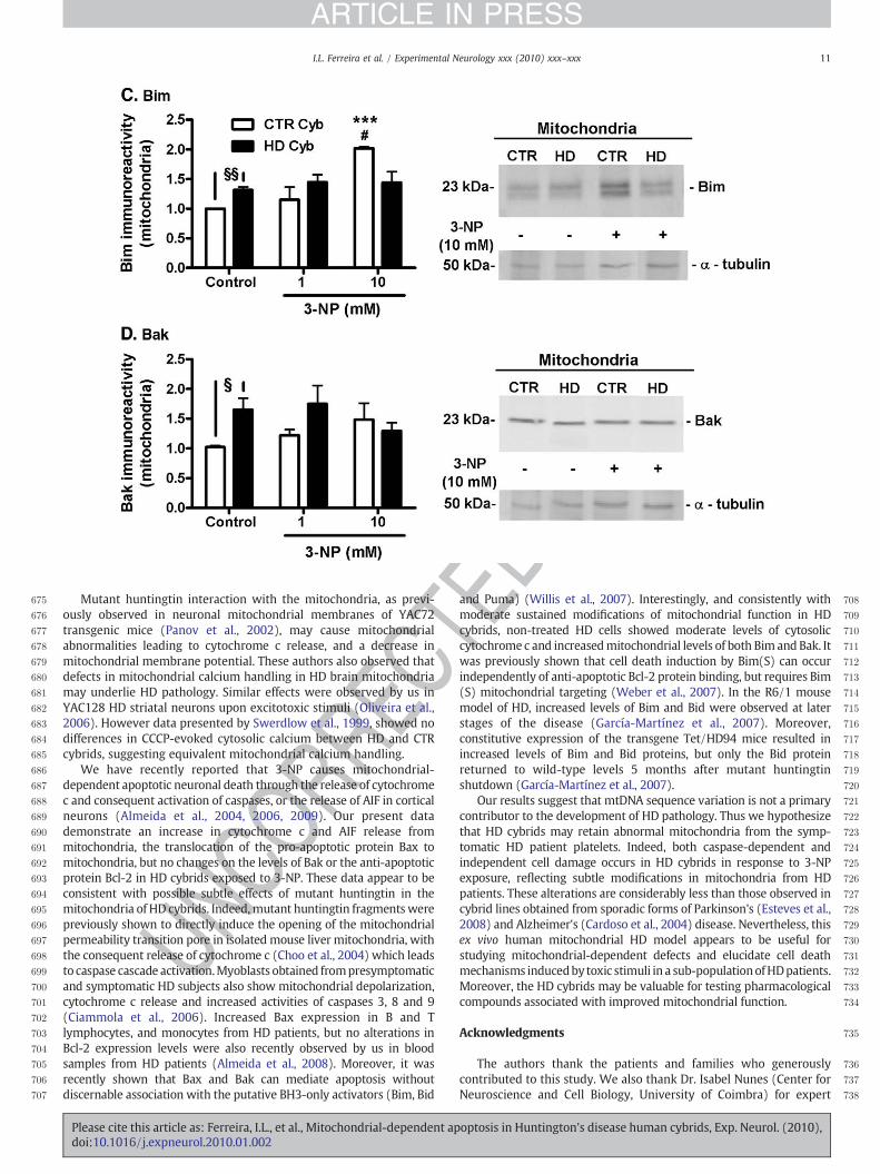
Effect of 3-NP on pro-apoptotic Cyt c, AIF, Bax, Bak, and Bim, andanti-apoptotic Bcl-2 protein levels
Because cell death, including DNA fragmentation, caspase-3-likeactivation and superoxide production were more evident in HD
Please cite this article as: Ferreira, I.L., et al., Mitochondrial-dependent apdoi:10.1016/j.expneurol.2010.01.002
cybrids exposed to 3-NP, we characterized in more detail the levels ofpro- and anti-apoptotic proteins involved in 3-NP-induced apoptosisin this model. The levels of both mitochondrial and cytosoliccytochrome c, Bax and Bcl-2 and mitochondrial Bak, Bim and AIFwere analyzed by western blotting in HD and CTR cybrids (Fig. 6).Cytochrome cwas released in a significantmanner frommitochondriaof HD cybrids subjected to 10 mM 3-NP, when compared to bothuntreated HD cybrids (control) (pb0.05) or CTR cybrids (pb0.01)(Fig. 6A). Interestingly, when statistical analysis was performed byunpaired Student's t test in control conditions, a significant increase incytochrome c in cytosol of HD vs. CTR cybrids was also detected(pb0.05). Slight changes in cytochrome c release under basalconditions seem to correlate with a small decrease in mitochondrialmembrane potential. AIF, which induces apoptotic cell death througha caspase-independent pathway, was released in a similar manner
optosis in Huntington's disease human cybrids, Exp. Neurol. (2010),

551
552
553
554
555
556
557
558
559
560
561
562
563
564
565
566
567
568
569
570
571
572
573
574
575
576
577
578
579
580
581
582
583
584
585
586
587
588
589
590
591
592
593
594
595
596
597
598
599
600
601
602
603
604
605
606
607
608
609
610
611
612
613
614
615
616
617
618
619
620
621
622
623
624
625
626
627
628
629
630
631
632
633
634
635
636
637
638
639
640
Fig. 5. Caspase-3 activation induced by 3-NP (A) or STS (B) in HD and CTR cybrids. Cellswere incubated in the absence or in the presence of 3-NP (1 and 10 mM) or STS (1 or10 nM) for 24 h and 15 h, respectively. Caspase-3-like activity was measured byfollowing the cleavage of the colorimetric substrate Ac-DEVD-pNA. The activity isexpressed as the increase in optic density values above the control (untreated cybrids).Results are expressed as the mean±S.E.M. from 10–12 independent experimentsperformed in duplicates. Statistical analysis was performed by two-way ANOVA,followed by Bonferroni post test. ⁎⁎pb0.01 when compared to CTR cybrids; ##pb0.01,###pb0.001 when compared to control (untreated) conditions.
9I.L. Ferreira et al. / Experimental Neurology xxx (2010) xxx–xxx
ARTICLE IN PRESS
UNCO
RRECfrom HD and CTR cybrids mitochondria upon exposure to 3-NP (Fig.
6B). The pro-apoptotic protein Bim was present in higher amounts inmitochondria derived from non-treated (control) HD, compared toCTR, cybrids (pb0.01 analyzed by t test), but exposure to 3-NP did nothighly affect Bim expression in HD cybrids (Fig. 6C). Interestingly, forthe highest concentration of 3-NP tested (10 mM), CTR cybridsshowed a significant increase in Bim levels in the mitochondria whencompared to untreated conditions (control) (pb0.05) or with HDcybrids exposed to 3-NP (pb0.001) (Fig. 6C). Incubation with 3-NPdid not significantly affect the levels of the proapoptotic protein Bak inmitochondria from both HD and CTR cybrids. However, our resultsdemonstrate that under basal conditions (untreated cybrids) thelevels of Bak are higher in HD than in CTR cybrids (pb0.05) (Fig. 6D).Treatment with 3-NP (1 and 10 mM) increased the translocation ofBax from the cytosol to the mitochondria in both CTR and HD cybrids(pb0.05); however, no significant differences were observed betweenHD and CTR cybrids (Fig. 6E). Finally, cytosolic or mitochondrial levelsof the anti-apoptotic protein Bcl-2 were unaffected under basal or 3-NP-treated conditions in both HD and CTR cybrids (Fig. 6F).
Discussion
Mitochondria are central in the process of apoptosis, a mechanismleading to neuronal loss in neurodegenerative disorders like HD(Kroemer and Reed, 2000; Beal, 2005; García-Martínez et al., 2007;Yang et al., 2008). In the present study we provide evidence that
Please cite this article as: Ferreira, I.L., et al., Mitochondrial-dependent apdoi:10.1016/j.expneurol.2010.01.002
TEDPR
OOF
human HD cybrids show subtle mitochondrial modifications. Indeed,HD cybrids are more susceptible than CTR cybrids to mitochondrial-dependent cell degeneration produced by the mitochondrial complexII inhibitor 3-NP and the classic apoptotic inducer STS. In HD cybrids,treatment with 3-NP caused the release of mitochondrial cytochromec, the subsequent activation of caspase-3, as well as the release ofmitochondrial AIF. This effect appears to be mediated by mitochon-drial translocation of Bax. Moreover, increasedmitochondrial levels ofBim and Bak, a slight decrease in mitochondrial membrane potentialconcomitant with the release of cytochrome c, and increasedhydroperoxide production in non-treated HD cybrids may explainthe increased susceptibility to apoptosis caused by exposure to stressinducers (3-NP or STS). Conversely, CTR cybrids are more vulnerableto necrotic cell death upon 3-NP treatment, and no changes incaspase-3 activation are observed. Increased mitochondrial Bax, andparticularly Bim, may contribute to promote a different mode of celldeath in 3-NP treated-CTR cybrids.
Previous evidence showed that both nuclear and mitochondrialgenomes are damaged in the 3-NP chemically inducedHDmousemodeland in the HD R6/2 transgenic mice (Acevedo-Torres et al., 2009).Lymphocytes from HD patients have higher frequencies of mtDNAdeletions and oxidative stress, suggesting that CAG repeat instability,and thus mutant huntingtin, are a causative factor in mtDNA damage(Banoei et al., 2007). Moreover, decreased mtDNA content wascorrelated with the length of CAG repeats in leukocytes from HDpatients (Liu et al., 2008). Data presented in Table 1 suggest that themtDNA sequence variations are not causal for HD in the patientsincluded in the present study, since some are also found in CTR cybrids.Additionally, there is no information regarding theCAG repeat genotypein the HD gene of CTR subjects, and thus we cannot exclude that theymay be carriers for the intermediate or expanded allele and that theymay develop any type of neurodegenerative disease later in life,including dementia. The presence of mtDNA variations, including an8656A N G variant in one patient, was recently shown in a screeningstudy for mutations in the tRNA(leu/lys) and MTATP6 genes of 20patients with HD (Kasraie et al., 2008). However, the nucleotides 8915-9207 of the same gene do not present any sequence variation in ourstudy. Table 1 also presents one HD cybrid line carrying the 3394T N Cmutation with status “unclear,” previously described in cases sufferingfrom Leber Hereditary Optic Neuropathy (LHON), which was shown tobe related with HD features (Morimoto et al., 2004). Despite theseobservations, we cannot exclude that other genes outside the regionsinvestigatedmay be involved in the disease or thatmtDNA involvementis either related to deletion events or copy number alterations.
UnchangedmtDNA sequence variations correlate with the fact thatno mitochondrial respiratory chain defects were found in HD,compared to CTR cybrids. These results are in agreement withprevious data showing no substantial modifications in mitochondrialcomplexes activity in cybrid cell lines containing mtDNA from HDpatients (Swerdlow et al., 1999). Furthermore, there were nosignificant changes in the activity of mitochondrial respiratorycomplexes (I–IV) or in superoxide formation among the six HDcybrid lines used in the current study. Thus, our data suggest thatother mitochondrial modifications induced by full-length and/orfragments of mutant huntingtin, such as protein post-translationalmodifications, are retained in HD cybrids, which may be related withan interaction of mutant huntingtin with the organelle in HD carriersplatelets. However, we could not detect huntingtin associated withthe mitochondrial fractions derived from HD cybrids, as detected bywestern blotting using the anti-huntingtin antibody MAB2166(Chemicon) (data not shown). Although HD cybrids do not expressmutant huntingtin and thus cannot be directly compared withmodelsexpressing mutant huntingtin, in striatal cell lines expressing full-lengthmutant huntingtin (derived from knock-inmice) no significanteffects on respiratory complexes activities were observed either(Milakovic and Johnson, 2005).
optosis in Huntington's disease human cybrids, Exp. Neurol. (2010),

641
642
643
644
645
646
647
648
649
650
651
652
653
654
655
656
657
658
659
660
661
662
663
664
665
666
667
668
669
670
671
672
673
674
10 I.L. Ferreira et al. / Experimental Neurology xxx (2010) xxx–xxx
ARTICLE IN PRESS
Mitochondrial dysfunction has been frequently associated withincreased generation of ROS, promoting intracellular oxidative stressand leading to protein, lipid and DNA oxidation. Indeed, oxidativedamage was shown to play an important role in the pathogenesis andprogression of HD in the R6/2 transgenic mousemodel (Perluigi et al.,2005) and also in post-mortem samples obtained from the striatumand cortex of human HD brain (Sorolla et al., 2008). Our data alsodemonstrate that, under basal conditions, HD cybrids are endowedwith a significant higher production of hydroperoxides whencompared to CTR cybrids. These data differ from a previous studyshowing no evidence of ROS generation, as measured with DCFH2-DAin untreated HD cybrids (Swerdlow et al., 1999); however, theseauthors did not exclude a subtle mitochondrial pathology in thesecells. In agreement, we show that HD cybrids are more vulnerablethan CTR cybrids to produce superoxide upon 3-NP or STS treatment,whereas increased hydroperoxide production was mainly evoked bySTS, suggesting that the presence of higher amounts of hydroper-
UNCO
RREC
Fig. 6. Changes in cytosolic and mitochondrial levels of cytochrome c, AIF, Bax, Bim, Bak, andNP (1 and 10 mM) for 24 h. Cytosolic and mitochondrial fractions were obtained as describeand Bcl-2 (F) protein levels were analyzed byWestern blotting. α-Tubulin or Cox1 were usedresults are expressed as the mean±S.E.M. from 3–10 independent experiments. Statistical aand ⁎⁎⁎pb0.001 when compared to CTR cybrids; #pb0.05, ##pb0.01 when compared to contwith CTR cybrids under control (untreated) conditions, and statistical analysis was perform
Please cite this article as: Ferreira, I.L., et al., Mitochondrial-dependent apdoi:10.1016/j.expneurol.2010.01.002
oxides in untreated HD cybrids masks the effect caused by 3-NP-induced mitochondrial inhibition.
The mitochondrial toxin 3-NP was shown to cause energydeficiency and cell death by necrosis and apoptosis in striatal, corticaland hippocampal cells (Behrens et al., 1995; Pang and Geddes, 1997;Almeida et al., 2004, 2006; Ruan et al., 2004; Brouillet et al., 2005), andboth processes of cell damage have been proven to involvemitochondria (Kroemer and Reed, 2000). In the present work,exposure of HD cybrid cell lines to 3-NP or STS caused DNAfragmentation and moderate caspase-3 activation, evidencing anincreased susceptibility of HD cybrids to apoptosis. However, 3-NPtreated CTR cybrids died predominantly by necrosis, not involvingcaspase-3 activation. Recent data obtained in our laboratory using thesame cybrid lines also showed that endogenous levels of ATP arehigher in HD cybrids compared to CTR cybrids (author's unpublisheddata). Preserved ATP levels in HD cybrids may explain the preferentialmode of cell death by apoptosis.
TEDPR
OOF
Bcl-2 in HD and CTR cybrids. Cells were incubated in the absence or in the presence of 3-d in the Material and Methods and cytochrome c (A), AIF (B), Bim (C), Bak (D), Bax (E),as loading controls for analysis of cytosolic or mitochondrial fractions, respectively. Thenalysis was performed by two-way ANOVA, followed by Bonferroni post test. ⁎⁎pb0.01rol (untreated) conditions. §pb0.05 and §§pb0.01 when the HD cybrids were compareded by using Student's t test.
optosis in Huntington's disease human cybrids, Exp. Neurol. (2010),
PROO
F675
676
677
678
679
680
681
682
683
684
685
686
687
688
689
690
691
692
693
694
695
696
697
698
699
700
701
702
703
704
705
706
707
708
709
710
711
712
713
714
715
716
717
718
719
720
721
722
723
724
725
726
727
728
729
730
731
732
733
734
735
736
737
738
11I.L. Ferreira et al. / Experimental Neurology xxx (2010) xxx–xxx
ARTICLE IN PRESS
UNCO
RREC
Mutant huntingtin interaction with the mitochondria, as previ-ously observed in neuronal mitochondrial membranes of YAC72transgenic mice (Panov et al., 2002), may cause mitochondrialabnormalities leading to cytochrome c release, and a decrease inmitochondrial membrane potential. These authors also observed thatdefects in mitochondrial calcium handling in HD brain mitochondriamay underlie HD pathology. Similar effects were observed by us inYAC128 HD striatal neurons upon excitotoxic stimuli (Oliveira et al.,2006). However data presented by Swerdlow et al., 1999, showed nodifferences in CCCP-evoked cytosolic calcium between HD and CTRcybrids, suggesting equivalent mitochondrial calcium handling.
We have recently reported that 3-NP causes mitochondrial-dependent apoptotic neuronal death through the release of cytochromec and consequent activation of caspases, or the release of AIF in corticalneurons (Almeida et al., 2004, 2006, 2009). Our present datademonstrate an increase in cytochrome c and AIF release frommitochondria, the translocation of the pro-apoptotic protein Bax tomitochondria, but no changes on the levels of Bak or the anti-apoptoticprotein Bcl-2 in HD cybrids exposed to 3-NP. These data appear to beconsistent with possible subtle effects of mutant huntingtin in themitochondria of HD cybrids. Indeed,mutant huntingtin fragmentswerepreviously shown to directly induce the opening of the mitochondrialpermeability transition pore in isolated mouse liver mitochondria, withthe consequent release of cytochrome c (Choo et al., 2004) which leadsto caspase cascade activation.Myoblasts obtained frompresymptomaticand symptomatic HD subjects also show mitochondrial depolarization,cytochrome c release and increased activities of caspases 3, 8 and 9(Ciammola et al., 2006). Increased Bax expression in B and Tlymphocytes, and monocytes from HD patients, but no alterations inBcl-2 expression levels were also recently observed by us in bloodsamples from HD patients (Almeida et al., 2008). Moreover, it wasrecently shown that Bax and Bak can mediate apoptosis withoutdiscernable association with the putative BH3-only activators (Bim, Bid
Please cite this article as: Ferreira, I.L., et al., Mitochondrial-dependent apdoi:10.1016/j.expneurol.2010.01.002
TED
and Puma) (Willis et al., 2007). Interestingly, and consistently withmoderate sustained modifications of mitochondrial function in HDcybrids, non-treated HD cells showed moderate levels of cytosoliccytochrome c and increasedmitochondrial levels of both Bim and Bak. Itwas previously shown that cell death induction by Bim(S) can occurindependently of anti-apoptotic Bcl-2 protein binding, but requires Bim(S) mitochondrial targeting (Weber et al., 2007). In the R6/1 mousemodel of HD, increased levels of Bim and Bid were observed at laterstages of the disease (García-Martínez et al., 2007). Moreover,constitutive expression of the transgene Tet/HD94 mice resulted inincreased levels of Bim and Bid proteins, but only the Bid proteinreturned to wild-type levels 5 months after mutant huntingtinshutdown (García-Martínez et al., 2007).
Our results suggest that mtDNA sequence variation is not a primarycontributor to the development of HD pathology. Thus we hypothesizethat HD cybrids may retain abnormal mitochondria from the symp-tomatic HD patient platelets. Indeed, both caspase-dependent andindependent cell damage occurs in HD cybrids in response to 3-NPexposure, reflecting subtle modifications in mitochondria from HDpatients. These alterations are considerably less than those observed incybrid lines obtained from sporadic forms of Parkinson's (Esteves et al.,2008) and Alzheimer's (Cardoso et al., 2004) disease. Nevertheless, thisex vivo human mitochondrial HD model appears to be useful forstudying mitochondrial-dependent defects and elucidate cell deathmechanisms inducedby toxic stimuli in a sub-populationofHDpatients.Moreover, the HD cybrids may be valuable for testing pharmacologicalcompounds associated with improved mitochondrial function.
Acknowledgments
The authors thank the patients and families who generouslycontributed to this study. We also thank Dr. Isabel Nunes (Center forNeuroscience and Cell Biology, University of Coimbra) for expert
optosis in Huntington's disease human cybrids, Exp. Neurol. (2010),

ECTEDPR
OOF
739
740
741
742
743
744
745
746
747
748749750751752753754755756757758759760
761762763764765766767768769770771
Fig. 6 (continued).
12 I.L. Ferreira et al. / Experimental Neurology xxx (2010) xxx–xxx
ARTICLE IN PRESS
ORRtechnical assistance with cell culture maintenance, and Dr. Russell H.
Swerdlow (University of Virginia, Charlottesville, VA, USA) for the giftof ρ° NT2 cells. This work was supported by Project III/BIO/49/2005(Instituto de Investigação Interdisciplinar, III, Universidade deCoimbra, Portugal), Project STARTER S-09 (Gabinete de Apoio àInvestigação, GAI, Faculdade de Medicina, Universidade de Coimbra)and Project POCI/SAU-NEU/57310/2004 (Fundação para a Ciência e aTecnologia, Portugal).
C 772773774775776777778779780781782783784785786787788
UNReferences
Acevedo-Torres, K., Berríos, L., Rosario, N., Dufault, V., Skatchkov, S., Eaton, M.J., Torres-Ramos, C.A., Ayala-Torres, S., 2009. Mitochondrial DNA damage is a hallmark ofchemically induced and the R6/2 transgenic model of Huntington's disease. DNARepair 8, 126–136.
Almeida, S., Domingues, A., Rodrigues, L., Oliveira, C.R., Rego, A.C., 2004. FK506 preventsmitochondrial-dependent apoptotic cell death induced by 3-nitropropionic acid inrat primary cortical cultures. Neurobiol. Dis. 17, 435–444.
Almeida, S., Brett, A.C., Góis, I.N., Oliveira, C.R., Rego, A.C., 2006. Caspase-dependent and-independent cell death induced by 3-nitropropionic acid in rat cortical neurons. J.Cell Biochem. 98, 93–101.
Almeida, S., Sarmento-Ribeiro, A.B., Januário, C., Rego, A.C., Oliveira, C.R., 2008. Evidenceof apoptosis and mitochondrial abnormalities in peripheral blood cells ofHuntington's disease patients. Biochem. Biophys. Res. Commun. 374, 599–603.
Please cite this article as: Ferreira, I.L., et al., Mitochondrial-dependent apdoi:10.1016/j.expneurol.2010.01.002
Almeida, S., Laço, M., Cunha-Oliveira, T., Oliveira, C.R., Rego, A.C., 2009. BDNF regulatesBIM expression levels in 3-nitropropionic acid-treated cortical neurons. Neurobiol.Dis. 35, 448–456.
Aronin, N., Chase, K., Young, C., Sapp, E., Schwarz, C., Matta, N., Kornreich, R.,Landwehrmeyer, B., Bird, E., Beal, M.F., Vonsattel, J.P., Smith, T., Carraway, R., Boyce,F.M., Young, A.B., Penney, J.B., DiFiglia, M., 1995. CAG expansion affects the expressionof mutant huntingtin in the Huntington's disease brain. Neuron 15, 1193–1201.
Banoei, M.M., Houshmand, M., Panahi, M.S.S., Shariati, P., Rostami, M., 2007.Huntington's disease and mitochondrial DNA deletions: Event or regularmechanism for mutant huntingtin protein and CAG repeats expansion?! CellMol. Neurobiol. 27, 867–875.
Beal, M.F., 2005. Mitochondria take center stage in aging and neurodegeneration. Ann.Neurol. 58, 495–505.
Behrens, M.I., Koh, J., Canzoniero, L.M., Sensi, S.L., Csernansky, C.A., Choi, D.W., 1995. 3-Nitropropionic acid induces apoptosis in cultured striatal and cortical neurons.Neuroreport 6, 545–548.
Bergmeyer, H.U., Bernt, E., 1974. UV-assay with pyruvate and NADH. Methods ofenzymatic analysis. Academic press, New York, pp. 574–579.
Bezprozvanny, I., Hayden, M.R., 2004. Deranged neuronal calcium signaling andHuntington disease. Biochem. Biophy. Res. Comm. 322, 1310–1317.
Bossy-Wetzel, E., Petrilli, A., Knott, A.B., 2008. Mutant huntingtin and mitochondrialdysfunction. Trends Neurosci. 31, 609–616.
Brouillet, E., Guyot, M.C., Mittoux, V., Altairac, S., Condé, F., Palfi, S., Hantraye, P., 1998.Partial inhibition of brain succinate dehydrogenase by 3-nitropropionic acid issufficient to initiate striatal degeneration in rat. J. Neurochem. 70, 794–804.
Brouillet, E., Jacquard, C., Bizat, N., Blum, D., 2005. 3-Nitropropionic acid: amitochondrial toxin to uncover physiopathological mechanisms underlying striataldegeneration in Huntington's disease. J. Neurochem. 95, 1521–1540.
optosis in Huntington's disease human cybrids, Exp. Neurol. (2010),

789790791792793794795796797798799800801802803804805806807808809810811812813814815816817818819820821822823824825826827828829830831832833834835836837838839840841842843844845846
847848849850851852853854855856857858859860861862863864865866867868869870871872873874875876877878879880881882883884885886887888889890891892893894895896897898899900901902903904
905
13I.L. Ferreira et al. / Experimental Neurology xxx (2010) xxx–xxx
ARTICLE IN PRESS
EC
Cardoso, S.M., Santana, I., Swerdlow, R.H., Oliveira, C.R., 2004. Mitochondria dysfunctionof Alzheimer's disease cybrids enhances Abeta toxicity. J. Neurochem. 89,1417–1426.
Carré, M., André, N., Carles, G., Borghi, H., Brichese, L., Briand, C., Braguer, D., 2002.Tubulin is an inherent component of mitochondrial membranes that interacts withthe voltage-dependent anion channel. J. Biol. Chem. 277, 33664–33679.
Choo, Y.S., Johnson, G.V., MacDonald,M., Detloff, P.J., Lesort,M., 2004.Mutant huntingtindirectly increases susceptibility of mitochondria to the calcium-induced perme-ability transition and cytochrome c release. Hum. Mol. Genet. 13, 1407–1420.
Ciammola, A., Sassone, J., Alberti, L., Meola, G., Mancinelli, E., Russo, M.A., Squitieri, F.,Silani, V., 2006. Increased apoptosis, Huntingtin inclusions and altered differenti-ation in muscle cell cultures from Huntington's disease subjects. Cell Death Differ.13, 2068–2078.
del Hoyo, P., García-Redondo, A., de Bustos, F., Molina, J.A., Sayed, Y., Alonso-Navarro, H.,Caballero, L., Arenas, J., Jiménez-Jiménez, F.J., 2006. Oxidative stress in skinfibroblasts cultures of patients with Huntington's disease. Neurochem. Res. 31,1103–1109.
Esteves, A.R., Domingues, A.F., Ferreira, I.L., Januário, C., Swerdlow, R.H., Oliveira, C.R.,Cardoso, S.M., 2008. Mitochondrial function in Parkinson's disease cybridscontaining an nt2 neuron-like nuclear background. Mitochondrion 8, 219–228.
Fan, M.M.Y., Raymond, L.A., 2007. N-methyl-D-aspartate (NMDA) receptor function andexcitotoxicity in Huntington's disease. Prog. Neurobiol. 81, 272–293.
García-Martínez, J.M., Pérez-Navarro, E., Xifró, X., Canals, J.M., Díaz-Hernández, M.,Trioulier, Y., Brouillet, E., Lucas, J.J., Alberch, J., 2007. BH3-only proteins Bid and Bim(EL) are differentially involved in neuronal dysfunction in mouse models ofHuntington's disease. J. Neurosci. Res. 85, 2756–2769.
Gil, J.M., Rego, A.C., 2008. Mechanisms of neurodegeneration in Huntington's disease.Eur. J. Neurosci. 27, 2803–2820.
Grazina, M., Pratas, J., Silva, F., Oliveira, S., Santana, I., Oliveira, C., 2006. Genetic basis ofAlzheimer's dementia: role ofmtDNAmutations. Genes Brain Behav. 5 (S2), 92–107.
Huntington Study Group, 1996. Unified Huntington's Disease Rating Scale: reliabilityand consistency. Mov. Disord. 11, 136–142.
Kasraie, S., Houshmand, M., Banoei, M.M., Ahari, S.E., Panahi, M.S., Shariati, P., Bahar, M.,Moin, M., 2008. Investigation of tRNA(Leu/Lys) and ATPase 6 Genes Mutations inHuntington's Disease. Cell. Mol. Neurobiol. 28, 933–938.
Kuhn, A., Goldstein, D.R., Hodges, A., Strand, A.D., Sengstag, T., Kooperberg, C., Becanovic,K., Pouladi, M.A., Sathasivam, K., Cha, J.H., Hannan, A.J., Hayden, M.R., Leavitt, B.R.,Dunnett, S.B., Ferrante, R.J., Albin, R., Shelbourne, P., Delorenzi,M., Augood, S.J., Faull,R.L., Olson, J.M., Bates, G.P., Jones, L., Luthi-Carter, R., 2007. Mutant huntingtin'seffects on striatal gene expression in mice recapitulate changes observed in humanHuntington's disease brain and do not differ withmutant huntingtin length orwild-type huntingtin dosage. Hum. Mol. Genet. 16, 1845–1861.
Kroemer, G., Reed, J.C., 2000. Mitochondrial control of cell death. Nat. Med. 6, 513–519.Liu, C.S., Cheng, W.L., Kuo, S.J., Li, J.Y., Soong, B.W., Wei, Y.H., 2008. Depletion of
mitochondrial DNA in leukocytes of patients with poly-Q diseases. J. Neurol. Sci.264, 18–21.
Mancuso, M., Orsucci, D., Siciliano, G., Murri, L., 2008. Mitochondria, mitochondrial DNAand Alzheimer's disease. What comes first? Curr. Alzheimer Res. 5, 457–468.
Milakovic, T., Johnson, G.V.W., 2005. Mitochondrial respiration and ATP production aresignificantly impaired in striatal cells expressing mutant huntingtin. J. Biol. Chem.35, 30773–30782.
Morimoto, N., Nagano, I., Deguchi, K., Murakami, T., Fushimi, S., Shoji, M., Abe, K., 2004.Leber hereditary optic neuropathy with chorea and dementia resemblingHuntington disease. Neurology 63, 2451–2552.
Oliveira, J.M., Chen, S., Almeida, S., Riley, R., Goncalves, J., Oliveira, C.R., Hayden, M.R.,Nicholls, D.G., Ellerby, L.M., Rego, A.C., 2006. Mitochondrial-dependent Ca2+handling in Huntington's disease striatal cells: effect of histone deacetylaseinhibitors. J. Neurosci. 26, 11174–11186.
UNCO
R
Please cite this article as: Ferreira, I.L., et al., Mitochondrial-dependent apdoi:10.1016/j.expneurol.2010.01.002
TEDPR
OOF
Onyango, I., Khan, S., Miller, B., Swerdlow, R., Trimmer, P., Bennett Jr., P., 2006.Mitochondrial genomic contribution to mitochondrial dysfunction in Alzheimer'sdisease. J. Alzheimers Dis. 9, 183–193.
Pang, Z., Geddes, J.W., 1997. Mechanisms of cell death induced by the mitochondrialtoxin 3-nitropropionic acid: acute excitotoxic necrosis and delayed apoptosis. J.Neurosci. 17, 3064–3073.
Panov, A.V., Gutekunst, C.A., Leavitt, B.R., Hayden, M.R., Burke, J.R., Strittmatter, W.J.,Greenamyre, J.T., 2002. Early mitochondrial calcium defects in Huntington's diseaseare a direct effect of polyglutamines. Nat. Neurosci. 5, 731–736.
Perluigi, M., Poon, H.F., Maragos, W., Pierce, W.M., Klein, J.B., Calabrese, V., Cini, C., DeMarco, C., Butterfield, D.A., 2005. Proteomic analysis of protein expression andoxidative modification in r6/2 transgenic mice: a model of Huntington disease.Mol. Cell Proteomics 4, 1849–1861.
Rego, A.C., de Almeida, L.P., 2005. Molecular targets and therapeutic strategies inHuntington's disease. Curr. Drug Targets 4, 361–381.
Ruan, Q., Lesort, M., MacDonald, M.E., Johnson, G.V., 2004. Striatal cells from mutanthuntingtin knock-in mice are selectively vulnerable to mitochondrial complex IIinhibitor-induced cell death through a non-apoptotic pathway. Hum. Mol. Genet.13, 669–681.
Sas, K., Robotka, H., Toldi, J., Vécsei, L., 2007. Mitochondria, metabolic disturbances,oxidative stress and kynurenine system, with focus on neurodegenerativedisorders. J. Neurol. Sci. 257, 221–239.
Sawa, A., Wiegand, G.W., Cooper, J., Margolis, R.L., Sharp, A.H., Lawler, J.F.Jr,Greenamyre, J.T., Snyder, S.H., Ross, C.A., 1999. Increased apoptosis of Huntingtondisease lymphoblasts associated with repeat length-dependent mitochondrialdepolarization. Nat Med. 10, 1194–1198.
Schapira, A.H., 1998. Mitochondrial dysfunction in neurodegenerative disorders.Biochim. Biophys. Acta 1366, 225–233.
Sorolla, M.A., Reverter-Branchat, G., Tamarit, J., Ferrer, I., Ros, J., Cabiscol, E., 2008.Proteomic and oxidative stress analysis in human brain samples of Huntingtondisease. Free Radic. Biol. Med. 45, 667–678.
Swerdlow, R.H., Parks, J.K., Cassarino, D.S., Shilling, A.T., Bennett Jr., J.P., Harrison, M.B.,Parker Jr., W.D., 1999. Characterization of cybrid cell lines containing mtDNA fromHuntington's disease patients. Biochem. Biophys. Res. Commun. 291, 701–704.
Tabrizi, S.J., Cleeter, M.W., Xuereb, J., Taanman, J.W., Cooper, J.M., Schapira, A.H., 1999.Biochemical abnormalities and excitotoxicity in Huntington's disease brain. Ann.Neurol. 45, 25–32.
Trottier, Y., Devys, D., Imbert, G., Saudou, F., An, I., Lutz, Y., Weber, C., Agid, Y., Hirsch, E.C., Mandel, J.L., 1995. Cellular localization of the Huntington's disease protein anddiscrimination of the normal and mutated form. Nat. Genet. 10, 104–110.
Turner, C., Cooper, J.M., Schapira, A.H., 2007. Clinical correlates of mitochondrialfunction in Huntington's disease muscle. Mov. Disord. 22, 1715–1721.
Vanlangenakker, N., Berghe, T.V., Krysko, D.V., Festjens, N., Vandenabeele, P., 2008.Molecular mechanisms and pathophysiology of necrotic cell death. Curr. Mol. Med.8, 207–220.
Ward, M.W., Rego, A.C., Frenguelli, B.G., Nicholls, D.G., 2000. Mitochondrial membranepotential and glutamate excitotoxicity in cultured cerebellar granule cells. J.Neurosci. 20, 7208–7219.
Weber, A., Paschen, S.A., Heger, K., Wilfling, F., Frankenberg, T., Bauerschmitt, H.,Seiffert, B.M., Kirschnek, S., Wagner, H., Häcker, G., 2007. BimS-induced apoptosisrequires mitochondrial localization but not interaction with anti-apoptotic Bcl-2proteins. J. Cell Biol. 177, 625–636.
Willis, S.N., Fletcher, J.I., Kaufmann, T., van Delft, M.F., Chen, L., Czabotar, P.E., Ierino, H.,Lee, E.F., Fairlie, W.D., Bouillet, P., Strasser, A., Kluck, R.M., Adams, J.M., Huang, D.C.,2007. Apoptosis initiated when BH3 ligands engage multiple Bcl-2 homologs, notBax or Bak. Science 315, 856–859.
Yang, J.-L., Weissman, L., Bohr, V., Mattson, M.P., 2008. Mitochondrial DNA damage andrepair in neurodegenerative disorders. DNA Repair 7, 1110–1120.
Roptosis in Huntington's disease human cybrids, Exp. Neurol. (2010),