Embed Size (px)
Citation preview
ARTICLE IN PRESS
InsectBiochemistry
andMolecularBiology
0965-1748/$ - se
doi:10.1016/j.ib
�CorrespondE-mail addr
Insect Biochemistry and Molecular Biology 37 (2007) 1241–1248
www.elsevier.com/locate/ibmb
Molecular characterization of an inhibitor of apoptosis inthe Egyptian armyworm, Spodoptera littoralis, and midgut
cell death during metamorphosis
Lluısa Vilaplana, Nuria Pascual, Nathalia Perera, Xavier Belles�
Department of Physiology and Molecular Biodiversity, Institut de Biologia Molecular de Barcelona (CSIC), Jordi Girona, 18-26, 08034 Barcelona, Spain
Received 22 May 2007; received in revised form 19 July 2007; accepted 23 July 2007
Abstract
The cDNA corresponding to an inhibitor of apoptosis (IAP) from the Egyptian armyworm, Spodoptera littoralis, was cloned by RT-
PCR. Sequence analysis showed that the IAP of S. littoralis (SlIAP) contains two baculoviral IAP repeat (BIR) motifs, followed by a
RING finger, an organization which is very similar to that of other lepidopteran IAPs. SlIAP mRNA was detected in ovary, testis,
salivary gland, fat body, epidermis, brain and midgut of S. littoralis. During the last larval instar, prepupal and pupal stages, brain
mRNA levels remained approximately constant, whereas those of midgut showed a large peak centred in the prepupal stage. Midgut
morphology changed during metamorphosis from a semi-transparent, cylindrical structure in last instar larvae to a brownish globular
mass in pupae. TUNEL assays, LysoTracker staining and caspase-3 immunohistochemistry, indicated that programmed cell death in
midgut starts actively at the onset of pupation process, coinciding with the dramatic decrease of SlIAP mRNA levels observed at the
same time.
r 2007 Elsevier Ltd. All rights reserved.
Keywords: Inhibitor of apoptosis (IAP), Midgut cell death, Spodoptera littoralis, TUNEL, Caspase, Apoptosis, Autophagy
1. Introduction
Programmed cell death plays an important role in thedevelopment and homeostasis of metazoans, and it is alsocritical in insect embryogenesis and metamorphosis (Vauxand Korsmeyer, 1999). Cells dying by this mechanism showdistinct morphological changes including cell shrinkage,membrane blebbing, DNA fragmentation and apoptoticbody formation (Wyllie et al., 1980). Programmed celldeath is precisely controlled, and regulatory mechanismshave been conserved across animal species during evolution(Fraser and Evan, 1996).
Inhibitor of apoptosis proteins (IAPs) are key regulatorsof cell death. The first IAPs were discovered in baculo-viruses thanks to their capacity to functionally replace thebaculoviral p35 protein, another viral apoptosis inhibitor(Crook et al., 1993; Birnbaum et al., 1994). Baculovirus
e front matter r 2007 Elsevier Ltd. All rights reserved.
mb.2007.07.013
ing author.
ess: [email protected] (X. Belles).
IAPs impair the apoptotic response of the host and extendtheir survival, thus optimizing viral replication in it.Specifically, IAPs inhibit the caspases that promote celldeath (Kumar and Cakouros, 2004), apparently interactingwith a particular subset of caspases (Deveraux et al., 1997;Roy et al., 1997; Chai et al., 2003; Zachariou et al., 2003).IAPs have been reported in all eukaryotic groups,including humans (Clem and Duckett, 1998; Deveraux,1998), and available sequences share a number of structuralmotifs. Structurally, IAPs are recognized by the presence of1–3 copies of a motif called baculovirus IAP repeat (BIR)at the amino-terminus (Salvesen and Duckett, 2002). TheBIR motif usually comprises approximately 70 amino acidsand contains a highly conserved arrangement of Cys/Hisresidues which forms a stable fold that can chelate zinc(Hinds et al., 1999; Sun et al., 1999). BIR motifs areessential for the anti-apoptotic function of IAPs (Vucicet al., 1998), and in the fruitfly Drosophila melanogaster
they can bind apoptotic inducers, like Grim, Reaperand Hid proteins (Vucic et al., 1997; Vucic et al., 1998;
ARTICLE IN PRESSL. Vilaplana et al. / Insect Biochemistry and Molecular Biology 37 (2007) 1241–12481242
Goyal et al., 2000; Yin and Thummel, 2004). In addition toBIR motifs, some IAPs contain a RING finger near thecarboxy-terminus, which is also present in other cellproteins (Freemont et al., 1991), although its relevance inthe context of cell death depends on the IAP or onthe nature of the apoptotic stimulus (Clem and Miller,1994; Hay et al., 1995). IAPs have been describedin a number of lepidopteran species, like Spodoptera
frugiperda (Huang et al., 2000), Bombyx mori (Huanget al., 2001), Trichoplusia ni (Liao et al., 2002), Spodoptera
exigua (GenBank Accession number DQ206460) andSpodoptera ( ¼ Prodenia) litura (GenBank Accession num-ber AY751528). A number of organisms have more thanone IAP, for example mammals have eight (Liston et al.,2003) and D. melanogaster two (Hay, 2000).
In holometabolous insects, programmed cell deathremoves obsolete larval tissues during larval–pupal transi-tion, and a fine-tuned balance between pro-death factorsand anti-death factors regulates the whole death process(Yin and Thummel, 2004). Caspases are typical pro-deathfactors, whereas IAPs, which directly interact withcaspases, are effective anti-death factors. In lepidopterans,in addition to the IAPs mentioned above, two effectorcaspases have been reported: caspase-1 (Liu et al., 2005;Liu and Chejanovsky, 2006) and another protease calledICE (GenBank Accession Number AY885228).
During insect metamorphosis, midgut tissue undergoesextensive remodelling, and this is especially important inthose species where feeding habits change dramatically inthe adult. For this reason, midgut remodelling duringmetamorphosis has been chosen as preferential model tostudy cell death (Jiang et al., 1997; Lee et al., 2002a; Uwoet al., 2002; Parthasarathy and Palli, 2007). In the presentpaper, we report the cDNA cloning and characterization ofan IAP from the Egyptian armyworm, Spodoptera littoralis
(SlIAP). Molecular characterization allowed studyingSlIAP gene expression in brain and gut tissues, whichhelped to understand the molecular basis of midgutremodelling during metamorphosis. In order to describethis remodelling in detail, we monitored the changesoccurring in midgut structure by using TUNEL assays,LysoTracker staining and caspase-3 immunohistochemistry.
2. Material and methods
2.1. Insect rearing and treatments
A colony of S. littoralis (Boisduval) (Lepidoptera,Noctuidae), was reared in the laboratory on artificial diet(Poitout and Bues, 1974). Just after egg hatching, larvaewere placed in groups of 40–50 into 20� 30 cm plasticboxes and maintained in a climatic chamber on a 16 h light:8 h dark regime, at 2571 1C and 60–70% relative humidity.Freshly ecdysed last instar larvae (L6) were separated andstaged every 24 h and used for experiments. At the end ofthis instar, prepupal stage (PP) starts. PP stage ischaracterized by a shortening of the body and suppression
of abdominal segments. Pupae were staged as white,freshly formed pupa (P0) and at intervals of 24 h after thismoment (P1, P2, P3y). Adults were fed with 20% sugarsolution in water. Last larval instar (L6) lasts 4 days,prepupal stage lasts 2 days (PP1, PP2) and pupae lasts 11days (P0–P11).
2.2. Cloning of SlIAP cDNA
An initial PCR was carried out using a degenerate pri-mer set (LepIAP-F3: TGGGTNGARGGNGAYGAYCC,LepIAP-R1: ATAYCDCACAAGCYTCAGAMA) de-signed on the basis of conserved BIR and RING sequencesof known lepidopteran IAPs. As a template we used 30
cDNA (Gibco, BRL RACE Kit) obtained from total RNAfrom young pupae of S. littoralis. This yielded a fragmentof approximately 460 bp, which was cloned and sequenced.Based on this fragment, specific primers were designed for50- and 30-rapid amplification of cDNA ends (RACE)(Gibco, BRL kits). Following this approach, we completedthe cDNA of the SlIAP according to previously describedmethods (Frohman et al., 1988). To make sure that allamplifications obtained with RACE corresponded to thesame molecule, the entire cDNA was amplified with aforward primer designed at the 50 end and a reverse primerdesigned at the 30 end just before the Poly (A)+ tail. PCRproducts were subjected to electrophoresis on 1.2%agarose gel and subcloned in pSTBlue-1 Acceptor vector(Novagen). Sequencing was carried out by the dideoxynu-cleotide chain termination method. Clones were sequencedon both strands using SP6 and T7 sequencing primers andinternal, specific primers, in an automated fluorescencesequencing system ABI (Perkin Elmer).
2.3. RT-PCR and Southern Blot analyses
Total RNA from tissues under study was isolated usingthe GenElute Mammalian Total RNA kit (Sigma). Anamount between 0.5 and 1 mg of each RNA preparationwas used for cDNA synthesis, as previously described (Zhuet al., 2000). For mRNA detection, cDNA samples weresubjected to PCR amplification with a number of cycleswithin the linear range of amplification, being in all casesbetween 28 and 33 cycles. The primer pair, forwardLepIAP-F1: CATCGGAAGCAGAACGTGATGT, andreverse LepIAP-R2: TGCATTCTGAAACGTCCTGCG,were used to amplify a 224 bp fragment of the SlIAP. As areference, cDNA corresponding to b-actin RNA of S.
littoralis (GenBank Accession number: Z46873, unpub-lished) using forward: 50-GTGATGGTTGGTATGGG-TCAGAA-30, and reverse: 50-GATCTGGGTCATCTT-CTCCCTGT-30, was amplified in parallel for each cDNAsample. cDNA probes for Southern Blot analyses weregenerated by PCR with the same primer pairs usingplasmid DNAs containing the corresponding cDNA clonesas templates. The probes were labelled with fluorescein,using the Gene Images random prime-labelling module
ARTICLE IN PRESSL. Vilaplana et al. / Insect Biochemistry and Molecular Biology 37 (2007) 1241–1248 1243
(Amersham Biosciences). RT-PCR followed by Southernblotting of total RNA without reverse transcription did notresult in amplification, thus indicating that there was nogenomic contamination.
2.4. TUNEL assay
Larval and pupal midguts from S. littoralis weredissected out of the developing adult gut under Ringer’ssaline. They were cut longitudinally and cleaned from theircontent in such a way that the epithelium which is incontact with the lumen was exposed to the treatment.TUNEL assays were carried out using the in situ CellDeath Detection Kit, Fluorescein (Roche), essentially asdescribed by the manufacturer. Dissected and cut midgutswere fixed for 30min at room temperature in 4%paraformaldehyde in PBS, washed in PT (0.1% Triton-PBS), and permeabilized by incubation in PT-SC (0.1%sodium citrate-0.1% PT) at room temperature for 30min.Samples were then rinsed in PBS and incubated in TUNELreaction mixture for 1 h at 37 1C. Finally they weremounted in Mowiol 4-88 (Calbiochem). Sections wereexamined under a Leica TCS-SP laser scanning confocalmicroscope and documented using Leica TCS software.
2.5. LysoTracker staining
Midguts from larvae at appropriate stages were dissectedin Ringer’s saline, and cut as above. First, they werepermeabilized during 45min in PT and then stained with1mM of the nuclear dye TOPRO-3 for 10min (far-redDNA dye excitable with a helium–neon laser; fromMolecular Probes, Eugene, OR, USA) diluted 1:500 inPT. After two washes with PBS, samples were stained withLysoTracker Red DND-99 (Molecular Probes), whichlabels acidic organelles, such as lysosomes, diluted 1:1000in PBS for 5min. Finally they were washed twice in PBSfor 10min, mounted in Mowiol 4-88, and examined byconfocal microscopy, as above.
2.6. Caspase-3 immunohistochemistry
To detect caspase activity by immunohistochemistry, weused mammalian caspase-3 antibody (Cleaved caspase-3(Asp 175) antibody from Cell Signalling Technology),which has been shown to reveal caspase homologues ininsects, for example in the lepidopteran Heliothis virescens
(Parthasarathy and Palli, 2007). Tissues were dissected inRinger’s saline and fixed for 30min with PEM-FA (PEM:100mM PIPES, 2mM EGTA, 1M MgSO4; PEM-10%formaldehyde (FA) 37%), and they were then washedin PBS 0.2% Tween-20. After three washes of 10mineach, samples were blocked with PBT-NGS (5% normalgoat serum, NGS, diluted in 0.1% BSA-0.1% PT) for1 h at room temperature. Samples were then incubatedwith caspase-3 antibody at 1:50 diluted in PBT-NGS for12 h at 41C. After three washes with PT of 20min each,
samples were incubated with Alexa Fluor 488-Anti-RabbitIgG (Molecular Probes). Nuclear staining was carried outwith 1mM TOPRO-3, and tissues were mounted asdescribed above. The specificity of signals was checked byincluding controls without primary antibody treatment.The sections were examined by confocal microscopy, asabove.
3. Results
3.1. Structure of Spodoptera littoralis IAP
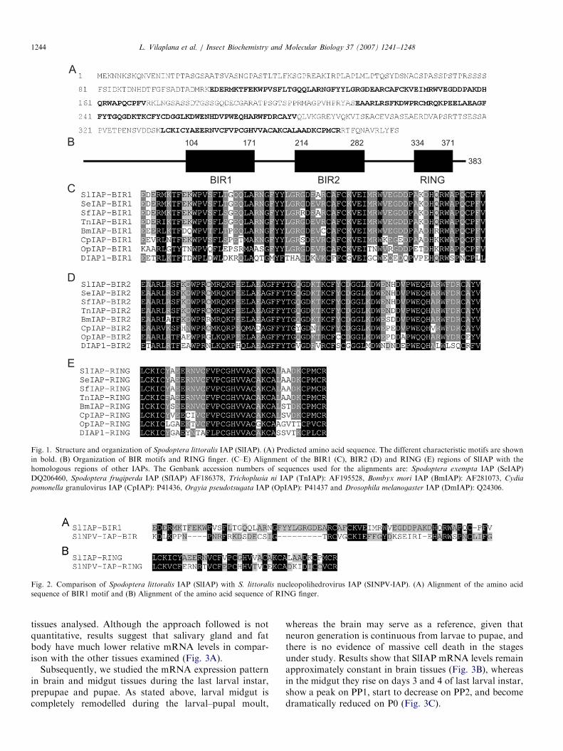
The complete cDNA of SlIAP is 382 amino acids long(Fig. 1A). It contains a putative start codon preceded by anin-frame stop codon, and a final stop codon followed by apoly(A)+ tail, which indicates that the sequence corre-sponds to a full-length ORF (GenBank Accession Number:AM709785). SlIAP sequence analysis reveals the presenceof two BIR motifs and one RING finger (Fig. 1B). The firstBIR motif extends between amino acids 104 and 171 andthe second one has the same size but extending betweenamino acids 214 and 282. Both BIR motifs exhibit thetypical distribution of cysteine and histidine residues:CX2CX6WX9HX6C. They are followed by a RING fingerdomain which is 37 amino acids long and bears thecharacteristic C3HC4 motif from Cys334 to Cys371.Considering the BIR and RING regions (Fig. 1C–E),
SlIAP shows 96% amino acid identity with SeIAP (from S.
exigua), 93% identity with SfIAP (from S. frugiperda) and83% and 67% identity with TnIAP (from T. ni) andBmIAP (from B. mori), respectively. The specific IAP of S.
littoralis nucleopolihedrovirus (SlNPV-IAP) has beendescribed by Liu et al. (2003) as a 15 kDa polypeptidewith only one BIR motif, a RING finger and a thirdspecific acidic-rich motif. The alignment of SlIAP and itsviral counterpart, SlNPV-IAP, plus other BIR motifs frombaculovirus IAPs (data not shown) indicates that theunique viral BIR motif is more similar to the BIR1 than tothe BIR2 motif of S. littoralis. Pairwise sequence analysis(Fig. 2) of lepidopteran and baculoviral IAP sequencesrevealed that they show a 22% amino acid identity whencomparing SlIAP BIR1 motif with the only SlNPV-IAPBIR motif and 54% within their RING finger. Regardingother baculoviral IAPs, SlIAP show 72% identity withCpGV-IAP (Cydia pomonella granulovirus: Crook et al.,1993) and 65% identity with OpNPV-IAP (Orgyia
pseudotsugata nucleopolihedrovirus: Birnbaum et al.,1994).
3.2. Expression levels of SlIAP gene in brain and midgut
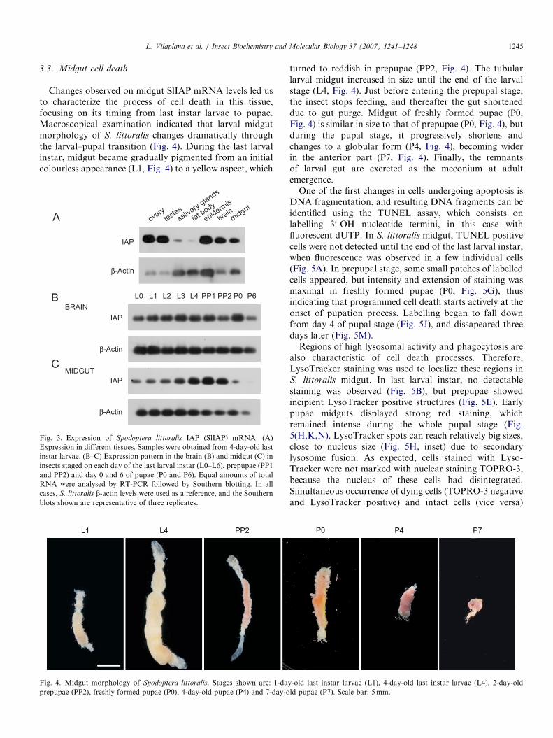
In order to study the tissue distribution of SlIAPexpression, we carried out a RT–PCR screening for SlIAPmRNA using as a template total mRNA from ovary,testis, salivary gland, fat body, epidermis, brain andmidgut tissues obtained from 4-day-old last larval instar.Results (Fig. 3A) indicated that SlIAP is expressed in all
ARTICLE IN PRESS
Fig. 1. Structure and organization of Spodoptera littoralis IAP (SlIAP). (A) Predicted amino acid sequence. The different characteristic motifs are shown
in bold. (B) Organization of BIR motifs and RING finger. (C–E) Alignment of the BIR1 (C), BIR2 (D) and RING (E) regions of SlIAP with the
homologous regions of other IAPs. The Genbank accession numbers of sequences used for the alignments are: Spodoptera exempta IAP (SeIAP)
DQ206460, Spodoptera frugiperda IAP (SfIAP) AF186378, Trichoplusia ni IAP (TnIAP): AF195528, Bombyx mori IAP (BmIAP): AF281073, Cydia
pomonella granulovirus IAP (CpIAP): P41436, Orgyia pseudotsugata IAP (OpIAP): P41437 and Drosophila melanogaster IAP (DmIAP): Q24306.
Fig. 2. Comparison of Spodoptera littoralis IAP (SlIAP) with S. littoralis nucleopolihedrovirus IAP (SINPV-IAP). (A) Alignment of the amino acid
sequence of BIR1 motif and (B) Alignment of the amino acid sequence of RING finger.
L. Vilaplana et al. / Insect Biochemistry and Molecular Biology 37 (2007) 1241–12481244
tissues analysed. Although the approach followed is notquantitative, results suggest that salivary gland and fatbody have much lower relative mRNA levels in compar-ison with the other tissues examined (Fig. 3A).
Subsequently, we studied the mRNA expression patternin brain and midgut tissues during the last larval instar,prepupae and pupae. As stated above, larval midgut iscompletely remodelled during the larval–pupal moult,
whereas the brain may serve as a reference, given thatneuron generation is continuous from larvae to pupae, andthere is no evidence of massive cell death in the stagesunder study. Results show that SlIAP mRNA levels remainapproximately constant in brain tissues (Fig. 3B), whereasin the midgut they rise on days 3 and 4 of last larval instar,show a peak on PP1, start to decrease on PP2, and becomedramatically reduced on P0 (Fig. 3C).
ARTICLE IN PRESSL. Vilaplana et al. / Insect Biochemistry and Molecular Biology 37 (2007) 1241–1248 1245
3.3. Midgut cell death
Changes observed on midgut SlIAP mRNA levels led usto characterize the process of cell death in this tissue,focusing on its timing from last instar larvae to pupae.Macroscopical examination indicated that larval midgutmorphology of S. littoralis changes dramatically throughthe larval–pupal transition (Fig. 4). During the last larvalinstar, midgut became gradually pigmented from an initialcolourless appearance (L1, Fig. 4) to a yellow aspect, which
ovary
teste
s
saliv
ary g
lands
fat b
ody
epidermis
brainm
idgut
L0
BRAIN
MIDGUT
IAP
β-Actin
IAP
β-Actin
IAP
β-Actin
L1 L2 L3 L4 PP1 PP2 P0 P6
Fig. 3. Expression of Spodoptera littoralis IAP (SlIAP) mRNA. (A)
Expression in different tissues. Samples were obtained from 4-day-old last
instar larvae. (B–C) Expression pattern in the brain (B) and midgut (C) in
insects staged on each day of the last larval instar (L0–L6), prepupae (PP1
and PP2) and day 0 and 6 of pupae (P0 and P6). Equal amounts of total
RNA were analysed by RT-PCR followed by Southern blotting. In all
cases, S. littoralis b-actin levels were used as a reference, and the Southern
blots shown are representative of three replicates.
L1 L4 PP2
Fig. 4. Midgut morphology of Spodoptera littoralis. Stages shown are: 1-da
prepupae (PP2), freshly formed pupae (P0), 4-day-old pupae (P4) and 7-day-o
turned to reddish in prepupae (PP2, Fig. 4). The tubularlarval midgut increased in size until the end of the larvalstage (L4, Fig. 4). Just before entering the prepupal stage,the insect stops feeding, and thereafter the gut shorteneddue to gut purge. Midgut of freshly formed pupae (P0,Fig. 4) is similar in size to that of prepupae (P0, Fig. 4), butduring the pupal stage, it progressively shortens andchanges to a globular form (P4, Fig. 4), becoming widerin the anterior part (P7, Fig. 4). Finally, the remnantsof larval gut are excreted as the meconium at adultemergence.One of the first changes in cells undergoing apoptosis is
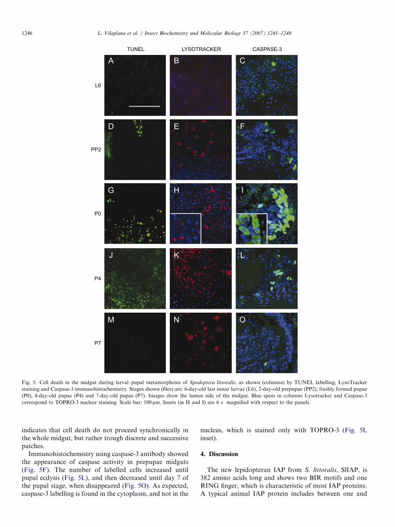
DNA fragmentation, and resulting DNA fragments can beidentified using the TUNEL assay, which consists onlabelling 30-OH nucleotide termini, in this case withfluorescent dUTP. In S. littoralis midgut, TUNEL positivecells were not detected until the end of the last larval instar,when fluorescence was observed in a few individual cells(Fig. 5A). In prepupal stage, some small patches of labelledcells appeared, but intensity and extension of staining wasmaximal in freshly formed pupae (P0, Fig. 5G), thusindicating that programmed cell death starts actively at theonset of pupation process. Labelling began to fall downfrom day 4 of pupal stage (Fig. 5J), and dissapeared threedays later (Fig. 5M).Regions of high lysosomal activity and phagocytosis are
also characteristic of cell death processes. Therefore,LysoTracker staining was used to localize these regions inS. littoralis midgut. In last larval instar, no detectablestaining was observed (Fig. 5B), but prepupae showedincipient LysoTracker positive structures (Fig. 5E). Earlypupae midguts displayed strong red staining, whichremained intense during the whole pupal stage (Fig.5(H,K,N). LysoTracker spots can reach relatively big sizes,close to nucleus size (Fig. 5H, inset) due to secondarylysosome fusion. As expected, cells stained with Lyso-Tracker were not marked with nuclear staining TOPRO-3,because the nucleus of these cells had disintegrated.Simultaneous occurrence of dying cells (TOPRO-3 negativeand LysoTracker positive) and intact cells (vice versa)
P0 P4 P7
y-old last instar larvae (L1), 4-day-old last instar larvae (L4), 2-day-old
ld pupae (P7). Scale bar: 5mm.
ARTICLE IN PRESS
P7
P4
P0
PP2
L6
TUNEL LYSOTRACKER CASPASE-3
A B C
D E F
G H I
J K L
M N O
Fig. 5. Cell death in the midgut during larval–pupal metamorphosis of Spodoptera littoralis, as shown (columns) by TUNEL labelling, LysoTracker
staining and Caspase-3 immunohistochemistry. Stages shown (files) are: 6-day-old last instar larvae (L6), 2-day-old prepupae (PP2), freshly formed pupae
(P0), 4-day-old pupae (P4) and 7-day-old pupae (P7). Images show the lumen side of the midgut. Blue spots in columns Lysotracker and Caspase-3
correspond to TOPRO-3 nuclear staining. Scale bar: 100mm. Insets (in H and I) are 6� magnified with respect to the panels.
L. Vilaplana et al. / Insect Biochemistry and Molecular Biology 37 (2007) 1241–12481246
indicates that cell death do not proceed synchronically inthe whole midgut, but rather trough discrete and successivepatches.
Immunohistochemistry using caspase-3 antibody showedthe appearance of caspase activity in prepupae midguts(Fig. 5F). The number of labelled cells increased untilpupal ecdysis (Fig. 5L), and then decreased until day 7 ofthe pupal stage, when disappeared (Fig. 5O). As expected,caspase-3 labelling is found in the cytoplasm, and not in the
nucleus, which is stained only with TOPRO-3 (Fig. 5I,inset).
4. Discussion
The new lepidopteran IAP from S. littoralis, SlIAP, is382 amino acids long and shows two BIR motifs and oneRING finger, which is characteristic of most IAP proteins.A typical animal IAP protein includes between one and
ARTICLE IN PRESSL. Vilaplana et al. / Insect Biochemistry and Molecular Biology 37 (2007) 1241–1248 1247
three BIR motifs and one RING finger, whereas baculo-viral IAPs contain one or two BIR motifs, and one or noneRING finger. With the description of the SlIAP presentedherein, S. littoralis becomes one of the few cases where thehost and the specific viral counterpart IAPs are available.Until now, the described IAPs of Spodoptera spp. containtwo BIR motifs and the RING finger. On the other hand,baculoviral SlNPV-IAP has been reported as a 15 kDapolypeptide with only one BIR motif, a RING finger and athird specific acidic-rich motif (Liu et al., 2003). SlNPV-IAP is able to delay, but not to suppress, programmed celldeath induced by replication of a recombinant AcMNPVdeficient in p35 (Liu et al., 2003). Sequence alignments andphylogenetic analyses have revealed that the BIR motif ofSlNPV-IAP is more similar to BIR1 than to BIR2 motifs ofSpodoptera spp. From a phylogenetical point of view, IAPsincluding only one BIR motif and the RING finger, form acluster separated from that of IAPs containing two BIRdomains and one RING finger, due to BIR motifdivergence (Liu et al., 2003). Accordingly, SlIAP sequenceis more similar to other baculoviral IAPs containing twoBIR motifs than to SlNPV-IAP which only contains one.
Our studies have shown that SlIAP is expressed in ovary,testes, salivary gland, fat body, epidermis, brain andmidgut tissues of S. littoralis last larval instar. Thus, IAPexpression seems to be required to prevent premature celldeath in these larval tissues. Interestingly, mRNA levels insalivary glands and fat body appeared to be lower incomparison with the other tissues. This may be related withthe fact that, during insect metamorphosis, salivary glandundergo rapid and massive cell death (Lee and Baehrecke,2001; Lee et al., 2002b) and larval fat body dissociates andpersists as individual fat cells that are eventually removedby a caspase cascade (Aguila et al., 2007).
Sequential expression studies on midgut and braintissues revealed that, whereas SlIAP mRNA levels in thebrain do not fluctuate significantly, those of the midgutshow a particular pattern, with a large peak centred in theprepupal stage. This probably reflects the distinct ways inwhich cell death process plays on them, given that midgutundergoes a dramatic remodelling during the larval–pupaltransition, whereas brain tissues show continuous produc-tion and degeneration of neurones throughout larval andpupal stages (Nordlander and Edwards, 1969). In the mothHeliothis virescens, expression patterns of caspase-1, ICEand IAP in the midgut during last larval and pupal stageshave been reported (Parthasarathy and Palli, 2007). IAPmRNA levels showed a narrow peak on day 2 of last larvalinstar, then decreased and remained low until day 1 of thepupal stage, where they showed a modest increase. Theactivation of caspase expression occurred after full IAPdown regulation (Parthasarathy and Palli, 2007). Theexpression of H. virescens IAP during the larval–pupaltransition differs with respect to that described herein forSlIAP, which suggests that the timing of midgut cell deathevents or the mechanisms of IAP regulation may bedifferent in the two moths.
Macroscopical examination indicated that midgut mor-phology changed during metamorphosis from a semi-transparent, cylindrical structure in last instar larvae to abrownish globular mass in pupae. Gross morphologicalchanges, which served us as a frame for more detailedmicroscopy studies, are similar to those described for otherlepidopterans, like Pieris brassicae (Komuves et al., 1985)or Galleria mellonella (Uwo et al., 2002).Microscopy studies using TUNEL assays, LysoTracker
staining and caspase-3 immunohistochemistry, indicatethat S. littoralis midgut epithelium death proceeds throughdiscrete and successive patches of dying cells, a feature thathas been reported in other insects, such as the fruitflyD. melanogaster and a number of lepidopteran species(Komuves et al., 1985; Lee et al., 2002a; Uwo et al., 2002).Autophagy has been shown to be the key mechanism forlarval midgut death in a number of insects (Nopanitayaand Misch, 1974; Komuves et al., 1985; Lee et al., 2002a).Dying larval midgut of S. littoralis, however, show markersof both authophagy and apoptosis, but data available donot allow to discriminate whether authophagy is requiredfor the death of these tissues or if it is just coincident withapoptosis. In this context, it is worth noting that auto-phagy and apoptosis share a number of mechanisms andsteps, for example, the involvement of effector caspases(Jiang et al., 1997; Lee et al., 2002a; Yin and Thummel,2004).The comparison of results from microscopy examination
with those from SlIAP expression studies in the midgut,indicates that cell death starts actively at the onset ofpupation, coinciding with the dramatical decrease of SlIAPmRNA levels. This coincidence points to an inhibitory roleof SlIAP on apoptosis, as expected.
Acknowledgement
Financial support from the Spanish Ministry of Scienceand Technology (project AGL2005-00773 to X. Belles) andthe Generalitat de Catalunya (2001 SGR 003245) isgratefully acknowledged. Ll. Vilaplana is recipient of apost-doctoral contract from and N. Perera is recipientof a technician contract, both from CSIC under the I3Pprogram.
References
Aguila, J.R., Suszko, J., Gibbs, A.G., Hoshizaki, D.K., 2007. The role of
larval fat cells in adult Drosophila melanogaster. J. Exp. Biol. 210,
956–963.
Birnbaum, M.J., Clem, R.J., Miller, L.K., 1994. An apoptosis-inhibiting
gene from a nuclear polyhedrosis virus encoding a polypeptide with
Cys/His sequence motifs. J. Virol. 68, 2521–2528.
Clem, R.J., Duckett, C.S., 1998. The IAP genes: unique arbiters of cell
death. Trends Biochem. Sci. 23, 159–162.
Clem, R.J., Miller, L.K., 1994. Control of programmed cell death by the
baculovirus genes p35 and iap. Mol. Cell. Biol. 14, 5212–5222.
Chai, J., Yan, N., Huh, J.R., Wu, J.W., Li, W., Hay, B.A., Shi, Y., 2003.
Molecular mechanism of Reaper-Grim-Hid-mediated suppression of
ARTICLE IN PRESSL. Vilaplana et al. / Insect Biochemistry and Molecular Biology 37 (2007) 1241–12481248
DIAP1-dependent Dronc ubiquitination. Nat. Struct. Biol. 10,
892–898.
Crook, N.E., Clem, R.J., Miller, L.K., 1993. An apoptosis-inhibiting
baculovirus gene with a zinc finger-like motif. J. Virol. 67, 2168–2174.
Deveraux, Q.L., Takahashi, R., Salvesen, G.S., Reed, J.C., 1997. X-linked
IAP is a direct inhibitor of cell-death proteases. Nature 388, 300–304.
Deveraux, Q.L.a.R.J.C., 1998. IAP family proteins: suppressors of
apoptosis. Genes Dev 13, 239–252.
Fraser, A., Evan, G., 1996. A license to kill. Cell 85, 781–784.
Freemont, P.S., Hanson, I.M., Trowsdale, J., 1991. A novel cysteine-rich
sequence motif. Cell 64, 483–484.
Frohman, M.A., Dush, M.K., Martin, G.R., 1988. Rapid production of
full-length cDNAs from rare transcripts: amplification using a single
gene-specific oligonucleotide primer. Proc. Natl. Acad. Sci. USA 85,
8998–9002.
Goyal, L., McCall, K., Agapite, J., Hartwieg, E., Steller, H., 2000.
Induction of apoptosis by Drosophila reaper, hid and grim through
inhibition of IAP function. Embo J. 19, 589–597.
Hay, B.A., 2000. Understanding IAP function and regulation: a view from
Drosophila. Cell Death Differ 7, 1045–1056.
Hay, B.A., Wassarman, D.A., Rubin, G.M., 1995. Drosophila homologs
of baculovirus inhibitor of apoptosis proteins function to block cell
death. Cell 83, 1253–1262.
Hinds, M.G., Norton, R.S., Vaux, D.L., Day, C.L., 1999. Solution
structure of a baculoviral inhibitor of apoptosis (IAP) repeat. Nat.
Struct. Biol. 6, 648–651.
Huang, Q., Deveraux, Q.L., Maeda, S., Salvesen, G.S., Stennicke, H.R.,
Hammock, B.D., Reed, J.C., 2000. Evolutionary conservation of
apoptosis mechanisms: lepidopteran and baculoviral inhibitor of
apoptosis proteins are inhibitors of mammalian caspase-9. Proc. Natl.
Acad. Sci. USA 97, 1427–1432.
Huang, Q., Deveraux, Q.L., Maeda, S., Stennicke, H.R., Hammock, B.D.,
Reed, J.C., 2001. Cloning and characterization of an inhibitor of
apoptosis protein (IAP) from Bombyx mori. Biochim. Biophys. Acta.
1499, 191–198.
Jiang, C., Baehrecke, E.H., Thummel, C.S., 1997. Steroid regulated
programmed cell death during Drosophila metamorphosis. Develop-
ment 124, 4673–4683.
Komuves, L.G., Sass, M., Kovacs, J., 1985. Autophagocytosis in the
larval midgut cells of Pieris brassicae during metamorphosis. Cell
Tissue Res. 240, 215–221.
Kumar, S., Cakouros, D., 2004. Transcriptional control of the core cell-
death machinery. Trends Biochem. Sci. 29, 193–199.
Lee, C.Y., Baehrecke, E.H., 2001. Steroid regulation of autophagic pro-
grammed cell death during development. Development 128, 1443–1455.
Lee, C.Y., Cooksey, B.A., Baehrecke, E.H., 2002a. Steroid regulation of
midgut cell death during Drosophila development. Dev. Biol. 250,
101–111.
Lee, C.Y., Simon, C.R., Woodard, C.T., Baehrecke, E.H., 2002b. Genetic
mechanism for the stage- and tissue-specific regulation of steroid
triggered programmed cell death in Drosophila. Dev. Biol. 252,
138–148.
Liao, W.T., Yang, Y., Wu, X.F., 2002. Expression and functional analysis
of an inhibitor of apoptosis protein from Trichoplusia ni. Biochem.
Biophys. Res. Commun. 293, 675–679.
Liston, P., Fong, W.G., Korneluk, R.G., 2003. The inhibitors of
apoptosis: there is more to life than Bcl2. Oncogene 22, 8568–8580.
Liu, Q., Chejanovsky, N., 2006. Activation pathways and signal-mediated
upregulation of the insect Spodoptera frugiperda caspase-1. Apoptosis
11, 487–496.
Liu, Q., Qi, Y., Chejanovsky, N., 2003. Identification and classification of
the Spodoptera littoralis nucleopolyhedrovirus inhibitor of apoptosis
gene. Virus Genes 26, 143–149.
Liu, Q., Qi, Y., Chejanovsky, N., 2005. Spodoptera littoralis caspase-1, a
Lepidopteran effector caspase inducible by apoptotic signaling.
Apoptosis 10, 787–795.
Nopanitaya, W., Misch, D.W., 1974. Developmental cytology of the
midgut in the flesh-fly, Sarcophaga bullata (Parker). Tissue Cell 6,
487–502.
Nordlander, R.H., Edwards, J.S., 1969. Postembryonic brain development
in the monarch butterfly, Danaus plexippus plexippus, L. Dev. genes
Evol. 162, 197–217.
Parthasarathy, R., Palli, S.R., 2007. Developmental and hormonal
regulation of midgut remodeling in a lepidopteran insect, Heliothis
virescens. Mech. Dev. 124, 23–34.
Poitout, S., Bues, R., 1974. Linolenic acid requirements of lepidoptera
Noctuidae Quadrifinae Plusiinae: Chrysodeixis chalcites Esp., Auto-
grapha gamma L. Macdunnoughia confusa Stph., Trichoplusia ni Hbn.
reared on artificial diets. Ann. Nutr. Aliment 28, 173–187.
Roy, N., Deveraux, Q.L., Takahashi, R., Salvesen, G.S., Reed, J.C., 1997.
The c-IAP-1 and c-IAP-2 proteins are direct inhibitors of specific
caspases. Embo J 16, 6914–6925.
Salvesen, G.S., Duckett, C.S., 2002. IAP proteins: blocking the road to
death’s door. Nat. Rev. Mol. Cell Biol. 3, 401–410.
Sun, C., Cai, M., Gunasekera, A.H., Meadows, R.P., Wang, H., Chen, J.,
Zhang, H., Wu, W., Xu, N., Ng, S.C., Fesik, S.W., 1999. NMR
structure and mutagenesis of the inhibitor-of-apoptosis protein XIAP.
Nature 401, 818–822.
Uwo, M.F., Ui-Tei, K., Park, P., Takeda, M., 2002. Replacement of
midgut epithelium in the greater wax moth, Galleria mellonella, during
larval-pupal moult. Cell Tissue Res 308, 319–331.
Vaux, D.L., Korsmeyer, S.J., 1999. Cell death in development. Cell 96,
245–254.
Vucic, D., Kaiser, W.J., Harvey, A.J., Miller, L.K., 1997. Inhibition of
reaper-induced apoptosis by interaction with inhibitor of apoptosis
proteins (IAPs). Proc. Natl. Acad. Sci. USA 94, 10183–10188.
Vucic, D., Kaiser, W.J., Miller, L.K., 1998. Inhibitor of apoptosis proteins
physically interact with and block apoptosis induced by Drosophila
proteins HID and GRIM. Mol. Cell. Biol. 18, 3300–3309.
Wyllie, A.H., Kerr, J.F., Currie, A.R., 1980. Cell death: the significance of
apoptosis. Int. Rev. Cytol. 68, 251–306.
Yin, V.P., Thummel, C.S., 2004. A balance between the diap1 death
inhibitor and reaper and hid death inducers controls steroid-triggered
cell death in Drosophila. Proc. Natl. Acad. Sci. USA 101, 8022–8027.
Zachariou, A., Tenev, T., Goyal, L., Agapite, J., Steller, H., Meier, P.,
2003. IAP-antagonists exhibit non-redundant modes of action through
differential DIAP1 binding. Embo J 22, 6642–6652.
Zhu, J., Miura, K., Chen, L., Raikhel, A.S., 2000. AHR38, a homolog of
NGFI-B, inhibits formation of the functional ecdysteroid receptor in
the mosquito Aedes aegypti. Embo J 19, 253–262.


![Spodoptera frugiperda (J.E.Smith) [Lepidoptera: …...G20 Discussion group on ‘Fall Armyworm Spodoptera frugiperda (J.E.Smith) [Lepidoptera: Noctuidae]’ Sengottaiyan Vennila1,](https://img.pdfslide.us/doc/110x75/5fd516c8ccd4990891330f0c/spodoptera-frugiperda-jesmith-lepidoptera-g20-discussion-group-on-afall.jpg)