Embed Size (px)
Citation preview
ARTICLE
1
Developing an aqueous approach for synthesizing Au
and M@Au (M = Pd, CuPt) hybrid nanostars with
plasmonic properties†
Jingshan Du, Junjie Yu, Yalin Xiong, Zhuoqing Lin, Hui Zhang* and Deren Yang
Anisotropic Au nanoparticles show unique localized surface plasmon resonance (LSPR) properties,
which make it attractive in optical, sensing, and biomedical applications. In this contribut ion, we report
a general and facile strategy towards aqueous synthesis of Au and M@Au (M = Pd, CuPt) hybrid
nanostars by reducing HAuCl4 with ethanolamine in the presence of cetyltrimethylammonium bromide
(CTAB). According to electron microscopic observati on and spectral monitoring, we found that the
layered epitaxial growth mode (i.e., Frank-van der Merwe mechanism) contributes to the enlargement
of the core, while, the random attachment of Au nanoclusters onto the cores accounts for the formation
of the branches. Both of them are indispensable for the formation of the nanostars. The LSPR
properties of the Au nanoparticles have been well investigated with morphology control via precursor
amount and growth temperature. The Au nanostars showed improved surfac e-enhanced Raman
spectroscopy (SERS) performance for rhodamine 6G due to their sharp edges and tips, which were
therefore confirmed as good SERS substrate to detect trace amount of molecules.
Introduction
Metallic nanoparticles (NPs) have received great attention due
to their distinguished properties in catalysis,1 optics,2 and
biomedicine.3 Among those NPs of various metal elements and
morphologies, anisotropic Au NPs have become extremely
significant due to their unique localized surface plasmon
resonance (LSPR) properties,4, 5 and thus widespread use in
biological imaging, photothermal therapy and optical sensing.6-8
The last decade has witnessed the significant progress in the
synthesis of anisotropic Au NPs, with notable examples
including nanorods,9-12 nanostars,13-15 and nanopods.16, 17 Such
Au nanostructures show tunable LSPR peaks in the range from
visible to near-infrared (IR) regions, which have been found
mainly dependent on their aspect ratios. In addition, these
anisotropic NPs can also create “hot spots” according to the
coupling between their electromagnetic fields, which provide
extremely large electric field enhancement around the
nanostructures.18, 19 All of such fascinating characteristics
facilitate the surface-enhanced Raman scattering where the
Raman signals of the small molecules adsorbed on the NPs are
significantly enhanced, thereby make it possible to sensing and
detecting these molecules at trace amount level.20
Of these anisotropic nanostructures, Au nanostars with
various branches are particularly interesting since their intrinsic
properties result from hybridization of plasmon focalized at
sharp edges and tips. Up to now, there are some synthetic
routes that have been exploited for Au nanostars. To this end,
Liz-Marzán et al. demonstrated the seed-mediated growth of
Au nanostars in high yield through the reduction of HAuCl4 in
a concentrated solution of polyvinylpyrrolidone (PVP) in N,N-
dimethylformamide (DMF).14, 21-23 The use of PVP with a weak
reducing power and DMF serving as a solvent played a key role
in the formation of Au nanostars. In aqueous systems, a silver
ion-induced strategy was also developed to generate Au
nanostars.13, 24-27 In these syntheses, the selective adsorption
effect of silver ions on the surface of Au NPs through
underpotential deposition (UPD) promoted their branched
growth. Recently, a surfactant-assisted approach has been
exploited for the synthesis of Au nanostars by taking
advantages of simple procedure and high efficiency. For
example, Pallavicini et al. reported the synthesis of branched
Au NPs with the use of laurylsulfobetaine as the Zwitterionic
surfactant.28, 29 In another study, many researchers
demonstrated successful synthesis of Au nanostars with various
branched arms in the presence of complex amine molecules
such as hydroxylamine,30, 31 melamine,32
hexamethylenetetramine (HMT),16 and bis(amidoethyl-
carbamoylethyl)octadecylamine (C18N3).33 Although significant
advances have been achieved on the synthesis of Au nanostars,
the origin and mechanism for the formation of these branched
structures have not been fully understood.
In parallel, bimetallic or multi-metallic NPs also show
significant potential in catalysis or other multifunctional
applications. Formation of core@shell bimetallic
ARTICLE Journal Name
2
nanostructures has been considered as an efficient way to alter
the electron structure of the shells due to a strong coupling
between these two metals, which can significantly affect their
catalytic performance and stability.34-36 Au-based bimetallic
NPs are also interesting candidates of plasmonic
multifunctional colloids since they bring the unique optical
response of Au to other functional metal NPs.37 Therefore,
developing a general and facile strategy for synthesizing
anisotropic Au and Au-based bimetallic or multi-metallic
nanostars, together with clarifying their formation mechanism,
is still of great importance to the scientific community.
Herein, we report a general, facile, and powerful strategy
towards aqueous synthesis of Au and M@Au (M = Pd, CuPt)
hybrid nanostars in a controlled manner. Star-like Au shells
were successfully grown on different as-preformed seeds
including Au nanospheres, Pd nanocubes and CuPt bimetallic
nanocubes in an aqueous solution containing HAuCl4 and
cetyltrimethylammonium bromide (CTAB) with ethanolamine
as both a capping and reducing agent. Quantitative studies
revealed that two concurrent mechanisms are responsible for
the anisotropic morphology and the corresponding LSPR-
induced absorption properties. The interior core of the NPs was
gradually grown up due to the conventional layered epitaxial
mechanism, i.e., Frank-van der Merwe (F-M) growth mode. In
contrast, the formation of the branches was attributed to the
random attachment of Au nanoclusters onto the core particles.
The as-prepared Au nanostars showed improved surface-
enhanced Raman spectroscopy (SERS) performance compared
to Au nanospheres in detecting trace amount of rhodamine 6G
molecules.
Experimental section
Chemicals
Chloroauric acid (HAuCl4·4H2O, AR),
cetyltrimethylammonium bromide (CTAB, AR), ethanolamine
(AR), potassium chloride (KCl, AR) and hydrochloric acid
(HCl, AR 36%-38%) were supplied by Sinopharm Chemical
Reagent. L-ascorbic acid (AA, BioXtra, ≥99.0%), potassium
tetrachloroplatinate(II) (K2PtCl4, 99.99% trace metals basis),
sodium tetrachloropalladate(II) (Na2PdCl4, 99.99% trace metals
basis), copper(II) chloride dihydrate (CuCl2·2H2O, reagent
grade), and polyvinylpyrrolidone (PVP, MW=40,000) were
supplied by Sigma-Aldrich. Sodium borohydride (NaBH4,
≥98.0%) was supplied by Aldrich. Potassium bromide (KBr,
AR) was supplied by Shanghai No. 4 Reagent. Ultrapure de-
ionized water (DI water) was produced by a Milli-Q Synergy
water purification system. All chemicals were used without
further purification.
Synthesis of 19-nm Au nanospheres
The synthetic procedure of Au nanospheres was adapted from a
previously published method.10 Firstly, 5 mL of 0.2 M CTAB
and 5 mL of 0.5 mM HAuCl4 aqueous solutions were mixed at
25 °C under stirring, followed by the quick injection of 0.6 mL
of 0.01 M ice-cold NaBH4 aqueous solution. The mixture was
under violent stirring for 2 min and then kept still for another
30 min. Secondly, 0.5 mL of the afore-mentioned mixture was
mixed with 50 mg of CTAB, 0.05 mL of ethanolamine, 2 mL of
2.3 mM HAuCl4 and 5 mL of DI water. The mixture was kept
overnight until it turned to fresh red color.
Synthesis of 14-nm Pd nanocubes
Pd nanocubes were synthesized according to a previously
reported method.38 Briefly, 105 mg of PVP, 60 mg of AA, 400
mg of KBr and 185 mg of KCl were dissolved in 8 mL of DI
water. The solution was transferred to a vial, heated to 80 °C
and kept for 10 min, followed by the addition of 3 mL of
aqueous solution containing 57 mg of Na2PdCl4 with a pipette.
The solution was kept at 80 °C for 10 min with a cap. After
that, the final product was obtained by centrifugation at 13,000
rpm and washed with a mixture of DI water and ethanol for
three times for further use.
Synthesis of 7-nm CuPt bimetallic nanocubes
CuPt bimetallic nanocubes were generated through a previously
reported method.39 Briefly, 0.03 mmol of K2PtCl4, 0.03 mmol
of CuCl2, 9 mmol of KBr and 100 mg of PVP were dissolved in
15 mL of DI water, followed by the addition of 0.15 mL of 1 M
HCl solution. The mixture was then transferred to a 25 mL
autoclave and heated at 160 °C for 4 h. After cooling down to
room temperature, the solution was centrifuged at 13,000 rpm
and the final product was washed with DI water for three times
for further use.
Synthesis of Au and M@Au (M = Pd, CuPt) hybrid nanostars
Au nanostars were synthesized by a seed-mediated approach in
an aqueous solution containing as-preformed Au seeds, CTAB,
and HAuCl4, with ethanolamine as both a capping and reducing
agent. In a standard procedure, 0.1 mL of 19-nm Au
nanosphere solution was mixed with 50 mg of CTAB, 0.05 mL
of ethanolamine and 1 mL of 2.3 mM HAuCl4 solution.
Additional 5.95 mL of DI water was added to keep the overall
volume to 7.1 mL. The mixture was stirred at 25 °C overnight
to ensure the completion of the reaction. The final product was
centrifuged at 5,000 rpm and washed with DI water for further
characterization. For the synthesis of hybrid nanostars, the
procedure was similar to that of Au nanostars except that Pd
nanocubes or CuPt bimetallic nanocubes were used instead of
Au nanospheres.
Characterizations
Transmission electron microscopy (TEM) images were
obtained with a Hitachi HT7700 at an accelerating voltage of
100 kV. Ultraviolet-visible (UV-vis) spectra were collected
with a Shimadzu UV-3150 spectrometer under absorption
mode.
Surface-enhanced Raman spectroscopy
Au nanoparticles were re-dispersed in DI water and dropped on
identical silicon wafers to form films. After the wafers were
dried, 5 μL of 10-6 M rhodamine 6G ethanol solution was
dropped and dispersed on the surface of the Au films. Raman
signals were collected by a Bruker SENTERRA dispersive
Raman microscope with excitation laser of 532 nm and 10 mW.
Journal Name ARTICLE
3
Results and discussion
Synthesis of Au nanostars
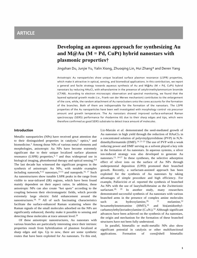
Figure 1a shows a schematic illustration of the strategy
used in synthesizing Au and M@Au (M = Pd, CuPt) hybrid
nanostars. The nanostars were typically synthesized by
reducing HAuCl4 with ethanolamine in an aqueous solution
containing as-preformed metallic NPs and CTAB serving as the
seeds and stabilizer, respectively. Obviously, the experimental
parameters such as precursor concentration and reaction
temperature have a great influence on the shape of the Au NPs
due to the different reaction kinetics. Using Au nanostars as an
example, we characterized their shapes, structures, and LSPR
properties, together with clarifying the growth mechanism
presented in the synthesis. Au nanospheres of 19 nm in size that
obtained by a two-step reduction procedure (see Figure S1)
were used as seeds for the synthesis of Au nanostars. Figure 1,
b and c show typical TEM images of the Au nanostars that
obtained using the standard procedure (see Experimental for
details). As observed from the TEM images, most of the Au
NPs consisted of various arms (like stars). The as-prepared Au
nanostars were uniform with a typical overall size of about 90
nm. To simplify the discussion in the next part of growth
mechanism, we introduced the core size D that defined as the
maximum incircle diameter of a nanostar (Figure 1c). The Au
NPs were used as the seeds in order to generate uniform
nanostars. In the absence of Au seeds, Au nanostars with
various sizes were generated while other parameters were kept
the same (Figure S2).
Figure 1. (a) Schematic illustration of controlled synthesis of Au and M@Au (M =
Pd, CuPt) hybrid nanostars. (b, c) TEM images of Au nanostars obtained by the
standard procedure. The dashed circle in (c) indicates the definition of the core
size D.
Compared our synthetic procedure with those in the
previous reports,40-42 it is clear that the use of ethanolamine as a
reducing agent is indispensable for the synthesis of the Au
nanostars. The role of ethanolamine has been identified by
replacing it with AA, which is a widely-used reducer for Au
NPs. More specifically, 3 mg of AA instead of ethanolamine
was used in the synthesis of Au seeds as well as in the
overgrowth procedure of Au NPs while keeping other
parameters unchanged. From the TEM images in Figure S3,
only inhomogeneous Au NPs with various shapes were formed,
indicating that ethanolamine plays a key role in facilitating a
highly anisotropic growth of Au. It is well-known that
ethanolamine is a weak base that contains hydroxyl and amino
functional groups. The amino group has an appropriate power
to reduce Au salt precursor. In addition, ethanolamine can also
serve as a capping agent to selectively bind onto some specific
planes of Au NPs by the amino group, and thereby promote the
highly anisotropic growth.32 More importantly, the alkaline
condition can extensively suppress the intraparticle ripening
induced by the chlorine ions that existed in the reaction system,
and thus stabilize such branched Au nanostructures.30 All of
these characteristics are beneficial to the formation of Au
nanostars. To better understand its role in the formation of Au
nanostars, we further chose different types of amines in place of
ethanolamine. Figure S4 a-c, shows TEM images of Au NPs
that obtained in the presence of ethylenediamine,
diethanolamine, and triethanolamine, respectively. Figure S4d
shows the corresponding UV-vis spectra. From these TEM
images, no Au nanostars were successfully observed. This
demonstration was also supported by UV-vis spectra, in which
only single LSPR peak was detected. It is clear that
ethylenediamine is a stronger complex former than
ethanolamine due to the two amino groups, thus much lower
reaction rate and smaller Au NPs were obtained.
Diethanolamine is a secondary amines while the hydroxyl
group is retained, which results in weaker capping ability to the
surface of Au due to its higher steric hindrance than
ethanolamine. In this case, Au NPs with minor protuberances
were achieved. Triethanolamine as a tertiary amine has even
weaker capping ability, thus big inhomogeneous NPs were
generated.
Formation mechanisms and LSPR properties
In order to further understand the process how Au
nanostars were formed, we performed quantitative experiments
with the aid of TEM observation and UV-vis monitoring. The
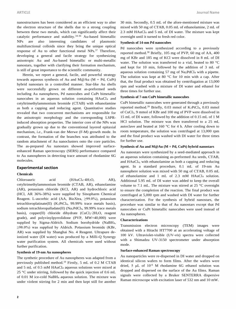
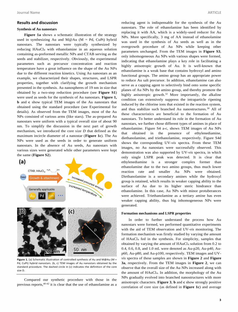
formation mechanism was firstly studied by varying the amount
of HAuCl4 fed in the synthesis. For simplicity, samples that
obtained by varying the amount of HAuCl4 solution from 0.2 to
0.4, 0.6, 0.8, and 1.0 mL were denoted as Au-p20, Au-p40, Au-
p60, Au-p80, and Au-p100, respectively. TEM images and UV-
vis spectra of these samples are shown in Figure 2 and Figure
3a, respectively. From the TEM images in Figure 2, we can
observe that the overall size of the Au NPs increased along with
the amount of HAuCl4. In addition, the morphology of the Au
NPs gradually evolved into branched nanostructures with more
anisotropic characters. Figure 3, b and c show strongly positive
correlation of core size (as defined in Figure 1c) and average
ARTICLE Journal Name
4
observed number of branches per nanostar with the amount of
gold precursor, respectively. The core size of the nanostars was
gradually increased from 19 nm (as-preformed Au seeds) to 53
nm when 1.0 mL of HAuCl4 solution was added. Meanwhile,
the observed number of Au branches increased to over 2.5 per
nanostar. As such, these TEM observations indicate that the
nanostars were generated through the overgrowth of the core in
combination with the anisotropic growth of the branches.
Figure 2. TEM images of (a) Au-p20, (b) Au-p40, (c) Au-p60 and (d) Au-p80
samples. Images share a scale bar of 100 nm.
Such morphology evolution of the Au NPs dependent on
the amount of gold precursor was also monitored by their LSPR
properties from UV-vis absorption spectra (Figure 3a). Since
the LSPR of Au NPs can be highly affected by surfactant and
the surrounding dielectric environment (e.g., solvent, capping
agent, etc.),18 the concentration of CTAB was kept the same in
all experiments. From Figure 3a, the Au nanostars that
obtained by the standard procedure (1.0 mL of HAuCl4 was
used) show a primary absorption peak at 734 nm, together with
a shoulder in the range of 500 to 600 nm. Obviously, this
shoulder refers to the LSPR from the core of the nanostars,
which represents the similar resonance mode as that in
spherical Au seeds at 523 nm.40 While, the primary absorption
peak in near-IR could be attributed to the longitudinal
resonance mode from the branches. The transverse mode from
the branches was not distinguished possibly because they are
too weak compared to that shown by the core, which also fell in
the visible range. This demonstration was confirmed by the
UV-vis spectra collected from the samples with different
amount of HAuCl4. The near-IR absorption peak raised
gradually when the amount of HAuCl4 increased, indicating the
formation of nanostructures with higher aspect ratio, i.e. the
overgrowth of Au with anisotropic morphology. The intensity
ratio of the two peaks dependent on precursor amount is also
shown in Figure 3c, which is in good agreement with the
correlation shown by observed branch number from TEM
observation. Meanwhile, the peak wavelength in the range of
500 to 600 nm also red-shifted when the core size increased
with the amount of gold precursor.
Figure 3. (a) UV-vis spectra of Au nanostars prepared with different amount of
HAuCl4, including the as-performed Au spherical seed. Spectra are offset for
clarity. (b) Average core size of these different Au nanostar samples. Error bars
indicate standard deviation. (c) Peak intensity ratio of absorbance in near-IR (NIR)
and visible ranges (red discs, left axis) and observed number of branches per
nanostar from TEM images (cyan squares, right axis) in these different Au
nanostar samples. In (b) and (c), sample Au-s appears as Au-p0.
We attribute these phenomena to the concurrence and
competition of two formation mechanisms. First, growing of
Au adatoms on Au seeds follows a conventional epitaxial
mechanism (i.e., Frank-van der Merwe growth mode)43 since
the contact angle of Au on Au seeds is zero (mechanism I). The
shift of visible light peaks reflects such growth of the core,
which was also revealed by the increase of the core size when
precursor amount was increased. This demonstration was also
supported by TEM imaging the products obtained at different
reaction times in the initial stage (Figure S5). As observed, the
Au cores gradually grew up with the reaction time, showing a
linear relation. In the seeded growth of heterogeneous
nanocrystals, there are generally three types of growth modes
that are responsible for the final products. Obviously, epitaxial
growth mechanism results in the core-shell nanostructures. In
this homogeneous growth, the epitaxial growth mechanism only
leads to the increase of the Au core in size. The similar results
were also observed in the heterogeneous growth of Pd@Au and
CuPt@Au nanostructures, and would be discussed in the next
section. Second, rising of the near-IR peak represents the
formation of Au branches, which possibly results from the
attachment of small nanoclusters onto the nanoparticles
Journal Name ARTICLE
5
(mechanism II). Careful observation shows that many
nanometer-sized clusters co-existed with the nanostars, for
example in Figure 1c, which confirms our demonstration.
When precursor concentration was increased, higher proportion
of gold atoms are nucleated into nanoclusters compared to that
grown on seeds, which facilitated the formation of branches.
This demonstration was further supported by the morphology
evolution of the aged samples after the reaction was completed.
After being aged at 0 °C, the branches on Au nanostars became
much longer as indicated in Figure S6, with nanoclusters
disappeared. This result indicates that the attachment
mechanism contributes to the formation of the branches.
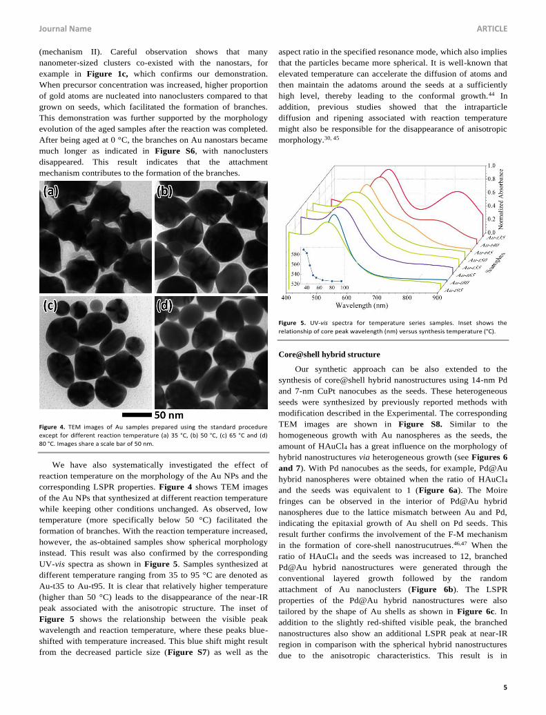
Figure 4. TEM images of Au samples prepared using the standard procedure
except for different reaction temperature (a) 35 °C, (b) 50 °C, (c) 65 °C and (d)
80 °C. Images share a scale bar of 50 nm.
We have also systematically investigated the effect of
reaction temperature on the morphology of the Au NPs and the
corresponding LSPR properties. Figure 4 shows TEM images
of the Au NPs that synthesized at different reaction temperature
while keeping other conditions unchanged. As observed, low
temperature (more specifically below 50 °C) facilitated the
formation of branches. With the reaction temperature increased,
however, the as-obtained samples show spherical morphology
instead. This result was also confirmed by the corresponding
UV-vis spectra as shown in Figure 5. Samples synthesized at
different temperature ranging from 35 to 95 °C are denoted as
Au-t35 to Au-t95. It is clear that relatively higher temperature
(higher than 50 °C) leads to the disappearance of the near-IR
peak associated with the anisotropic structure. The inset of
Figure 5 shows the relationship between the visible peak
wavelength and reaction temperature, where these peaks blue-
shifted with temperature increased. This blue shift might result
from the decreased particle size (Figure S7) as well as the
aspect ratio in the specified resonance mode, which also implies
that the particles became more spherical. It is well-known that
elevated temperature can accelerate the diffusion of atoms and
then maintain the adatoms around the seeds at a sufficiently
high level, thereby leading to the conformal growth.44 In
addition, previous studies showed that the intraparticle
diffusion and ripening associated with reaction temperature
might also be responsible for the disappearance of anisotropic
morphology.30, 45
Figure 5. UV-vis spectra for temperature series samples. Inset shows the
relationship of core peak wavelength (nm) versus synthesis temperature (°C).
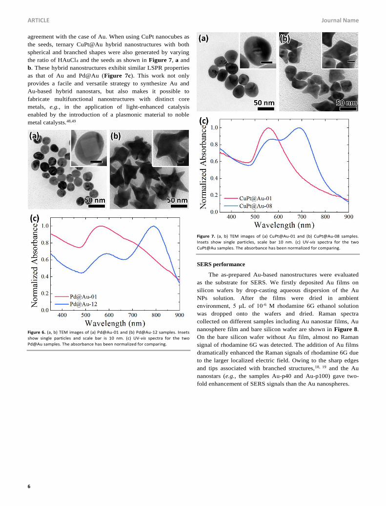
Core@shell hybrid structure
Our synthetic approach can be also extended to the
synthesis of core@shell hybrid nanostructures using 14-nm Pd
and 7-nm CuPt nanocubes as the seeds. These heterogeneous
seeds were synthesized by previously reported methods with
modification described in the Experimental. The corresponding
TEM images are shown in Figure S8. Similar to the
homogeneous growth with Au nanospheres as the seeds, the
amount of HAuCl4 has a great influence on the morphology of
hybrid nanostructures via heterogeneous growth (see Figures 6
and 7). With Pd nanocubes as the seeds, for example, Pd@Au
hybrid nanospheres were obtained when the ratio of HAuCl4
and the seeds was equivalent to 1 (Figure 6a). The Moire
fringes can be observed in the interior of Pd@Au hybrid
nanospheres due to the lattice mismatch between Au and Pd,
indicating the epitaxial growth of Au shell on Pd seeds. This
result further confirms the involvement of the F-M mechanism
in the formation of core-shell nanostrucutrues.46,47 When the
ratio of HAuCl4 and the seeds was increased to 12, branched
Pd@Au hybrid nanostructures were generated through the
conventional layered growth followed by the random
attachment of Au nanoclusters (Figure 6b). The LSPR
properties of the Pd@Au hybrid nanostructures were also
tailored by the shape of Au shells as shown in Figure 6c. In
addition to the slightly red-shifted visible peak, the branched
nanostructures also show an additional LSPR peak at near-IR
region in comparison with the spherical hybrid nanostructures
due to the anisotropic characteristics. This result is in
ARTICLE Journal Name
6
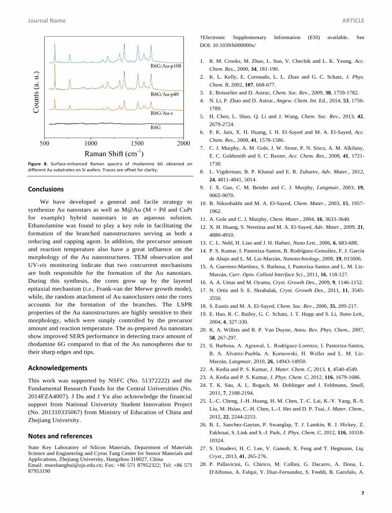
agreement with the case of Au. When using CuPt nanocubes as
the seeds, ternary CuPt@Au hybrid nanostructures with both
spherical and branched shapes were also generated by varying
the ratio of HAuCl4 and the seeds as shown in Figure 7, a and
b. These hybrid nanostructures exhibit similar LSPR properties
as that of Au and Pd@Au (Figure 7c). This work not only
provides a facile and versatile strategy to synthesize Au and
Au-based hybrid nanostars, but also makes it possible to
fabricate multifunctional nanostructures with distinct core
metals, e.g., in the application of light-enhanced catalysis
enabled by the introduction of a plasmonic material to noble
metal catalysts.48,49
Figure 6. (a, b) TEM images of (a) Pd@Au-01 and (b) Pd@Au-12 samples. Insets
show single particles and scale bar is 10 nm. (c) UV-vis spectra for the two
Pd@Au samples. The absorbance has been normalized for comparing.
Figure 7. (a, b) TEM images of (a) CuPt@Au-01 and (b) CuPt@Au-08 samples.
Insets show single particles, scale bar 10 nm. (c) UV-vis spectra for the two
CuPt@Au samples. The absorbance has been normalized for comparing.
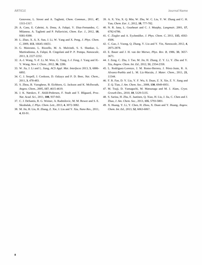
SERS performance
The as-prepared Au-based nanostructures were evaluated
as the substrate for SERS. We firstly deposited Au films on
silicon wafers by drop-casting aqueous dispersion of the Au
NPs solution. After the films were dried in ambient
environment, 5 μL of 10-6 M rhodamine 6G ethanol solution
was dropped onto the wafers and dried. Raman spectra
collected on different samples including Au nanostar films, Au
nanosphere film and bare silicon wafer are shown in Figure 8.
On the bare silicon wafer without Au film, almost no Raman
signal of rhodamine 6G was detected. The addition of Au films
dramatically enhanced the Raman signals of rhodamine 6G due
to the larger localized electric field. Owing to the sharp edges
and tips associated with branched structures,18, 19 and the Au
nanostars (e.g., the samples Au-p40 and Au-p100) gave two-
fold enhancement of SERS signals than the Au nanospheres.
Journal Name ARTICLE
7
Figure 8. Surface-enhanced Raman spectra of rhodamine 6G obtained on
different Au substrates on Si wafers. Traces are offset for clarity.
Conclusions
We have developed a general and facile strategy to
synthesize Au nanostars as well as M@Au (M = Pd and CuPt
for example) hybrid nanostars in an aqueous solution.
Ethanolamine was found to play a key role in facilitating the
formation of the branched nanostructures serving as both a
reducing and capping agent. In addition, the precursor amount
and reaction temperature also have a great influence on the
morphology of the Au nanostructures. TEM observation and
UV-vis monitoring indicate that two concurrent mechanisms
are both responsible for the formation of the Au nanostars.
During this synthesis, the cores grow up by the layered
epitaxial mechanism (i.e., Frank-van der Merwe growth mode),
while, the random attachment of Au nanoclusters onto the cores
accounts for the formation of the branches. The LSPR
properties of the Au nanostructures are highly sensitive to their
morphology, which were simply controlled by the precursor
amount and reaction temperature. The as-prepared Au nanostars
show improved SERS performance in detecting trace amount of
rhodamine 6G compared to that of the Au nanospheres due to
their sharp edges and tips.
Acknowledgements
This work was supported by NSFC (No. 51372222) and the
Fundamental Research Funds for the Central Universities (No.
2014FZA4007). J Du and J Yu also acknowledge the financial
support from National University Student Innovation Project
(No. 201310335067) from Ministry of Education of China and
Zhejiang University.
Notes and references
State Key Laboratory of Silicon Materials, Department of Materials Science and Engineering and Cyrus Tang Center for Sensor Materials and Applications, Zhejiang University, Hangzhou 310027, China Email: [email protected]; Fax: +86 571 87952322; Tel: +86 571 87953190
†Electronic Supplementary Information (ESI) available. See
DOI: 10.1039/b000000x/
1. R. M. Crooks, M. Zhao, L. Sun, V. Chechik and L. K. Yeung, Acc.
Chem. Res., 2000, 34, 181-190.
2. K. L. Kelly, E. Coronado, L. L. Zhao and G. C. Schatz, J. Phys.
Chem. B, 2002, 107, 668-677.
3. E. Boisselier and D. Astruc, Chem. Soc. Rev., 2009, 38, 1759-1782.
4. N. Li, P. Zhao and D. Astruc, Angew. Chem. Int. Ed., 2014, 53, 1756-
1789.
5. H. Chen, L. Shao, Q. Li and J. Wang, Chem. Soc. Rev., 2013, 42,
2679-2724.
6. P. K. Jain, X. H. Huang, I. H. El-Sayed and M. A. El-Sayed, Acc.
Chem. Res., 2008, 41, 1578-1586.
7. C. J. Murphy, A. M. Gole, J. W. Stone, P. N. Sisco, A. M. Alkilany,
E. C. Goldsmith and S. C. Baxter, Acc. Chem. Res., 2008, 41, 1721-
1730.
8. L. Vigderman, B. P. Khanal and E. R. Zubarev, Adv. Mater., 2012,
24, 4811-4841, 5014.
9. J. X. Gao, C. M. Bender and C. J. Murphy, Langmuir, 2003, 19,
9065-9070.
10. B. Nikoobakht and M. A. El-Sayed, Chem. Mater., 2003, 15, 1957-
1962.
11. A. Gole and C. J. Murphy, Chem. Mater., 2004, 16, 3633-3640.
12. X. H. Huang, S. Neretina and M. A. El-Sayed, Adv. Mater., 2009, 21,
4880-4910.
13. C. L. Nehl, H. Liao and J. H. Hafner, Nano Lett., 2006, 6, 683-688.
14. P. S. Kumar, I. Pastoriza-Santos, B. Rodríguez-González, F. J. García
de Abajo and L. M. Liz-Marzán, Nanotechnology, 2008, 19, 015606.
15. A. Guerrero-Martínez, S. Barbosa, I. Pastoriza-Santos and L. M. Liz-
Marzán, Curr. Opin. Colloid Interface Sci., 2011, 16, 118-127.
16. A. A. Umar and M. Oyama, Cryst. Growth Des., 2009, 9, 1146-1152.
17. N. Ortiz and S. E. Skrabalak, Cryst. Growth Des., 2011, 11, 3545-
3550.
18. S. Eustis and M. A. El-Sayed, Chem. Soc. Rev., 2006, 35, 209-217.
19. E. Hao, R. C. Bailey, G. C. Schatz, J. T. Hupp and S. Li, Nano Lett.,
2004, 4, 327-330.
20. K. A. Willets and R. P. Van Duyne, Annu. Rev. Phys. Chem., 2007,
58, 267-297.
21. S. Barbosa, A. Agrawal, L. Rodriguez-Lorenzo, I. Pastoriza-Santos,
R. A. Alvarez-Puebla, A. Kornowski, H. Weller and L. M. Liz-
Marzan, Langmuir, 2010, 26, 14943-14950.
22. A. Kedia and P. S. Kumar, J. Mater. Chem. C, 2013, 1, 4540-4549.
23. A. Kedia and P. S. Kumar, J. Phys. Chem. C, 2012, 116, 1679-1686.
24. T. K. Sau, A. L. Rogach, M. Doblinger and J. Feldmann, Small,
2011, 7, 2188-2194.
25. L.-C. Cheng, J.-H. Huang, H. M. Chen, T.-C. Lai, K.-Y. Yang, R.-S.
Liu, M. Hsiao, C.-H. Chen, L.-J. Her and D. P. Tsai, J. Mater. Chem.,
2012, 22, 2244-2253.
26. B. L. Sanchez-Gaytan, P. Swanglap, T. J. Lamkin, R. J. Hickey, Z.
Fakhraai, S. Link and S.-J. Park, J. Phys. Chem. C, 2012, 116, 10318-
10324.
27. S. Umadevi, H. C. Lee, V. Ganesh, X. Feng and T. Hegmann, Liq.
Cryst., 2013, 41, 265-276.
28. P. Pallavicini, G. Chirico, M. Collini, G. Dacarro, A. Dona, L.
D'Alfonso, A. Falqui, Y. Diaz-Fernandez, S. Freddi, B. Garofalo, A.
ARTICLE Journal Name
8
Genovese, L. Sironi and A. Taglietti, Chem. Commun., 2011, 47,
1315-1317.
29. A. Casu, E. Cabrini, A. Dona, A. Falqui, Y. Diaz-Fernandez, C.
Milanese, A. Taglietti and P. Pallavicini, Chem. Eur. J., 2012, 18,
9381-9390.
30. L. Zhao, X. Ji, X. Sun, J. Li, W. Yang and X. Peng, J. Phys. Chem.
C, 2009, 113, 16645-16651.
31. G. Maiorano, L. Rizzello, M. A. Malvindi, S. S. Shankar, L.
Martiradonna, A. Falqui, R. Cingolani and P. P. Pompa, Nanoscale,
2011, 3, 2227-2232.
32. A.-J. Wang, Y.-F. Li, M. Wen, G. Yang, J.-J. Feng, J. Yang and H.-
Y. Wang, New J. Chem., 2012, 36, 2286.
33. W. Jia, J. Li and L. Jiang, ACS Appl. Mat. Interfaces 2013, 5, 6886-
6892.
34. C. J. Serpell, J. Cookson, D. Ozkaya and P. D. Beer, Nat. Chem.,
2011, 3, 478-483.
35. S. Zhou, B. Varughese, B. Eichhorn, G. Jackson and K. McIlwrath,
Angew. Chem., 2005, 117, 4615-4619.
36. J. K. Nørskov, F. Abild-Pedersen, F. Studt and T. Bligaard, Proc.
Nat. Acad. Sci., 2011, 108, 937-943.
37. C. J. DeSantis, R. G. Weiner, A. Radmilovic, M. M. Bower and S. E.
Skrabalak, J. Phys. Chem. Lett., 2013, 4, 3072-3082.
38. M. Jin, H. Liu, H. Zhang, Z. Xie, J. Liu and Y. Xia, Nano Res., 2011,
4, 83-91.
39. A. X. Yin, X. Q. Min, W. Zhu, W. C. Liu, Y. W. Zhang and C. H.
Yan, Chem. Eur. J., 2012, 18, 777-782.
40. N. R. Jana, L. Gearheart and C. J. Murphy, Langmuir, 2001, 17,
6782-6786.
41. C. Ziegler and A. Eychmuller, J. Phys. Chem. C, 2011, 115, 4502-
4506.
42. C. Gao, J. Vuong, Q. Zhang, Y. Liu and Y. Yin, Nanoscale, 2012, 4,
2875-2878.
43. E. Bauer and J. H. van der Merwe, Phys. Rev. B, 1986, 33, 3657-
3671.
44. J. Zeng, C. Zhu, J. Tao, M. Jin, H. Zhang, Z. Y. Li, Y. Zhu and Y.
Xia, Angew. Chem. Int. Ed., 2012, 51, 2354-2358.
45. L. Rodríguez-Lorenzo, J. M. Romo-Herrera, J. Pérez-Juste, R. A.
Alvarez-Puebla and L. M. Liz-Marzán, J. Mater. Chem., 2011, 21,
11544.
46. F. R. Fan, D. Y. Liu, Y. F. Wu, S. Duan, Z. X. Xie, Z. Y. Jiang and
Z. Q. Tian, J. Am. Chem. Soc., 2008, 130, 6949-6951.
47. M. Tsuji, D. Yamaguchi, M. Matsunaga and M. J. Alam, Cryst.
Growth Des., 2010, 10, 5129-5135.
48. S. Sarina, H. Zhu, E. Jaatinen, Q. Xiao, H. Liu, J. Jia, C. Chen and J.
Zhao, J. Am. Chem. Soc., 2013, 135, 5793-5801.
49. X. Huang, Y. Li, Y. Chen, H. Zhou, X. Duan and Y. Huang, Angew.
Chem. Int. Ed., 2013, 52, 6063-6067.


![Phase stability of dispersions of hollow silica nanocubes ...theoretically [42], experimental studies on the phase be-haviour of stable dispersions of colloidal nanocubes mixed with](https://img.pdfslide.us/doc/110x75/611b8326f18c574a142c3931/phase-stability-of-dispersions-of-hollow-silica-nanocubes-theoretically-42.jpg)


![CASE STUDY - ICDD...presented in this case study are AuPd nanocubes synthesized by -mediated growth method described in Ref. [1]. Figure 1 (a) shows a transmission electron microscopy](https://img.pdfslide.us/doc/110x75/60d70c1851b30a0c640031d8/case-study-icdd-presented-in-this-case-study-are-aupd-nanocubes-synthesized.jpg)