Embed Size (px)
Citation preview
518
Arteriovenous Fistulas of the Cervicomedullary Junction as a Cause of Myelopathy: Radiographic Findings in Two Cases Erik H. L. Gaensler, 1 Donald E. Jackson, Jr., and Van V. Halbach
Spinal dural arteriovenous fistulas (SDAVFs) represent an abnormal connection between a dural artery and an intradural radicular vein. The radicular vein drains the medullary venous plexus , which dilates in response to arterial pressure, resulting in enlarged serpiginous vessels , usually posterior to the spinal cord. This finding is not specific for SDAVF, as it can be found in most spinal arteriovascular malformations (AVMs) as well as hemangioblastomas. SDAVFs can be differentiated from intramedullary arteriovenous malformations in that the arteriovenous connection in SDAVF is extramedullary, lying outside the cord substance within the dura. Clinically, SDAVFs usually present as a slowly progressive myelopathy or radiculopathy in middle-aged to elderly patients, that can mimic other spinal pathology [1].
Since most SDAVFs are located in the lower thoracic and lumbar regions, with symptoms referable to the conus, angiegraphic searches may be limited to the thoracic, lumbar, and sacral contribution to the dural arterial supply [1]. However, the site of the SDAVF may be far removed from the portion of the spinal cord affected cl inically. As illustrated in the two cases that follow, conus-related symptoms can result from an arteriovenous fistula located as high as the cervical canal. In these two cases , as well as six others from the literature, exhaustive angiographic evaluation of all possible vascular pedicles was required to identify and treat the SDAVF.
Case Reports
Case 1
This 50-year-old man first noted bilateral upper extremity weakness in 1983, and went on to develop rapidly progressive weakness of his legs in 1985. This was followed by urinary incontinence in 1986, dysesthesias in all extremities. and impotence. Physical examination in 1987 revealed upper extremity muscle wasting , with knee and ankle clonus. Babinski reflexes were upgoing bilaterally, with a spastic gait.
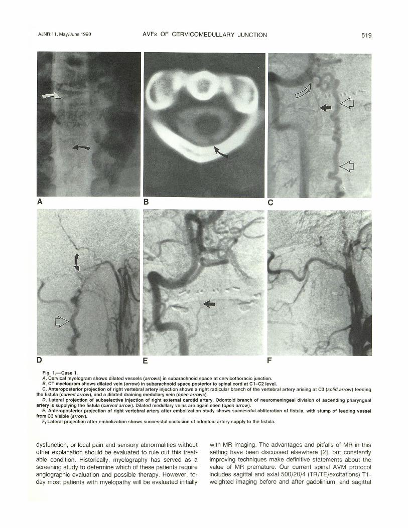
Myelography revealed dilated vascular structures posterior to the entire spinal cord , extending up to the foramen magnum (Figs. 1 A
and 1 B). Angiography demonstrated a radiculomedullary fistula located at the anterior foramen magnum, with supply from a C3 radicular branch of the vertebral artery (Fig. 1 C), and from the odontoid branch of the neuromeningeal division of the ascending pharyngeal artery (Fig. 1 D). Embolization of the vertebral contribution with polyvinyl alcohol (PVA, Pacific Medical Co. , La Jolla, CA) particles and isobutyl 2-cyanoacrylate (IBCA, Ethicon Inc., Sommerville, NJ) resulted in obliteration of the AVM (Figs 1E and 1F). Subsequently, the patient showed steady improvement in upper extremity strength and bladder control , and erectile function returned . His long-standing lower extremity spasticity did not resolve, but has not progressed over the subsequent 2 years .
Case 2
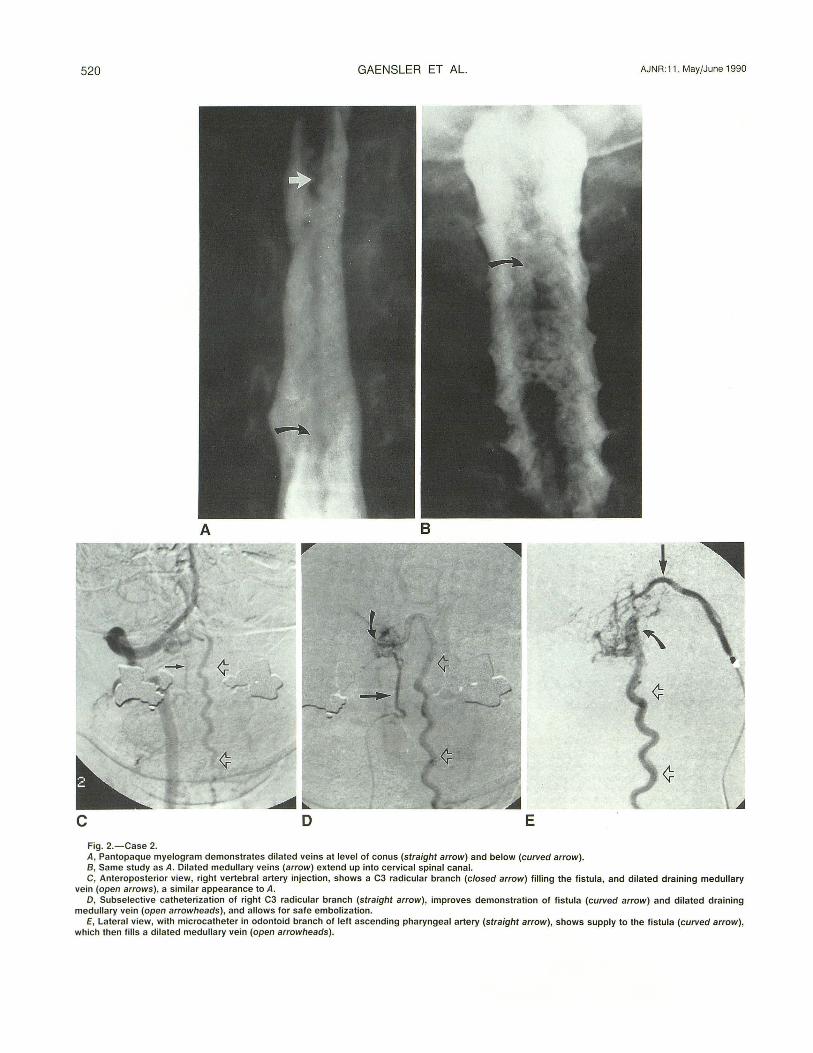
This 69-year-old woman had a history of lower back pain for 2 years. She developed rapidly progressive motor and sensory deficits in her lower extremities over 2 days, and a myelogram demonstrated dilated medullary veins in the lumbar region (Fig. 2A), which extended up into the thoracic and cervical regions (Fig. 2B). Lumbar and thoracic spinal angiography was negative.
Subsequent cerebral arteriography revealed a dural fistula located at C1 , supplied by the right C3 radicular branch from the vertebral artery (Fig. 2C), the odontoid branch of the ascending pharyngeal artery. and posterior inferior cerebellar artery branches. The ascending pharyngeal artery and radicular branches from the vertebral artery were selectively catheterized (Figs. 2D and 2E) and embolized with PVA particles. The remaining supply from the posterior inferior cerebellar artery could not be embolized safely. An MR study obtained at this time showed persistently dilated veins posterior to the cervical spinal cord . The patient was treated surgically, and the dural fistula was identified and removed . A postoperative angiogram showed complete obliteration of the fistula. Despite some initial improvement, the patient had no long-term change in her neurologic deficits.
Discussion
SDAVF is better understood than in the past and more frequently recognized as a cause of progressive paraparesis. Adults with progressive leg weakness, bowel and bladder
Received May 22, 1989; revision requested May 25, 1989; revision received August 21, 1989; accepted August 29 , 1989. 'All authors: Department of Radiology, Neuroradiology Section, University of California, San Francisco Medical Center, 505 Parnassus Ave., San Francisco, CA
94143. Address reprint requests to E. H. L. Gaensler.
AJNR 11:518-521 , Mayf June 1990 01 95-6108/90/1103- 0518 © American Society of Neuroradiology
AJNR :11 , May/June 1990 AVFs OF CERVICOMEDULLARY JUNCTION 519
Fig. 1.-Case 1. A, Cervical myelogram shows dilated vessels (arrows) in subarachnoid space at cervicothoracic junction. B, CT myelogram shows dilated vein (arrow) in subarachnoid space posterior to spinal cord at C1-C2 level. C, Anteroposterior projection of right vertebral artery injection shows a right radicular branch of the vertebral artery arising at C3 (solid arrow) feeding
the fistula (curved arrow), and a dilated draining medullary vein (open arrows). D, Lateral projection of subselective injection of right external carotid artery. Odontoid branch of neuromeningeal division of ascending pharyngeal
artery is supplying the fistula (curved arrow). Dilated medullary veins are again seen (open arrow). E, Anteroposterior projection of right vertebral artery after embolization study shows successful obliteration of fistula, with stump of feeding vessel
from C3 visible (arrow). F, Lateral projection after embolization shows successful occlusion of odontoid artery supply to the fistula .
dysfunction , or local pain and sensory abnormalities without other explanation should be evaluated to rule out this treatable condition. Historically, myelography has served as a screening study to determine which of these patients require angiographic evaluation and possible therapy. However, today most patients with myelopathy will be evaluated initially
with MR imaging. The advantages and pitfalls of MR in this setting have been discussed elsewhere [2], but constantly improving techniques make definitive statements about the value of MR premature. Our current spinal AVM protocol includes sagittal and axial 500/20/4 (TRfTEjexcitations) T1-weighted imaging before and after gadolinium, and sagittal
520 GAENSLER ET AL.
c
A 8
D E
Fig. 2.-Case 2. A, Pantopaque myelogram demonstrates dilated veins at level of conus (straight arrow) and below (curved arrow). B, Same study as A. Dilated medullary veins (arrow) extend up into cervical spinal canal.
AJNR :11 , May/June 1990
C, Anteroposterior view, right vertebral artery injection, shows a C3 radicular branch (closed arrow) filling the fistula, and dilated draining medullary vein (open arrows), a similar appearance to A.
D, Subselective catheterization of right C3 radicular branch (straight arrow), improves demonstration of fistula (curved arrow) and dilated draining medullary vein (open arrowheads), and allows for safe embolization.
E, Lateral view, with microcatheter in odontoid branch of left ascending pharyngeal artery (straight arrow), shows supply to the fistula (curved arrow), which then fills a dilated medullary vein (open arrowheads).
AJNR :11 , May/June 1990 AVFs OF CERVICOMEDULLARY JUNCTION 521
cardiac-gated T2-weighted imaging with a TE 9f 30{80 prior to gadolinium. Coronal sequences have also proved helpful , as the medullary vessels are in the plane of section and less prone to entry slice and flow artifact than on axial and sagittal images. Flow compensation is essential to prevent CSF flow artifacts that can mimic dilated medullary veins. If the level of an AVM is known from prior studies , a 5-in . surface coil can be used to give better definition , but in the screening setting a 12-in. license-plate coil is used, with a 20-24-cm field of view and 192 x 256 matrix.
However, even when using an optimal MR technique, we and others have encountered dilated vessels so subtle that they escaped detection on screening MR , only to be finally identified on myelography (2 , 3] . Therefore, if MR is negative, we always perform total spinal myelography. Films are taken with the patient in the supine as well as the prone position in order to maximally pool contrast around any dilated vessels posterior to the cord. An additional benefit of myelography is that the relationship of the major draining vein to a given pedicle can be established , suggesting a site at which to begin searching for the fistula .
Once dilated vessels in the medullary venous plexus have been identified by myelography or MR, angiographic evaluation is needed. Sometimes a complete search at all lumbar and thoracic levels fails to reveal the source of the fistula. Once possibility is that the dilated vessels have spontaneously thrombosed (Foix-Aiajouanine syndrome). In the two patients in this study, however, a further search to include all possible connections to the medullary venous plexus was rewarded by the discovery of a treatable lesion. In both cases , feeding vessels off the vertebral artery, and off the external carotid artery via the ascending pharyngeal artery, were identified. As a result of these cases , our angiographic evaluation of patients with suspected SDAVF includes cerebral arteriography if spinal angiography is negative.
Six similar cases have been reported . Woimant et al. [4] described a 35-year-old woman with myelopathy resulting from an arteriovenous fistula located at the bend of a distally thrombosed left transverse sinus. The fistula was supplied by the occipital artery, with subtentorial and medullary cortical venous drainage. These veins were discovered on myelography, and extended to the conus. Picard et al. [5] reported two cases of intracranial dural fistulas with medullary venous drainage presenting as ascending myelopathy. Only one was described in complete detail, and was due to a pial fistula at the foramen ovale, supplied by meningeal branches of both the internal and external carotid artery. More recently, Wrobel et al. (3] reported three cases of intracranial dural arteriovenous fistulas draining into spinal medullary veins. All three patients presented with lower extremity symptoms and showed dilated vessels posterior to the cord on myelography. In one case, these vessels had been noted on MR, but in another, initial MR had been read as normal. Spinal angiegrams were negative in all three. Cerebral arteriography revealed dural arteriovenous fistulas in the petrous area in two cases , with both internal and external carotid supply, and a tentorial fistula supplied by meningohypophyseal , occipital , and ascending pharyngeal branches in the third . All three patients ' myelopathy improved with interruption of the fistulas .
The fact that fistulas at or above the foramen magnum cause reversible conus symptoms helps explain the pathophysiology of SDAVFs. As proposed by Aminoff et al. (6] , myelopathy in these cases is due to congestive hypoxia. This hypoxia is probably secondary to perimedullary venous hypertension , caused by the direct arterial inflow from the fistula . Alternative mechanisms, such as cord compression , arterial steal with ischemia, and hemorrhage (with or without arachnoiditis) do not adequately explain how a chronic low-flow lesion at the cerivomedullary junction can result in conus symptoms.
Recent MR studies (2 , 7] have described increased conus signal on T2-weighted images in SDAVFs, consistent with edema, further support for the hypothesis that the associated myelopathy is caused by venous congestion. Treatment of the SDAVF can relieve venous hypertension and reverse the myelopathy. In case 1, the patient had some recovery of function , presumably on this basis. However, not all deficits appear to be reversible. In case 1 , lower extremity spasticity persisted , and in case 2 no long-term improvement occurred . Whether or not therapy reverses the myelopathy may depend on whether the cord is merely ischemic or if infarction has occurred . Even if deficits are not reversed , therapy can be beneficial in preventing accelerating deterioration , which is the natural history of SDAVFs. Neither case presented here developed further deficits on follow up.
Our two cases differ from those previously reported in that the fistulas were in the region of the craniocervical junction , with significant supply from cervical radicular branches of the vertebral artery. All the feeding vessels in the previous six reports came exclusively from the intracranial circulation. We found no prior reports of SDAVF in the cervical region . The eight cases combined illustrate an important clinical point: all potential sources of arterial supply must be evaluated when there is suspicion of an SDAVF, including the intracranial circulation and external carotid branches.
ACKNOWLEDGMENT
We thank S. N. Bishara, Director of Neurosurgery, Dunedin Hospital, New Zealand, for his help in referral and follow up of case 1.
REFERENCES
1. Rosenblum B, Oldfield EH , Doppman JL, Di Chiro G. Spinal arteriovenous malformations: a comparison of dural arteriovenous fistulas and intradural AVMs in 81 patients. J Neurosurg 1987;67:795-802
2. Gaensler EHL, Dillon WP, Tsuruda JS , Halbach VV, Higashida RT, Hieshima GB. Arteriovenous malformations of the spinal cord: MR spectrum. AJNR 1989;10:880 (abstr)
3. Wrobel CJ, Oldfield EH , Di Chiro G, Tarlov EC , Baker RA, Doppman JL. Myelopathy due to intracranial dural arteriovenous fistulas draining intrathecally into spinal medullary veins. J Neurosurg 1988;69 :934-939
4. Woimant F, Merland JJ, Riche MC, et al. Syndrome bulbo-medullaire en rapport avec une fistule arterio-veineuse meningee du sinus lateral a drainage veneux medullaire. Rev Neural 1982; 138:559-566
5. Picard L, Bracard S, Moret J, Per A, Giacobbe MD, Roland J. Spontaneous dural arteriovenous fistulas . Semin lntervent Radio/ 1987;4:219-241
6. AminoH MJ , Barnard RO, Logue V. The pathophysiology of spinal vascular malformations. J Neurol Sci 1974;23 :255-263
7. Masaryk TJ, Ross JS, Modic MT, Ruff RL, Selman WR , Ratcheson RA. Radiculomeningeal vascular malformations of the spinal cord: MR imaging. Radiology 1987; 164 : 845-849


![Pathophysiology: Heart Failure - Columbia University Heart Failure ... – TPR = [MAP - CVP] / CO, and ... – Anemia – Systemic arteriovenous fistulas – Hyperthyroidism](https://img.pdfslide.us/doc/110x75/5aa356057f8b9ab4208e3286/pathophysiology-heart-failure-columbia-heart-failure-tpr-map-cvp.jpg)