Embed Size (px)
Citation preview
1
Arterial & Venous Ulcers A Comprehensive Review
Assessment & Management
Arterial & Venous Ulcers A Comprehensive Review
Assessment & Management
2015 AXXESS. UNAUTHORIZED USE IS PROHIBITED
2
ObjectivesObjectives
Understand Arterial & Venous disease
Understand the etiology of lower extremities
ulcers
Understand assessment of lower extremities
ulcers
Understand lower extremities ulcer treatment
plan
Identify best practices in home care setting for
the management of patients with lower
extremity ulcers.
3
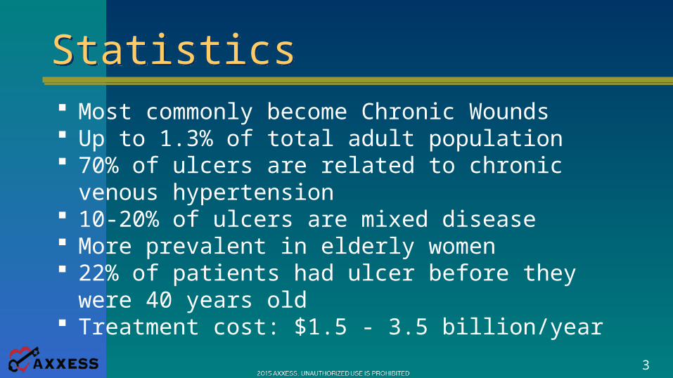
StatisticsStatistics Most commonly become Chronic Wounds Up to 1.3% of total adult population 70% of ulcers are related to chronic venous
hypertension 10-20% of ulcers are mixed disease More prevalent in elderly women 22% of patients had ulcer before they were 40
years old Treatment cost: $1.5 - 3.5 billion/year
4
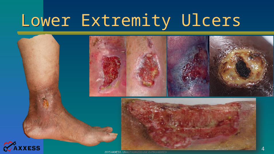
Lower Extremity UlcersLower Extremity Ulcers
5
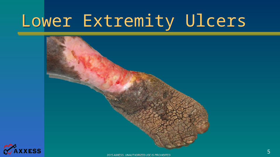
Lower Extremity UlcersLower Extremity Ulcers
6
Associated ConditionsAssociated Conditions Venous Hypertension Arterial Ischemia Diabetes or Neuropathy Cardiovascular disease Infection Lymphedema Insect Bites Vasculitis Trauma
7
Assessment of Lower ExtremitiesAssessment of Lower Extremities
Color Changes with Limb Elevation and Dependence
Supine, raise leg to 60ºCount the time until color changes
Place leg dependent positionNote development of rubor
8
Assessment of Lower ExtremitiesAssessment of Lower Extremities
Venous Filling TimeElevate the limb to provide for venous
drainagePlace limb in dependent position
Record the time required for venous fillingProlonged venous filling is independently
predictive of PADGreater than 20 seconds usually indicates
occlusive diseaseAuscultate all major pulses for evidenceof bruits, which can indicate occlusion
9
Assessment of Lower ExtremitiesAssessment of Lower Extremities
Ankle Brachial Index Test (ABI)Using a BP Cuff and a handheld DopplerMeasure the brachial systolic pressures
Place the cuff around the ankleMeasure the systolic pressure
Dorsalis PedisPosterior Tibial
10
Assessment of Lower ExtremitiesAssessment of Lower Extremities
Ankle Brachial Index Test (ABI)Ankle Pressures =120
Brachial Pressures =120120/120 = 1
-or-Ankle Pressures = 60
Brachial Pressures =12060/120 = .5
11
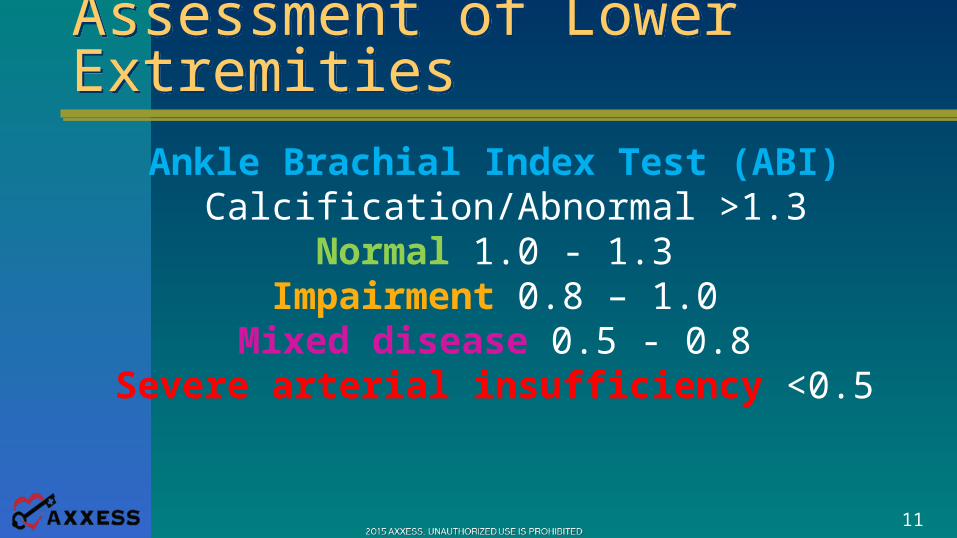
Assessment of Lower ExtremitiesAssessment of Lower Extremities
Ankle Brachial Index Test (ABI) Calcification/Abnormal >1.3
Normal 1.0 - 1.3Impairment 0.8 – 1.0
Mixed disease 0.5 - 0.8Severe arterial insufficiency <0.5
12
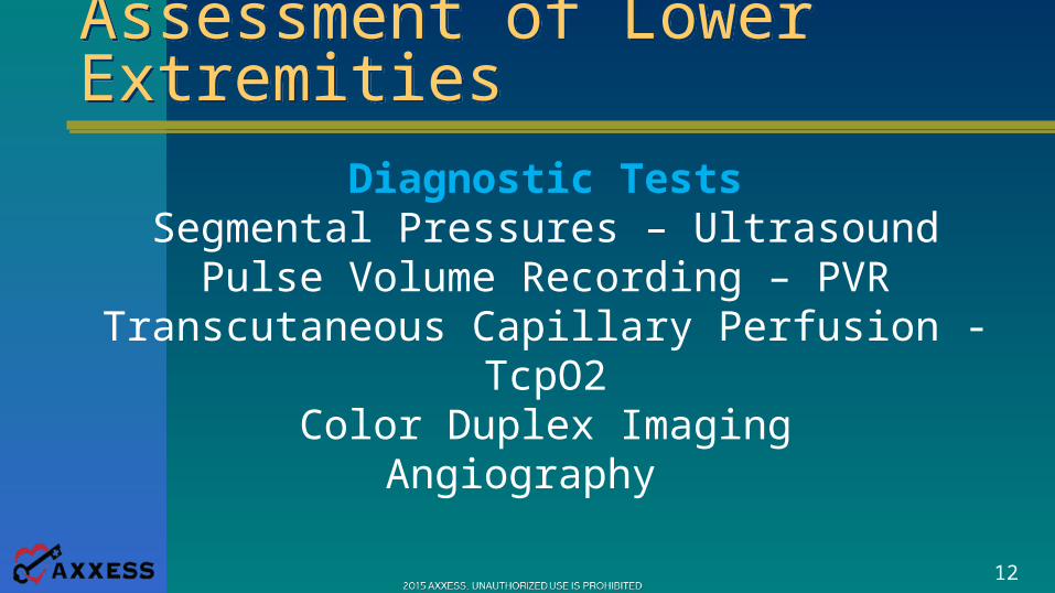
Assessment of Lower ExtremitiesAssessment of Lower Extremities
Diagnostic TestsSegmental Pressures – Ultrasound
Pulse Volume Recording – PVRTranscutaneous Capillary Perfusion - TcpO2
Color Duplex ImagingAngiography
13
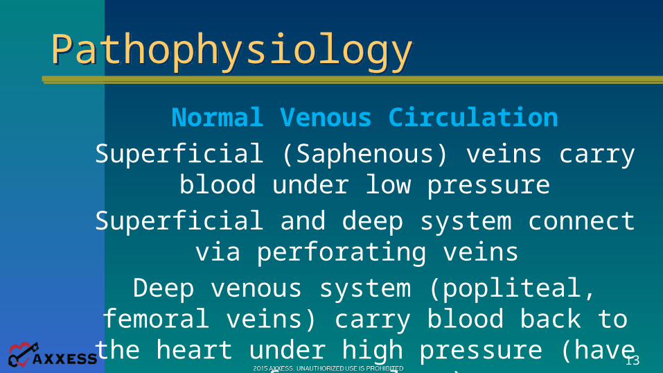
PathophysiologyPathophysiology
Normal Venous CirculationSuperficial (Saphenous) veins carry blood
under low pressureSuperficial and deep system connect via
perforating veins Deep venous system (popliteal, femoral
veins) carry blood back to the heart under high pressure (have fewer valves)
14
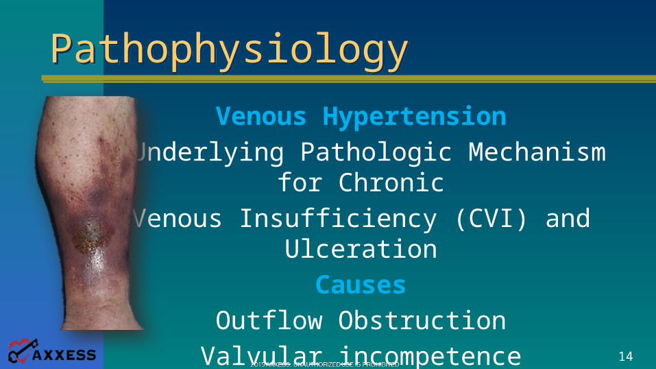
PathophysiologyPathophysiology
Venous Hypertension•Underlying Pathologic Mechanism for
ChronicVenous Insufficiency (CVI) and Ulceration
CausesOutflow Obstruction
Valvular incompetenceMuscle pump failure
15
Etiology VLUEtiology VLU
Fibrin Cuff Theory• Venous Hypertension - Capillary dilation• Fibrinogen leaks into dermal tissue• Fibrinogen hardens and forms a cuff - Barrier to O2/nutrients • Fibrin cuffs may indicate endothelial cell damage and affect wound healing by inhibiting collagen formation, prolong inflammation, or block growth factors
16
Etiology VLUEtiology VLU
White Cell Trapping Theory• Velocity of blood flow through capillary becomes sluggish• White cell adhere to capillary wall, plugging capillariescausing tissue ischemia• White cell activation• Toxic metabolites/proteolytic enzymes• Local occlusion, ischemia, ulceration
17
Clinical Signs & SymptomsClinical Signs & Symptoms
• Gaiter Distribution• Edema | 1+• Hemosiderin Staining | Discoloration of skin• Venous Dermatitis | Marked Redness• Atrophie blanche | Sluggish capillary refill• Varicose veins | Lack of hairs on the legs• Atrophy of the skin | Lipodermatosclerosis
18
Clinical Signs & SymptomsClinical Signs & Symptoms
• Usual location : Medial malleolus | Irregular edges• Wound bed- ruddy red, yellow adherent or loose slough,undermining or tunneling uncommon• Usually shallow, full thickness, heavily draining• Heavily contaminated• Surrounding skin- macerated; crusted, and scaling• Pain is variable-severe; dull, aching or bursting
19
Treatment PhilosophyTreatment Philosophy
Identify and treat the underlying cause ofthe ulcer and the factors that affect
wound closureRestricted mobility | Edema in the limbMalnutrition | Psychosocial problems
Minimize colonization | Apply Compression
20
Treatment PhilosophyTreatment Philosophy
TYPES OF COMPRESSIONShort Stretch BandagesPaste Boot/Unna Boot
Long Stretch BandagesBandaging “Systems”
Compression StockingsDynamic Compression Pumps
21
Treatment PhilosophyTreatment Philosophy
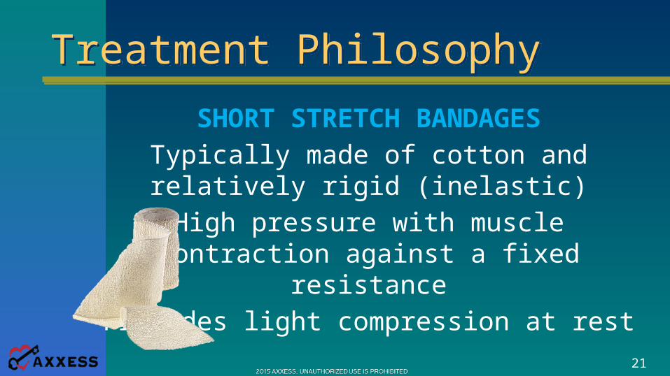
SHORT STRETCH BANDAGESTypically made of cotton and relatively
rigid (inelastic)High pressure with muscle contraction
against a fixed resistanceProvides light compression at rest
22
Treatment PhilosophyTreatment Philosophy
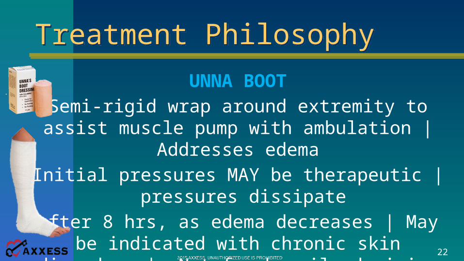
UNNA BOOTSemi-rigid wrap around extremity to assist muscle pump with ambulation | Addresses
edemaInitial pressures MAY be therapeutic |
pressures dissipateafter 8 hrs, as edema decreases | May be
indicated with chronic skin disorders | Not for heavily draining wounds | Comes in a variety
of styles with zinc oxide, calamine, gelatin and lanolin
23
Treatment PhilosophyTreatment Philosophy
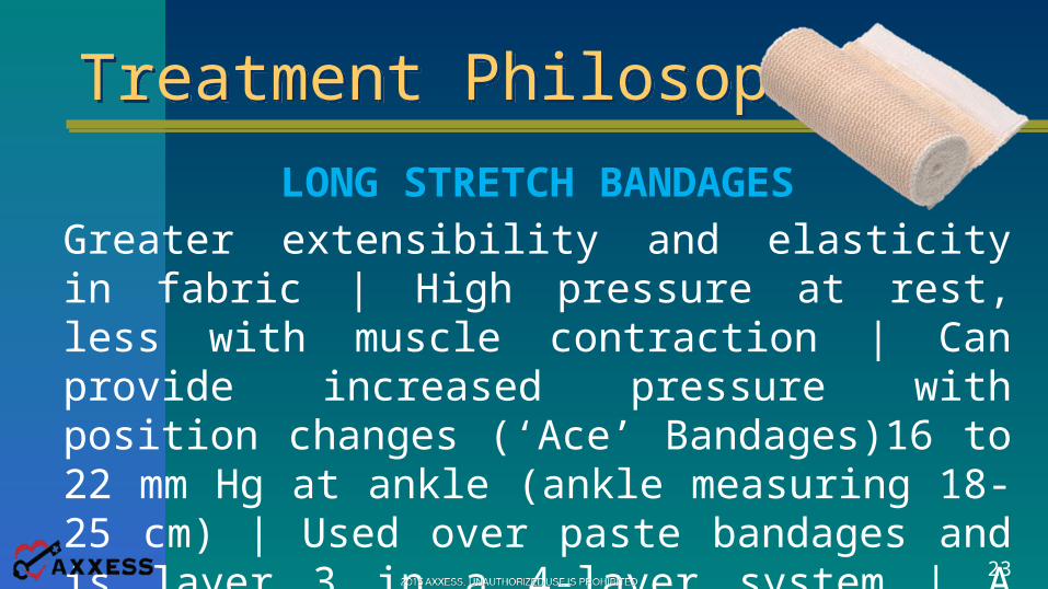
LONG STRETCH BANDAGESGreater extensibility and elasticity in fabric | High pressure at rest, less with muscle contraction | Can provide increased pressure with position changes (‘Ace’ Bandages)16 to 22 mm Hg at ankle (ankle measuring 18-25 cm) | Used over paste bandages and is layer 3 in a 4-layer system | A single wash reduces pressure by 20% | some brands have rectangles woven into the dressing turn into squares when bandage is stretched | Potential risk for ischemia with over stretching
24
Treatment PhilosophyTreatment Philosophy
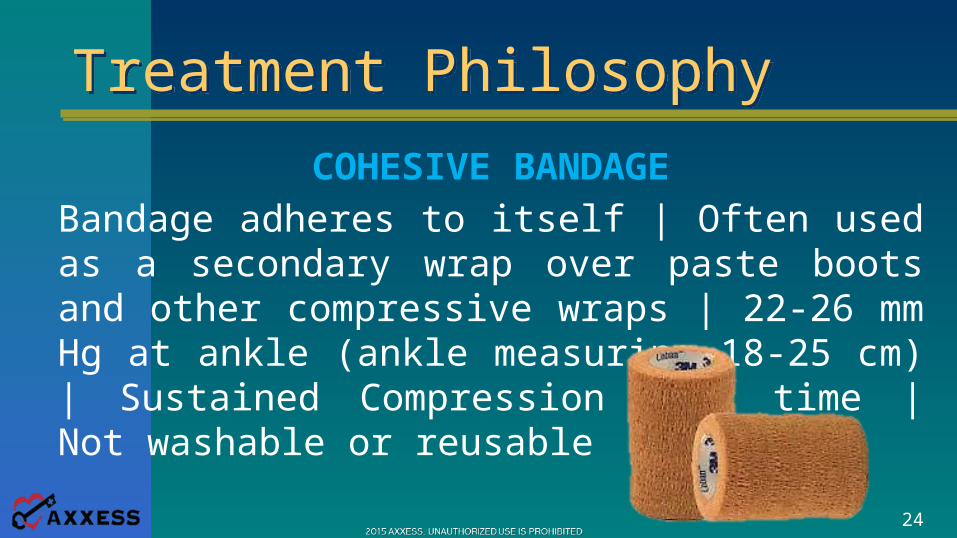
COHESIVE BANDAGEBandage adheres to itself | Often used as a secondary wrap over paste boots and other compressive wraps | 22-26 mm Hg at ankle (ankle measuring 18-25 cm) | Sustained Compression over time | Not washable or reusable
25
Treatment PhilosophyTreatment Philosophy
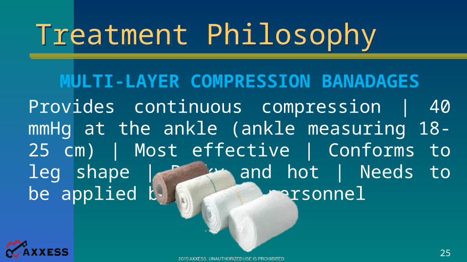
MULTI-LAYER COMPRESSION BANADAGESProvides continuous compression | 40 mmHg at the ankle (ankle measuring 18-25 cm) | Most effective | Conforms to leg shape | Bulky and hot | Needs to be applied by trained personnel
26
Treatment PhilosophyTreatment Philosophy
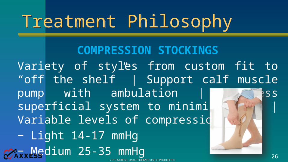
COMPRESSION STOCKINGSVariety of styles from custom fit to “off the shelf” | Support calf muscle pump with ambulation | Compress superficial system to minimize edema | Variable levels of compression:− Light 14-17 mmHg− Medium 25-35 mmHg− High 35-45 mmHg
27
Arterial DiseaseArterial Disease
Clotting | Shower of clots (small/large vessel)Rheumatoid arthritis (arteritis) | Diabetes mellitus (atherosclerosis)Degenerative changes with advancing age (atherosclerosis)Raynaud’s disease (vasospastic disease)
28
Arterial DiseaseArterial Disease
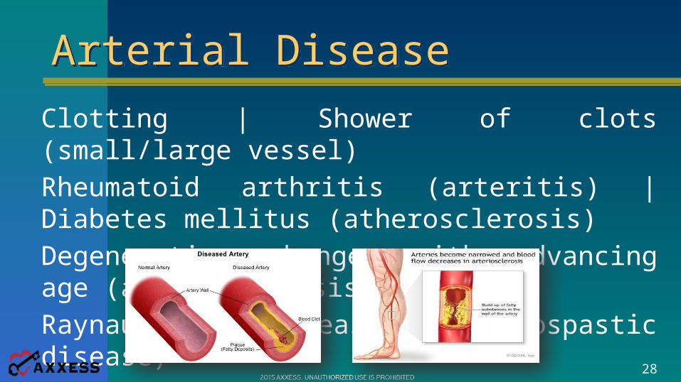
Clotting | Shower of clots (small/large vessel)Rheumatoid arthritis (arteritis) | Diabetes mellitus (atherosclerosis)Degenerative changes with advancing age (atherosclerosis)Raynaud’s disease (vasospastic disease)
29
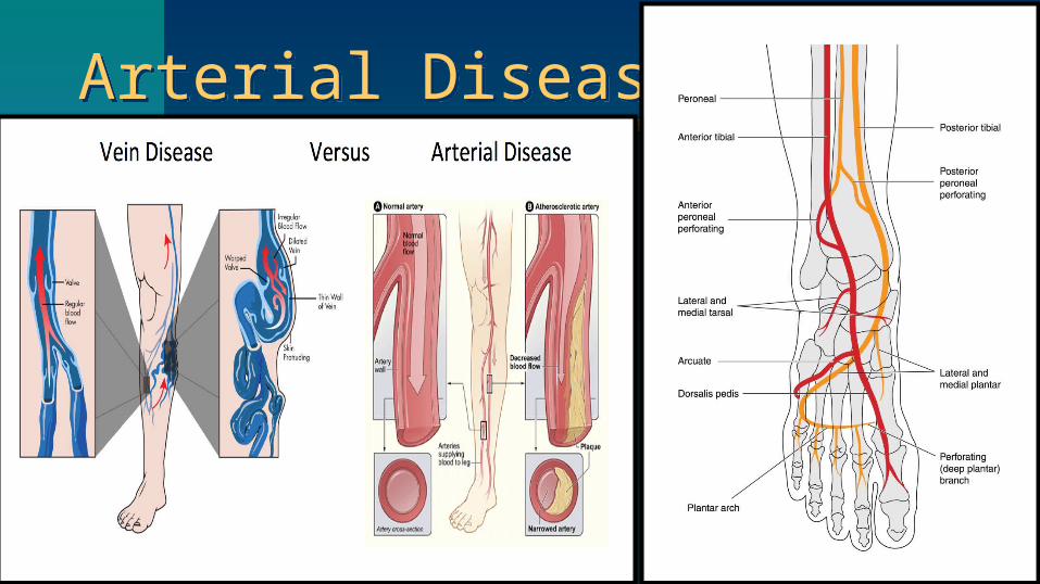
Arterial DiseaseArterial Disease
30
Arterial DiseaseArterial Disease
Ischemic rest pain | Pain relief w/dependency | Loss of hairAtrophic, shiny skin | Muscle wasting calf or thigh Trophic nail changes | Poor tissue perfusionColor changes | Coldness of the foot | Gangrene of toesAbsence of palpable pulse
31
Arterial DiseaseArterial Disease
Pain of sudden onset and severe intensity | PallorParaesthesia (numbness) | Pulselessness (absence of pulses below the occlusion) | Paralysis (sudden weakness in the limb) Extremity cool to touch
32
Management PhilosophyManagement Philosophy
Pain Perfusion is Insufficient for Wound Healing• Revascularization• Amputation• If patient is not appropriate for surgical intervention Keep the wounds clean, dry and free from infection No compression !!! No Elastic or stretchable gauze rolls
33
Management PhilosophyManagement Philosophy
Pain Perfusion is Insufficient for Wound Healing• Revascularization | Hyperbaric Oxygen Therapy• Amputation• If patient is not appropriate for surgical intervention Keep the wounds clean, dry and free from infection No compression !!! No Elastic or stretchable gauze rolls
34
ReferencesReferences1. Pressure ulcer staging. (2013). www.npuap.org2. Home Health Potentially Avoidable Event Measures (2013).
Centers for Medicare & Medicaid Services. www.cms.gov. 3. www.woundcarenurses.org4. Ferrell BA, Josephson K, Norvid P, Alcorn H. Pressure ulcers
among patients admitted to home care. J Am Geriatr Soc 2000; 48(9):1042-1047.
5. Bergquist S. Subscales, subscores, or summative score: evaluating the contribution of Braden Scale items for predicting pressure ulcer risk in older adults receiving home health care. J Wound Ostomy Continence Nurs 2001; 28(6):279-289.
6. Gorecki C, Brown JM, Nelson EA, Briggs M, Schoonhoven L, Dealey C et al. Impact of pressure ulcers on quality of life in older patients: a systematic review. J Am Geriatr Soc 2009; 57(7):1175-1183.