Embed Size (px)
Citation preview
Arginine dephosphorylation propels spore germinationin bacteriaBing Zhoua,b, Maja Semanjskic, Natalie Orlovetskieb, Saurabh Bhattacharyab, Sima Alonb, Liron Argamanb,Nayef Jarrousb, Yan Zhanga, Boris Macekc, Lior Sinaib,1, and Sigal Ben-Yehudab,1
aCollege of Food Science and Nutritional Engineering, National Engineering Research Center for Fruits and Vegetables Processing, China AgriculturalUniversity, 10083 Beijing, China; bDepartment of Microbiology and Molecular Genetics, Institute for Medical Research Israel-Canada, The HebrewUniversity-Hadassah Medical School, The Hebrew University of Jerusalem, 91120 Jerusalem, Israel; and cProteome Center Tuebingen, University ofTuebingen, 72076 Tuebingen, Germany
Edited by Caroline S. Harwood, University of Washington, Seattle, WA, and approved May 6, 2019 (received for review October 16, 2018)
Bacterial spores can remain dormant for years but possess the re-markable ability to germinate, within minutes, once nutrients be-come available. However, it still remains elusive how such instantawakening of cellular machineries is achieved. Utilizing Bacillussubtilis as a model, we show that YwlE arginine (Arg) phosphataseis crucial for spore germination. Accordingly, the absence of theArg kinase McsB accelerated the process. Arg phosphoproteome ofdormant spores uncovered a unique set of Arg-phosphorylatedproteins involved in key biological functions, including translationand transcription. Consequently, we demonstrate that during ger-mination, YwlE dephosphorylates an Arg site on the ribosome-associated chaperone Tig, enabling its association with the ribosometo reestablish translation. Moreover, we show that Arg dephosphor-ylation of the housekeeping σ factor A (SigA), mediated by YwlE,facilitates germination by activating the transcriptional machin-ery. Subsequently, we reveal that transcription is reinitiated atthe onset of germination and its recommencement precedes thatof translation. Thus, Arg dephosphorylation elicits the most crit-ical stages of spore molecular resumption, placing this unusualpost-translational modification as a major regulator of a devel-opmental process in bacteria.
Bacillus subtilis | spore dormancy | germination | arginine phosphorylation
In response to nutrient deprivation, the Gram-positive bacte-rium Bacillus subtilis (B. subtilis) and its relatives have the ca-
pacity to initiate a developmental process, termed sporulation,which culminates in the formation of a highly resilient durablespore (1–4). Sporulation commences with the formation of apolar septum, asymmetrically dividing the cell into a small fore-spore and a larger mother cell compartment. Subsequently, theforespore is engulfed by the mother cell, and through sequentialactivation of cell type specific σ factors, the spore matures withinthe mother cell (5, 6). Spore development is accompanied by theformation of a peptidoglycan layer called the cortex, surroundedby two proteinaceous coats, conferring spore robustness (7–12).The forespore core then undergoes dehydration, mainly by accu-mulating pyridine-2, 6-dicarboxylic acid [dipicolinic acid or(DPA)] in complex with Ca2+ (13). Eventually, the phase-brightspore is liberated by lysis of the mother cell but intriguingly canstill undergo significant molecular changes, at least for a few days,influencing its emergence from quiescence (14–16).Spores can maintain dormancy for years yet possess the re-
markable ability to rapidly resume a vegetative life form oncenutrients become available. This revival process includes threekey phases: germination, ripening, and outgrowth (17–19). Ger-mination, the earliest revival event, is characterized by transitionfrom a phase-bright spore to a phase-dark cell (20), whereas nomorphological changes are evident during the intermediate rip-ening period, exploited by the spore for molecular reorganization(18). The last phase of revival is outgrowth, in which the sporeintensively synthesizes macromolecules to rebuild a rod shapedcell that emerges from the disintegrating spore shells (17, 21).
Germination is considered the most enigmatic event in theprocess of spore revival, lasting only for a few minutes (19).During germination, the spore undergoes rehydration, release ofDPA, hydrolysis of the cortex, and coat disassembly (17, 21, 22).This phase is associated with the loss of heat resistance and adecrease in optical density (20). Recently, we demonstrated thattranslation is rapidly activated after DPA release and is requiredfor the subsequent events of germination (23, 24), challengingthe traditional view that germination occurs without the need forany macromolecule synthesis (13, 16, 17, 25, 26). Furthermore,we showed that protein synthesis during germination was facili-tated by the bona fide translational factors RpmE, a ribosomalsubunit, and Tig, a peptidyl-prolyl cis–trans isomerase, that is aribosome-associated chaperone (23, 27–29).Germination is triggered by binding of nutrients, termed ger-
minants, to multiple germination receptors located in the sporemembrane (30). Germinant factors include single amino acids,sugars, purine nucleosides, and cell wall muropeptides (17, 19,31). In B. subtilis, the GerA receptor binds L-alanine (L-Ala) toinduce germination, whereas GerB and GerK receptors respondjointly to a mixture of germinants consisting of aspargine (Asp),glucose, fructose, and potassium (AGFK) ions (17, 21, 22). Thisgerminant sensing stimulates downstream effectors activatinggermination; however, relatively little is known about this signaltransmission pathway. A potential mechanism for the fast re-sumption of multiple cellular processes in the germinating spore
Significance
Dormant bacterial spores can survive long periods of time,withstanding extreme conditions, but can rapidly resume avegetative life form once nutrients become accessible. The keyevent of this revival process is termed germination, the earliestphase, lasting only for a few minutes, during which cellularawakening is established. Yet, the molecular events propellingthis enigmatic phase remain unknown. Here, we revealed thatdephosphorylation of Arg residues on key target proteins triggersgermination in the model bacterium B. subtilis. We further dem-onstrate that Arg dephosphorylation of a ribosome-associatedfactor reactivates translation, whereas dephosphorylation of themajor housekeeping σ factor reinitiates transcription. Our resultsprovide a whole mechanism for spore awakening in bacteria.
Author contributions: B.Z., M.S., N.O., S.B., S.A., L.A., N.J., Y.Z., B.M., L.S., and S.B.-Y.designed research; B.Z., M.S., N.O., and L.S. performed research; B.Z., M.S., N.O., B.M.,L.S., and S.B.-Y. analyzed data; and B.Z., L.S., and S.B.-Y. wrote the paper.
The authors declare no conflict of interest.
This article is a PNAS Direct Submission.
Published under the PNAS license.1To whom correspondence may be addressed. Email: [email protected] or [email protected].
This article contains supporting information online at www.pnas.org/lookup/suppl/doi:10.1073/pnas.1817742116/-/DCSupplemental.
Published online June 20, 2019.
14228–14237 | PNAS | July 9, 2019 | vol. 116 | no. 28 www.pnas.org/cgi/doi/10.1073/pnas.1817742116
is protein phosphorylation, a ubiquitous means for mediatingrapid cellular responses to various external stimuli in bacteria(32, 33). Evidence has been provided that peptidoglycan-derivedmuropeptides induce germination by binding to the Ser/Thr ki-nase PrkC, which appears to phosphorylate the translationelongation factor G (EF-G) (31, 34). However, the impact of thisphosphorylation event on germination is still unexplored, and theabsence of PrkC has no effect on the process of germinationdriven by nutrient germinants (31, 35). More recently, we de-scribed the dynamic of the Ser/Thr/Tyr phosphoproteome duringgermination, identifying a plethora of 155 phosphorylation sites,modified during germination, providing evidence that phos-phorylation events facilitate spore revival. However, no role inpromoting germination was assigned to these events (35).Arg phosphorylation, an emerging protein modification, has
been lately described to occur in B. subtilis and Staphylococcusaureus (36, 37). The kinase responsible for Arg phosphorylationin B. subtilis was shown to be McsB, which counteracts the proteinArg phosphatase YwlE (36–41). Here, we demonstrate that YwlEdrives the progression of spore germination by dephosphorylatingArg phosphosites of target proteins involved in key cellular pro-cesses. Furthermore, we show that YwlE mediates the rapidreactivation of the translational machinery by dephosphorylatingthe translational component Tig, enabling its association with theribosome. Surprisingly, we found that Arg dephosphorylation ofthe housekeeping factor SigA by YwlE is crucial for the executionof germination, and subsequently, we discovered that transcriptionis reestablished at the onset of the process.
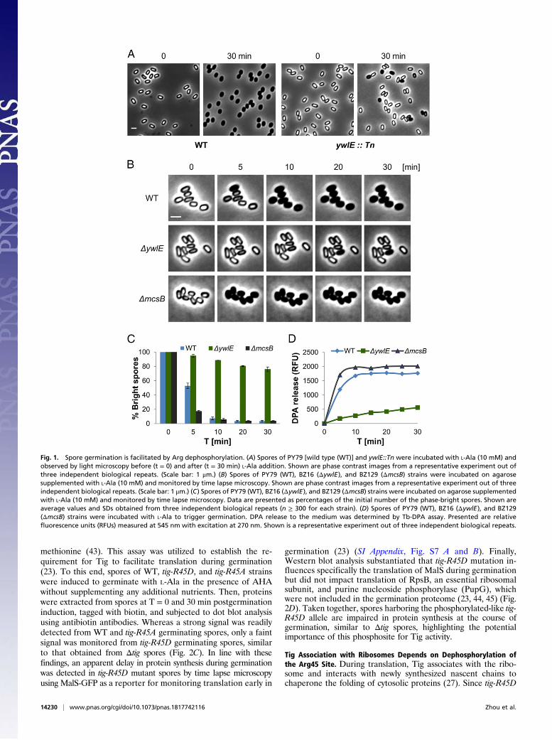
ResultsSpore Germination Is Driven by Arg Dephosphorylation. To identifygenes required for spore germination, we designed a transposon-based genetic screen, searching for mutants that are able to formmature spores but deficient in the conversion from a phase-bright to a phase-dark state following germinant addition (SIAppendix, Fig. S1A). Using this approach, we identified a mutantdisrupted in the ywlE gene, encoding an Arg phosphatase (39),exhibiting severe germination deficiency (Fig. 1A). Constructinga strain, fully deleted of ywlE, revealed that it was able to grow andsporulate normally but produced spores that were perturbed ingermination as indicated by only a slight decrease in optical densityachieved in response to the efficient L-Ala germinant (SI Appendix,Figs. S1 B and C and S2A). Monitoring germination efficiency bytime lapse microscopy at a single spore resolution showed that30 min following L-Ala addition only 20% of the ΔywlE sporescould convert from a phase-bright to a phase-dark state in com-parison with 96% of the wild type (WT) spores (Fig. 1 B and C).Similar results were obtained when germination was induced bysupplementing the spores with AGFK and even with LB (SI Ap-pendix, Fig. S2 B, C, and E). In line with these findings, ΔywlEmutant spores were slower to release DPA and lose their heatresistance relative to the WT spores (Fig. 1D and SI Appendix, Fig.S2D), signifying that YwlE is a key factor in promptingspore germination.The role of YwlE as an Arg phosphatase brings about the
possibility that some crucial factors required for germination arephosphorylated on their Arg sites during sporulation, a modifi-cation that should be removed to allow germination. Notably,only a single kinase, McsB, is known to be responsible for Argphosphorylation in B. subtilis (38, 40). Hence, we hypothesizedthat, in ΔmcsB spores, these factors would be constantly in theirdephosphorylated active form, and consequently, these sporeswould germinate rapidly. Indeed, ΔmcsB mutant spores germi-nated faster than WT spores as evidenced by their remarkablerapid transition into the phase-dark state in all tested conditions(Fig. 1 B and C and SI Appendix, Fig. S2). Furthermore, germi-nating ΔmcsB spores turned heat sensitive and released DPA fasterthan WT spores (Fig. 1D and SI Appendix, Fig. S2D). Examining
germination of ΔmcsB ΔywlE double mutant spores revealedkinetics similar to that of ΔmcsB spores (SI Appendix, Fig. S3).Taken together, we infer that Arg phosphorylation of targetproteins is executed by McsB during sporulation and is removedby YwlE to propel spore germination.
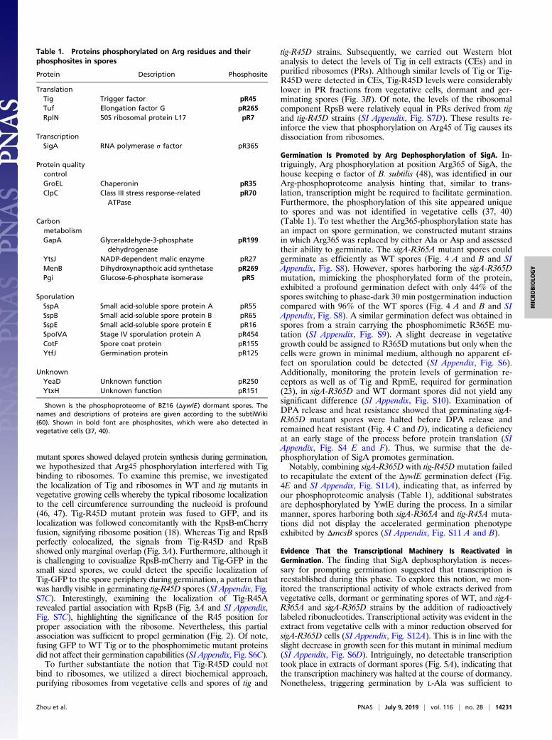
Revealing the Arg Phosphoproteome of Dormant Spores. Havingestablished that Arg dephosphorylation is required for spore ger-mination, we next sought to identify Arg-phosphorylated sites in thedormant spore proteomes that could serve as substrates for YwlEduring spore germination. As Arg phosphorylation was shown to bemore stable and enriched in the ywlE mutant (37, 40), we carriedout Arg-phosphoproteomic analysis of ΔywlE dormant spores. Intotal, we identified 18 Arg-phosphorylation sites located in 18 pro-teins with very high confidence using stringent quality criteria forthe validation of the phosphorylation sites (Table 1 and SI Ap-pendix, Table S1). A comparison with the Arg phosphoproteome ofvegetative cells (37, 40) revealed that Arg-phosphorylation eventsin spores are less frequent and largely differ from those of vege-tative cells with 10 identified sites unique to spores (Table 1 and SIAppendix, Table S1). The detected Arg-phosphorylated proteinsare involved in key biological functions, including carbon metabo-lism, sporulation, translation, and transcription (Table 1). Thus, weuncovered a unique set of protein Arg-phosphorylation sitesexisting in dormant spores that can be potentially dephosphorylatedby YwlE to allow germination. Among the identified phosphory-lated factors, the most intriguing were those involved in transcrip-tion and translation, raising the possibility that they provide themeans for reestablishment of these basic cellular processes.
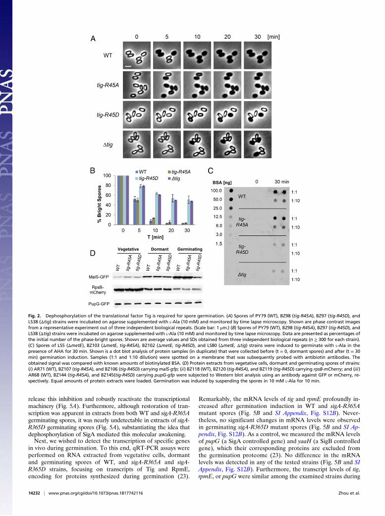
Dephosphorylation of an Arg Site on the Translational Factor TigPowers Spore Germination. Previously, we revealed that proteinsynthesis takes place during germination and relies on the bonafide translational factor Tig (23, 27), identified in our phospho-proteomic analysis to harbor an Arg45-phosphorylated site(Table 1). Interestingly, the occurrence of this phosphorylationevent was monitored in vegetative cells following exposure toheat stress (40). We reasoned that phosphorylation of Tig inspores blocks its activity, whereas dephosphorylation by YwlErestores the factor utility during germination. To investigate theimpact of the Arg45 site on the activity of Tig, we introducedmutations that would either abolish the phosphorylation potentialby replacing Arg with Ala or mimic a constitutive phosphorylationstate by replacing Arg with Asp (42). Each mutation was insertedas a sole copy at the native tig chromosomal locus, replacing theoriginal WT allele. Remarkably, tig-R45Dmutant spores exhibitedgermination defects similar to that of Δtig spores, whereas tig-R45A mutant spores germinated similarly to WT spores as indi-cated by optical density and time lapse microscopy analyses (Fig. 2A and B and SI Appendix, Fig. S4 A–D). Consistently, mimickingconstitutive phosphorylation by replacing the Arg45 site with Glu(R45E), harboring a larger side chain group, led to a germinationdefect similar to that of R45D protein (SI Appendix, Fig. S5).Importantly, both tig-R45A and tig-R45D mutations had no effecton vegetative growth or sporulation (SI Appendix, Fig. S6 A andB). Further analysis of the tig mutant spores showed that, upongermination induction, tig-R45D spores were capable of releasingDPA and accordingly lost their heat resistance (SI Appendix, Fig.S4 E and F). Hence, similar to Δtig spores, tig-R45D spores ini-tiated germination normally but were subsequently stalled in theirphase-bright state.To corroborate that the Tig Arg-phosphorylation state influ-
ences the function of Tig, we examined its impact on proteinsynthesis during germination. We employed the BioOrthogonalNon-Canonical Amino acid Tagging (BONCAT) protein taggingtechnique that enables specific labeling of newly synthesizedproteins due to the incorporation of an artificial amino acidtermed azidohomoalanine (AHA), which is a substitute for
Zhou et al. PNAS | July 9, 2019 | vol. 116 | no. 28 | 14229
MICRO
BIOLO
GY
methionine (43). This assay was utilized to establish the re-quirement for Tig to facilitate translation during germination(23). To this end, spores of WT, tig-R45D, and tig-R45A strainswere induced to germinate with L-Ala in the presence of AHAwithout supplementing any additional nutrients. Then, proteinswere extracted from spores at T = 0 and 30 min postgerminationinduction, tagged with biotin, and subjected to dot blot analysisusing antibiotin antibodies. Whereas a strong signal was readilydetected from WT and tig-R45A germinating spores, only a faintsignal was monitored from tig-R45D germinating spores, similarto that obtained from Δtig spores (Fig. 2C). In line with thesefindings, an apparent delay in protein synthesis during germinationwas detected in tig-R45D mutant spores by time lapse microscopyusing MalS-GFP as a reporter for monitoring translation early in
germination (23) (SI Appendix, Fig. S7 A and B). Finally,Western blot analysis substantiated that tig-R45D mutation in-fluences specifically the translation of MalS during germinationbut did not impact translation of RpsB, an essential ribosomalsubunit, and purine nucleoside phosphorylase (PupG), whichwere not included in the germination proteome (23, 44, 45) (Fig.2D). Taken together, spores harboring the phosphorylated-like tig-R45D allele are impaired in protein synthesis at the course ofgermination, similar to Δtig spores, highlighting the potentialimportance of this phosphosite for Tig activity.
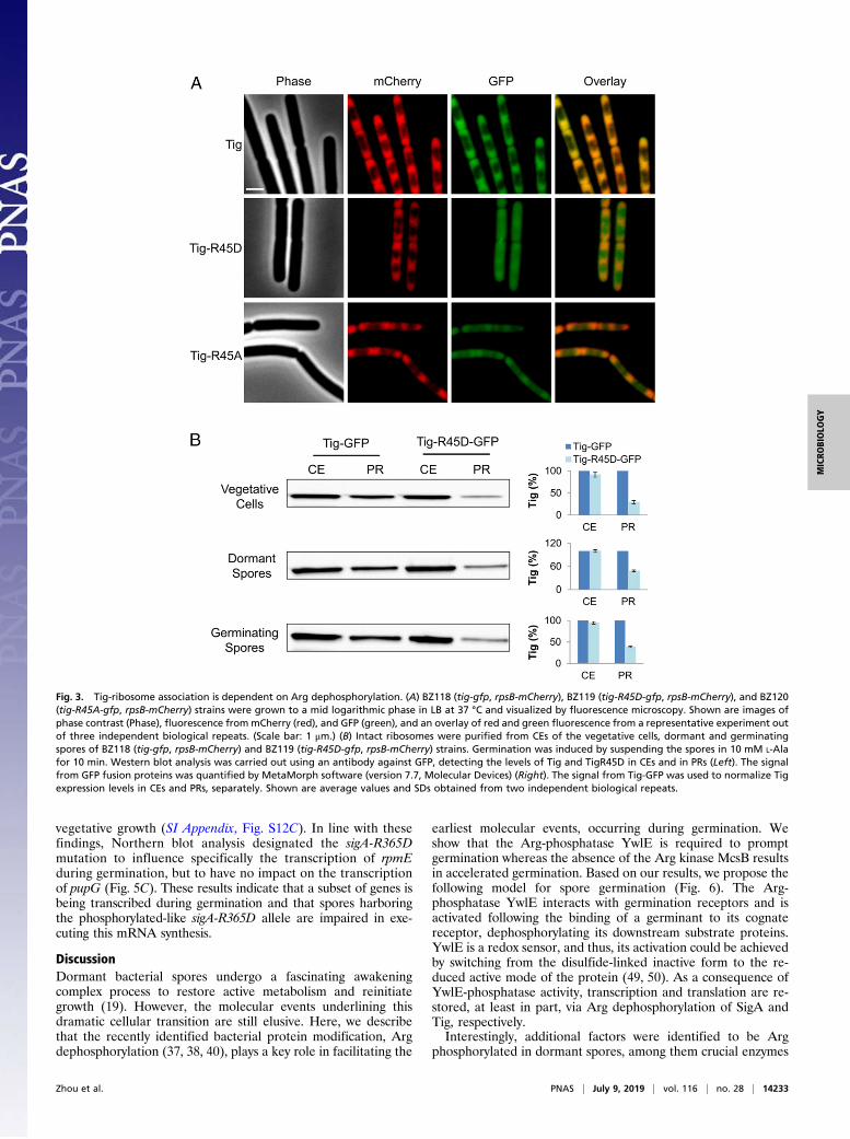
Tig Association with Ribosomes Depends on Dephosphorylation ofthe Arg45 Site. During translation, Tig associates with the ribo-some and interacts with newly synthesized nascent chains tochaperone the folding of cytosolic proteins (27). Since tig-R45D
Fig. 1. Spore germination is facilitated by Arg dephosphorylation. (A) Spores of PY79 [wild type (WT)] and ywlE::Tn were incubated with L-Ala (10 mM) andobserved by light microscopy before (t = 0) and after (t = 30 min) L-Ala addition. Shown are phase contrast images from a representative experiment out ofthree independent biological repeats. (Scale bar: 1 μm.) (B) Spores of PY79 (WT), BZ16 (ΔywlE), and BZ129 (ΔmcsB) strains were incubated on agarosesupplemented with L-Ala (10 mM) and monitored by time lapse microscopy. Shown are phase contrast images from a representative experiment out of threeindependent biological repeats. (Scale bar: 1 μm.) (C) Spores of PY79 (WT), BZ16 (ΔywlE), and BZ129 (ΔmcsB) strains were incubated on agarose supplementedwith L-Ala (10 mM) and monitored by time lapse microscopy. Data are presented as percentages of the initial number of the phase-bright spores. Shown areaverage values and SDs obtained from three independent biological repeats (n ≥ 300 for each strain). (D) Spores of PY79 (WT), BZ16 (ΔywlE), and BZ129(ΔmcsB) strains were incubated with L-Ala to trigger germination. DPA release to the medium was determined by Tb-DPA assay. Presented are relativefluorescence units (RFUs) measured at 545 nm with excitation at 270 nm. Shown is a representative experiment out of three independent biological repeats.
14230 | www.pnas.org/cgi/doi/10.1073/pnas.1817742116 Zhou et al.
mutant spores showed delayed protein synthesis during germination,we hypothesized that Arg45 phosphorylation interfered with Tigbinding to ribosomes. To examine this premise, we investigatedthe localization of Tig and ribosomes in WT and tig mutants invegetative growing cells whereby the typical ribosome localizationto the cell circumference surrounding the nucleoid is profound(46, 47). Tig-R45D mutant protein was fused to GFP, and itslocalization was followed concomitantly with the RpsB-mCherryfusion, signifying ribosome position (18). Whereas Tig and RpsBperfectly colocalized, the signals from Tig-R45D and RpsBshowed only marginal overlap (Fig. 3A). Furthermore, although itis challenging to covisualize RpsB-mCherry and Tig-GFP in thesmall sized spores, we could detect the specific localization ofTig-GFP to the spore periphery during germination, a pattern thatwas hardly visible in germinating tig-R45D spores (SI Appendix, Fig.S7C). Interestingly, examining the localization of Tig-R45Arevealed partial association with RpsB (Fig. 3A and SI Appendix,Fig. S7C), highlighting the significance of the R45 position forproper association with the ribosome. Nevertheless, this partialassociation was sufficient to propel germination (Fig. 2). Of note,fusing GFP to WT Tig or to the phosphomimetic mutant proteinsdid not affect their germination capabilities (SI Appendix, Fig. S6C).To further substantiate the notion that Tig-R45D could not
bind to ribosomes, we utilized a direct biochemical approach,purifying ribosomes from vegetative cells and spores of tig and
tig-R45D strains. Subsequently, we carried out Western blotanalysis to detect the levels of Tig in cell extracts (CEs) and inpurified ribosomes (PRs). Although similar levels of Tig or Tig-R45D were detected in CEs, Tig-R45D levels were considerablylower in PR fractions from vegetative cells, dormant and ger-minating spores (Fig. 3B). Of note, the levels of the ribosomalcomponent RpsB were relatively equal in PRs derived from tigand tig-R45D strains (SI Appendix, Fig. S7D). These results re-inforce the view that phosphorylation on Arg45 of Tig causes itsdissociation from ribosomes.
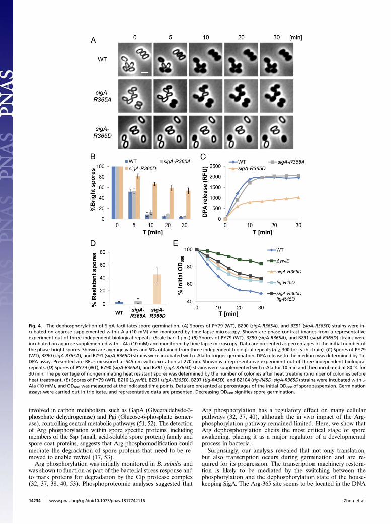
Germination Is Promoted by Arg Dephosphorylation of SigA. In-triguingly, Arg phosphorylation at position Arg365 of SigA, thehouse keeping σ factor of B. subtilis (48), was identified in ourArg-phosphoproteome analysis hinting that, similar to trans-lation, transcription might be required to facilitate germination.Furthermore, the phosphorylation of this site appeared uniqueto spores and was not identified in vegetative cells (37, 40)(Table 1). To test whether the Arg365-phosphorylation state hasan impact on spore germination, we constructed mutant strainsin which Arg365 was replaced by either Ala or Asp and assessedtheir ability to germinate. The sigA-R365A mutant spores couldgerminate as efficiently as WT spores (Fig. 4 A and B and SIAppendix, Fig. S8). However, spores harboring the sigA-R365Dmutation, mimicking the phosphorylated form of the protein,exhibited a profound germination defect with only 44% of thespores switching to phase-dark 30 min postgermination inductioncompared with 96% of the WT spores (Fig. 4 A and B and SIAppendix, Fig. S8). A similar germination defect was obtained inspores from a strain carrying the phosphomimetic R365E mu-tation (SI Appendix, Fig. S9). A slight decrease in vegetativegrowth could be assigned to R365D mutations but only when thecells were grown in minimal medium, although no apparent ef-fect on sporulation could be detected (SI Appendix, Fig. S6).Additionally, monitoring the protein levels of germination re-ceptors as well as of Tig and RpmE, required for germination(23), in sigA-R365D and WT dormant spores did not yield anysignificant difference (SI Appendix, Fig. S10). Examination ofDPA release and heat resistance showed that germinating sigA-R365D mutant spores were halted before DPA release andremained heat resistant (Fig. 4 C and D), indicating a deficiencyat an early stage of the process before protein translation (SIAppendix, Fig. S4 E and F). Thus, we surmise that the de-phosphorylation of SigA promotes germination.Notably, combining sigA-R365D with tig-R45D mutation failed
to recapitulate the extent of the ΔywlE germination defect (Fig.4E and SI Appendix, Fig. S11A), indicating that, as inferred byour phosphoproteomic analysis (Table 1), additional substratesare dephosphorylated by YwlE during the process. In a similarmanner, spores harboring both sigA-R365A and tig-R45A muta-tions did not display the accelerated germination phenotypeexhibited by ΔmcsB spores (SI Appendix, Fig. S11 A and B).
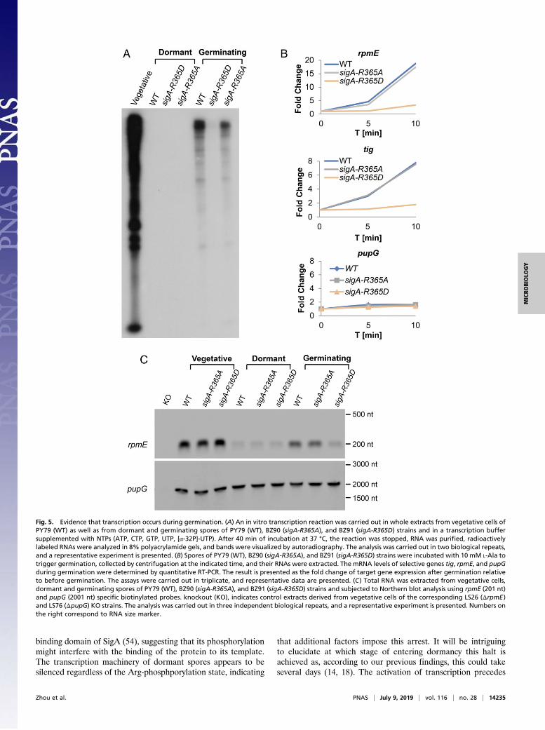
Evidence That the Transcriptional Machinery Is Reactivated inGermination. The finding that SigA dephosphorylation is neces-sary for prompting germination suggested that transcription isreestablished during this phase. To explore this notion, we mon-itored the transcriptional activity of whole extracts derived fromvegetative cells, dormant or germinating spores of WT, and sigA-R365A and sigA-R365D strains by the addition of radioactivelylabeled ribonucleotides. Transcriptional activity was evident in theextract from vegetative cells with a minor reduction observed forsigA-R365D cells (SI Appendix, Fig. S12A). This is in line with theslight decrease in growth seen for this mutant in minimal medium(SI Appendix, Fig. S6D). Intriguingly, no detectable transcriptiontook place in extracts of dormant spores (Fig. 5A), indicating thatthe transcription machinery was halted at the course of dormancy.Nonetheless, triggering germination by L-Ala was sufficient to
Table 1. Proteins phosphorylated on Arg residues and theirphosphosites in spores
Protein Description Phosphosite
TranslationTig Trigger factor pR45Tuf Elongation factor G pR265RplN 50S ribosomal protein L17 pR7
TranscriptionSigA RNA polymerase σ factor pR365
Protein qualitycontrolGroEL Chaperonin pR35ClpC Class III stress response-related
ATPasepR70
CarbonmetabolismGapA Glyceraldehyde-3-phosphate
dehydrogenasepR199
YtsJ NADP-dependent malic enzyme pR27MenB Dihydroxynapthoic acid synthetase pR269Pgi Glucose-6-phosphate isomerase pR5
SporulationSspA Small acid-soluble spore protein A pR55SspB Small acid-soluble spore protein B pR65SspE Small acid-soluble spore protein E pR16SpoIVA Stage IV sporulation protein A pR454CotF Spore coat protein pR155YtfJ Germination protein pR125
UnknownYeaD Unknown function pR250YtxH Unknown function pR151
Shown is the phosphoproteome of BZ16 (ΔywlE) dormant spores. Thenames and descriptions of proteins are given according to the subtiWiki(60). Shown in bold font are phosphosites, which were also detected invegetative cells (37, 40).
Zhou et al. PNAS | July 9, 2019 | vol. 116 | no. 28 | 14231
MICRO
BIOLO
GY
release this inhibition and robustly reactivate the transcriptionalmachinery (Fig. 5A). Furthermore, although restoration of tran-scription was apparent in extracts from both WT and sigA-R365Agerminating spores, it was nearly undetectable in extracts of sigA-R365D germinating spores (Fig. 5A), substantiating the idea thatdephosphorylation of SigA mediated this molecular awakening.Next, we wished to detect the transcription of specific genes
in vivo during germination. To this end, qRT-PCR assays wereperformed on RNA extracted from vegetative cells, dormantand germinating spores of WT, and sigA-R365A and sigA-R365D strains, focusing on transcripts of Tig and RpmE,encoding for proteins synthesized during germination (23).
Remarkably, the mRNA levels of tig and rpmE profoundly in-creased after germination induction in WT and sigA-R365Amutant spores (Fig. 5B and SI Appendix, Fig. S12B). Never-theless, no significant changes in mRNA levels were observedin germinating sigA-R365D mutant spores (Fig. 5B and SI Ap-pendix, Fig. S12B). As a control, we measured the mRNA levelsof pupG (a SigA controlled gene) and yaaH (a SigB controlledgene), which their corresponding proteins are excluded fromthe germination proteome (23). No difference in the mRNAlevels was detected in any of the tested strains (Fig. 5B and SIAppendix, Fig. S12B). Furthermore, the transcript levels of tig,rpmE, or pupG were similar among the examined strains during
Fig. 2. Dephosphorylation of the translational factor Tig is required for spore germination. (A) Spores of PY79 (WT), BZ98 (tig-R45A), BZ97 (tig-R45D), andLS38 (Δtig) strains were incubated on agarose supplemented with L-Ala (10 mM) and monitored by time lapse microscopy. Shown are phase contrast imagesfrom a representative experiment out of three independent biological repeats. (Scale bar: 1 μm.) (B) Spores of PY79 (WT), BZ98 (tig-R45A), BZ97 (tig-R45D), andLS38 (Δtig) strains were incubated on agarose supplemented with L-Ala (10 mM) and monitored by time lapse microscopy. Data are presented as percentages ofthe initial number of the phase-bright spores. Shown are average values and SDs obtained from three independent biological repeats (n ≥ 300 for each strain).(C) Spores of LS5 (ΔmetE), BZ103 (ΔmetE, tig-R45A), BZ102 (ΔmetE, tig-R45D), and LS80 (ΔmetE, Δtig) strains were induced to germinate with L-Ala in thepresence of AHA for 30 min. Shown is a dot blot analysis of protein samples (in duplicate) that were collected before (t = 0, dormant spores) and after (t = 30min) germination induction. Samples (1:1 and 1:10 dilution) were spotted on a membrane that was subsequently probed with antibiotin antibodies. Theobtained signal was compared with known amounts of biotinylated BSA. (D) Protein extracts from vegetative cells, dormant and germinating spores of strains:(i) AR71 (WT), BZ107 (tig-R45A), and BZ106 (tig-R45D) carryingmalS-gfp; (ii) BZ118 (WT), BZ120 (tig-R45A), and BZ119 (tig-R45D) carrying rpsB-mCherry; and (iii)AR68 (WT), BZ144 (tig-R45A), and BZ145(tig-R45D) carrying pupG-gfp were subjected to Western blot analysis using an antibody against GFP or mCherry, re-spectively. Equal amounts of protein extracts were loaded. Germination was induced by suspending the spores in 10 mM L-Ala for 10 min.
14232 | www.pnas.org/cgi/doi/10.1073/pnas.1817742116 Zhou et al.
vegetative growth (SI Appendix, Fig. S12C). In line with thesefindings, Northern blot analysis designated the sigA-R365Dmutation to influence specifically the transcription of rpmEduring germination, but to have no impact on the transcriptionof pupG (Fig. 5C). These results indicate that a subset of genes isbeing transcribed during germination and that spores harboringthe phosphorylated-like sigA-R365D allele are impaired in exe-cuting this mRNA synthesis.
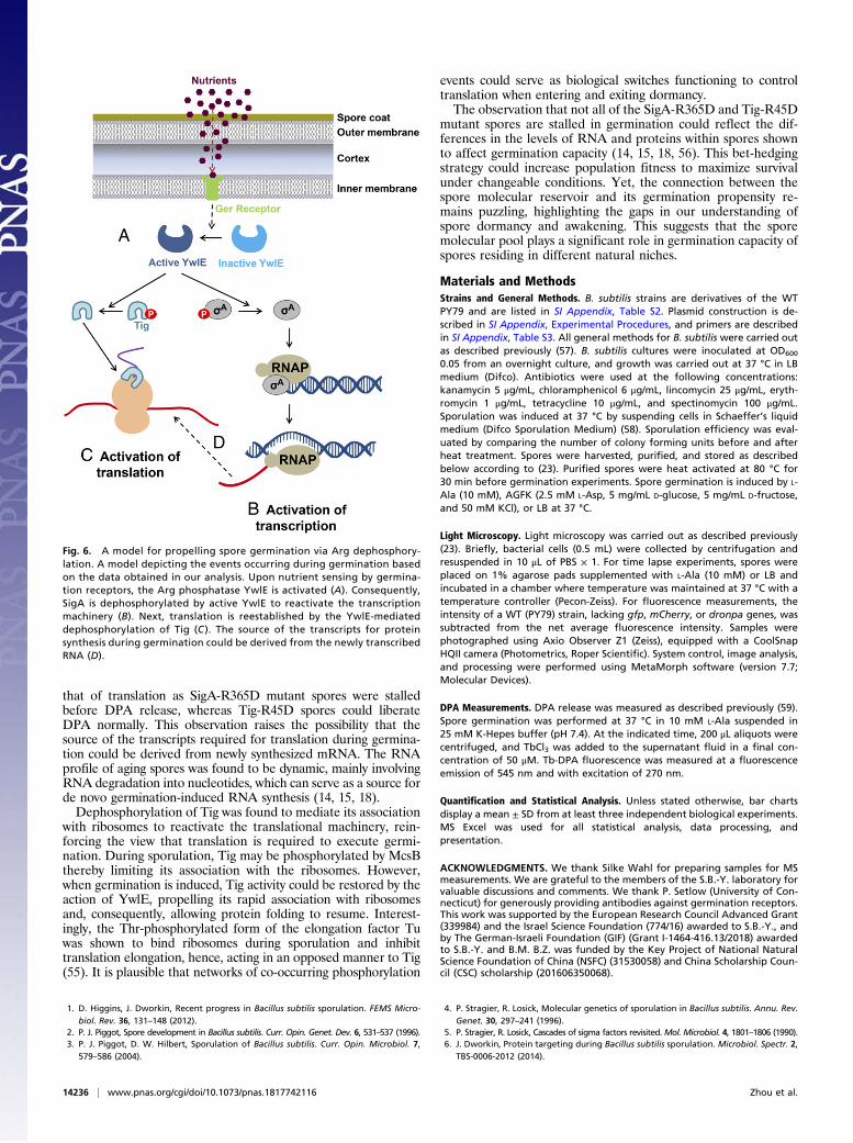
DiscussionDormant bacterial spores undergo a fascinating awakeningcomplex process to restore active metabolism and reinitiategrowth (19). However, the molecular events underlining thisdramatic cellular transition are still elusive. Here, we describethat the recently identified bacterial protein modification, Argdephosphorylation (37, 38, 40), plays a key role in facilitating the
earliest molecular events, occurring during germination. Weshow that the Arg-phosphatase YwlE is required to promptgermination whereas the absence of the Arg kinase McsB resultsin accelerated germination. Based on our results, we propose thefollowing model for spore germination (Fig. 6). The Arg-phosphatase YwlE interacts with germination receptors and isactivated following the binding of a germinant to its cognatereceptor, dephosphorylating its downstream substrate proteins.YwlE is a redox sensor, and thus, its activation could be achievedby switching from the disulfide-linked inactive form to the re-duced active mode of the protein (49, 50). As a consequence ofYwlE-phosphatase activity, transcription and translation are re-stored, at least in part, via Arg dephosphorylation of SigA andTig, respectively.Interestingly, additional factors were identified to be Arg
phosphorylated in dormant spores, among them crucial enzymes
Fig. 3. Tig-ribosome association is dependent on Arg dephosphorylation. (A) BZ118 (tig-gfp, rpsB-mCherry), BZ119 (tig-R45D-gfp, rpsB-mCherry), and BZ120(tig-R45A-gfp, rpsB-mCherry) strains were grown to a mid logarithmic phase in LB at 37 °C and visualized by fluorescence microscopy. Shown are images ofphase contrast (Phase), fluorescence frommCherry (red), and GFP (green), and an overlay of red and green fluorescence from a representative experiment outof three independent biological repeats. (Scale bar: 1 μm.) (B) Intact ribosomes were purified from CEs of the vegetative cells, dormant and germinatingspores of BZ118 (tig-gfp, rpsB-mCherry) and BZ119 (tig-R45D-gfp, rpsB-mCherry) strains. Germination was induced by suspending the spores in 10 mM L-Alafor 10 min. Western blot analysis was carried out using an antibody against GFP, detecting the levels of Tig and TigR45D in CEs and in PRs (Left). The signalfrom GFP fusion proteins was quantified by MetaMorph software (version 7.7, Molecular Devices) (Right). The signal from Tig-GFP was used to normalize Tigexpression levels in CEs and PRs, separately. Shown are average values and SDs obtained from two independent biological repeats.
Zhou et al. PNAS | July 9, 2019 | vol. 116 | no. 28 | 14233
MICRO
BIOLO
GY
involved in carbon metabolism, such as GapA (Glyceraldehyde-3-phosphate dehydrogenase) and Pgi (Glucose-6-phosphate isomer-ase), controlling central metabolic pathways (51, 52). The detectionof Arg phosphorylation within spore specific proteins, includingmembers of the Ssp (small, acid-soluble spore protein) family andspore coat proteins, suggests that Arg phosphomodification couldmediate the degradation of spore proteins that need to be re-moved to enable revival (17, 53).Arg phosphorylation was initially monitored in B. subtilis and
was shown to function as part of the bacterial stress response andto mark proteins for degradation by the Clp protease complex(32, 37, 38, 40, 53). Phosphoproteomic analyses suggested that
Arg phosphorylation has a regulatory effect on many cellularpathways (32, 37, 40), although the in vivo impact of the Arg-phosphorylation pathway remained limited. Here, we show thatArg dephosphorylation elicits the most critical stage of sporeawakening, placing it as a major regulator of a developmentalprocess in bacteria.Surprisingly, our analysis revealed that not only translation,
but also transcription occurs during germination and are re-quired for its progression. The transcription machinery restora-tion is likely to be mediated by the switching between thephosphorylation and the dephosphorylation state of the house-keeping SigA. The Arg-365 site seems to be located in the DNA
Fig. 4. The dephosphorylation of SigA facilitates spore germination. (A) Spores of PY79 (WT), BZ90 (sigA-R365A), and BZ91 (sigA-R365D) strains were in-cubated on agarose supplemented with L-Ala (10 mM) and monitored by time lapse microscopy. Shown are phase contrast images from a representativeexperiment out of three independent biological repeats. (Scale bar: 1 μm.) (B) Spores of PY79 (WT), BZ90 (sigA-R365A), and BZ91 (sigA-R365D) strains wereincubated on agarose supplemented with L-Ala (10 mM) and monitored by time lapse microscopy. Data are presented as percentages of the initial number ofthe phase-bright spores. Shown are average values and SDs obtained from three independent biological repeats (n ≥ 300 for each strain). (C) Spores of PY79(WT), BZ90 (sigA-R365A), and BZ91 (sigA-R365D) strains were incubated with L-Ala to trigger germination. DPA release to the medium was determined by Tb-DPA assay. Presented are RFUs measured at 545 nm with excitation at 270 nm. Shown is a representative experiment out of three independent biologicalrepeats. (D) Spores of PY79 (WT), BZ90 (sigA-R365A), and BZ91 (sigA-R365D) strains were supplemented with L-Ala for 10 min and then incubated at 80 °C for30 min. The percentage of nongerminating heat resistant spores was determined by the number of colonies after heat treatment/number of colonies beforeheat treatment. (E) Spores of PY79 (WT), BZ16 (ΔywlE), BZ91 (sigA-R365D), BZ97 (tig-R45D), and BZ104 (tig-R45D, sigA-R365D) strains were incubated with L-Ala (10 mM), and OD600 was measured at the indicated time points. Data are presented as percentages of the initial OD600 of spore suspension. Germinationassays were carried out in triplicate, and representative data are presented. Decreasing OD600 signifies spore germination.
14234 | www.pnas.org/cgi/doi/10.1073/pnas.1817742116 Zhou et al.
binding domain of SigA (54), suggesting that its phosphorylationmight interfere with the binding of the protein to its template.The transcription machinery of dormant spores appears to besilenced regardless of the Arg-phosphporylation state, indicating
that additional factors impose this arrest. It will be intriguingto elucidate at which stage of entering dormancy this halt isachieved as, according to our previous findings, this could takeseveral days (14, 18). The activation of transcription precedes
Fig. 5. Evidence that transcription occurs during germination. (A) An in vitro transcription reaction was carried out in whole extracts from vegetative cells ofPY79 (WT) as well as from dormant and germinating spores of PY79 (WT), BZ90 (sigA-R365A), and BZ91 (sigA-R365D) strains and in a transcription buffersupplemented with NTPs (ATP, CTP, GTP, UTP, [α-32P]-UTP). After 40 min of incubation at 37 °C, the reaction was stopped, RNA was purified, radioactivelylabeled RNAs were analyzed in 8% polyacrylamide gels, and bands were visualized by autoradiography. The analysis was carried out in two biological repeats,and a representative experiment is presented. (B) Spores of PY79 (WT), BZ90 (sigA-R365A), and BZ91 (sigA-R365D) strains were incubated with 10 mM L-Ala totrigger germination, collected by centrifugation at the indicated time, and their RNAs were extracted. The mRNA levels of selective genes tig, rpmE, and pupGduring germination were determined by quantitative RT-PCR. The result is presented as the fold change of target gene expression after germination relativeto before germination. The assays were carried out in triplicate, and representative data are presented. (C) Total RNA was extracted from vegetative cells,dormant and germinating spores of PY79 (WT), BZ90 (sigA-R365A), and BZ91 (sigA-R365D) strains and subjected to Northern blot analysis using rpmE (201 nt)and pupG (2001 nt) specific biotinylated probes. knockout (KO), indicates control extracts derived from vegetative cells of the corresponding LS26 (ΔrpmE)and LS76 (ΔpupG) KO strains. The analysis was carried out in three independent biological repeats, and a representative experiment is presented. Numbers onthe right correspond to RNA size marker.
Zhou et al. PNAS | July 9, 2019 | vol. 116 | no. 28 | 14235
MICRO
BIOLO
GY
that of translation as SigA-R365D mutant spores were stalledbefore DPA release, whereas Tig-R45D spores could liberateDPA normally. This observation raises the possibility that thesource of the transcripts required for translation during germina-tion could be derived from newly synthesized mRNA. The RNAprofile of aging spores was found to be dynamic, mainly involvingRNA degradation into nucleotides, which can serve as a source forde novo germination-induced RNA synthesis (14, 15, 18).Dephosphorylation of Tig was found to mediate its association
with ribosomes to reactivate the translational machinery, rein-forcing the view that translation is required to execute germi-nation. During sporulation, Tig may be phosphorylated by McsBthereby limiting its association with the ribosomes. However,when germination is induced, Tig activity could be restored by theaction of YwlE, propelling its rapid association with ribosomesand, consequently, allowing protein folding to resume. Interest-ingly, the Thr-phosphorylated form of the elongation factor Tuwas shown to bind ribosomes during sporulation and inhibittranslation elongation, hence, acting in an opposed manner to Tig(55). It is plausible that networks of co-occurring phosphorylation
events could serve as biological switches functioning to controltranslation when entering and exiting dormancy.The observation that not all of the SigA-R365D and Tig-R45D
mutant spores are stalled in germination could reflect the dif-ferences in the levels of RNA and proteins within spores shownto affect germination capacity (14, 15, 18, 56). This bet-hedgingstrategy could increase population fitness to maximize survivalunder changeable conditions. Yet, the connection between thespore molecular reservoir and its germination propensity re-mains puzzling, highlighting the gaps in our understanding ofspore dormancy and awakening. This suggests that the sporemolecular pool plays a significant role in germination capacity ofspores residing in different natural niches.
Materials and MethodsStrains and General Methods. B. subtilis strains are derivatives of the WTPY79 and are listed in SI Appendix, Table S2. Plasmid construction is de-scribed in SI Appendix, Experimental Procedures, and primers are describedin SI Appendix, Table S3. All general methods for B. subtilis were carried outas described previously (57). B. subtilis cultures were inoculated at OD600
0.05 from an overnight culture, and growth was carried out at 37 °C in LBmedium (Difco). Antibiotics were used at the following concentrations:kanamycin 5 μg/mL, chloramphenicol 6 μg/mL, lincomycin 25 μg/mL, eryth-romycin 1 μg/mL, tetracycline 10 μg/mL, and spectinomycin 100 μg/mL.Sporulation was induced at 37 °C by suspending cells in Schaeffer’s liquidmedium (Difco Sporulation Medium) (58). Sporulation efficiency was eval-uated by comparing the number of colony forming units before and afterheat treatment. Spores were harvested, purified, and stored as describedbelow according to (23). Purified spores were heat activated at 80 °C for30 min before germination experiments. Spore germination is induced by L-Ala (10 mM), AGFK (2.5 mM L-Asp, 5 mg/mL D-glucose, 5 mg/mL D-fructose,and 50 mM KCl), or LB at 37 °C.
Light Microscopy. Light microscopy was carried out as described previously(23). Briefly, bacterial cells (0.5 mL) were collected by centrifugation andresuspended in 10 μL of PBS × 1. For time lapse experiments, spores wereplaced on 1% agarose pads supplemented with L-Ala (10 mM) or LB andincubated in a chamber where temperature was maintained at 37 °C with atemperature controller (Pecon-Zeiss). For fluorescence measurements, theintensity of a WT (PY79) strain, lacking gfp, mCherry, or dronpa genes, wassubtracted from the net average fluorescence intensity. Samples werephotographed using Axio Observer Z1 (Zeiss), equipped with a CoolSnapHQII camera (Photometrics, Roper Scientific). System control, image analysis,and processing were performed using MetaMorph software (version 7.7;Molecular Devices).
DPA Measurements. DPA release was measured as described previously (59).Spore germination was performed at 37 °C in 10 mM L-Ala suspended in25 mM K-Hepes buffer (pH 7.4). At the indicated time, 200 μL aliquots werecentrifuged, and TbCl3 was added to the supernatant fluid in a final con-centration of 50 μM. Tb-DPA fluorescence was measured at a fluorescenceemission of 545 nm and with excitation of 270 nm.
Quantification and Statistical Analysis. Unless stated otherwise, bar chartsdisplay a mean ± SD from at least three independent biological experiments.MS Excel was used for all statistical analysis, data processing, andpresentation.
ACKNOWLEDGMENTS. We thank Silke Wahl for preparing samples for MSmeasurements. We are grateful to the members of the S.B.-Y. laboratory forvaluable discussions and comments. We thank P. Setlow (University of Con-necticut) for generously providing antibodies against germination receptors.This work was supported by the European Research Council Advanced Grant(339984) and the Israel Science Foundation (774/16) awarded to S.B.-Y., andby The German-Israeli Foundation (GIF) (Grant I-1464-416.13/2018) awardedto S.B.-Y. and B.M. B.Z. was funded by the Key Project of National NaturalScience Foundation of China (NSFC) (31530058) and China Scholarship Coun-cil (CSC) scholarship (201606350068).
1. D. Higgins, J. Dworkin, Recent progress in Bacillus subtilis sporulation. FEMS Micro-biol. Rev. 36, 131–148 (2012).
2. P. J. Piggot, Spore development in Bacillus subtilis. Curr. Opin. Genet. Dev. 6, 531–537 (1996).3. P. J. Piggot, D. W. Hilbert, Sporulation of Bacillus subtilis. Curr. Opin. Microbiol. 7,
579–586 (2004).
4. P. Stragier, R. Losick, Molecular genetics of sporulation in Bacillus subtilis. Annu. Rev.Genet. 30, 297–241 (1996).
5. P. Stragier, R. Losick, Cascades of sigma factors revisited.Mol. Microbiol. 4, 1801–1806 (1990).6. J. Dworkin, Protein targeting during Bacillus subtilis sporulation. Microbiol. Spectr. 2,
TBS-0006-2012 (2014).
Fig. 6. A model for propelling spore germination via Arg dephosphory-lation. A model depicting the events occurring during germination basedon the data obtained in our analysis. Upon nutrient sensing by germina-tion receptors, the Arg phosphatase YwlE is activated (A). Consequently,SigA is dephosphorylated by active YwlE to reactivate the transcriptionmachinery (B). Next, translation is reestablished by the YwlE-mediateddephosphorylation of Tig (C ). The source of the transcripts for proteinsynthesis during germination could be derived from the newly transcribedRNA (D).
14236 | www.pnas.org/cgi/doi/10.1073/pnas.1817742116 Zhou et al.
7. A. Driks, Bacillus subtilis spore coat. Microbiol. Mol. Biol. Rev. 63, 1–20 (1999).8. D. L. Popham, Specialized peptidoglycan of the bacterial endospore: The inner wall of
the lockbox. Cell. Mol. Life Sci. 59, 426–433 (2002).9. A. O. Henriques, C. P. Moran, Jr, Structure, assembly, and function of the spore surface
layers. Annu. Rev. Microbiol. 61, 555–588 (2007).10. P. T. McKenney, A. Driks, P. Eichenberger, The Bacillus subtilis endospore: Assembly
and functions of the multilayered coat. Nat. Rev. Microbiol. 11, 33–44 (2013).11. D. L. Popham, C. B. Bernhards, Spore peptidoglycan. Microbiol. Spectr. 3, TBS-0005-
2012 (2015).12. A. Driks, P. Eichenberger, The spore coat. Microbiol. Spectr. 4, TBS-0023-2016 (2016).13. P. Setlow, Spores of Bacillus subtilis: Their resistance to and killing by radiation, heat
and chemicals. J. Appl. Microbiol. 101, 514–525 (2006).14. E. Segev, Y. Smith, S. Ben-Yehuda, RNA dynamics in aging bacterial spores. Cell 148,
139–149 (2012).15. S. Ghosh, G. Korza, M. Maciejewski, P. Setlow, Analysis of metabolism in dormant
spores of Bacillus species by 31P nuclear magnetic resonance analysis of low-molecular-weight compounds. J. Bacteriol. 197, 992–1001 (2015).
16. G. Korza, B. Setlow, L. Rao, Q. Li, P. Setlow, Changes in Bacillus spore small molecules,rRNA, germination, and outgrowth after extended sublethal exposure to varioustemperatures: Evidence that protein synthesis is not essential for spore germination.J. Bacteriol. 198, 3254–3264 (2016).
17. P. Setlow, Spore germination. Curr. Opin. Microbiol. 6, 550–556 (2003).18. E. Segev, A. Rosenberg, G. Mamou, L. Sinai, S. Ben-Yehuda, Molecular kinetics of
reviving bacterial spores. J. Bacteriol. 195, 1875–1882 (2013).19. P. Setlow, Germination of spores of Bacillus species: What we know and do not know.
J. Bacteriol. 196, 1297–1305 (2014).20. A. Moir, G. Cooper, Spore germination. Microbiol. Spectr. 3, TBS-0014-2012 (2015).21. A. Moir, How do spores germinate? J. Appl. Microbiol. 101, 526–530 (2006).22. P. Setlow, S. Wang, Y. Q. Li, Germination of spores of the orders Bacillales and
Clostridiales. Annu. Rev. Microbiol. 71, 459–477 (2017).23. L. Sinai, A. Rosenberg, Y. Smith, E. Segev, S. Ben-Yehuda, The molecular timeline of a
reviving bacterial spore. Mol. Cell 57, 695–707 (2015).24. L. Sinai, S. Ben-Yehuda, Commentary: Changes in Bacillus spore small molecules,
rRNA, germination, and outgrowth after extended sublethal exposure to varioustemperatures: Evidence that protein synthesis is not essential for spore germination.Front. Microbiol. 7, 2043 (2016).
25. W. Steinberg, H. O. Halvorson, A. Keynan, E. Weinberg, Timing of protein synthesisduring germination and outgrowth of spores of Bacillus cereus strain T. Nature 208,710 (1965).
26. V. Vinter, Symposium on bacterial spores: V. Germination and outgrowth: Effect ofinhibitors. J. Appl. Bacteriol. 33, 50–59 (1970).
27. G. Kramer et al., L23 protein functions as a chaperone docking site on the ribosome.Nature 419, 171–174 (2002).
28. M. A. Lauber, W. E. Running, J. P. Reilly, B. subtilis ribosomal proteins: Structuralhomology and post-translational modifications. J. Proteome Res. 8, 4193–4206 (2009).
29. S. F. Göthel, C. Scholz, F. X. Schmid, M. A. Marahiel, Cyclophilin and trigger factorfrom Bacillus subtilis catalyze in vitro protein folding and are necessary for viabilityunder starvation conditions. Biochemistry 37, 13392–13399 (1998).
30. D. Paredes-Sabja, P. Setlow, M. R. Sarker, Germination of spores of Bacillales andClostridiales species: Mechanisms and proteins involved. Trends Microbiol. 19, 85–94(2011).
31. I. M. Shah, M. H. Laaberki, D. L. Popham, J. Dworkin, A eukaryotic-like Ser/Thr kinasesignals bacteria to exit dormancy in response to peptidoglycan fragments. Cell 135,486–496 (2008).
32. I. Mijakovic, C. Grangeasse, K. Turgay, Exploring the diversity of protein modifica-tions: Special bacterial phosphorylation systems. FEMS Microbiol. Rev. 40, 398–417(2016).
33. J. Dworkin, Ser/Thr phosphorylation as a regulatory mechanism in bacteria. Curr.Opin. Microbiol. 24, 47–52 (2015).
34. J. Dworkin, I. M. Shah, Exit from dormancy in microbial organisms. Nat. Rev. Micro-biol. 8, 890–896 (2010).
35. A. Rosenberg et al., Phosphoproteome dynamics mediate revival of bacterial spores.BMC Biol. 13, 76 (2015).
36. K. Bäsell et al., The phosphoproteome and its physiological dynamics in Staphylo-coccus aureus. Int. J. Med. Microbiol. 304, 121–132 (2014).
37. A. K. Elsholz et al., Global impact of protein arginine phosphorylation on the physi-ology of Bacillus subtilis. Proc. Natl. Acad. Sci. U.S.A. 109, 7451–7456 (2012).
38. J. Fuhrmann et al., McsB is a protein arginine kinase that phosphorylates and inhibitsthe heat-shock regulator CtsR. Science 324, 1323–1327 (2009).
39. J. Fuhrmann et al., Structural basis for recognizing phosphoarginine and evolvingresidue-specific protein phosphatases in gram-positive bacteria. Cell Rep. 3, 1832–1839 (2013).
40. A. Schmidt et al., Quantitative phosphoproteomics reveals the role of protein argi-nine phosphorylation in the bacterial stress response. Mol. Cell. Proteomics 13, 537–550 (2014).
41. S. Junker et al., Spectral library based analysis of arginine phosphorylations inStaphylococcus aureus. Mol. Cell. Proteomics 17, 335–348 (2018).
42. N. Dephoure, K. L. Gould, S. P. Gygi, D. R. Kellogg, Mapping and analysis of phos-phorylation sites: A quick guide for cell biologists. Mol. Biol. Cell 24, 535–542 (2013).
43. D. C. Dieterich et al., Labeling, detection and identification of newly synthesizedproteomes with bioorthogonal non-canonical amino-acid tagging. Nat. Protoc. 2,532–540 (2007).
44. R. Schuch, A. Garibian, H. H. Saxild, P. J. Piggot, P. Nygaard, Nucleosides as a carbonsource in Bacillus subtilis: Characterization of the drm-pupG operon. Microbiology145, 2957–2966 (1999).
45. G. Akanuma et al., Inactivation of ribosomal protein genes in Bacillus subtilis revealsimportance of each ribosomal protein for cell proliferation and cell differentiation. J.Bacteriol. 194, 6282–6291 (2012).
46. P. J. Lewis, S. D. Thaker, J. Errington, Compartmentalization of transcription andtranslation in Bacillus subtilis. EMBO J. 19, 710–718 (2000).
47. A. Rosenberg, L. Sinai, Y. Smith, S. Ben-Yehuda, Dynamic expression of the trans-lational machinery during Bacillus subtilis life cycle at a single cell level. PLoS One 7,e41921 (2012).
48. W. G. Haldenwang, The sigma factors of Bacillus subtilis. Microbiol. Rev. 59, 1–30(1995).
49. J. Fuhrmann, V. Subramanian, D. J. Kojetin, P. R. Thompson, Activity-based profilingreveals a regulatory link between oxidative stress and protein arginine phosphory-lation. Cell Chem. Biol. 23, 967–977 (2016).
50. J. Fuhrmann, V. Subramanian, P. R. Thompson, Targeting the arginine phosphataseYwlE with a catalytic redox-based inhibitor. ACS Chem. Biol. 8, 2024–2032 (2013).
51. C. Prasad, E. Freese, Cell lysis of Bacillus subtilis caused by intracellular accumulationof glucose-1-phosphate. J. Bacteriol. 118, 1111–1122 (1974).
52. A. DeLoughery, J. B. Lalanne, R. Losick, G. W. Li, Maturation of polycistronic mRNAs bythe endoribonuclease RNase Y and its associated Y-complex in Bacillus subtilis. Proc.Natl. Acad. Sci. U.S.A. 115, E5585–E5594 (2018).
53. D. B. Trentini et al., Arginine phosphorylation marks proteins for degradation by a Clpprotease. Nature 539, 48–53 (2016).
54. S. P. Haugen, W. Ross, R. L. Gourse, Advances in bacterial promoter recognition and itscontrol by factors that do not bind DNA. Nat. Rev. Microbiol. 6, 507–519 (2008).
55. S. F. Pereira, R. L. Gonzalez, Jr, J. Dworkin, Protein synthesis during cellular quies-cence is inhibited by phosphorylation of a translational elongation factor. Proc. Natl.Acad. Sci. U.S.A. 112, E3274–E3281 (2015).
56. A. Mutlu et al., Phenotypic memory in Bacillus subtilis links dormancy entry and exitby a spore quantity-quality tradeoff. Nat. Commun. 9, 69 (2018).
57. C. R. Harwood, S. M. Cutting, Molecular Biological Methods for Bacillus (Wiley, Chichester,New York, 1990).
58. P. Schaeffer, [Problems of spore formation in bacteria]. Zentralblatt fur Bakteriologie,Parasitenkunde, Infektionskrankheiten und Hygiene. 1. Abt. Medizinisch-hygienischeBakteriologie, Virusforschung und Parasitologie. Originale 198, 72–75 (1965).
59. A. Perez-Valdespino et al., Isolation and characterization of Bacillus subtilis sporesthat are superdormant for germination with dodecylamine or Ca2+ -dipicolinic acid. J.Appl. Microbiol. 114, 1109–1119 (2013).
60. B. Zhu, J. Stülke, SubtiWiki in 2018: from genes and proteins to functional networkannotation of the model organism Bacillus subtilis. Nucleic Acids Res. 46, D743–D748 (2018).
Zhou et al. PNAS | July 9, 2019 | vol. 116 | no. 28 | 14237
MICRO
BIOLO
GY