Embed Size (px)
Citation preview
Ardipithecus ramidus and the evolution of the humancranial baseWilliam H. Kimbela,1, Gen Suwab, Berhane Asfawc, Yoel Raka,d, and Tim D. Whitee,1
aInstitute of Human Origins and School of Human Evolution and Social Change, Arizona State University, Tempe, AZ 85287; bThe University Museum,University of Tokyo, Bunkyo-ku, Tokyo 113-0033, Japan; cRift Valley Research Service, Addis Ababa, Ethiopia; dDepartment of Anatomy and Anthropology,Sackler School of Medicine, Tel Aviv University, 69978 Ramat Aviv, Israel; and eDepartment of Integrative Biology, Human Evolution Research Center,University of California, Berkeley, CA 94720
Contributed by Tim D. White, December 5, 2013 (sent for review October 14, 2013)
The early Pliocene African hominoid Ardipithecus ramidus was di-agnosed as a having a unique phylogenetic relationship with theAustralopithecus + Homo clade based on nonhoning canine teeth,a foreshortened cranial base, and postcranial characters related tofacultative bipedality. However, pedal and pelvic traits indicatingsubstantial arboreality have raised arguments that this taxon mayinstead be an example of parallel evolution of human-like traitsamong apes around the time of the chimpanzee–human split. Herewe investigated the basicranial morphology of Ar. ramidus for ad-ditional clues to its phylogenetic position with reference to Africanapes, humans, and Australopithecus. Besides a relatively anteriorforamen magnum, humans differ from apes in the lateral shift ofthe carotid foramina, mediolateral abbreviation of the lateral tym-panic, and a shortened, trapezoidal basioccipital element. Thesetraits reflect a relative broadening of the central basicranium, a de-rived condition associated with changes in tympanic shape and theextent of its contact with the petrous. Ar. ramidus shares with Aus-tralopithecus each of these human-like modifications. We usedthe preserved morphology of ARA-VP 1/500 to estimate the missingbasicranial length, drawing on consistent proportional relationshipsin apes and humans. Ar. ramidus is confirmed to have a relativelyshort basicranium, as in Australopithecus and Homo. Reorganiza-tion of the central cranial base is among the earliest morphologicalmarkers of the Ardipithecus + Australopithecus + Homo clade.
human origins | fossil record | skull | occipital bone | temporal bone
As the confluence of the neural, locomotor, and masticatorysystems, the cranial base has been the site of profound struc-
tural change in human evolution. The modern human basicraniumdiffers from that of our closest living relatives, the great apes, innumerous aspects of shape and morphological detail (1–4). Inhumans, the foramen magnum and occipital condyles are moreanteriorly located, the midline basicranial axis is relatively shortanteroposteriorly and strongly “flexed” internally, and the bilateralstructures marking vascular and neural pathways through the cen-tral part of the base are more widely separated. This organizationalters the relationships between the petrous and tympanic parts ofthe temporal bone. These phylogenetically derived features arealready seen in the earliest known skulls of Australopithecus, ca. 3.0–3.4 Ma (5, 6).The cranium of Ardipithecus ramidus, an early Pliocene (4.4
Ma) hominoid from Ethiopia, was shown to have a relativelyanterior foramen magnum on a short basicranium, corroboratingevidence of nonhoning canine teeth and terrestrial bipedality forphylogenetic attribution of this taxon. These sets of derivedcharacters are shared uniquely with the Australopithecus + Homoclade (7–10). At the same time, pelvic and pedal characters in-dicate that Ar. ramidus also retained considerable arboreal capa-bilities (11–14). Despite the evidence for a unique phylogeneticrelationship with the Australopithecus + Homo clade, it has beenargued that Ar. ramidus may be an example of putatively wide-spread parallel evolution (homoplasy) of human-like traits amonggreat apes around the time of the split between the chimpanzeeand human lineages (15–17).
We report here results of a metrical and morphological studyof the Ar. ramidus basicranium as another test of its hypothesizedphylogenetic affinity with Australopithecus and Homo. We ana-lyzed the length and breadth of the external cranial base and thestructural relationship between the petrous and tympanic elementsof the temporal bone in Ar. ramidus, Australopithecus (includingParanthropus of some authors), and mixed-sex samples of extantAfrican hominoid (Gorilla gorilla, Pan troglodytes, Pan paniscus)and modern human skulls (SI Text, Note 1). The finding of ad-ditional shared basicranial modifications would support the hy-pothesis of phylogenetic affinity and weaken the alternativehypothesis of homoplasy as an explanation for human-like basi-cranial morphology. The outcome has important implications forunderstanding the functional-adaptive foundations of basicranialevolution in Australopithecus and Homo.The best-preserved basicranial specimen of Ar. ramidus, ARA-
VP 1/500, comprises two nonarticulating temporo-occipital por-tions spanning the skull’s midline. This preservation permits re-construction of distances between bilateral landmarks, includingthe carotid canal and the lateral margins of the tympanic elements(7, 10) (Fig. 1 and SI Text, Note 2). The petrous elements are in-complete but their articulation with the tympanics is preserved. Themargin of the foramen magnum includes the anterior midline point(basion), constituting the posterior end of the external basicraniallength. The specimen is insufficiently complete to permit directmeasurement of external cranial base length, from basion forwardto hormion (the posterior midline point of the vomer’s intersectionwith the basisphenoid). Suwa et al. (10) estimated the position ofthe foramen ovale to reconstruct the anterior terminus of a relatively
Significance
The Pliocene (4.4 Ma) hominoid species Ardipithecus ramidushas been linked phylogenetically to the Australopithecus +Homo clade by nonhoning canines, a short basicranium, andpostcranial features related to bipedality. However, aspects ofthe foot and pelvis indicative of arboreal locomotion haveraised arguments that this taxon may instead exemplify par-allel evolution of human-like traits among apes around thetime of the chimpanzee-human split. Our investigation of thebasicranium shows that Ar. ramidus shares with Australopithecusand Homo a relatively short, broad central cranial base and re-lated modifications of the tympanic, petrous, and basioccipitalelements. These similarities support the proposed relationship ofAr. ramidus to Australopithecus + Homo. Reorganization of thecentral basicranium is among the earliest morphological attributesof this group.
Author contributions: W.H.K. and G.S. designed and performed research; W.H.K., G.S.,B.A., Y.R., and T.D.W. analyzed data and wrote the paper.
The authors declare no conflict of interest.1To whom correspondence may be addressed. E-mail: [email protected] or [email protected].
This article contains supporting information online at www.pnas.org/lookup/suppl/doi:10.1073/pnas.1322639111/-/DCSupplemental.
948–953 | PNAS | January 21, 2014 | vol. 111 | no. 3 www.pnas.org/cgi/doi/10.1073/pnas.1322639111
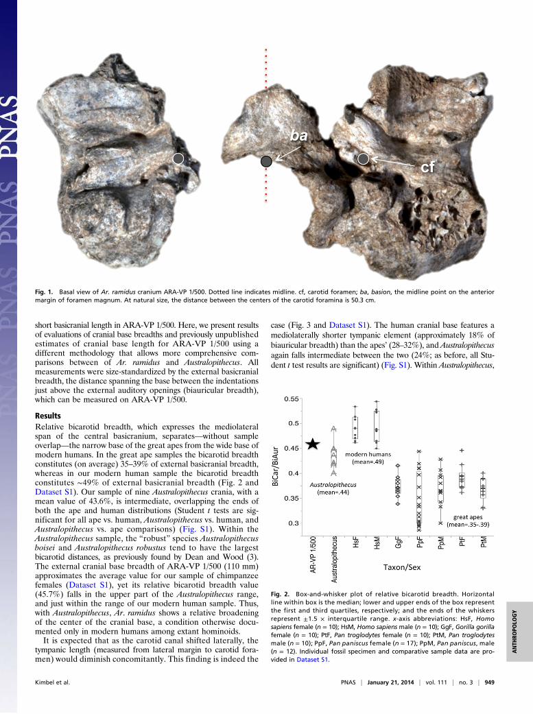
short basicranial length in ARA-VP 1/500. Here, we present resultsof evaluations of cranial base breadths and previously unpublishedestimates of cranial base length for ARA-VP 1/500 using adifferent methodology that allows more comprehensive com-parisons between of Ar. ramidus and Australopithecus. Allmeasurements were size-standardized by the external basicranialbreadth, the distance spanning the base between the indentationsjust above the external auditory openings (biauricular breadth),which can be measured on ARA-VP 1/500.
ResultsRelative bicarotid breadth, which expresses the mediolateralspan of the central basicranium, separates—without sampleoverlap—the narrow base of the great apes from the wide base ofmodern humans. In the great ape samples the bicarotid breadthconstitutes (on average) 35–39% of external basicranial breadth,whereas in our modern human sample the bicarotid breadthconstitutes ∼49% of external basicranial breadth (Fig. 2 andDataset S1). Our sample of nine Australopithecus crania, with amean value of 43.6%, is intermediate, overlapping the ends ofboth the ape and human distributions (Student t tests are sig-nificant for all ape vs. human, Australopithecus vs. human, andAustralopithecus vs. ape comparisons) (Fig. S1). Within theAustralopithecus sample, the “robust” species Australopithecusboisei and Australopithecus robustus tend to have the largestbicarotid distances, as previously found by Dean and Wood (3).The external cranial base breadth of ARA-VP 1/500 (110 mm)approximates the average value for our sample of chimpanzeefemales (Dataset S1), yet its relative bicarotid breadth value(45.7%) falls in the upper part of the Australopithecus range,and just within the range of our modern human sample. Thus,with Australopithecus, Ar. ramidus shows a relative broadeningof the center of the cranial base, a condition otherwise docu-mented only in modern humans among extant hominoids.It is expected that as the carotid canal shifted laterally, the
tympanic length (measured from lateral margin to carotid fora-men) would diminish concomitantly. This finding is indeed the
case (Fig. 3 and Dataset S1). The human cranial base features amediolaterally shorter tympanic element (approximately 18% ofbiauricular breadth) than the apes’ (28–32%), and Australopithecusagain falls intermediate between the two (24%; as before, all Stu-dent t test results are significant) (Fig. S1). Within Australopithecus,
Fig. 1. Basal view of Ar. ramidus cranium ARA-VP 1/500. Dotted line indicates midline. cf, carotid foramen; ba, basion, the midline point on the anteriormargin of foramen magnum. At natural size, the distance between the centers of the carotid foramina is 50.3 cm.
Fig. 2. Box-and-whisker plot of relative bicarotid breadth. Horizontalline within box is the median; lower and upper ends of the box representthe first and third quartiles, respectively; and the ends of the whiskersrepresent ±1.5 × interquartile range. x-axis abbreviations: HsF, Homosapiens female (n = 10); HsM, Homo sapiens male (n = 10); GgF, Gorilla gorillafemale (n = 10); PtF, Pan troglodytes female (n = 10); PtM, Pan troglodytesmale (n = 10); PpF, Pan paniscus female (n = 17); PpM, Pan paniscus, male(n = 12). Individual fossil specimen and comparative sample data are pro-vided in Dataset S1.
Kimbel et al. PNAS | January 21, 2014 | vol. 111 | no. 3 | 949
ANTH
ROPO
LOGY
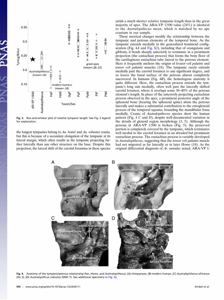
the longest tympanics belong to Au. boisei and Au. robustus crania,but this is because of a secondary elongation of the tympanic at itslateral margin, which often results in the tympanic projecting far-ther laterally than any other structure on the base. Despite thisprojection, the lateral shift of the carotid foramina in these species
yields a much shorter relative tympanic length than in the greatmajority of apes. The ARA-VP 1/500 value (24%) is identicalto the Australopithecus mean, which is matched by no apecranium in our sample.These metrical changes modify the relationship between the
tympanic and petrous elements of the temporal bone. As thetympanic extends medially in the generalized hominoid config-uration (Fig. 4A and Fig. S2), including that of orangutans andgibbons, it bends sharply anteriorly to terminate in a prominentprojection (the eustachian process) that forms the bony floor ofthe cartilaginous eustachian tube lateral to the petrous element.Here it frequently anchors the origins of levator veli palatini andtensor veli palatini muscles (18). The tympanic rarely extendsmedially past the carotid foramen to any significant degree, andso leaves the basal surface of the petrous almost completelyuncovered. In humans (Fig. 4B), the homologous anatomy isquite different. Here, the eustachian process extends the tym-panic’s long axis medially, often well past the laterally shiftedcarotid foramen, where it overlaps some 30–40% of the petrouselement’s length. In place of the anteriorly projecting eustachianprocess observed in the apes, a prominent posterior angle of thesphenoid bone (bearing the sphenoid spine) abuts the petrouslaterally and makes a substantial contribution to the entoglenoidprocess of the temporal squama, bounding the mandibular fossamedially. Crania of Australopithecus species show the humanpattern (Fig. 4 C and D), despite well-documented variation inthe details of glenoid region morphology (3, 5). Although thepetrous in ARA-VP 1/500 is broken (Fig. 5), the preservedportion is completely covered by the tympanic, which terminateswell medial to the carotid foramen in an abraded but prominenteustachian process. The eustachian process is variably developedin Australopithecus, suggesting that the tensor veli palatini musclehad not migrated as far laterally as in later Homo (18). As theoriginal differential diagnosis of Ar. ramidus noted, ARA-VP 1/
Fig. 3. Box-and-whisker plot of relative tympanic length. See Fig. 2 legendfor explanation.
Fig. 4. Anatomy of the tympanic/petrous relationship Pan, Homo, and Australopithecus: (A) chimpanzee, (B) modern human, (C) Australopithecus africanus(Sts 5), (D) Australopithcus robustus (DNH 7). See additional specimens in Fig. S2.
950 | www.pnas.org/cgi/doi/10.1073/pnas.1322639111 Kimbel et al.
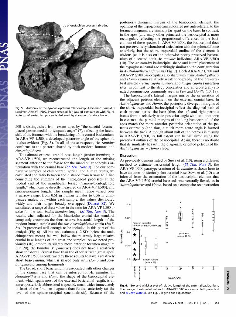
500 is distinguished from extant apes by “the carotid foramenplaced posteromedial to tympanic angle” (7), reflecting the lateralshift of the foramen with the broadening of the central basicranium.In ARA-VP 1/500, a developed posterior angle of the sphenoidis also evident (Fig. 5). In all of these respects, Ar. ramidusconforms to the pattern shared by both modern humans andAustralopithecus.To estimate external cranial base length (basion-hormion) in
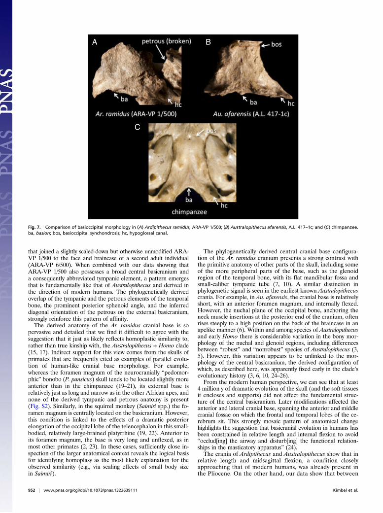
ARA-VP 1/500, we reconstructed the length of the missingsegment anterior to the fossae for the mandibular condyle’s ar-ticulation with the cranial base (SI Text, Note 3). For our com-parative samples of chimpanzee, gorilla, and human crania, wecalculated the ratio between the distance from basion to a lineconnecting the summits of the entoglenoid processes at themedial end of the mandibular fossae (“basion-bientoglenoidlength,” which can be directly measured on ARA-VP 1/500), andbasion-hormion length. The sample mean ratios varied overa narrow range, from 0.61 in human females to 0.56 in chim-panzee males, but within each sample, the values distributedwidely and their ranges broadly overlapped (Dataset S2). Wesubstituted a range of these values in the ratio for ARA-VP 1/500 tosolve for the total basion-hormion length (SI Text, Note 3). Theresults, when adjusted for the biauricular cranial size standard,completely encompass the short relative basicranial lengths of themodern human sample and the two Australopithecus crania (Sts 5,Sts 19) preserved well enough to be included in this part of theanalysis (Fig. 6). All but one estimate (−2 SDs below the malechimpanzee mean) fall well below the relatively large relativecranial base lengths of the great ape samples. As we noted pre-viously (10), despite its slightly more anterior foramen magnum(19, 20), the bonobo (P. paniscus) does not have a relativelyshorter external cranial base than the other African great apes.ARA-VP 1/500 is confirmed by these results to have a relativelyshort basicranium, which is shared only with Homo and Aus-tralopithecus among hominoids.The broad, short basicranium is associated with other changes
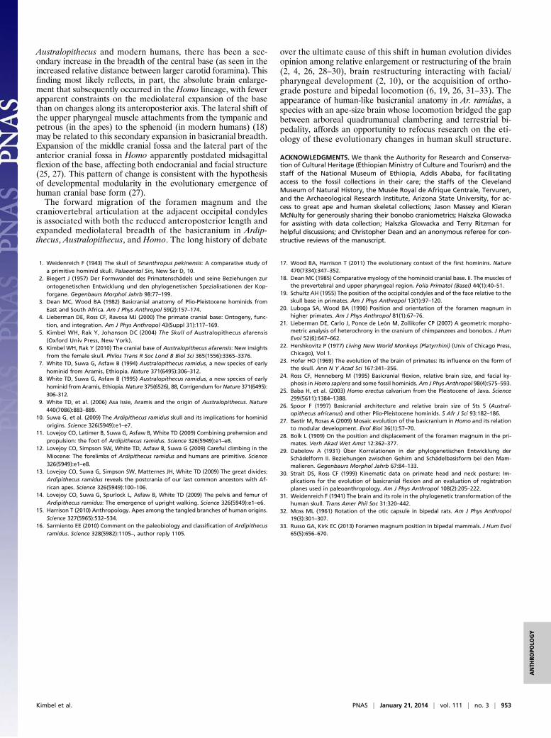
in the cranial base that can be inferred for Ar. ramidus. InAustralopithecus and Homo the shape of the basioccipital ele-ment, which spans most of the external basicranial length, is ananteroposteriorly abbreviated trapezoid, much wider immediatelyin front of the foramen magnum than further anteriorly (at thelevel of the spheno-occipital synchondrosis). Because of the
posteriorly divergent margins of the basioccipital element, theopenings of the hypoglossal canals, located just anterolateral to theforamen magnum, are similarly far apart on the base. In contrast,in the apes (and many other primates) the basioccipital is morerectangular, reflecting the proportional differences in the basi-cranium of these species. In ARA-VP 1/500, the basioccipital doesnot preserve its synchondrosal articulation with the sphenoid boneanteriorly, but the short, trapezoidal outline of the element isobvious (as it is also on the otherwise poorly preserved basicra-nium of a second adult Ar. ramidus individual, ARA-VP 6/500)(10). The Ar. ramidus basioccipital shape and lateral placement ofthe hypoglossal canal are strikingly similar to the configurationin Australopithecus afarensis (Fig. 7). Both ARA-VP 1/500 andARA-VP 6/500 basioccipitals also share with many Australopithecusand Homo crania relatively weak topography of the preverte-bral muscle (rectus capitis anterior and longus capitis) insertionsites, in contrast to the deep concavities and anterolaterally sit-uated prominences commonly seen in Pan and Gorilla (10, 18).The basioccipital’s lateral margins mirror the orientation of
the adjacent petrous element on the external cranial base. InAustralopithecus and Homo, the posteriorly divergent margins ofthe short, trapezoidal basioccipital reflect the diagonal path ofeach petrous across the base (thus, the left and right petrousbones form a relatively wide posterior angle with one another);in contrast, the parallel margins of the long basioccipital of theapes match the more anterior–posterior orientation of the pe-trous externally (and thus, a much more acute angle is formedbetween the two). Although about half of the petrous is missingin ARA-VP 1/500, its full extent can be visualized using thepreserved outlines of the basioccipital. Again, there is no doubtthat its similarity lies with the diagonally oriented petrous of theAustralopithecus + Homo clade.
DiscussionAs previously demonstrated by Suwa et al. (10), using a differentmethod to estimate basicranial length (SI Text, Note 3), theARA-VP 1/500 paratype cranium of Ar. ramidus is shown here tohave an anteroposteriorly short cranial base. Suwa et al. (10) alsoinferred from the orientation of the basioccipital element thatthe ARA-VP 1/500 cranial base axis was ventrally flexed, as inAustralopithecus and Homo, based on a composite reconstruction
Fig. 5. Anatomy of the tympanic/petrous relationship: Ardipithecus ramidusspecimen ARA-VP 1/500, image reversed for ease of comparison with Fig. 4.Note tip of eustachian process is darkened by abrasion of surface bone.
Fig. 6. Box-and-whisker plot of relative length of the external basicranium.Then range of estimated values for ARA-VP 1/500 is shown at left (main textand SI Text, Note 3). See Fig. 2 legend for explanation.
Kimbel et al. PNAS | January 21, 2014 | vol. 111 | no. 3 | 951
ANTH
ROPO
LOGY
that joined a slightly scaled-down but otherwise unmodified ARA-VP 1/500 to the face and braincase of a second adult individual(ARA-VP 6/500). When combined with our data showing thatARA-VP 1/500 also possesses a broad central basicranium anda consequently abbreviated tympanic element, a pattern emergesthat is fundamentally like that of Australopithecus and derived inthe direction of modern humans. The phylogenetically derivedoverlap of the tympanic and the petrous elements of the temporalbone, the prominent posterior sphenoid angle, and the inferreddiagonal orientation of the petrous on the external basicranium,strongly reinforce this pattern of affinity.The derived anatomy of the Ar. ramidus cranial base is so
pervasive and detailed that we find it difficult to agree with thesuggestion that it just as likely reflects homoplastic similarity to,rather than true kinship with, the Australopithecus + Homo clade(15, 17). Indirect support for this view comes from the skulls ofprimates that are frequently cited as examples of parallel evolu-tion of human-like cranial base morphology. For example,whereas the foramen magnum of the neurocranially “pedomor-phic” bonobo (P. pansicus) skull tends to be located slightly moreanterior than in the chimpanzee (19–21), its external base isrelatively just as long and narrow as in the other African apes, andnone of the derived tympanic and petrous anatomy is present(Fig. S2). Similarly, in the squirrel monkey (Saimiri spp.) the fo-ramen magnum is centrally located on the basicranium. However,this condition is linked to the effects of a dramatic posteriorelongation of the occipital lobe of the telencephalon in this small-bodied, relatively large-brained platyrrhine (19, 22). Anterior toits foramen magnum, the base is very long and unflexed, as inmost other primates (2, 23). In these cases, sufficiently close in-spection of the larger anatomical context reveals the logical basisfor identifying homoplasy as the most likely explanation for theobserved similarity (e.g., via scaling effects of small body sizein Saimiri).
The phylogenetically derived central cranial base configura-tion of the Ar. ramidus cranium presents a strong contrast withthe primitive anatomy of other parts of the skull, including someof the more peripheral parts of the base, such as the glenoidregion of the temporal bone, with its flat mandibular fossa andsmall-caliber tympanic tube (7, 10). A similar distinction inphylogenetic signal is seen in the earliest known Australopithecuscrania. For example, in Au. afarensis, the cranial base is relativelyshort, with an anterior foramen magnum, and internally flexed.However, the nuchal plane of the occipital bone, anchoring theneck muscle insertions at the posterior end of the cranium, oftenrises steeply to a high position on the back of the braincase in anapelike manner (6). Within and among species of Australopithecusand early Homo there is considerable variation in the bony mor-phology of the nuchal and glenoid regions, including differencesbetween “robust” and “nonrobust” species of Australopithecus (3,5). However, this variation appears to be unlinked to the mor-phology of the central basicranium, the derived configuration ofwhich, as described here, was apparently fixed early in the clade’sevolutionary history (3, 6, 10, 24–26).From the modern human perspective, we can see that at least
4 million y of dramatic evolution of the skull (and the soft tissuesit encloses and supports) did not affect the fundamental struc-ture of the central basicranium. Later modifications affected theanterior and lateral cranial base, spanning the anterior and middlecranial fossae on which the frontal and temporal lobes of the ce-rebrum sit. This strongly mosaic pattern of anatomical changehighlights the suggestion that basicranial evolution in humans hasbeen constrained in relative length and internal flexion to avoid“occlud[ing] the airway and disturb[ing] the functional relation-ships in the masticatory apparatus” (24).The crania of Ardipithecus and Australopithecus show that in
relative length and midsagittal flexion, a condition closelyapproaching that of modern humans, was already present inthe Pliocene. On the other hand, our data show that between
Fig. 7. Comparison of basioccipital morphology in (A) Ardipithecus ramidus, ARA-VP 1/500; (B) Australopithecus afarensis, A.L. 417–1c; and (C) chimpanzee.ba, basion; bos, basioccipital synchondrosis; hc, hypoglossal canal.
952 | www.pnas.org/cgi/doi/10.1073/pnas.1322639111 Kimbel et al.
Australopithecus and modern humans, there has been a sec-ondary increase in the breadth of the central base (as seen in theincreased relative distance between larger carotid foramina). Thisfinding most likely reflects, in part, the absolute brain enlarge-ment that subsequently occurred in the Homo lineage, with fewerapparent constraints on the mediolateral expansion of the basethan on changes along its anteroposterior axis. The lateral shift ofthe upper pharyngeal muscle attachments from the tympanic andpetrous (in the apes) to the sphenoid (in modern humans) (18)may be related to this secondary expansion in basicranial breadth.Expansion of the middle cranial fossa and the lateral part of theanterior cranial fossa in Homo apparently postdated midsagittalflexion of the base, affecting both endocranial and facial structure(25, 27). This pattern of change is consistent with the hypothesisof developmental modularity in the evolutionary emergence ofhuman cranial base form (27).The forward migration of the foramen magnum and the
craniovertebral articulation at the adjacent occipital condylesis associated with both the reduced anteroposterior length andexpanded mediolateral breadth of the basicranium in Ardip-thecus, Australopithecus, and Homo. The long history of debate
over the ultimate cause of this shift in human evolution dividesopinion among relative enlargement or restructuring of the brain(2, 4, 26, 28–30), brain restructuring interacting with facial/pharyngeal development (2, 10), or the acquisition of ortho-grade posture and bipedal locomotion (6, 19, 26, 31–33). Theappearance of human-like basicranial anatomy in Ar. ramidus, aspecies with an ape-size brain whose locomotion bridged the gapbetween arboreal quadrumanual clambering and terrestrial bi-pedality, affords an opportunity to refocus research on the eti-ology of these evolutionary changes in human skull structure.
ACKNOWLEDGMENTS. We thank the Authority for Research and Conserva-tion of Cultural Heritage (Ethiopian Ministry of Culture and Tourism) and thestaff of the National Museum of Ethiopia, Addis Ababa, for facilitatingaccess to the fossil collections in their care; the staffs of the ClevelandMuseum of Natural History, the Musée Royal de Afrique Centrale, Tervuren,and the Archaeological Research Institute, Arizona State University, for ac-cess to great ape and human skeletal collections; Jason Massey and KieranMcNulty for generously sharing their bonobo craniometrics; Halszka Glowackafor assisting with data collection; Halszka Glowacka and Terry Ritzman forhelpful discussions; and Christopher Dean and an anonymous referee for con-structive reviews of the manuscript.
1. Weidenreich F (1943) The skull of Sinanthropus pekinensis: A comparative study ofa primitive hominid skull. Palaeontol Sin, New Ser D, 10.
2. Biegert J (1957) Der Formwandel des Primatenschädels und seine Beziehungen zurontogenetischen Entwicklung und den phylogenetischen Spezialisationen der Kop-forgane. Gegenbaurs Morphol Jahrb 98:77–199.
3. Dean MC, Wood BA (1982) Basicranial anatomy of Plio-Pleistocene hominids fromEast and South Africa. Am J Phys Anthropol 59(2):157–174.
4. Lieberman DE, Ross CF, Ravosa MJ (2000) The primate cranial base: Ontogeny, func-tion, and integration. Am J Phys Anthropol 43(Suppl 31):117–169.
5. Kimbel WH, Rak Y, Johanson DC (2004) The Skull of Australopithecus afarensis(Oxford Univ Press, New York).
6. Kimbel WH, Rak Y (2010) The cranial base of Australopithecus afarensis: New insightsfrom the female skull. Philos Trans R Soc Lond B Biol Sci 365(1556):3365–3376.
7. White TD, Suwa G, Asfaw B (1994) Australopithecus ramidus, a new species of earlyhominid from Aramis, Ethiopia. Nature 371(6495):306–312.
8. White TD, Suwa G, Asfaw B (1995) Australopithecus ramidus, a new species of earlyhominid from Aramis, Ethiopia. Nature 375(6526), 88, Corrigendum forNature 371(6495):306–312.
9. White TD, et al. (2006) Asa Issie, Aramis and the origin of Australopithecus. Nature440(7086):883–889.
10. Suwa G, et al. (2009) The Ardipithecus ramidus skull and its implications for hominidorigins. Science 326(5949):e1–e7.
11. Lovejoy CO, Latimer B, Suwa G, Asfaw B, White TD (2009) Combining prehension andpropulsion: the foot of Ardipithecus ramidus. Science 326(5949):e1–e8.
12. Lovejoy CO, Simpson SW, White TD, Asfaw B, Suwa G (2009) Careful climbing in theMiocene: The forelimbs of Ardipithecus ramidus and humans are primitive. Science326(5949):e1–e8.
13. Lovejoy CO, Suwa G, Simpson SW, Matternes JH, White TD (2009) The great divides:Ardipithecus ramidus reveals the postcrania of our last common ancestors with Af-rican apes. Science 326(5949):100–106.
14. Lovejoy CO, Suwa G, Spurlock L, Asfaw B, White TD (2009) The pelvis and femur ofArdipithecus ramidus: The emergence of upright walking. Science 326(5949):e1–e6.
15. Harrison T (2010) Anthropology. Apes among the tangled branches of human origins.Science 327(5965):532–534.
16. Sarmiento EE (2010) Comment on the paleobiology and classification of Ardipithecusramidus. Science 328(5982):1105–, author reply 1105.
17. Wood BA, Harrison T (2011) The evolutionary context of the first hominins. Nature470(7334):347–352.
18. Dean MC (1985) Comparative myology of the hominoid cranial base. II. The muscles ofthe prevertebral and upper pharyngeal region. Folia Primatol (Basel) 44(1):40–51.
19. Schultz AH (1955) The position of the occipital condyles and of the face relative to theskull base in primates. Am J Phys Anthropol 13(1):97–120.
20. Luboga SA, Wood BA (1990) Position and orientation of the foramen magnum inhigher primates. Am J Phys Anthropol 81(1):67–76.
21. Lieberman DE, Carlo J, Ponce de León M, Zollikofer CP (2007) A geometric morpho-metric analysis of heterochrony in the cranium of chimpanzees and bonobos. J HumEvol 52(6):647–662.
22. Hershkovitz P (1977) Living New World Monkeys (Platyrrhini) (Univ of Chicago Press,Chicago), Vol 1.
23. Hofer HO (1969) The evolution of the brain of primates: Its influence on the form ofthe skull. Ann N Y Acad Sci 167:341–356.
24. Ross CF, Henneberg M (1995) Basicranial flexion, relative brain size, and facial ky-phosis in Homo sapiens and some fossil hominids. Am J Phys Anthropol 98(4):575–593.
25. Baba H, et al. (2003) Homo erectus calvarium from the Pleistocene of Java. Science299(5611):1384–1388.
26. Spoor F (1997) Basicranial architecture and relative brain size of Sts 5 (Austral-opithecus africanus) and other Plio-Pleistocene hominids. S Afr J Sci 93:182–186.
27. Bastir M, Rosas A (2009) Mosaic evolution of the basicranium in Homo and its relationto modular development. Evol Biol 36(1):57–70.
28. Bolk L (1909) On the position and displacement of the foramen magnum in the pri-mates. Verh Akad Wet Amst 12:362–377.
29. Dabelow A (1931) Über Korrelationen in der phylogenetischen Entwicklung derSchädelform II. Beziehungen zwischen Gehirn and Schädelbasisform bei den Mam-malieren. Gegenbaurs Morphol Jahrb 67:84–133.
30. Strait DS, Ross CF (1999) Kinematic data on primate head and neck posture: Im-plications for the evolution of basicranial flexion and an evaluation of registrationplanes used in paleoanthropology. Am J Phys Anthropol 108(2):205–222.
31. Weidenreich F (1941) The brain and its role in the phylogenetic transformation of thehuman skull. Trans Amer Phil Soc 31:320–442.
32. Moss ML (1961) Rotation of the otic capsule in bipedal rats. Am J Phys Anthropol19(3):301–307.
33. Russo GA, Kirk EC (2013) Foramen magnum position in bipedal mammals. J Hum Evol65(5):656–670.
Kimbel et al. PNAS | January 21, 2014 | vol. 111 | no. 3 | 953
ANTH
ROPO
LOGY