Embed Size (px)
Citation preview
Special Issue: Chromatin Dynamics
Architectural proteins: regulators of 3Dgenome organization in cell fateElena Gomez-Dıaz and Victor G. Corces
Department of Biology, Emory University, Atlanta, GA 30322, USA
Review
The relation between alterations in chromatin structureand changes in gene expression during cell differentia-tion has served as a paradigm to understand the linkbetween genome organization and function. Yet, thefactors involved and the mechanisms by which the 3Dorganization of the nucleus is established remain poorlyunderstood. The use of Chromosome Conformation-Capture (3C)-based approaches has resulted in a newappreciation of the role of architectural proteins in theestablishment of 3D genome organization. Architecturalproteins orchestrate higher-order chromatin organiza-tion through the establishment of interactions betweenregulatory elements across multiple spatial scales. Theregulation of these proteins, their interaction with DNA,and their co-occurrence in the genome, may be responsiblefor the plasticity of 3D chromatin architecture that dictatescell and time-specific blueprints of gene expression.
Nuclear organizationChromosomes are tightly packed in the nucleus within chro-mosome territories [1–4]. The 3D arrangement of the chro-matin fiber in these territories during interphase is notrandom and, in principle, could be either a consequence ofgenome function or a pre-established effector of nuclearactivity [5]. Nuclear processes, such as transcription andreplication, require the assembly of large multiprotein com-plexes at promoters, enhancers, and replication origins [5–7].These proteins often contain multiple interacting domainsand, therefore, may drive the formation of intra- and inter-chromosomal contacts that contribute to the establishment ofa specific 3D arrangement of the chromatin fiber. Given thatthis arrangement may be a consequence of genome function,it should be, at least in part, cell type specific, correlating withthe transcriptional state of the cell. In addition to this tran-scription-driven organization, the cell appears to also usespecific protein complexes whose main role is to establishcontacts between distant sites in the genome to facilitate its3D organization and allow the execution of specific functionaloutcomes. These proteins, generally referred to as insulatorproteins, were originally characterized for their ability tointerfere with enhancer–promoter interactions and to shieldthe expression of transgenes from the effects of adjacent
0962-8924/
� 2014 Elsevier Ltd. All rights reserved. http://dx.doi.org/10.1016/j.tcb.2014.08.003
Corresponding author: Corces, V.G. ([email protected]).Keywords: CTCF; transcription; chromatin; development; differentiation.
sequences [8]. More recent results suggest that insulatorsequences and their associated proteins not only inhibit,but also facilitate enhancer–promoter interactions, as wellas regulating other aspects of transcription, in addition tomore general roles in chromosome organization [9]. Given thevaried, and sometimes contradictory, functions mediated bythese proteins, we refer to them here as architectural insteadof insulator proteins.
Starting with the premise that architectural proteinscan mediate interactions between different sequences toregulate genome function, here we discuss mechanisms bywhich the interaction of these proteins with DNA or otherproteins can be regulated to create specific patterns ofnuclear 3D organization to elicit distinct functional out-comes that may contribute to the establishment of specificcell lineages during development.
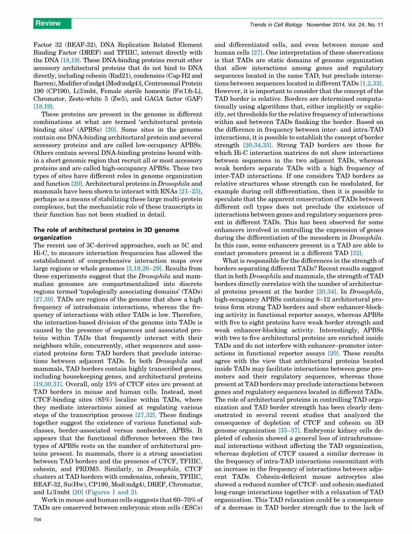
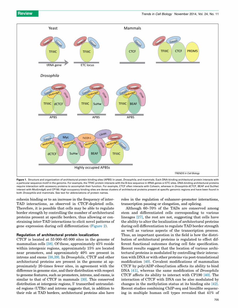
Architectural proteins: structure and organizationArchitectural proteins have been described in organismsranging from yeast to humans [10]. In Saccharomyces cere-visiae and Saccharomyces pombe, the main architecturalprotein characterized to date is the RNA polymerase III-associated factor TFIIIC, which is present at genes tran-scribed by this polymerase, such as tRNA genes, as well asat many nontranscribed regions of the genome known asextra TFIIIC (ETC) loci [11,12]. TFIIIC colocalizes withcohesin and condensin, which have been shown to be requiredfor its function in protecting against the spreading of histonecovalent modifications associated with transcription silenc-ing [13]. The best-characterized architectural protein in ver-tebrates is CCCTC-binding factor (zinc finger protein)(CTCF), which also requires association with cohesin forits enhancer-blocking function [10]. Recent experimentsshowing that tRNA genes can block enhancer function andthat TFIIIC colocalizes with CTCF at many ETC loci throughthe mouse and human genomes suggest a conservation in thefunction of TFIIIC as an architectural protein from yeast tohumans [14]. Other proteins shown to colocalize or directlyinteract with CTCF in vertebrates include Yin Yang 1 (YY1),Kaiso, chromodomain helicase DNA-binding protein 8(CHD8), poly(ADP-ribose) polymerase 1 (PARP1), MYC-as-sociated zinc-finger protein (MAZ), JUND, zinc-finger protein143 (ZNF143), PR domain zinc-finger protein 5 (PRDM5), andnucleophosmin [15–17]. Drosophila has also been a richsource of information aimed at understanding the structureand organization of this class of proteins. Several DNA-binding architectural proteins, including CTCF, Suppressorof Hairy-wing [Su(Hw)], Boundary Element Associated
Trends in Cell Biology, November 2014, Vol. 24, No. 11 703
Review Trends in Cell Biology November 2014, Vol. 24, No. 11
Factor 32 (BEAF-32), DNA Replication Related ElementBinding Factor (DREF) and TFIIIC, interact directly withthe DNA [18,19]. These DNA-binding proteins recruit otheraccessory architectural proteins that do not bind to DNAdirectly, including cohesin (Rad21), condensins (Cap-H2 andBarren), Modifier of mdg4 [Mod(mdg4)], Centrosomal Protein190 (CP190), L(3)mbt, Female sterile homeotic [Fs(1)h-L],Chromator, Zeste-white 5 (Zw5), and GAGA factor (GAF)[18,19].
These proteins are present in the genome in differentcombinations at what are termed ‘architectural proteinbinding sites’ (APBSs) [20]. Some sites in the genomecontain one DNA-binding architectural protein and severalaccessory proteins and are called low-occupancy APBSs.Others contain several DNA-binding proteins bound with-in a short genomic region that recruit all or most accessoryproteins and are called high-occupancy APBSs. These twotypes of sites have different roles in genome organizationand function [20]. Architectural proteins in Drosophila andmammals have been shown to interact with RNAs [21–25],perhaps as a means of stabilizing these large multi-proteincomplexes, but the mechanistic role of these transcripts intheir function has not been studied in detail.
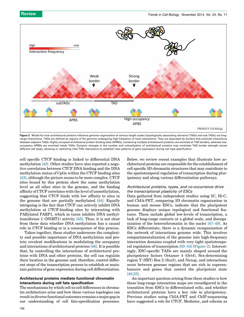
The role of architectural proteins in 3D genomeorganizationThe recent use of 3C-derived approaches, such as 5C andHi-C, to measure interaction frequencies has allowed theestablishment of comprehensive interaction maps overlarge regions or whole genomes [3,19,26–29]. Results fromthese experiments suggest that the Drosophila and mam-malian genomes are compartmentalized into discreteregions termed ‘topologically associating domains’ (TADs)[27,30]. TADs are regions of the genome that show a highfrequency of intradomain interactions, whereas the fre-quency of interactions with other TADs is low. Therefore,the interaction-based division of the genome into TADs iscaused by the presence of sequences and associated pro-teins within TADs that frequently interact with theirneighbors while, concurrently, other sequences and asso-ciated proteins form TAD borders that preclude interac-tions between adjacent TADs. In both Drosophila andmammals, TAD borders contain highly transcribed genes,including housekeeping genes, and architectural proteins[19,30,31]. Overall, only 15% of CTCF sites are present atTAD borders in mouse and human cells. Instead, mostCTCF-binding sites (85%) localize within TADs, wherethey mediate interactions aimed at regulating varioussteps of the transcription process [27,32]. These findingstogether suggest the existence of various functional sub-classes, border-associated versus nonborder, APBSs. Itappears that the functional difference between the twotypes of APBSs rests on the number of architectural pro-teins present. In mammals, there is a strong associationbetween TAD borders and the presence of CTCF, TFIIIC,cohesin, and PRDM5. Similarly, in Drosophila, CTCFclusters at TAD borders with condensins, cohesin, TFIIIC,BEAF-32, Su(Hw), CP190, Mod(mdg4), DREF, Chromator,and L(3)mbt [20] (Figures 1 and 2).
Work in mouse and human cells suggests that 60–70% ofTADs are conserved between embryonic stem cells (ESCs)
704
and differentiated cells, and even between mouse andhuman cells [27]. One interpretation of these observationsis that TADs are static domains of genome organizationthat allow interactions among genes and regulatorysequences located in the same TAD, but preclude interac-tions between sequences located in different TADs [1,2,33].However, it is important to consider that the concept of theTAD border is relative. Borders are determined computa-tionally using algorithms that, either implicitly or explic-itly, set thresholds for the relative frequency of interactionswithin and between TADs flanking the border. Based onthe difference in frequency between inter- and intra-TADinteractions, it is possible to establish the concept of borderstrength [20,34,35]. Strong TAD borders are those forwhich Hi-C interaction matrices do not show interactionsbetween sequences in the two adjacent TADs, whereasweak borders separate TADs with a high frequency ofinter-TAD interactions. If one considers TAD borders asrelative structures whose strength can be modulated, forexample during cell differentiation, then it is possible tospeculate that the apparent conservation of TADs betweendifferent cell types does not preclude the existence ofinteractions between genes and regulatory sequences pres-ent in different TADs. This has been observed for someenhancers involved in controlling the expression of genesduring the differentiation of the mesoderm in Drosophila.In this case, some enhancers present in a TAD are able tocontact promoters present in a different TAD [32].
What is responsible for the differences in the strength ofborders separating different TADs? Recent results suggestthat in both Drosophila and mammals, the strength of TADborders directly correlates with the number of architectur-al proteins present at the border [20,34]. In Drosophila,high-occupancy APBSs containing 8–12 architectural pro-teins form strong TAD borders and show enhancer-block-ing activity in functional reporter assays, whereas APBSswith five to eight proteins have weak border strength andweak enhancer-blocking activity. Interestingly, APBSswith two to five architectural proteins are enriched insideTADs and do not interfere with enhancer–promoter inter-actions in functional reporter assays [20]. These resultsagree with the view that architectural proteins locatedinside TADs may facilitate interactions between gene pro-moters and their regulatory sequences, whereas thosepresent at TAD borders may preclude interactions betweengenes and regulatory sequences located in different TADs.The role of architectural proteins in controlling TAD orga-nization and TAD border strength has been clearly dem-onstrated in several recent studies that analyzed theconsequence of depletion of CTCF and cohesin on 3Dgenome organization [35–37]. Embryonic kidney cells de-pleted of cohesin showed a general loss of intrachromoso-mal interactions without affecting the TAD organization,whereas depletion of CTCF caused a similar decrease inthe frequency of intra-TAD interactions concomitant withan increase in the frequency of interactions between adja-cent TADs. Cohesin-deficient mouse astrocytes alsoshowed a reduced number of CTCF- and cohesin-mediatedlong-range interactions together with a relaxation of TADorganization. This TAD relaxation could be a consequenceof a decrease in TAD border strength due to the lack of
Highly occupied APBSs
BEAF
BEAF
L3mbt
TFIIIC
TFIIIC
TFIIIC TFIIIC TFIIICCTCF CTCF PRDM5
TFIIIC TFIIIC
SuHw
SuHw
APBS
tRNA gene
Drosophila
Yeast Mammals
ETC locus
APBS APBS
Myc Fs(1)h-L
CP190
CP190
CP190
CP190
CP190
Mod(mdg4)Mod(mdg4)
Mod(mdg4)Mod(mdg4)
Mod(mdg4) CP190 od(mdg4)
dCTCF
dCTCF
TRENDS in Cell Biology
Figure 1. Structure and organization of architectural protein binding sites (APBS) in yeast, Drosophila, and mammals. Each DNA-binding architectural protein interacts with
a particular sequence motif in the genome. For example, the TFIIIC protein interacts with the B-box sequence in tRNA genes or ETC sites. DNA-binding architectural proteins
require interaction with accessory proteins to accomplish their function. For example, CTCF often interacts with Cohesin, whereas in Drosophila dCTCF, BEAF and Su(Hw)
interact with Mod(mdg4) and CP190. High-occupancy binding sites are dense clusters of architectural proteins present at specific genomic regions and have been found in
both Drosophila and mammals. See text for abbreviations of protein names.
Review Trends in Cell Biology November 2014, Vol. 24, No. 11
cohesin binding or to an increase in the frequency of inter-TAD interactions, as observed in CTCF-depleted cells.Therefore, it is possible that cells may be able to regulateborder strength by controlling the number of architecturalproteins present at specific borders, thus allowing or con-straining inter-TAD interactions to elicit novel patterns ofgene expression during cell differentiation (Figure 2).
Regulation of architectural protein localizationCTCF is located at 55 000–65 000 sites in the genome ofmammalian cells [38]. Of these, approximately 45% residewithin intergenic regions, approximately 15% are locatednear promoters, and approximately 40% are present inintrons and exons [38,39]. In Drosophila, CTCF and otherarchitectural proteins are present in the genome at ap-proximately 20-times fewer sites, in agreement with thedifference in genome size, and their distribution with respectto genome features, such as promoters, introns, and exons, issimilar to that of CTCF in mammals [30]. This conserveddistribution at intergenic regions, 50 transcribed untranslat-ed regions (UTRs) and introns suggests that, in addition totheir role at TAD borders, architectural proteins also have
roles in the regulation of enhancer–promoter interactions,transcription pausing or elongation, and splicing.
Although 60–70% of the TADs are conserved amongstem and differentiated cells corresponding to variouslineages [27], the rest are not, suggesting that cells havethe ability to alter the localization of architectural proteinsduring cell differentiation to regulate TAD border strengthas well as various aspects of the transcription process.Thus, an important question in the field is how the distri-bution of architectural proteins is regulated to effect dif-ferent functional outcomes during cell fate specification.Recent results suggest that the location of various archi-tectural proteins is modulated by controlling their interac-tion with DNA or with other proteins via post-translationalmodification [40]. Covalent modifications of mammalianCTCF by poly(ADP-ribosyl)ation affects its ability to bindDNA [41], whereas the same modification of DrosophilaCTCF affects its ability to interact with CP190 [40]. Theinteraction of CTCF with DNA can be also modulated bychanges in the methylation status at its binding site [42].Recent studies combining ChIP-seq and bisulfite sequenc-ing in multiple human cell types revealed that 41% of
705
High Low
Interac�on frequency
Weakborder
Strongborder
subTADs TAD
APBS High occupancyAPBS
TRENDS in Cell Biology
Figure 2. Model for how architectural proteins influence genome organization at various length scales [topologically associating domains (TADs) and sub-TADs] via long-
range interactions. TADs are defined as regions of the genome undergoing high frequency of local interactions. They are separated by borders that preclude interactions
between adjacent TADs. Highly occupied architectural protein binding sites (APBSs), containing multiple architectural proteins, are enriched at TAD borders, whereas low-
occupancy APBSs are enriched inside TADs. Dynamic changes in the number and colocalization of architectural proteins may modulate TAD border strength across
different cell types, allowing or restricting inter-TAD interactions to establish new patterns of gene expression during cell type specification.
Review Trends in Cell Biology November 2014, Vol. 24, No. 11
cell specific CTCF binding is linked to differential DNAmethylation [42]. Other studies have also reported a nega-tive correlation between CTCF DNA binding and the DNAmethylation status of CpGs within the CTCF binding sites[43], although the picture seems to be more complex. CTCFsites bound by this protein show the same methylationlevel as all other sites in the genome, and the bindingaffinity of CTCF correlates with the level of unmethylation,suggesting that CTCF binds with low affinity to sites inthe genome that are partially methylated [44]. Equallyintriguing is the fact that CTCF can actively inhibit DNAmethylation at CTCF-binding sites by interacting withPARylated PARP1, which in turns inhibits DNA methyl-transferase 1 (DNMT1) activity [45]. Thus, it is not clearfrom these data whether DNA methylation has a causalrole in CTCF binding or is a consequence of this process.
Taken together, these studies underscore the complexi-ty and possible importance of DNA methylation and pro-tein covalent modifications in modulating the occupancyand interactions of architectural proteins [46]. It is possiblethat, by controlling the interactions of architectural pro-teins with DNA and other proteins, the cell can regulatetheir location in the genome and, therefore, control differ-ent steps of the transcription process to establish or main-tain patterns of gene expression during cell differentiation.
Architectural proteins mediate functional chromatininteractions during cell fate specificationThe mechanisms by which cell-to-cell differences in chroma-tin architecture arise and how these various topologies canresult in diverse functional outcomes remains a major gap inour understanding of cell fate-specification processes.
706
Below, we review recent examples that illustrate how ar-chitectural proteins are responsible for the establishment ofcell specific 3D chromatin structures that may contribute tothe spatiotemporal regulation of transcription during plur-ipotency and along various differentiation pathways.
Architectural proteins, types, and co-occurrence drive
the transcriptional plasticity of ESCs
Data gathered from independent studies using 5C, Hi-C,and ChIA-PET, comparing 3D chromatin organization inhuman and mouse ESCs, indicate that the pluripotentgenome displays unique topological and functional fea-tures. These include global low-levels of transcription, alack of long-range contacts at a global scale, and disorga-nization of the heterochromatin in the nuclei [47–49]. AsESCs differentiate, there is a dynamic reorganization ofthe network of interactions genome wide. This involvescompartmentalization of the genome into high-frequencyinteraction domains coupled with very tight spatiotempo-ral regulation of transcription [50–54] (Figure 2). Interest-ingly, ESC-specific TADs are mainly shaped around thepluripotency factors Octamer 4 (Oct4), Sex-determiningregion Y (SRY) Box 2 (Sox2), and Nanog, and interactionsoccur between genome regions that are rich in superen-hancers and genes that control the pluripotent state[48,55].
An important question arising from these studies is howthese long-range interaction maps are reconfigured in thetransition from ESCs to differentiated cells, and whetherarchitectural proteins have a key role in pluripotency.Previous studies using ChIA-PET and ChIP-sequencinghave suggested a role for CTCF, Mediator, and cohesin as
Review Trends in Cell Biology November 2014, Vol. 24, No. 11
chromatin organizers in ESCs, showing that they engagein functional interactions with pluripotent genes and tran-scription factors [56,57]. This is supported by resultsobtained during the differentiation of ESCs into the endo-dermal lineage, where CTCF has been shown to directlyrecruit TAF3, a TBP-associated core promoter factor, todistal regulatory sequences. TAF3 present at CTCF/cohe-sin sites cooperates with these two proteins in mediatinginteractions between these enhancers and promoters dur-ing the differentiation of ESCs into endoderm [58,59].Changes in CTCF occupancy during differentiation ofESCs are associated with alterations in nucleosome posi-tioning and DNA demethylation [60,61].
A comprehensive analysis of APBS occupancy patternsin the context of ESC differentiation was obtained bycomparing 5C interaction maps in ESCs and neuralprogenitor cells (NPCs). The study revealed two classesof interactions: ESC-specific enhancer–promoter short-range contacts involving cohesin and Mediator but notCTCF, and larger loops coinciding with CTCF and cohe-sin binding. Loops at the sub-megabase scale show clearreorganization during differentiation, whereas CTCF-mediated megabase loops remain invariant and wereproposed to have a role in chromosome folding [62]. Theseobservations can be interpreted in the context of a modelin which the regulation in the occupancy of varioussubclasses of architectural proteins results in changesin chromatin organization that allow the cell to switchbetween various transcription programs. Consistent withthis hypothesis, two independent studies in ESCs usingconditional knockdowns in cohesin and Mediator, foundeither an artificial induction of differentiation of ESCs orimpaired reprogramming into induced pluripotent stemcells (iPSCs) [50,63]. Whether architectural protein bind-ing alone defines pluripotency, or whether pluripotency isinstead driven by state-specific transcription factors andenhancers, remains unanswered.
Together, these findings support a key role of architec-tural proteins in the dynamic folding of the genome duringcell fate specification. Yet, several important issues re-main. For example, how general is the relationship be-tween architectural proteins, pluripotency transcriptionfactors, and/or enhancers? Are architectural proteinscausal to changes in the pluripotent state, or a conse-quence of the binding of pluripotency transcription fac-tors? Do TADs and TAD borders have a regulatory role inthe transition between pluripotent and differentiatedchromatin states?
CTCF and cohesin regulate lymphocyte differentiation
Lymphocyte differentiation provides a compelling exampleof the role of architectural proteins and chromatin 3Darchitecture in generating cell diversity. B and T lympho-cytes have a unique antigen receptor that is highly variableand cell specific, and the basis of adaptive immunity. Thevariable portion of the B cell immunoglobulin (Ig) and Tcell receptor (Tcr) loci is encoded by multiple copies ofvariable (V), diversity (D), and joining (J) gene segmentsthat span across large genomic regions. Antigenic diversityin B and T lymphocytes is generated by gene rearrange-ments of these V, D, and J gene segments catalyzed by the
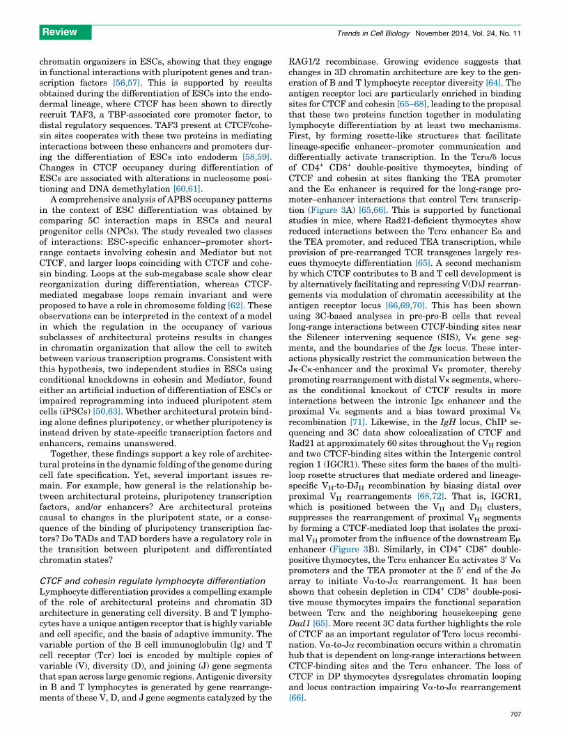
RAG1/2 recombinase. Growing evidence suggests thatchanges in 3D chromatin architecture are key to the gen-eration of B and T lymphocyte receptor diversity [64]. Theantigen receptor loci are particularly enriched in bindingsites for CTCF and cohesin [65–68], leading to the proposalthat these two proteins function together in modulatinglymphocyte differentiation by at least two mechanisms.First, by forming rosette-like structures that facilitatelineage-specific enhancer–promoter communication anddifferentially activate transcription. In the Tcra/d locusof CD4+ CD8+ double-positive thymocytes, binding ofCTCF and cohesin at sites flanking the TEA promoterand the Ea enhancer is required for the long-range pro-moter–enhancer interactions that control Tcrk transcrip-tion (Figure 3A) [65,66]. This is supported by functionalstudies in mice, where Rad21-deficient thymocytes showreduced interactions between the Tcra enhancer Ea andthe TEA promoter, and reduced TEA transcription, whileprovision of pre-rearranged TCR transgenes largely res-cues thymocyte differentiation [65]. A second mechanismby which CTCF contributes to B and T cell development isby alternatively facilitating and repressing V(D)J rearran-gements via modulation of chromatin accessibility at theantigen receptor locus [66,69,70]. This has been shownusing 3C-based analyses in pre-pro-B cells that reveallong-range interactions between CTCF-binding sites nearthe Silencer intervening sequence (SIS), Vk gene seg-ments, and the boundaries of the Igk locus. These inter-actions physically restrict the communication between theJk-Ck-enhancer and the proximal Vk promoter, therebypromoting rearrangement with distal Vk segments, where-as the conditional knockout of CTCF results in moreinteractions between the intronic Igk enhancer and theproximal Vk segments and a bias toward proximal Vk
recombination [71]. Likewise, in the IgH locus, ChIP se-quencing and 3C data show colocalization of CTCF andRad21 at approximately 60 sites throughout the VH regionand two CTCF-binding sites within the Intergenic controlregion 1 (IGCR1). These sites form the bases of the multi-loop rosette structures that mediate ordered and lineage-specific VH-to-DJH recombination by biasing distal overproximal VH rearrangements [68,72]. That is, IGCR1,which is positioned between the VH and DH clusters,suppresses the rearrangement of proximal VH segmentsby forming a CTCF-mediated loop that isolates the proxi-mal VH promoter from the influence of the downstream Em
enhancer (Figure 3B). Similarly, in CD4+ CD8+ double-positive thymocytes, the Tcra enhancer Ea activates 30 Va
promoters and the TEA promoter at the 50 end of the Ja
array to initiate Va-to-Ja rearrangement. It has beenshown that cohesin depletion in CD4+ CD8+ double-posi-tive mouse thymocytes impairs the functional separationbetween Tcrk and the neighboring housekeeping geneDad1 [65]. More recent 3C data further highlights the roleof CTCF as an important regulator of Tcra locus recombi-nation. Va-to-Ja recombination occurs within a chromatinhub that is dependent on long-range interactions betweenCTCF-binding sites and the Tcra enhancer. The loss ofCTCF in DP thymocytes dysregulates chromatin loopingand locus contraction impairing Va-to-Ja rearrangement[66].
707
CTCF BS
(A)
(B)
TEA Jα Cα Eα Dad1CTCF + CohesinCohesin BS
VH distal VH proximal DH JH CH 3’RRCTCF BS
RAG center
EmIGCR1
TRENDS in Cell Biology
Figure 3. CCCTC-binding factor (zinc finger protein) (CTCF) and cohesin regulate antigen receptor diversity in T and B lymphocytes. Antigen receptor diversity of B and T
cells is generated by the rearrangement of different variable (V), diversity (D), and joining (J) gene segments in individual lymphocytes. CTCF influences the outcome of
V(D)J recombination by regulating enhancer–promoter interactions and locus compaction. The general organization of the T cell receptor (TCRa) and immunoglobulin (Ig)H
loci are shown. (A) In the TCRa locus of thymocytes, cobinding of the CTCF/cohesin complex at the TEA promoter and the Ea enhancer results in a DNA loop that is required
to activate transcription of the nearby housekeeping gene Dad1. (B) In the IgH locus of pre-pro-B cells, CTCF-mediated looping between the Em enhancer and 30 regulatory
region (30RR) with distinct DH-JH-CH gene segments is required for ordered (DH–JH) recombination. CTCF binding at intergenic control region 1 (IGCR1) blocks the influence
of the Em enhancer on proximal variable (VH) regions.
Review Trends in Cell Biology November 2014, Vol. 24, No. 11
CTCF/cohesin mediate monoallelic gene expression in
neuronal differentiation
The differentiation of the hundreds of specialized neuronaltypes present in the brain requires the establishment ofspecific patterns of gene expression. Among the manygenes that are transcribed in a neuron-specific manner,the mechanisms underlying the expression of protocadher-ins have been studied in great detail. Protocadherins (Pcd)are part of the larger family of calcium-dependent celladhesion molecules in the central nervous system. Inmammals, there are more than 50 protocadherin isoformsgrouped into three gene clusters named a, b, and g. Inter-estingly, the genomic organization of the Pcd gene clustersresembles that of the immunoglobulin and T cell receptorgenes, albeit the mechanism of regulation differs slightly inthat it does not involve somatic rearrangements. In neu-rons, single cell diversity results from the monoallelic geneexpression of a protocadherin gene cluster, so that only oneisoform is transcribed at a time. This is achieved bystochastic promoter choice from the 15-variable first exons,followed by alternative pre-mRNA cis-splicing of the
708
chosen alternative exons to three downstream constantexons [73]. Two independent studies in human and mousecell lines provide evidence that CTCF and cohesin-mediat-ed interactions are ultimately responsible for the mono-allelic expression at the protocadherin a cluster [74,75]. Inthe first of these studies, Maniatis and colleagues used ahuman diploid neuroblastoma cell line SK-N-SH expres-sing a select number of alternative Pcdh isoforms. In thecase of Pcdha, the cluster comprises a set of 14–15 variableexons, each containing its own promoter and two cis-regu-latory elements with enhancer activity (HS7 and HS5-1).CTCF/cohesin co-bound sites interact with the TSS andfirst exon of a4, a8, and a12 isoforms, and activate specifictranscription of these isoforms. In the second study, thesame authors showed that DNA looping at Pcdha requiresspecific cobinding of the CTCF/cohesin complex to twosymmetrically aligned binding sites in both the transcrip-tionally active promoters and the HS5-1 enhancer. Inaddition, this study identified a unique regulatory rolefor cohesin, which binds to another enhancer (HS7) inde-pendently of CTCF. Functional analyses demonstrated
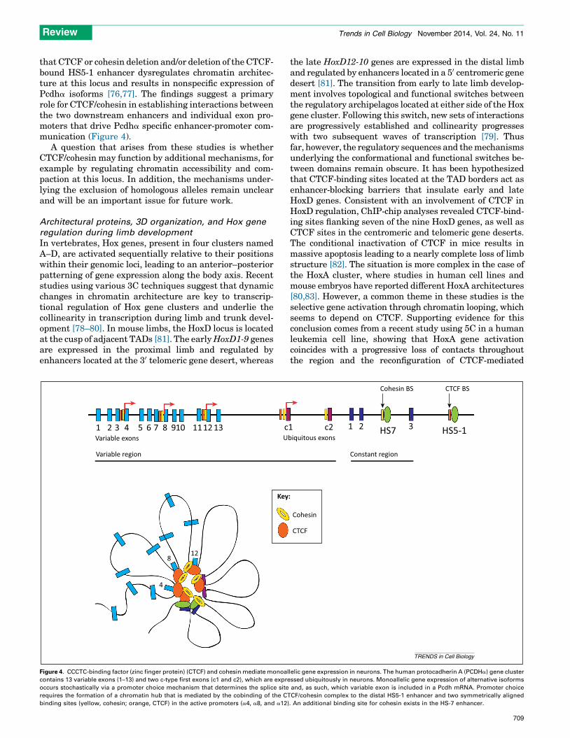
Review Trends in Cell Biology November 2014, Vol. 24, No. 11
that CTCF or cohesin deletion and/or deletion of the CTCF-bound HS5-1 enhancer dysregulates chromatin architec-ture at this locus and results in nonspecific expression ofPcdha isoforms [76,77]. The findings suggest a primaryrole for CTCF/cohesin in establishing interactions betweenthe two downstream enhancers and individual exon pro-moters that drive Pcdha specific enhancer-promoter com-munication (Figure 4).
A question that arises from these studies is whetherCTCF/cohesin may function by additional mechanisms, forexample by regulating chromatin accessibility and com-paction at this locus. In addition, the mechanisms under-lying the exclusion of homologous alleles remain unclearand will be an important issue for future work.
Architectural proteins, 3D organization, and Hox gene
regulation during limb development
In vertebrates, Hox genes, present in four clusters namedA–D, are activated sequentially relative to their positionswithin their genomic loci, leading to an anterior–posteriorpatterning of gene expression along the body axis. Recentstudies using various 3C techniques suggest that dynamicchanges in chromatin architecture are key to transcrip-tional regulation of Hox gene clusters and underlie thecollinearity in transcription during limb and trunk devel-opment [78–80]. In mouse limbs, the HoxD locus is locatedat the cusp of adjacent TADs [81]. The early HoxD1-9 genesare expressed in the proximal limb and regulated byenhancers located at the 30 telomeric gene desert, whereas
1 2 3 4 5 6 7 8 910 1112 13 cU
Variable region
Key
128
4
Variable exons
Figure 4. CCCTC-binding factor (zinc finger protein) (CTCF) and cohesin mediate monoal
contains 13 variable exons (1–13) and two c-type first exons (c1 and c2), which are expre
occurs stochastically via a promoter choice mechanism that determines the splice site
requires the formation of a chromatin hub that is mediated by the cobinding of the CT
binding sites (yellow, cohesin; orange, CTCF) in the active promoters (a4, a8, and a12)
the late HoxD12-10 genes are expressed in the distal limband regulated by enhancers located in a 50 centromeric genedesert [81]. The transition from early to late limb develop-ment involves topological and functional switches betweenthe regulatory archipelagos located at either side of the Hoxgene cluster. Following this switch, new sets of interactionsare progressively established and collinearity progresseswith two subsequent waves of transcription [79]. Thusfar, however, the regulatory sequences and the mechanismsunderlying the conformational and functional switches be-tween domains remain obscure. It has been hypothesizedthat CTCF-binding sites located at the TAD borders act asenhancer-blocking barriers that insulate early and lateHoxD genes. Consistent with an involvement of CTCF inHoxD regulation, ChIP-chip analyses revealed CTCF-bind-ing sites flanking seven of the nine HoxD genes, as well asCTCF sites in the centromeric and telomeric gene deserts.The conditional inactivation of CTCF in mice results inmassive apoptosis leading to a nearly complete loss of limbstructure [82]. The situation is more complex in the case ofthe HoxA cluster, where studies in human cell lines andmouse embryos have reported different HoxA architectures[80,83]. However, a common theme in these studies is theselective gene activation through chromatin looping, whichseems to depend on CTCF. Supporting evidence for thisconclusion comes from a recent study using 5C in a humanleukemia cell line, showing that HoxA gene activationcoincides with a progressive loss of contacts throughoutthe region and the reconfiguration of CTCF-mediated
1 c2 1 2 3
Cohesin BS
biquitous exons
Constant region
Cohesin
:
CTCF
CTCF BS
HS7 HS5-1
TRENDS in Cell Biology
lelic gene expression in neurons. The human protocadherin A (PCDHa) gene cluster
ssed ubiquitously in neurons. Monoallelic gene expression of alternative isoforms
and, as such, which variable exon is included in a Pcdh mRNA. Promoter choice
CF/cohesin complex to the distal HS5-1 enhancer and two symmetrically aligned
. An additional binding site for cohesin exists in the HS-7 enhancer.
709
Review Trends in Cell Biology November 2014, Vol. 24, No. 11
interactions between the two TAD boundaries [83,84]. How-ever, CTCF-dependent chromatin looping at the HoxA/Dgene clusters is still insufficient to explain the topologicaland functional changes that preclude the transition betweenearly and late regulation during limb development. Fur-thermore, the fact that Hox gene clusters display differenttopologies and apparently different transcription regulatorymechanisms across cell types and developmental processes[78,80,81,84] questions the role of CTCF as the sole player inthis process. In fact, Polycomb complexes have been shownto be directly involved in regulating changes in the topologyof Hox loci in different developmental settings [85]. There-fore, it is likely that the presence of both Pc-G and architec-tural proteins at TAD borders and within TADs might beresponsible for shaping the 3D organization of Hox geneclusters.
Concluding remarksBy mediating communication between distant DNAsequences, architectural proteins contribute to the organi-zation of the genome into topological and functionaldomains. However, the particulars of the different classesof architectural proteins associated with these domains,and how they facilitate or preclude interactions, remainobscure. In the context of cell differentiation, an emergingtheme from recent studies is that the dynamic regulation ofthe localization of architectural proteins, and their inter-actions with DNA and other proteins, modulate the net-work of contacts that result in cell specific chromatinconfigurations. This provides a novel mechanism for cellstate-specific regulation of transcription in pluripotencyand cell fate specification. During the transition from ESCto differentiated cells, genome-wide interaction maps arereshaped around cell type-specific enhancers and mastertranscription factors, at the same time that the bindinglandscapes of various architectural proteins are disrupted.However, whether architectural proteins are directly re-sponsible for these changes is unclear. Filling this gap willrequire understanding the dynamics of architectural pro-tein co-occupancy and their integration with TFs through-out the genome. Meanwhile, locus-specific studies, such asthose in lymphocytes and neurons, have provided compel-ling and direct evidence of the importance of chromatinlooping mediated by architectural proteins (CTCF/cohesin)in regulating differentiation. Much of our current knowl-edge is based on data obtained in different cell lines ortissue types that primarily lack functional validation.Thus, whether architectural protein binding is sufficientand necessary to engage in functional chromatin loops,remains unclear. Future research should investigate themechanisms regulating architectural protein localizationand cooperative binding, as well as the dynamics of 3Dlandscapes across various cell types and differentiationstages. Answers to these questions are key to our under-standing of the regulation of differentiation and develop-mental processes.
References1 Nora, E.P. et al. (2013) Segmental folding of chromosomes: a basis for
structural and regulatory chromosomal neighborhoods? Bioessays 35,818–828
710
2 Bickmore, W.A. (2013) The spatial organization of the human genome.Annu. Rev. Genomics Hum. Genet. 14, 67–84
3 Lieberman-Aiden, E. et al. (2009) Comprehensive mapping of long-range interactions reveals folding principles of the human genome.Science 326, 289–293
4 Bauer, C.R. et al. (2012) Condensin II promotes the formation ofchromosome territories by inducing axial compaction of polyploidinterphase chromosomes. PLoS Genet. 8, e1002873
5 Van Bortle, K. and Corces, V.G. (2012) Nuclear organization andgenome function. Annu. Rev. Cell Dev. Biol. 28, 163–187
6 Gilbert, D. et al. (2010) Space and time in the nucleus developmentalcontrol of replication timing and chromosome architecture. Cold SpringHarb. Symp. Quant. Biol. 75, 143–153
7 Li, B. et al. (2007) The role of chromatin during transcription. Cell 128,707–719
8 Kellum, R. and Schedl, P. (1991) A position-effect assay for boundariesof higher order chromosomal domains. Cell 64, 941–950
9 Phillips-Cremins, J.E. and Corces, V.G. (2013) Chromatin insulators:linking genome organization to cellular function. Mol. Cell 50, 461–474
10 Ong, C.T. and Corces, V.G. (2014) CTCF: an architectural proteinbridging genome topology and function. Nat. Rev. Genet. 15, 234–246
11 Hiraga, S. et al. (2012) TFIIIC localizes budding yeast ETC sites to thenuclear periphery. Mol. Biol. Cell 23, 2741–2754
12 Moqtaderi, Z. et al. (2010) Genomic binding profiles of functionallydistinct RNA polymerase III transcription complexes in human cells.Nat. Struct. Mol. Biol. 17, 635–640
13 D’Ambrosio, C. et al. (2008) Identification of cis-acting sites forcondensin loading onto budding yeast chromosomes. Genes Dev. 22,2215–2227
14 Van Bortle, K. and Corces, V.G. (2012) tDNA insulators and theemerging role of TFIIIC in genome organization. Transcription 3,277–284
15 Galli, G.G. et al. (2013) Genomic and proteomic analyses of Prdm5reveal interactions with insulator binding proteins in embryonic stemcells. Mol. Cell. Biol. 33, 4504–4516
16 Zlatanova, J. and Caiafa, P. (2009) CTCF and its protein partners:divide and rule? J. Cell Sci. 122, 1275–1284
17 Xie, W. et al. (2013) Epigenomic analysis of multilineage differentiationof human embryonic stem cells. Cell 153, 1134–1148
18 Schwartz, Y.B. et al. (2012) Nature and function of insulatorprotein binding sites in the Drosophila genome. Genome Res. 22,2188–2198
19 Sexton, T. et al. (2012) Three-dimensional folding and functionalorganization principles of the Drosophila genome. Cell 148, 458–472
20 Van Bortle, K. et al. (2014) Insulator function and topological domainborder strength scale with architectural protein occupancy. GenomeBiol. 15, R82
21 Lei, E.P. and Corces, V.G. (2006) RNA interference machineryinfluences the nuclear organization of a chromatin insulator. Nat.Genet. 38, 936–941
22 Moshkovich, N. et al. (2011) RNAi-independent role for Argonaute2 inCTCF/CP190 chromatin insulator function. Genes Dev. 25, 1686–1701
23 Saldana-Meyer, R. et al. (2014) CTCF regulates the human p53 genethrough direct interaction with its natural antisense transcript,Wrap53. Genes Dev. 28, 723–734
24 Sun, S. et al. (2013) Jpx RNA activates Xist by evicting CTCF. Cell 153,1537–1551
25 Yao, H. et al. (2010) Mediation of CTCF transcriptional insulation byDEAD-box RNA-binding protein p68 and steroid receptor RNAactivator SRA. Genes Dev. 24, 2543–2555
26 Belton, J.M. et al. (2012) Hi-C: a comprehensive technique to capturethe conformation of genomes. Methods 58, 268–276
27 Dixon, J.R. et al. (2012) Topological domains in mammalian genomesidentified by analysis of chromatin interactions. Nature 485, 376–380
28 Simonis, M. et al. (2007) An evaluation of 3C-based methods to captureDNA interactions. Nat. Methods 4, 895–901
29 de Wit, E. and de Laat, W. (2012) A decade of 3C technologies: insightsinto nuclear organization. Genes Dev. 26, 11–24
30 Hou, C. et al. (2012) Gene density, transcription, and insulatorscontribute to the partition of the Drosophila genome into physicaldomains. Mol. Cell 48, 471–484
31 Nora, E.P. et al. (2012) Spatial partitioning of the regulatory landscapeof the X-inactivation centre. Nature 485, 381–385
Review Trends in Cell Biology November 2014, Vol. 24, No. 11
32 Ghavi-Helm, Y. et al. (2014) Enhancer loops appear stable duringdevelopment and are associated with paused polymerase. Naturehttp://dx.doi.org/10.1038/nature13417
33 Gibcus, J.H. and Dekker, J. (2013) The hierarchy of the 3D genome.Mol. Cell 49, 773–782
34 Van Bortle, K. et al. (2012) Drosophila CTCF tandemly aligns withother insulator proteins at the borders of H3K27me3 domains. GenomeRes. 22, 2176–2187
35 Sofueva, S. et al. (2013) Cohesin-mediated interactions organizechromosomal domain architecture. EMBO J. http://dx.doi.org/10.1038/emboj.2013.237
36 Zuin, J. et al. (2013) Cohesin and CTCF differentially affect chromatinarchitecture and gene expression in human cells. Proc. Natl. Acad. Sci.U.S.A. http://dx.doi.org/10.1073/pnas.1317788111
37 Seitan, V.C. et al. (2013) Cohesin-based chromatin interactions enableregulated gene expression within preexisting architecturalcompartments. Genome Res. http://dx.doi.org/10.1101/gr.161620.113
38 Kim, T.H. et al. (2007) Analysis of the vertebrate insulator proteinCTCF-binding sites in the human genome. Cell 128, 1231–1245
39 Chen, H. et al. (2012) Comprehensive identification and annotation ofcell type-specific and ubiquitous CTCF-binding sites in the humangenome. PLoS ONE 7, e41374
40 Ong, C.T. et al. (2013) Poly(ADP-ribosyl)ation regulates insulator functionand intrachromosomal interactions in Drosophila. Cell 155, 148–159
41 Yu, W. et al. (2004) Poly(ADP-ribosyl)ation regulates CTCF-dependentchromatin insulation. Nat. Genet. 36, 1105–1110
42 Wang, H. et al. (2012) Widespread plasticity in CTCF occupancy linkedto DNA methylation. Genome Res. 22, 1680–1688
43 Mukhopadhyay, R. et al. (2004) The binding sites for the chromatininsulator protein CTCF map to DNA methylation-free domainsgenome-wide. Genome Res. 14, 1594–1602
44 Feldmann, A. et al. (2013) Transcription factor occupancy can mediateactive turnover of DNA methylation at regulatory regions. PLoS Genet.9, e1003994
45 Zampieri, M. et al. (2012) ADP-ribose polymers localized on CTCF–Parp1–Dnmt1 complex prevent methylation of CTCF target sites.Biochem. J. 441, 645–652
46 Jones, P.A. (2012) Functions of DNA methylation: islands, start sites,gene bodies and beyond. Nat. Rev. Genet. 13, 484–492
47 Efroni, S. et al. (2008) Global transcription in pluripotent embryonicstem cells. Cell Stem Cell 2, 437–447
48 de Wit, E. et al. (2013) The pluripotent genome in three dimensions isshaped around pluripotency factors. Nature 501, 227–231
49 Denholtz, M. and Plath, K. (2012) Pluripotency in 3D: genomeorganization in pluripotent cells. Curr. Opin. Cell Biol. 24, 793–801
50 Apostolou, E. et al. (2013) Genome-wide chromatin interactions of theNanog locus in pluripotency, differentiation, and reprogramming. CellStem Cell 12, 699–712
51 Wang, P. et al. (2012) Higher-order genomic organization in pluripotentstem cells. Protein Cell 3, 483–486
52 Wei, Z. et al. (2013) Klf4 organizes long-range chromosomalinteractions with the oct4 locus in reprogramming and pluripotency.Cell Stem Cell 13, 36–47
53 Zhang, Y. et al. (2013) Chromatin connectivity maps reveal dynamicpromoter-enhancer long-range associations. Nature 504, 306–310
54 Peric-Hupkes, D. et al. (2010) Molecular maps of the reorganization ofgenome-nuclear lamina interactions during differentiation. Mol. Cell 38,603–613
55 Whyte, W.A. et al. (2013) Master transcription factors and mediatorestablish super-enhancers at key cell identity genes. Cell 153, 307–319
56 Kagey, M.H. et al. (2010) Mediator and cohesin connect gene expressionand chromatin architecture. Nature 467, 430–435
57 Handoko, L. et al. (2011) CTCF-mediated functional chromatininteractome in pluripotent cells. Nat. Genet. 43, 630–638
58 Liu, Z. et al. (2011) Control of embryonic stem cell lineage commitmentby core promoter factor, TAF3. Cell 146, 720–731
59 Delgado-Olguın, P. et al. (2011) CTCF promotes muscle differentiationby modulating the activity of myogenic regulatory factors. J. Biol.Chem. 286, 12483–12494
60 Plasschaert, R.N. et al. (2013) CTCF binding site sequence differencesare associated with unique regulatory and functional trends during
embryonic stem cell differentiation. Nucleic Acids Res. http://dx.doi.org/10.1093/nar/gkt910
61 Teif, V.B. et al. (2014) Nucleosome repositioning links DNA(de)methylation and differential CTCF binding during stem celldevelopment. Genome Res. http://dx.doi.org/10.1101/gr.164418.113
62 Phillips-Cremins, J.E. et al. (2013) Architectural protein subclassesshape 3D organization of genomes during lineage commitment. Cell153, 1281–1295
63 Zhang, H. et al. (2013) Intrachromosomal looping is required foractivation of endogenous pluripotency genes during reprogramming.Cell Stem Cell 13, 30–35
64 Seitan, V.C. et al. (2012) Cohesin, CTCF and lymphocyte antigenreceptor locus rearrangement. Trends Immunol. 33, 153–159
65 Seitan, V.C. et al. (2011) A role for cohesin in T-cell-receptorrearrangement and thymocyte differentiation. Nature 476, 467–471
66 Shih, H.Y. et al. (2012) Tcra gene recombination is supported by a Tcraenhancer- and CTCF-dependent chromatin hub. Proc. Natl. Acad. Sci.U.S.A. 109, E3493–E3502
67 Degner, S.C. et al. (2009) Cutting edge: developmental stage-specificrecruitment of cohesin to CTCF sites throughout immunoglobulin lociduring B lymphocyte development. J. Immunol. 182, 44–48
68 Degner, S.C. et al. (2011) CCCTC-binding factor (CTCF) and cohesininfluence the genomic architecture of the Igh locus and antisensetranscription in pro-B cells. Proc. Natl. Acad. Sci. U.S.A. 108, 9566–9571
69 Guo, C. et al. (2011) Two forms of loops generate the chromatinconformation of the immunoglobulin heavy-chain gene locus. Cell147, 332–343
70 Guo, C. et al. (2011) CTCF-binding elements mediate control of V(D)Jrecombination. Nature 477, 424–430
71 Ribeiro de Almeida, C. et al. (2011) The DNA-binding protein CTCFlimits proximal Vkappa recombination and restricts kappa enhancerinteractions to the immunoglobulin kappa light chain locus. Immunity35, 501–513
72 Guo, C.G. et al. (2011) CTCF-binding elements mediate control of V(D)Jrecombination. Nature 477, 424
73 Chen, W.V. and Maniatis, T. (2013) Clustered protocadherins.Development 140, 3297–3302
74 Guo, Y. et al. (2012) CTCF/cohesin-mediated DNA looping is requiredfor protocadherin alpha promoter choice. Proc. Natl. Acad. Sci. U.S.A.109, 21081–21086
75 Monahan, K. et al. (2012) Role of CCCTC binding factor (CTCF)and cohesin in the generation of single-cell diversity ofprotocadherin-alpha gene expression. Proc. Natl. Acad. Sci. U.S.A.109, 9125–9130
76 Hirayama, T. et al. (2012) CTCF is required for neural developmentand stochastic expression of clustered Pcdh genes in neurons. Cell Rep.2, 345–357
77 Kehayova, P. et al. (2011) Regulatory elements required for theactivation and repression of the protocadherin-a gene cluster. Proc.Natl. Acad. Sci. U.S.A. 108, 17195–17200
78 Noordermeer, D. et al. (2011) The dynamic architecture of Hox geneclusters. Science 334, 222–225
79 Andrey, G. et al. (2013) A switch between topological domains underliesHoxD genes collinearity in mouse limbs. Science 340, 1234167
80 Berlivet, S. et al. (2013) Clustering of tissue-specific sub-TADsaccompanies the regulation of HoxA genes in developing limbs.PLoS Genet. 9, e1004018
81 Montavon, T. et al. (2011) A regulatory archipelago controls Hox genestranscription in digits. Cell 147, 1132–1145
82 Soshnikova, N. et al. (2010) Functional analysis of CTCF duringmammalian limb development. Dev. Cell 19, 819–830
83 Kim, Y.J. et al. (2011) Conserved, developmentally regulatedmechanism couples chromosomal looping and heterochromatinbarrier activity at the homeobox gene A locus. Proc. Natl. Acad. Sci.U.S.A. 108, 7391–7396
84 Rousseau, M. et al. (2013) Hox in motion: tracking HoxA clusterconformation during differentiation. Nucleic Acids Res. http://dx.doi.org/10.1093/nar/gkt998
85 Eskeland, R. et al. (2010) Ring1B compacts chromatin structure andrepresses gene expression independent of histone ubiquitination. Mol.Cell 38, 452–464
711