Embed Size (px)
Citation preview
Archives of Disease in Childhood, 1989, 64, 1653-1655
Arteriovenous malformations involving thevein of Galen
Vein of Galen aneurysms are rare but since the firstdescription in 1937 nearly 250 cases have beenreported. They have been described as bothaneurysms and arteriovenous malformations involv-ing the Galenic system. The number of variants is infavour of the latter description.
Anatomy
The vein of Galen is the largest of the deep cerebralveins formed posterior to the pineal body byconfluence of the two internal cerebral veins. Itcurves sharply upward around the splenium of thecorpus collosum and drains the deep mediallylocated veins of the diencephalon, basal ganglia, andmid brain.
Dilatation of the vein of Galen results from theforce of arterial blood either directly from an arteryor by way of a tributary vein or veins which receiveblood from an artery.2 The dilated vein of Galen istherefore a varix associated with an arteriovenousmalformation. In most cases a venous (dural)anomaly can be demonstrated downstream from thedraining vein,3 and this, together with the highblood flow into the vein of Galen causes itsdilatation. Hoffman et a!4 have described fourprominent angiographic patterns of Galenic arterio-venous malformations and related these to theclinical presentation treatment and outcome.
PAlTERN IMany vessels including anterior cerebral arteries,thalamic perforating arteries, and superior cerebellararteries discharge into the vein of Galen.
PAlTERN IIA single posterior choroidal artery draining into thevein of Galen.
PATrERN IIIOne or both posterior choroidal plus one or bothanterior cerebral arteries drain directly into theGalenic system.
PATTERN IVAn angiomatous network of posterior choroidal and
thalamic perforating arteries enter the vein of Galendirectly.We have recently seen a fifth pattern in which a
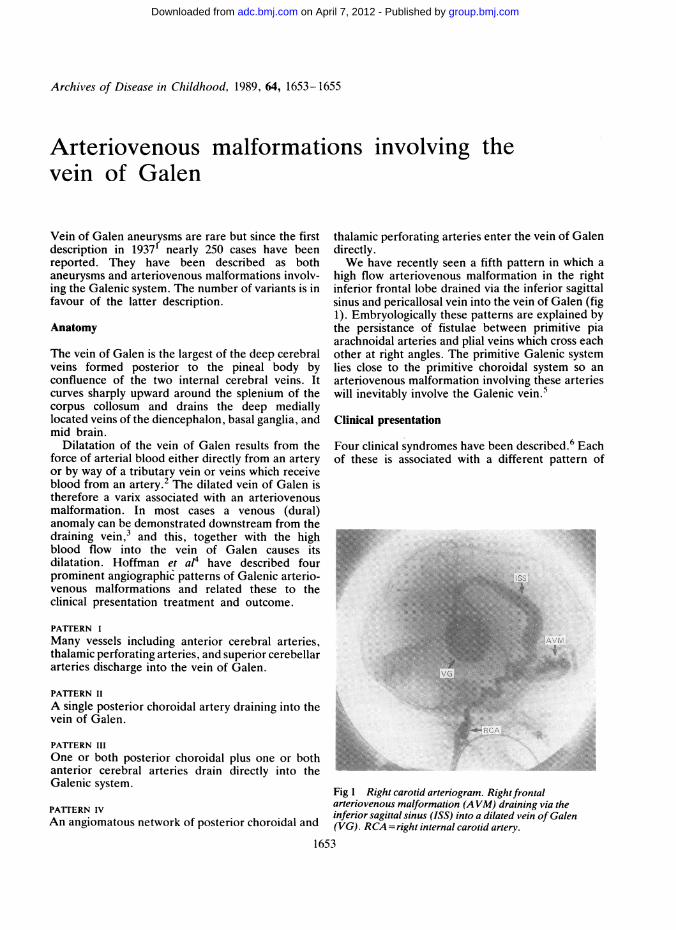
high flow arteriovenous malformation in the rightinferior frontal lobe drained via the inferior sagittalsinus and pericallosal vein into the vein of Galen (fig1). Embryologically these patterns are explained bythe persistance of fistulae between primitive piaarachnoidal arteries and plial veins which cross eachother at right angles. The primitive Galenic systemlies close to the primitive choroidal system so anarteriovenous malformation involving these arterieswill inevitably involve the Galenic vein.
Clinical presentation
Four clinical syndromes have been described.6 Eachof these is associated with a different pattern of
Fig 1 Right carotid arteriogram. Rightfrontalarteriovenous malformation (A VM) draining via theinferior sagittal sinus (ISS) into a dilated vein of Galen(VG). RCA =right internal carotid artery.
1653
group.bmj.com on April 7, 2012 - Published by adc.bmj.comDownloaded from
1654 Nicholson, Hourihan, and Hayward
Galenic arteriovenous malformation.4 The size ofthe fistula dictates whether haemodynamic featurespredominate over local mass effect. The larger thearteriovenous shunt, the greater the haemodynamiceffect and the earlier the patient presents.Neonates presenting with severe cardiac failure
and often a cranial bruit (pattern I) account for 44%of cases. These children are rarely hydrocephalicand subarachnoid haemorrhage is uncommon. Theheart failure that occurs in this group is due to thesize of the arteriovenous shunt which can steal 80%or more of the cardiac output, with large volumes ofblood under high pressure returning to the rightheart and pulmonary circulation.
Infants presenting with hydrocephalus, often acranial bruit, occasionally epilepsy, and sometimes aprogressive neurological defect caused by 'steal'phenomenon and compression of brain parenchymaby the aneurysm (patterns II and III) account for30% of cases. The hydrocephalus is caused mainlyby the mechanical effect of the dilated thick walledvein of Galen on the aqueduct of Sylvius. Com-municating hydrocephalus can also occur if thevenous (dural) obstruction is remote from the veinof Galen, thus affecting cerebral venous drainageand return of cerebrospinal fluid.
Children and adults presenting with headache,exercise syncope, neurological deterioration, cranialvenous hypertension (distended scalp and facialveins plus epistaxis), and subarachnoid haemorrhage(pattern IV) account for 26% of cases.More unusual presentations include various psy-
choses7 8 and precocious puberty. Subarachnoidhaemorrhage is relatively rare, occurring in 12% ofreported cases. This is because of the great thicknessof the wall of the dilated vein of Galen. Whenhaemorrhage does occur it is often in young adultswith a pattern IV arteriovenous malformation, thebleeding coming from the angiomatous arteriovenousmalformation.
Diagnosis
The recognition of the presenting features, inparticular cranial bruits and distended scalp and faceveins, is the basis of diagnosis. However, these arenot always present.9
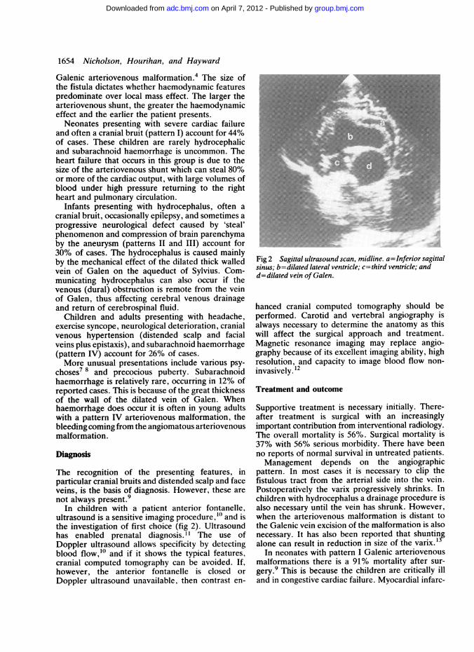
In children with a patient anterior fontanelle,ultrasound is a sensitive imaging procedure,'1' and isthe investigation of first choice (fig 2). Ultrasoundhas enabled prenatal diagnosis. " The use ofDoppler ultrasound allows specificity by detectingblood flow,'0 and if it shows the typical features,cranial computed tomography can be avoided. If,however, the anterior fontanelle is closed orDoppler ultrasound unavailable, then contrast en-
Fig 2 Sagittal ultrasound scan, midline. a=Inferiorsagittalsinus; b=dilated lateral ventricle; c=third ventricle; andd=dilated vein of Galen.
hanced cranial computed tomography should beperformed. Carotid and vertebral angiography isalways necessary to determine the anatomy as thiswill affect the surgical approach and treatment.Magnetic resonance imaging may replace angio-graphy because of its excellent imaging ability, highresolution, and capacity to image blood flow non-invasively.12
Treatment and outcome
Supportive treatment is necessary initially. There-after treatment is surgical with an increasinglyimportant contribution from interventional radiology.The overall mortality is 56%. Surgical mortality is37% with 56% serious morbidity. There have beenno reports of normal survival in untreated patients.Management depends on the angiographic
pattern. In most cases it is necessary to clip thefistulous tract from the arterial side into the vein.Postoperatively the varix progressively shrinks. Inchildren with hydrocephalus a drainage procedure isalso necessary until the vein has shrunk. However,when the arteriovenous malformation is distant tothe Galenic vein excision of the malformation is alsonecessary. It has also been reported that shuntingalone can result in reduction in size of the varix.13
In neonates with pattern I Galenic arteriovenousmalformations there is a 91% mortality after sur-gery.9 This is because the children are critically illand in congestive cardiac failure. Myocardial infarc-
group.bmj.com on April 7, 2012 - Published by adc.bmj.comDownloaded from
Arteriovenous malformations involving the vein of Galen 1655
tion is common due to appreciably reduced coronaryblood flow, and previously survivors were those inwhom hypotension was avoided during surgery.4However, it is now possible to embolise the arterio-venous malformation percutaneously under radio-logical control using giant urco coils,14 reducing flowsufficiently to correct heart failure, and allowingelective surgery when the child is older and fitter.Where this is possible there is a significant reductionin mortality and morbidity.9
In older infants and children with pattern II, III,and IV Galenic arteriovenous malformations themortality after surgery varies between 26% and32%. Postsurgical serious morbidity in these groupsvaries between 42% and 50% depending on pre-operative effects of the malformation and post-surgical complications. In infants with a pattern IIarteriovenous malformation and a single feedingartery, embolisation may be effective, reducing bothmorbidity and mortality.9
Conclusion
Arteriovenous malformations involving the vein ofGalen are being seen with increasing frequency dueto improved diagnostic techniques and awareness ofthe entity. In neonates early treatment is vital toprevent the blood supply to the cerebral parenchymaand myocardium being compromised by the flowthrough the malformation. In some cases this can bedone effectively by embolisation. Early diagnosisand treatment in older children will similarly reduceserious morbidity and mortality. A multidisciplinaryapproach to investigation and management allowsfor satisfactory treatment in many patients.
We would like to acknowledge the advice given by Mr J Vafadis(consultant neurosurgeon) and Dr A Day (consultant paediatrician)in the preparation of this article. We would also like to acknowledgethe secretarial help given by Miss D Webb.
References
Jaeger JR, Forbes RP, Dandy WE. Bilateral congenital cerebralarteriovenous communication aneurysm. Transactions of theAmerican Neurology Association 1937;63:173-6.
2 'O'Brien MS, Schecter MM. Arteriovenous malformationsinvolving the Galenic system. AJR 1970;110:50-5.Lasjaunias P, Ter-Brugge K, Lopez-Ibor L, et al. The role ofdural anomalies in vein of Galen aneurysms: report of six casesand review of the literature. AJNR 1987;8:185-92.
4 Hoffman HJ, Chuang S, Hendrick EB, et al. Aneurysm of thevein of Galen. Experience at the Hospital For Sick Children,Toronto. J Neurosurg 1982;57:316-22.
s Padget DH. Cranial venous system in man in reference todevelopment, adult configuration and relation to arteries. Am JAnat 1956;98:307-55.
6 Amacher AL, Shillito J. The syndromes and surgical treatmentof aneurysms of the great vein of Galen. J Neurosurg 1973;39:89-98.
7 Aleem A, Knesevich MA. Schizophrenia like psychosis associ-ated with vein of Galen malformation. Can J Psychiatry1987;32:226-7.
8 Ventureyra EC, Badejo A. Galenic arteriovenous malformationwith precocious puberty. Surg Neurol 1984;2:45-8.Johnson IH, Whittle IR, Besser M, et al. Vein of Galenmalformation: diagnosis and management. Neurosurgery 1987;20:747-58.
10 Vergesslich KA, Ponhold W, Wimmer M, et al. Diagnosis of ananeurysm of the great cerebral vein of Galen using duplexsonography. Pediatr Radiol 1986;21:377-84.
1 Vintzileos AM, Eisenfeld LI, Campbell WA, et al. Prenatalultrasonic diagnosis of arteriovenous malformation of the veinof Galen. Am J Perinatol 1986;3:209-1 1.
12 Roosen N, Schirmer M, Lins E, et al. MRI of an aneurysm of thevein of Galen. AJNR 1986;7:733-5.
13 Pun KK, Yu YL, Huang CY, et al. Ventriculo-peritonealshunting of acute hydrocephalus in vein of Galen malformation.Clin Exp Neurol 1987;23:209-12.
14 McCord FB, Shields MD, McNeil A, et al. Cerebral arterio-venous malformation in a neonate. Treatment by embolisation.Arch Dis Child 1987;62:1273-5.
A A NICHOLSON, M D HOURIHAN, andC HAYWARD
Department of Radiology,University Hospital of Wales,
Heath Park,Cardiff CF4 4XN
group.bmj.com on April 7, 2012 - Published by adc.bmj.comDownloaded from

doi: 10.1136/adc.64.12.1653 1989 64: 1653-1655Arch Dis Child
A A Nicholson, M D Hourihan and C Hayward involving the vein of Galen.Arteriovenous malformations
http://adc.bmj.com/content/64/12/1653.citationUpdated information and services can be found at:
These include:
serviceEmail alerting
the online article.article. Sign up in the box at the top right corner of Receive free email alerts when new articles cite this
Notes
http://group.bmj.com/group/rights-licensing/permissionsTo request permissions go to:
http://journals.bmj.com/cgi/reprintformTo order reprints go to:
http://group.bmj.com/subscribe/To subscribe to BMJ go to:
group.bmj.com on April 7, 2012 - Published by adc.bmj.comDownloaded from